Surface-Expressed Enolase Contributes to the Pathogenesis of ClinicalIsolate SSU of Aeromonas hydrophila�
Jian Sha,1,3 Tatiana E. Erova,1,3 Rebecca A. Alyea,1,3 Shaofei Wang,1,3 Juan P. Olano,2,3
Vijay Pancholi,4 and Ashok K. Chopra1,3*Departments of Microbiology and Immunology1 and Pathology,2 Institute of Human Infections and Immunity, and
Sealy Center for Vaccine Development,3 University of Texas Medical Branch, Galveston, Texas 77555, andOhio State University College, Columbus, Ohio 432104
Received 2 January 2009/Accepted 17 February 2009
In this study, we demonstrated that the surface-expressed enolase from diarrheal isolate SSU of Aeromonashydrophila bound to human plasminogen and facilitated the latter’s tissue-type plasminogen activator-medi-ated activation to plasmin. The bacterial surface-bound plasmin was more resistant to the action of its specificphysiological inhibitor, the antiprotease �2-antiplasmin. We found that immunization of mice with purifiedrecombinant enolase significantly protected the animals against a lethal challenge dose of wild-type (WT) A.hydrophila. Minimal histological changes were noted in organs from mice immunized with enolase and thenchallenged with WT bacteria compared to severe pathological changes found in the infected and nonimmunizedgroup of animals. This correlated with the smaller bacterial load of WT bacteria in the livers and spleens ofenolase-immunized mice than that found in the nonimmunized controls. We also showed that the enolase genecould potentially be important for the viability of A. hydrophila SSU as we could delete the chromosomal copyof the enolase gene only when another copy of the targeted gene was supplied in trans. By site-directedmutagenesis, we altered five lysine residues located at positions 343, 394, 420, 427, and 430 of enolase in A.hydrophila SSU; the mutated forms of enolase were hyperexpressed in Escherichia coli, and the proteins werepurified. Our results indicated that lysine residues at positions 420 and 427 of enolase were crucial inplasminogen-binding activity. We also identified a stretch of amino acid residues (252FYDAEKKEY260) in theA. hydrophila SSU enolase involved in plasminogen binding. To our knowledge, this is the first report of thedirect involvement of surface-expressed enolase in the pathogenesis of A. hydrophila SSU infections and of anygram-negative bacteria in general.
Aeromonas hydrophila is an emerging human pathogen thatcauses gastroenteritis and other extraintestinal infections, suchas septicemia, meningitis, pneumonia, cellulitis, bullous le-sions, and ecthyma gangrenosum (8, 43). The increasing isola-tion of this organism from water and food, as well as its resis-tance to water chlorination specifically in biofilms and tovarious antibiotics, could be a public health threat (16, 43).
Aeromonas spp. produce an array of virulence factors thatare associated with bacterial virulence (8). In terms of gastro-enteritis, three enterotoxins have been identified in our labo-ratory from a clinical isolate, SSU, of A. hydrophila (33). Inaddition, other virulence and regulatory genes that coded forthe glucose-inhibited division protein (GidA), DNA adeninemethyltransferase (Dam), hemolysin (Hly), ToxR-regulatedlipoprotein (TagA), and the cold shock exoribonuclease R(VacB) were recently identified and characterized from theabove-mentioned isolate (12–15, 29, 34). Further, we identifiedtype III and VI secretion systems, as well as their effectors in A.hydrophila SSU, and their roles in the pathogenesis of A. hy-drophila infections were established (16, 36–39).
While searching for new virulence factors in A. hydrophilaSSU, we previously used a murine peritoneal culture model in
which bacteria were grown in a dialysis bag implanted in theperitoneal cavity of mice (32). By restriction fragment differ-ential display PCR using RNA as a template, we identified aglycolytic enzyme enolase in A. hydrophila SSU, which wasdifferentially expressed under in vivo but not in vitro growthconditions (32). Enolase is a ubiquitous enzyme that catalyzesthe reversible conversion of 2-phosphoglycerate (2-PGE) tophosphoenolpyruvate (PEP) (26). In addition to its metabolicrole, enolase has been implicated for its contribution to severalbiological and pathophysiological processes by acting as a heatshock protein and in modulating gene transcription, as well asfor its involvement in microbial diseases and autoimmunity(26). Although mainly localized in the cytoplasm, enolase hasalso been identified on the surface of neuronal, cancer, andsome hematopoietic cells, as well as on the cell surfaces ofseveral bacterial species, where it binds plasminogen (6, 17, 20,21, 23, 25).
Plasminogen is a single-chain glycoprotein that is abundantin human plasma and extracellular fluid (20). Its conversion toa proteolytic, active two-chain form, namely plasmin, can bemediated either by eukaryotic activators such as the host-de-rived tissue plasminogen activator (tPA) and urokinase or bythe prokaryotic activators staphylokinase and streptokinase(17, 20). tPA is a principal activator of plasminogen in plasmaand intercellular fluid. The immobilization of plasminogenwith its receptors dramatically enhances tPA-mediated activa-tion of plasminogen, and the generated plasmin that continuesto bind to its receptors is more resistant to the action of its
* Corresponding author. Mailing address: Department of Microbi-ology and Immunology, 301 University Blvd., Medical Research Build-ing, UTMB, Galveston, TX 77555-1070. Phone: (409) 747-0578. Fax:(409) 747-6869. E-mail: [email protected].
specific physiological inhibitors (e.g., the antiprotease �2-anti-plasmin [�2AP]) (17, 20). Plasmin is a trypsin-like serine pro-tease with broad substrate specificity; it is physiologically in-volved in fibrinolysis by degradation of fibrin and also degradesnoncollagenous glycoproteins of mammalian extracellular ma-trices and basement membranes. Thus, surface-expressed plas-minogen receptors (e.g., enolase) provide a niche for host cellsor bacteria to interact with the mammalian proteolytic plas-minogen-plasmin system, which presumably facilitates themetasis process or promotes microbial pathogens to migratewithin the host to fulfill their nutritional demands (18, 19, 21,23, 25). Indeed, several recent studies demonstrated that eno-lase in certain pathogenic bacteria (e.g., Streptococci and Pneu-mococci) and microbiota (e.g., Lactobacilli and Bifidobacteria)not only is essential for bacterial viability because of its enzy-matic activity but also promotes pathogen-host interactionsand contributes to bacterial colonization and pathogenesis bymeans of its surface or extracellular location (1, 4, 6, 17, 19, 20,22, 26, 27). In our previous study, we reported the presence ofsurface-expressed enolase in the gram-negative bacterium A.hydrophila SSU (32) and demonstrated that crude, whole-bac-terial-cell lysates of A. hydrophila could bind human plasmin-ogen (32). However, the direct role of enolase in Aeromonasinfections has not yet been established.
In our present study, we provided direct evidence that thewashed whole bacterial cells of A. hydrophila SSU bound hu-man plasminogen/plasmin and facilitated tPA-mediated acti-vation of plasminogen. We also found that this plasminogen/plasmin-bacterial cell interaction was, in part, mediated byenolase. We immunized mice with purified recombinant eno-lase and showed that such immunized animals were protectedagainst mortality from a lethal challenge dose of wild-type(WT) A. hydrophila. However, we were unable to develop anenolase-negative mutant of A. hydrophila SSU, which suggeststhat this enzyme might be essential for the viability of thisorganism. Further, we studied enolase’s plasminogen-bindingactivity in A. hydrophila SSU and identified lysine residues atpositions 420 and 427 that contributed to this activity. We alsoidentified an internal stretch of amino acid residues (252FYDAEKKEY260) in the enolase (which has 434 amino acid resi-dues) of A. hydrophila SSU that was responsible for this plas-minogen-binding activity. This is the first detailed studyillustrating the role of enolase in the pathogenesis of A. hy-drophila SSU infections.
MATERIALS AND METHODS
Bacterial strains, plasmids, media, and DNA techniques. The bacterial strainsand plasmids (10, 24, 33) used in this study are listed in Table 1. A. hydrophilaSSU, a diarrheal isolate, was obtained from the Centers for Disease Control andPrevention, Atlanta, GA. SSU-R is a spontaneous rifampin-resistant (Rifr) strainof SSU that was prepared previously in our laboratory (35). Luria Bertani (LB)broth (Difco Laboratories, Detroit, MI) was used to grow A. hydrophila andEscherichia coli cultures at 37°C with aeration (180 rpm). The antibiotics ampi-cillin (Ap), tetracycline (Tc), kanamycin (Km), streptomycin (Sp), spectinomycin(Sm), and Rif were obtained from Sigma (St. Louis, MO) and were used atconcentrations of 100, 15, 50, 25, 25, and 40 �g/ml, respectively. Restrictionendonucleases were purchased from Promega (Madison, WI). All of the basicmolecular biology techniques used in this study were previously described (33).
Plasminogen binding to A. hydrophila SSU cells and activation of plasmino-gen. The tPA-mediated activation of plasminogen in the presence of bacteria wasmeasured as described by Lahteenmaki et al. (20). Briefly, 4 � 108 log-phase-grown, phosphate-buffered saline (PBS)-washed A. hydrophila SSU cells were
incubated with 4 �g of human plasminogen (Sigma), 2 ng of tPA (MolecularInnovations, Inc., Novi, MI) and a 0.45 mM concentration of the chromogenicplasmin substrate S-2251 (DiaPharma Group, Inc., West Chester, OH) in a finalvolume of 200 �l in PBST buffer (PBS containing 0.02% Tween 80). In somereaction mixture groups, 2 mM ε-aminocaproic acid (EACA; Sigma) or purifiedimmunoglobulin G (IgG) at concentrations of 0.125 mg/ml and 0.025 mg/ml fromenolase-immunized and preimmune mouse serum (as stated in the animal ex-periment section below) was added. The reaction mixture groups that did notcontain either the bacterial cells or the tPA served as negative controls. Thereaction mixtures were incubated at 37°C with slow rotation, and the increasedplasmin activity was assessed at a 30-min interval for a total of 3 h by measuringabsorbance at 405 nm with a VERSAmax tunable microplate reader (MolecularDevices Corporation, Sunnyvale, CA).
For the binding assay, 8 � 108 A. hydrophila SSU cells treated similarly asabove were incubated with either 4 �g of human plasminogen plus 2 ng of tPAor 4 �g of human plasmin only (Sigma) in a final volume of 200 �l in PBSTbuffer. The reaction mixtures were incubated at 37°C with slow rotation for 3 h.In some reaction mixture groups, 2 mM EACA or purified IgG (0.125 mg/ml and0.025 mg/ml) from enolase-immunized and preimmune mouse serum was addedinto the reaction mixture. The reaction mixture groups that contained onlybacterial cells without the addition of plasminogen and plasmin served as neg-ative controls. After incubation, the bacterial cells were collected and washedtwice with PBST buffer and were finally resuspended in 300 �l of PBST buffercontaining 0.45 mM S-2251. We incubated the reaction mixtures at 37°C for 2 hand measured plasmin activity at 405 nm. The plasmin activity levels in thenegative control reaction mixture groups were used as blanks in the measure-ment.
For analyzing the protection of plasmin from �2AP in the presence of bacterialcells, plasmin (0.25 �g or 2.5 �g) was incubated with or without 4 � 108 log-phase-grown, PBS-washed A. hydrophila SSU cells for 30 to 60 min at roomtemperature prior to the addition of 0.45 mM S-2251 and �2AP in increasingconcentrations (1, 8, and 16 �g). Plasmin activity was measured after 1 h ofincubation at 37°C. The reaction mixture groups that were omitted from eitherthe �2AP or both bacterial cells and �2AP cultures were used as controls. Theviability of bacterial cells was monitored by serial dilution and plating at the endof each of the above experiments.
Generation and characterization of enolase gene mutant of A. hydrophila SSU.Two pairs of primers, enoup5/enoup3 and enodn5/enodn3 (Table 2), were syn-thesized and used to amplify the upstream (796 bp) and downstream (718 bp)flanking DNA sequences to the enolase gene of A. hydrophila. These fragmentswere then ligated together through the introduced common BglII site and clonedinto a pET30a vector (37) at the NotI/XbaI restriction sites, resulting in therecombinant plasmid pETUD (Table 1). An Sm/Sp gene cassette flanked bythe BamHI site was removed from the plasmid pHP45� (33) and inserted at theBglII site (compatible with the BamHI site) of pETUD to generate the recom-binant plasmid pETUDSm/Sp (Table 1). After digestion with NotI/XbaI restric-tion enzymes, the DNA fragment (3.5 kb) from the above plasmid was removedand ligated to a pBluescript vector (33) at the compatible restriction enzyme sitesto generate the recombinant plasmid pBUDSm/Sp (Table 1). By using KpnI/XbaI restriction enzymes, the DNA fragment (3.5 kb) was removed from theplasmid pBUDSm/Sp and ligated to the pDMS197 suicide vector at the compat-ible restriction enzyme sites, and the resulting plasmid (pDMSUDSm/Sp) (Table1) was transformed into E. coli SM10 (24, 33). The recombinant E. coli[pDMSUDSm/Sp] was conjugated with the parental Rifr A. hydrophila strain(33). The transconjugants were selected based on their resistance to appropriateantibiotics and sucrose and subjected to further analysis (33).
Supplying an additional copy of the native enolase gene into WT A. hydrophilaSSU. Primers eno5 and eno3 (Table 2) were used to PCR amplify the codingregion of the enolase gene from A. hydrophila. The amplified DNA fragment(1,313 bp) was cloned into the pBR322 vector at the ScaI/PstI restriction enzymesites, generating the recombinant plasmid pBReno (Table 1). In this plasmid, theenolase gene was under the control of an Ap resistance gene promoter of thevector, and, therefore, it was constitutively expressed. The recombinant plasmidpBReno was subsequently electroporated into WT A. hydrophila (14). The pres-ence of recombinant plasmid pBReno in WT A. hydrophila was verified byplasmid isolation and digestion with ScaI and PstI restriction enzymes.
Generation of various mutated forms of the enolase gene by site-directedmutagenesis. An Altered Sites in vitro Mutagenesis System (Promega) was usedfor site-directed mutagenesis as we described previously (12). Briefly, the codingregion of the native enolase gene was PCR amplified from the genomic DNA ofWT A. hydrophila with the primer set enoF/enoR (Table 2) and subsequentlycloned into the plasmid pALTER-1 (Tcr Aps), generating the recombinant plas-mid pALTER-1/eno (Tcr Aps) (Table 1). Subsequently, E. coli JM109 cells
containing the pALTER-1/eno recombinant plasmid were infected with the R408helper bacteriophage, and the phagemid single-stranded DNA was isolated asdescribed by the manufacturer.
This single-stranded DNA was used as a template for the mutagenesis reac-tions with the individual mutagenic primers enoK-Q343, enoK-M394, enoK-L420, enoK-N427, and enoK-R430, respectively (Table 2). For example, theprimer enoK-Q343 was used to replace the lysine residue (K) at position 343 witha glutamine residue (Q) in the enolase. Each of the mutagenesis reactionsgenerated a specific mutated enolase gene in plasmid pALTER-1 and was trans-formed into E. coli ES1301 mutS competent cells. The plasmid DNA isolatedfrom these bacterial strains was then transformed into E. coli JM109, and thetransformants were screened for Tcs and Apr. Mutations within the enolase genein plasmid pALTER-1 were confirmed by DNA sequence analysis, and themutated enolase genes were designated eno(K343Q), eno(K394M), eno(K420L),eno(K427N), and eno(K430R) (Table 1). Different amino acids were used to
replace the lysine residues in enolase as our intent was to achieve a reasonableexpression level of the mutated enolases in the E. coli system while maintainingtheir solubility for easy purification and biological/functional activity measure-ments.
Hyperexpression and purification of different forms of A. hydrophila SSUenolase. By using the primer set enoN/enoC (Table 2), we PCR amplified nativeand mutated enolase genes [eno(K343Q), eno(K394M), and eno(K420L)] of A.hydrophila SSU from template plasmid DNA generated from the site-directedmutagenesis step above (Table 1). Similarly, the mutated enolase geneseno(K427N)and eno(K430R)were PCR amplified with the primer sets enoN/enoC427 and enoN/enoC430, respectively (Table 2). These amplified enolasegenes were cloned into a bacteriophage T7 polymerase/promoter-based pET-30a(�) expression vector (Novagen, San Diego, CA) at the NdeI/XhoI restric-tion enzyme sites, and their downstream sequences were fused in frame with theHis tag DNA sequence of the vector (Table 1). The resulting recombinant
TABLE 1. Strains and plasmids used in this study
Strain or plasmid Relevant characteristic(s) and/or construction Source or reference
StrainsA. hydrophila SSU CDC, Atlanta, GA
SSU-R Rifr strain of A. hydrophila SSU Laboratory stockWT/pBReno WT A. hydrophila SSU-R transformed with pBReno plasmid; Rifr Tcr This studyEnolase mutant The chromosomal enolase gene was deleted from WT A. hydrophila SSU-R that was
transformed with the plasmid pBReno; Sm/Spr Rifr TcrThis study
E. coliSM10 Kmr �pir 24JM109 endA1 recA1 gyrA96 thi-1 hsdR17(rK
� mK�) relA1 supE44 �(lac-proAB) F traD36 proAB
lacIqZ�M15�Promega
ES1301 mutS lacZ53 mutS201::Tn5 thyA36 rha-5 metB1 deoC PromegaHMS174 (DE3) RecA-mutated K-12 background E. coli strain that carries a chromosomal copy of the T7
RNA polymerase gene under the control of the lacUV5 promoter; used as the hoststrain for the pET expression system
Novagen
PlasmidspHP�45 Contains a 2.0-kb Sm/Spr gene cassette 33pET30a pBR322-derived expression vector with T7 lac promoter and up- and down-stream His
tags; KmrNovagen
pDMS197 A suicide vector; R6K ori sacB Tcr 10pALTER-1 Vector for the site mutagenesis; Tcr Aps PromegapBReno The coding region of A. hydrophila enolase gene was cloned in pBR322 at the ScaI/PstI
sites and under the control of the Ap promoter of the vector; TcrThis study
pETUD pET30a vector containing up- and downstream DNA flanking sequences to the enolasegene; Kmr
This study
pETUDSm/Sp The Sm/Sp cassette was ligated with the up- and downstream DNA flanking sequences tothe enolase gene in pET30a vector; Sm/Spr Kmr
This study
pBUDSm/Sp The Sm/Sp cassette was ligated with the up- and downstream DNA flanking sequences tothe enolase gene in pBluescript vector; Sm/Spr Apr
This study
pDMSUDSm/Sp Suicide vector pDMS197 containing the Sm/Sp cassette ligated with up- and downstreamDNA flanking sequences to the enolase gene; used to generate the enolase genedeletion mutant of A. hydrophila; Kmr Tcr
This study
pALTER-1/eno The native enolase gene of A. hydrophila was cloned into pALTER-1 vector at the BamHI/HindIII sites for the site mutagenesis; Tcr Aps
This study
pALTER/eno343K/Q pALTER-1 vector containing the mutated A. hydrophila enolase gene eno(K343Q); Tcs Apr This studypALTER/eno394K/M pALTER-1 vector containing the mutated A. hydrophila enolase gene eno(K394M); Tcs Apr This studypALTER/eno420K/L pALTER-1 vector containing the mutated A. hydrophila enolase gene eno(K420L); Tcs Apr This studypALTER/eno427K/N pALTER-1 vector containing the mutated A. hydrophila enolase gene eno(K427N); Tcs Apr This studypALTER/eno430K/R pALTER-1 vector containing the mutated A. hydrophila enolase gene eno(K430R); Tcs Apr This studypET30aeno The native enolase gene of A. hydrophila was cloned in pET30a and fused with the
downstream His tags of the vector for hyperexpression; KmrThis study
pET30aeno343K/Q The mutated enolase gene of A. hydrophila eno(K343Q) was cloned in pET30a and fusedwith the downstream His tags of the vector for hyperexpression; Kmr
This study
pET30aeno394K/M The mutated enolase gene of A. hydrophila eno(K394M) was cloned in pET30a and fusedwith the downstream His tags of the vector for hyperexpression; Kmr
This study
pET30aeno420K/L The mutated enolase gene of A. hydrophila eno(K420L) was cloned in pET30a and fusedwith the downstream His tags of the vector for hyperexpression; Kmr
This study
pET30aeno427K/N The mutated enolase gene of A. hydrophila eno(K427N) was cloned in pET30a and fusedwith the downstream His tags of the vector for hyperexpression; Kmr
This study
pET30aeno430K/R The mutated enolase gene of A. hydrophila eno(K430R) was cloned in pET30a and fusedwith the downstream His tags of the vector for hyperexpression; Kmr
This study
VOL. 191, 2009 ROLE OF ENOLASE IN AEROMONAS HYDROPHILA INFECTIONS 3097
plasmids were transformed into the E. coli HMS174 (DE3) strain for hyperex-pression. Each of these expressing strains was grown in 250 to 500 ml of LBmedium with shaking (180 rpm) to an optical density at 600 nm of 0.6 beforeinduction with 1 mM IPTG (isopropyl-�-D-thiogalactopyranoside) for 4 h at30°C.
The IPTG-induced bacterial cells were harvested, and the cell lysates fromthese enolase-expressing strains were first passed through a ProBond nickelchelating column (Invitrogen, Carlsbad, CA) and eluted according to the man-ufacturer’s instructions. The eluted fractions were monitored for the presence ofenolase by performing sodium dodecyl sulfate (SDS)–12% polyacrylamide gelelectrophoresis (PAGE) and Coomassie blue staining. Fractions containing theprotein of interest (42 to 45 kDa) were pooled and dialyzed (25- to 30-kDamolecular-mass-cutoff tubing) for 12 h at 4°C in a buffer containing l20 mM Tris,50 mM NaCl, 5 mM MgSO4, and 1 mM dithiothreitol (DTT), pH 6.5. Thedialyzed fractions were filtered through a 22-�m-pore-size syringe filter (Corn-ing, Lowell, MA) and loaded onto an ion exchange Resource Q column (6 ml;GE Healthcare, Piscataway, NJ) connected to the fast protein liquid chromatog-raphy ACTA Purifier 10/100. The proteins were eluted from the column by usinga salt gradient (20 mM Tris, 1 M NaCl, 5 mM MgSO4, and 1 mM DTT, pH 6.5)at a flow rate of 2 ml/min. The protein peaks were analyzed by Unicorn, version4.00.16, software (GE Healthcare) and monitored for the presence of pureenolase by SDS-PAGE and Coomassie blue staining of the gel. Fractions con-taining enolase were pooled and concentrated using a Centriprep instrumentfitted with an Ultracel YM-10 membrane (Millipore, Billerica, MA), and theprotein concentration was determined by using a Bradford dye protein assay(Bio-Rad, Hercules, CA) (5).
Enzymatic activity of various mutated forms of recombinant enolase. Theenzymatic activity of different forms of recombinant enolase was measured by theirability to catalyze the reversible conversion of 2-PGE to PEP (26, 32). In ourexperiments, we measured the reverse reaction (PEP to 2-PGE) due to the avail-ability of the substrate 2-PGE. The reactions were started by the addition of 2 �g ofeach of the purified recombinant enolase proteins into 100 �l of the reaction buffer(100 mM HEPES, pH 7.0, 10 mM MgSO4, and 7.7 mM KCl) containing differentconcentrations (0.2, 0.4, 0.6, 0.8 and 1.0 mM) of PEP (Sigma). The reactions weremonitored by measuring the reduction of PEP absorbance at A240 nm as a result of
conversion of PEP to PGE by spectrophotometry (Ultropec 2000; GE Healthcare)every 5 s for a total of 3 min per reaction.
Plasminogen-binding activity of various mutated forms of recombinant eno-lase. A sandwich enzyme-linked immunosorbent assay (ELISA) was used todetect binding of enolase to human plasminogen, as described previously (32).Briefly, microtiter plates (Evergreen Scientific, Los Angeles, CA) were coatedwith 2 �g per well of purified and different forms of recombinant enolaseovernight at 4°C. For the positive control, the plate wells were coated with 2 �gof human plasminogen (Sigma) while wells coated with PBS only were used asnegative controls. Eight wells were used for each tested recombinant enolase andcontrol. The overnight coated plates were washed and then blocked overnightagain at 4°C with buffer A (PBS containing 0.05% Tween 20, 1 mM EDTA, and0.25% gelatin) (40). An aliquot (2 �g) of plasminogen in 50 �l of PBS was addedto the coated wells and incubated for 2 h at room temperature. In some exper-iments, EACA or peptides (see below) were mixed with 2 �g of human plas-minogen before being added to the microtiter plate wells to allow us to studytheir abilities to block enolase binding to the plasminogen. We used EACA atconcentrations of 2, 4, 6, and 8 mM and the three peptides Seno (FYDKERKVY), Aeno (FYDAEKKEY), and AenoM (FYDAGKLEY), representing re-gions within the Streptococcus pneumoniae and A. hydrophila SSU enolases, atconcentrations of 100 to 200 �g/well. The AenoM peptide had two amino acidsubstitutions (underlined) compared to the peptide Aeno.
Peptide Seno represented an internal plasminogen-binding motif (amino acidresidues 248 to 256) identified in the enolase of S. pneumoniae (3, 11), whilepeptide Aeno (amino acid residues 252 to 260) represented a correspondingstretch of Seno in the enolase of A. hydrophila SSU (11). Peptide AenoM was themutated form of Aeno, and the substituted amino acid residues were indicatedby underlining in the sequence shown above. These peptides were synthesized bythe Biomatik Corporation (Toronto, Ontario, Canada) and reconstituted inwater at a concentration of 5 mg/ml. After a 2-h incubation, the plates werewashed three times with buffer A and incubated for 1 h with goat antiplasmino-gen antibodies (primary antibody; 1:1,500 dilution) followed by horseradish per-oxidase-labeled mouse, anti-goat IgG (secondary antibody; 1:2,000 dilution)(Santa Cruz Biotechnology, Inc.) (32). TMB (3,3, 5,5-tetramethyl-benzidine;Sigma) substrate was used for color development, and the optical density (sub-
TABLE 2. Sequences of the primers used in this study
Primer name Sequence (restriction enzyme)a Purpose
enoup5 5-TTGCGGCCGCTCAAGCACGGCGGTCTG-3 (NotI) PCR amplification of the upstream flanking DNAfragment of the enolase gene from A.hydrophilaenoup3 5-CCAGATCTATGTGTATTTCCTCAGGT-3 (BglII)
enodn5 5-GTAGATCTATCGTCGCCGGTTCTCTTG-3 (BglII) PCR amplification of the downstream flankingDNA fragment of the enolase gene from A.hydrophilaenodn3 5-TTTCTAGAGGATCCTCGGATCGGCGG-3 XbaI
eno5 5-GTAGTACTATGTCCAAGATCGTTAAAGTG-3 (ScaI) PCR amplification of the DNA fragmentencoding the enolase gene from A. hydrophilafor cloning into plasmid pBR322eno3 5-TCCTGCAGTTAAGCCTGGTTCTTCACTTC-3 (PstI)
Sm5 5-ATGCGCTCACGCAACTGGTC-3 Identification of the deletion of the enolase geneon the chromosome of A. hydrophilaSm3 5-TTATTTGCCGACTACCTTGG-3
ENT5 5-CCTACAAGTCCGTCAACGAG-3ENT3 5-ACGTGCAGCGCATTGAGCAC-3enoF 5-CGCGGATCCATGTCCAAGATCGTTAAAGTGATCGG-3 (BamHI) Cloning of the native eno gene into pALTER-1
a The underlining indicates the restriction endonuclease site. Boldface indicates mutated nucleotide(s) in the enolase gene of A. hydrophila; the original nucleotideis shown in parentheses. All primers were developed for this study.
tracted from the blanks, i.e., the wells coated with PBS and used as the negativecontrol) was read at 370 nm by using a VERSAmax tunable microplate reader.
Animal experiments. Groups of 15 Swiss Webster mice (Taconic Farms, Ger-mantown, NY) were first immunized with purified native (unmutated) recombi-nant enolase produced from E. coli. Briefly, animals were injected intraperito-neally (i.p.) with 10 �g of purified enolase mixed with monophosphoryl lipidA-trehalose dicorynomycolate-cell wall skeleton adjuvant(Sigma). The animalswere bled before immunization, boosted twice with the antigen (10 �g) at 15-dayintervals, and finally bled after 1 month. The antibody titers in the pooled serawere determined by using an ELISA having recombinant enolase as the antigensource (33). The immunized and nonimmunized mice (given the adjuvant alonewithout the antigen) were challenged i.p. with the WT A. hydrophila SSU at twoto three times the 50% lethal dose ([LD50] 5 � 107 to 1 � 108 CFU) after 1month of immunization. Organs (lungs, spleen, and liver) from five mice in eachgroup were removed at day 3 of the challenge and subjected to analyses forhistopathology and bacterial load (liver and spleen), as we recently described(31). Five noninfected mice were used as controls for the histopathological study.The deaths of mice were recorded, and the animals were monitored for 3 weekspostinfection.
The IgGs from the enolase-immunized and preimmune mouse sera collectedfrom the above animal experiment were purified by using Nab Spin Kits (ThermoFisher Scientific Inc., Rockford, IL) and further desalted with a Zeba Desalt Spincolumn (Thermo). The enolase antibody titers in the purified IgGs were furtherevaluated by ELISA using purified native recombinant enolase as the antigensource. The amount of purified IgG in both preimmune and immune sera wasdetermined by using a Bradford dye protein assay (5). The purified IgGs werestored at 4°C and employed in the phagocytosis assay and plasminogen-bindingand activation assays as described above.
Phagocytosis assay. An aliquot (1 � 108) of log-phase-grown, PBS-washed A.hydrophila SSU cells was incubated with purified IgGs from either enolase-immunized or preimmune mouse sera at concentrations of 0.125 mg/ml and0.025 mg/ml in a final volume of 500 �l in PBS. The bacterial cells incubated withPBS served only as negative controls. After a 1-h incubation at 37°C, the bacterialcells were washed three times with PBST buffer, and a portion of the bacterialcells was serially diluted and plated to examine viability of the bacterial cells afterthey were coated with the antibodies. The rest of the bacterial cells were used toinfect RAW 264.7 murine macrophages (in six-well tissue culture plates) at amultiplicity of infection of 1 (37). The wells were done in triplicate for eachinfection group. After 3 h of incubation at 37°C with 5% CO2, the infectedmacrophages were washed twice with PBS and then incubated an additional hourin fresh Dulbecco’s modified essential medium containing 200 �g/ml gentamicin.The gentamicin treatment killed extracellular bacteria but did not affect theviability of intracellular organisms. The macrophages were then washed threetimes with PBS to remove gentamicin and lysed with 200 �l of 1% (vol/vol)Triton X-100 to release intracellular bacteria. A 10-fold serial dilution was made,and bacteria were enumerated on LB agar plates in duplicate.
Statistical analysis of the data. Where applicable, a minimum of three inde-pendent experiments were performed, and the data were analyzed using either aStudent’s t test or Fisher’s exact test.
RESULTS
Immobilization and conversion of plasminogen to plasminon the cell surface of A. hydrophila SSU cells. In our previousstudy, we detected strong enolase activity associated with theintact A. hydrophila cells and demonstrated that crude, whole-bacterial-cell lysates of A. hydrophila could bind human plas-minogen (32). To further investigate whether plasminogenindeed binds to the bacterial surface, the log phase and PBS-washed A. hydrophila SSU cells were first incubated with hu-man plasminogen. After the unbound plasminogen was washedaway, the surface-bound plasminogen was further converted tothe active form of plasmin by tPA, and the plasmin activity wasmeasured by adding the plasmin-specific chromogenic sub-strate S-2251.
As shown in Fig. 1A, plasmin activity was detected on thecell surface of A. hydrophila SSU cells (column a), and additionof the lysine analog EACA decreased bacterial surface-associ-ated plasmin activity by 56% (column b). These data indicated
that plasminogen binding to the bacterial surface was specificand that lysine residues were involved. Importantly, we ob-served a dose-dependent decrease in plasmin activity (29 to46%) in the reaction mixture groups in which purified enolase-specific antibodies were added (columns c and d). As expected,there was no decrease in plasmin activity compared to that ofthe control (column a) when purified IgGs from preimmunesera were added to the reaction mixtures (columns e and f).Overall, these data indicated to us that the bacterial surface-plasminogen interaction was mediated in part by enolase. Asimilar pattern was noticed when human plasmin was directlyincubated with A. hydrophila SSU cells (Fig. 1B). No significantchanges in the number of bacterial cells were observed beforeor after the experiment in each group and among the groups,which suggested to us that the viability of A. hydrophila SSUwas not affected during the experiment.
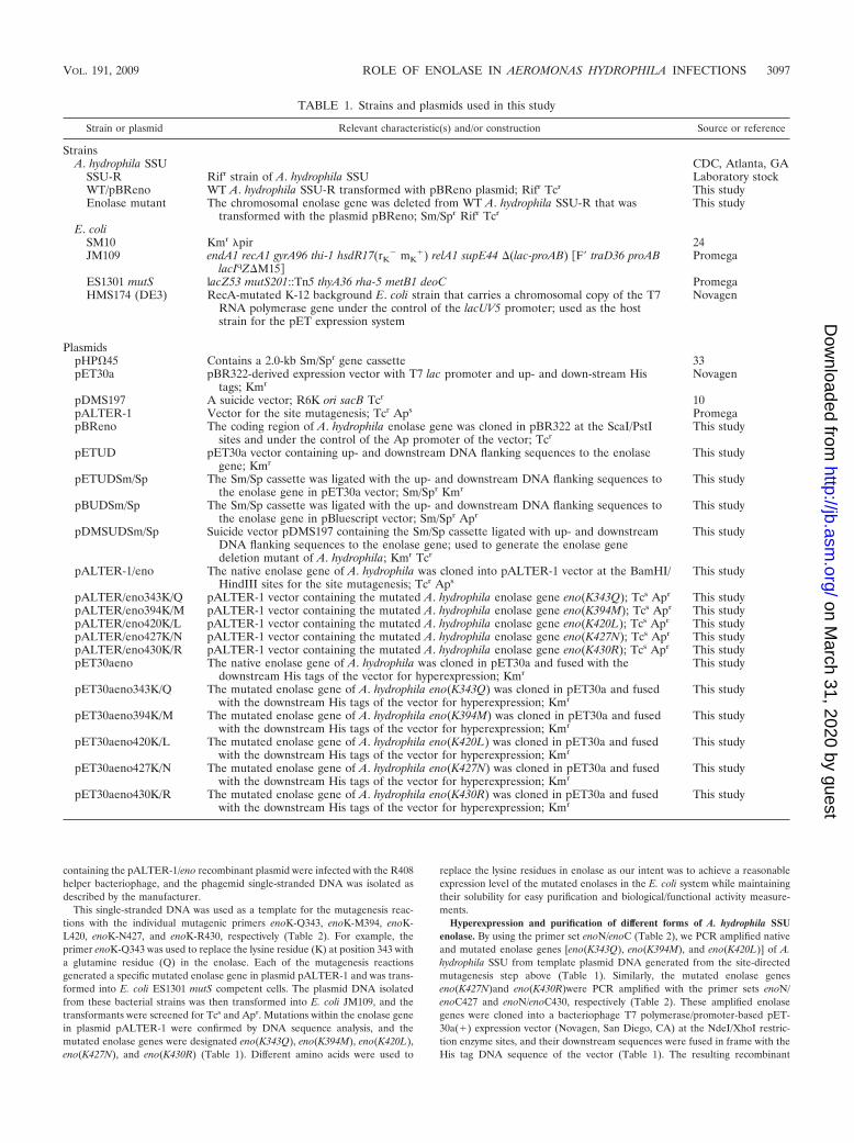
Plasminogen binding on the surface of A. hydrophila SSUcells promoted its tPA-mediated activation and protected plas-min from its physiological inhibitor �2AP. As shown in Fig. 2,there was a significant, time-dependent (30 to 180 min) for-mation of plasmin from plasminogen mediated by tPA (groupA) in the presence of A. hydrophila SSU cells. In contrast, only
FIG. 1. Plasminogen and plasmin binding to the surface of A. hy-drophila SSU. The log phase and PBS-washed A. hydrophila SSU cellswere first incubated with 4 �g of either plasminogen (A) or plasmin (B).After washing away the unbound plasminogen/plasmin, we converted thesurface-bound plasminogen to the active form plasmin by tPA and mea-sured the plasmin activity by adding the plasmin-specific chromogenicsubstrate S-2251. The reaction mixtures that contained only bacterial cellswithout plasminogen and plasmin served as negative controls and were setas blanks when the absorbance at 405 nm was recorded. Columns a,bacterial cells incubated with plasminogen (A) or plasmin (B); columns b,addition of 2 mM EACA to the binding reaction mixture; columns c andd, addition of 0.125 mg/ml and 0.025 mg/ml, respectively, of the purifiedIgGs from enolase-immunized mouse serum; columns e and f; addition of0.125 mg/ml and 0.025 mg/ml, respectively, of purified IgGs from preim-mune mouse serum. The asterisk denotes statistically significant differ-ences (P 0.05) compared to columns a by a Student’s t test.
VOL. 191, 2009 ROLE OF ENOLASE IN AEROMONAS HYDROPHILA INFECTIONS 3099
marginal plasmin activity was observed in the group in whichno bacterial cells were added (group E). Plasmin formationwas greatly inhibited (32 to 61%) over a period of 3 h ofincubation in the presence of EACA (group B) compared tothat in the control group A, a finding that further emphasizedlysine residue involvement and the importance of plasmino-gen’s binding to the bacterial surface prior to its activation.
Compared to the groups of reaction mixtures with the ad-dition of purified IgGs from preimmune sera (group D), thepresence of enolase-specific antibodies in the reaction mixturessignificantly and dose-dependently decreased (12 to 43%) tPA-mediated plasmin formation (group C), with a maximum de-crease (43%) occurring at the 60-min time point. In Fig. 2, datarepresent only experiments with high (0.125 mg/ml) enolaseantibody concentrations. When a lower concentration of eno-lase-specific antibodies (0.025 mg/ml) was used in the reactionmixtures, the decrease in plasmin activity was in the range of 8to 31% over a period of 3 h, with statistical differences com-pared to the preimmune sera only at 30- and 60-min timepoints (data not shown). These data further confirmed thatenolase was involved in plasminogen binding and its activation.Minimal plasmin formation was noticed in the reaction mix-ture in which no tPA was added (group F), suggesting to usthat there was no apparent plasminogen activator secreted byA. hydrophila SSU.
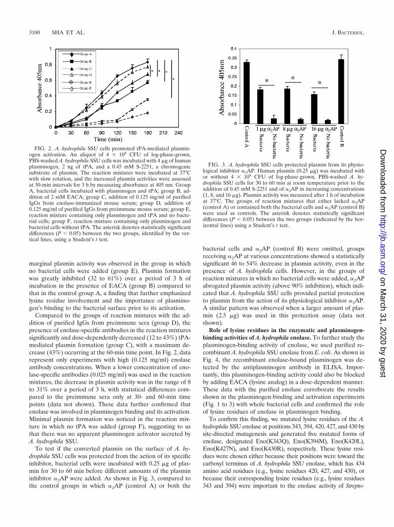
To test if the converted plasmin on the surface of A. hy-drophila SSU cells was protected from the action of its specificinhibitor, bacterial cells were incubated with 0.25 �g of plas-min for 30 to 60 min before different amounts of the plasmininhibitor �2AP were added. As shown in Fig. 3, compared tothe control groups in which �2AP (control A) or both the
bacterial cells and �2AP (control B) were omitted, groupsreceiving �2AP at various concentrations showed a statisticallysignificant 46 to 54% decrease in plasmin activity, even in thepresence of A. hydrophila cells. However, in the groups ofreaction mixtures in which no bacterial cells were added, �2APabrogated plasmin activity (above 90% inhibition), which indi-cated that A. hydrophila SSU cells provided partial protectionto plasmin from the action of its physiological inhibitor �2AP.A similar pattern was observed when a larger amount of plas-min (2.5 �g) was used in this protection assay (data notshown).
Role of lysine residues in the enzymatic and plasminogen-binding activities of A. hydrophila enolase. To further study theplasminogen-binding activity of enolase, we used purified re-combinant A. hydrophila SSU enolase from E. coli. As shown inFig. 4, the recombinant enolase-bound plasminogen was de-tected by the antiplasminogen antibody in ELISA. Impor-tantly, this plasminogen-binding activity could also be blockedby adding EACA (lysine analog) in a dose-dependent manner.These data with the purified enolase corroborate the resultsshown in the plasminogen-binding and activation experiments(Fig. 1 to 3) with whole bacterial cells and confirmed the roleof lysine residues of enolase in plasminogen binding.
To confirm this finding, we mutated lysine residues of the A.hydrophila SSU enolase at positions 343, 394, 420, 427, and 430 bysite-directed mutagenesis and generated five mutated forms ofenolase, designated Eno(K343Q), Eno(K394M), Eno(K420L),Eno(K427N), and Eno(K430R), respectively. These lysine resi-dues were chosen either because their positions were toward thecarboxyl terminus of A. hydrophila SSU enolase, which has 434amino acid residues (e.g., lysine residues 420, 427, and 430), orbecause their corresponding lysine residues (e.g., lysine residues343 and 394) were important to the enolase activity of Strepto-
FIG. 2. A. hydrophila SSU cells promoted tPA-mediated plasmin-ogen activation. An aliquot of 4 � 108 CFU of log-phase-grown,PBS-washed A. hydrophila SSU cells was incubated with 4 �g of humanplasminogen, 2 ng of tPA, and a 0.45 mM S-2251, a chromogenicsubstrate of plasmin. The reaction mixtures were incubated at 37°Cwith slow rotation, and the increased plasmin activities were assessedat 30-min intervals for 3 h by measuring absorbance at 405 nm. GroupA, bacterial cells incubated with plasminogen and tPA; group B, ad-dition of 2 mM EACA; group C, addition of 0.125 mg/ml of purifiedIgGs from enolase-immunized mouse serum; group D, addition of0.125 mg/ml of purified IgGs from preimmune mouse serum; group E,reaction mixture containing only plasminogen and tPA and no bacte-rial cells; group F, reaction mixture containing only plasminogen andbacterial cells without tPA. The asterisk denotes statistically significantdifferences (P 0.05) between the two groups, identified by the ver-tical lines, using a Student’s t test.
FIG. 3. A. hydrophila SSU cells protected plasmin from its physio-logical inhibitor �2AP. Human plasmin (0.25 �g) was incubated withor without 4 � 108 CFU of log-phase-grown, PBS-washed A. hy-drophila SSU cells for 30 to 60 min at room temperature prior to theaddition of 0.45 mM S-2251 and of �2AP in increasing concentrations(1, 8, and 16 �g). Plasmin activity was measured after 1 h of incubationat 37°C. The groups of reaction mixtures that either lacked �2AP(control A) or contained both the bacterial cells and �2AP (control B)were used as controls. The asterisk denotes statistically significantdifferences (P 0.05) between the two groups (indicated by the hor-izontal lines) using a Student’s t test.
coccus pyogenes (26). As with the native form of recombinantenolase, the mutated enolase genes were hyperexpressed, and theresulting recombinant proteins were purified. The integrity andpurity of the recombinant enolase were examined by SDS–12%PAGE and Coomassie blue staining (Fig. 5A, inset). As shown inFig. 5A, compared to the native recombinant enolase, the plas-minogen-binding activities of Eno(K420L) and Eno(K427N)were decreased by 49 to 60%, respectively, while the bindingactivity remained similar to that of the native recombinant eno-lase in Eno(K343Q), Eno(K394M), and Eno(K430R). Interest-ingly, the enzymatic activity in all of the mutated forms of enolasewas severely affected, with minimal activities (21%, 10%, and35% of the native recombinant enolase activity, respectively) de-tected in Eno(K343Q), Eno(K427N), and Eno(K430R) (Fig. 5B).Importantly, Eno(K394M)and Eno(K420L) exhibited no detect-able enolase activity. These findings were interesting asEno(K394M)affected enolase activity but not the plasminogen-binding activity of enolase, while Eno(K420L) affected both ofthese activities.
Plasminogen-binding motif of enolase in A. hydrophila SSU.It has been recently reported that a 9-amino-acid stretch ofenolase (248FYDKERKVY256) in S. pneumoniae is also re-sponsible for plasminogen-binding activity (3). By sequencecomparison, a corresponding stretch (FYDAEKKEY) wasidentified at amino acid residues 252 to 260 in the enolase of A.hydrophila SSU (11). The latter shared a 60% identity withsimilar stretches identified in S. pneumoniae and Leishmaniamexicana enolase (Fig. 6A) (11, 40). To delineate the role ofthis stretch in the binding of enolase to plasminogen, peptidescorresponding to these stretches (for both S. pneumoniae andA. hydrophila) were synthesized. These peptides were Seno(FYDKERKVY), Aeno (FYDAEKKEY), and AenoM (FYDAGKLEY). Peptide AenoM is a mutated form of Aeno, and in
the forging sequence, the two amino acid substitutions areunderlined.
As shown in Fig. 6B, when wells were coated with the nativerecombinant A. hydrophila SSU enolase, the addition of pep-tides Seno and Aeno to the reaction mixtures led to a 40 to54% reduction in the enolase-plasminogen-binding activitiescompared to the reaction mixtures without the addition ofpeptides. These data indicated that the peptides Seno andAeno competed with the coated enolase, thus inhibiting eno-lase-plasminogen-binding activity. However, when the mutated
FIG. 4. EACA blocked plasminogen binding of recombinant eno-lase of A. hydrophila SSU hyperproduced in E. coli. In a sandwichELISA, 2 �g of purified recombinant enolase of A. hydrophila wasprecoated in each well, and plasminogen (2 �g) with or without EACA(2 to 8 mM) was then added to the wells for a 2-h incubation at roomtemperature. After a washing step, we detected plasminogen bindingto the coated enolase by using antiplasminogen antibody. The wellscoated with 2 �g of plasminogen were used as positive controls for thesystem while those coated with PBS only were used as negative con-trols and set as blanks when the absorbance readings were taken. Atotal of eight wells were used per group. The asterisk denotes statis-tically significant differences compared to the group without the addi-tion of EACA (Student’s t test).
FIG. 5. Role of lysine residues in plasminogen-binding and enzy-matic activities of A. hydrophila SSU enolase. (A) Sandwich ELISAshowing plasminogen-binding activities of mutated forms of Aeromo-nas enolase (with mutations of the indicated lysine residues). Wells ofthe microtiter plates were precoated with either native recombinantenolase (unmutated) or mutated forms of enolase [Eno(K343KQ),Eno(K394M), Eno(K420L), Eno(K427N), and Eno(K430R)]. An ali-quot (2 �g) of plasminogen was then added to the wells for a 2-hincubation at room temperature. After a washing step, binding ofplasminogen was detected by using antiplasminogen antibody. Wellscoated with 2 �g of plasminogen were used as positive controls for thesystem while the wells coated with PBS only were used as negativecontrols and were set as blanks when the absorbance readings weretaken. A total of eight wells were used for each group. The integrityand purity of these recombinant enolases were examined by SDS–12%PAGE with Coomassie blue staining, shown here in the inset. (B) En-zymatic activity of different forms of recombinant enolase was evalu-ated by measuring their ability to convert PEP to 2-PGE. Differentconcentrations of substrate PEP were used, and the results of a typicalreaction at a 0.6 mM concentration of PEP are shown. The asteriskdenotes statistically significant differences compared to the native eno-lase group (Student’s t test).
VOL. 191, 2009 ROLE OF ENOLASE IN AEROMONAS HYDROPHILA INFECTIONS 3101
peptide (AenoM) was used, no such blocking effects werenoticed, thus showing that the blocking effects of peptides Senoand Aeno were specific and that the corresponding amino acidstretch (positions 252 to 260) in the enolase of A. hydrophilaSSU had a function similar to that of the plasminogen-bindingmotif identified in the enolase of S. pneumoniae (3, 11).
Interestingly, when peptide Aeno was added to the wellscoated with the mutated forms of enolase [i.e., Eno(K420L)and Eno(K427N)], we noted a further 57 to 60% reduction inplasminogen binding to mutated enolase in comparison to thewells receiving the mutated peptide AenoM or no peptides(Fig. 6B). These data indicated that the lysine residue at po-sition 420 or 427, along with the amino acid stretch FYDAEKKEY of A. hydrophila SSU enolase, had an additive effect onplasminogen binding.
Potential role of the enolase gene in the viability of A. hy-drophila SSU. To study the contribution of enolase in thepathogenesis of Aeromonas infections, we first attempted togenerate an enolase-negative isogenic mutant of A. hydrophila.However, after several attempts, we failed to obtain the eno-lase knockout mutant of A. hydrophila SSU, thus raising thepossibility that enolase might be essential for the viability of A.hydrophila. To test this, an additional copy of the A. hydrophilaenolase gene was supplied via plasmid pBR322 to the WT A.hydrophila before conjugation (Fig. 7). Consequently, we ob-tained four colonies on the selected LB agar plates (containing5% sucrose, 25 �g of Sp/Sm, and 200 �g of Rif). To confirmthe deletion of the chromosomal copy of the enolase gene inthese A. hydrophila mutant clones, two pairs of primers weresynthesized. One set was Sm5/Sm3 (Table 2 and Fig. 7), which
corresponded to the coding region of the Sm/Sp gene cassette,and another was ENT5/ENT3 (Table 2 and Fig. 7), represent-ing regions of the genomic DNA just outside of the sequencesto which primers enoup5 and enodn3, respectively, were de-signed. Therefore, primers ENT5 and ENT3 were not withinthe flanking DNA sequences used for homologous recombina-tion to generate the enolase mutant of A. hydrophila.
By using primers Sm5/Sm3, we PCR amplified a 1.0-kbsegment of the Sm/Sp gene-coding region from the testedcolonies, which was evidence of the integration of the Sm/Spcassette in these clones (data not shown). The primersENT5/Sm3 and ENT3/Sm5 were used to examine the DNAsequences adjacent to the Sm/Sp integration site after ho-mologous recombination. As the corresponding primerbinding sequences for ENT5 and ENT3 existed only on thechromosome of A. hydrophila (Fig. 7) and neither the plas-mid pBReno nor pDMSUDSm/Sp contained the bindingsequences for ENT5/ENT3, we expected specific PCR am-plifications from the chromosome of the tested colonies. Asshown in Fig. 7B, lanes 2 to 5, 2.3-kb and 2.1-kb DNAfragments were PCR amplified from the chromosomes ofthese tested clones when primer sets ENT5/Sm3 (panel I)and ENT5/Sm3 (panel II) were used. No band was amplifiedfrom WT A. hydrophila transformed with pBReno (Fig. 7B,lane 6), which indicated to us the correct location of theintegration, which resulted in the replacement of a chromo-somal copy of the enolase gene with an Sm/Sp cassette. Theintegrity of the native enolase gene on the plasmid pBRenoin these colonies was confirmed by plasmid isolation andrestriction enzyme analysis (data not shown).
FIG. 6. Identification of the plasminogen-binging motif in the enolase of A. hydrophila SSU. (A) Comparison of the plasminogen-binding motifin the enolase of S. pneumoniae with the corresponding strectches identified in the enolase of A. hydrophila and L. mexicana. The conserved aminoacid residues are in bold and underlined. (B) Sandwich ELISA was used for measuring the plasminogen-binding activity. Wells of the microtiterplates were precoated with either native recombinant enolase (unmutated) or mutated forms of enolase [Eno(K420L) and Eno(K427N)]. Analiquot (2 �g) of plasminogen with or without the synthetic peptides (Seno, Aeno, and AenoM) was added to the wells for a 2-h incubation at roomtemperature. After a washing step, binding of plasminogen was detected by using antiplasminogen antibody. The wells coated with 2 �g ofplasminogen were used as positive controls for the system while the wells coated with PBS only were used as negative controls and were set asblanks when the absorbance readings were taken. A total of eight wells were used for each group. The asterisk denotes statistically significantdifferences between the two groups, identified by the horizontal lines (Student’s t test).
Role of enolase in the pathogenesis of A. hydrophila SSUinfection. To study enolase’s role in the pathogenesis of A.hydrophila infections, we immunized mice with the purifiedrecombinant enolase and subsequently challenged them with alethal dose (5 � 107 to 1 � 108 CFU) of WT A. hydrophila. At3 weeks postinfection, we observed 100% mortality in the non-immunized group of mice compared to only 20% in the im-munized group (Fig. 8A). The average bacterial load (perorgan) of the nonimmunized mice at day 3 postchallenge was5.5 � 107 CFU for the liver and 1.5 � 107 CFU for the spleenwhile the corresponding numbers in the immunized group ofmice dropped to 1.4 � 103 and 1.6 � 103 CFU, respectively(Fig. 8B).
The mouse organs (lungs, liver, and spleen) from both theimmunized and nonimmunized groups were histopathologi-cally analyzed. The noninfected mice were used as controls. Asshown in Fig. 9, the noninfected control mice had normalorgan architecture with no lesions in the lungs, liver, andspleen (Fig. 9a, b, and c). In the nonimmunized group of miceinfected with the WT bacteria (Fig. 9d, e, and f), the lungsection had marked vascular congestion, alveolar hemorrhage,and widening of the interstitium. The liver section (panel e)showed prominent coagulative necrosis of the hepatic paren-chyma. Several inflammatory cells were seen in the sinusoids.
FIG. 7. Construction of the chromosomal enolase gene deletionmutant of A. hydrophila SSU. (A) The flow diagram showing con-struction of the mutant. The primer pairs enoup5/enoup3 andenodn5/enodn3 (Table 2) were used to PCR amplify the flankingDNA fragments (open boxes) to the enolase gene (dotted box). TheSm/Sp gene cassette was removed from the plasmid pHP45�. Thesuicide vector pDMS197 was used for delivering the construct intoWT A. hydrophila that contained an additional copy of the enolasegene on the plasmid pBReno. By homologous recombination, theenolase gene on the chromosome of A. hydrophila was replaced withthe Sm/Sp cassette. The arrows represent the direction and positionof different primers. (The figure is not drawn to scale). (B) PCRidentification of the potential enolase gene deletion mutants of A.hydrophila. In the experiment shown in panel I, the primer setENT5/Sm3 was used while primer set ENT3/Sm5 was employed forthe experiment shown in panel II. Lanes 2 to 5, potential enolasemutant colonies. Other lanes are as indicated.
FIG. 8. Role of enolase in a mouse model of A. hydrophila SSUinfection. The recombinant enolase-immunized Swiss Webster miceas well as the nonimmunized mice were intraperitoneally chal-lenged with a lethal dose (2 to 3 LD50s) of WT A. hydrophila. Thedeaths were recorded, and mice were observed for 3 weeks afterchallenge. The mortality rates from both the groups (immunizedand nonimmunized) are shown in panel A while the bacterial loadsin the livers and spleens of both the groups are shown in panel B.The asterisk denotes statistically significant differences betweenimmunized and nonimmunized groups by a Fisher’s exact test(A) or a Student’s t test (B).
VOL. 191, 2009 ROLE OF ENOLASE IN AEROMONAS HYDROPHILA INFECTIONS 3103
In the spleen section of nonimmunized mice (panel f), thesplenic follicle exhibited necrosis and apoptotic cells in the redpulp in proximity to the lymphoid follicle.
In the immunized group (Fig. 9g, h, and i), the lungsection had focal thickening of alveolar septa with lympho-histiocytic infiltrates and neutrophils, but there was no evi-dence of alveolar hemorrhage or vascular congestion. In theliver section, occasional lobular and perivascular lymphohis-tiocytic infiltrates with focal areas of necrosis were observed.The increased macrophages in the red pulp and markedlymphoid activation in the splenic follicles, with germinal
center formation, were present in the spleen section (paneli). Overall, architectural analysis revealed to us that miceinfected with WT A. hydrophila without immunization withthe recombinant enolase exhibited more severe pathologies.
These in vivo data pointed to a disseminative role of the A.hydrophila SSU enolase in the infected animals. We also exploredwhether enolase-specific antibodies altered the phagocytic abilityof A. hydrophila SSU. For those studies, we precoated bacterialcells with either enolase-specific IgGs or IgGs from preimmunemouse serum. These bacterial cells were then used to infect mu-rine RAW 264.7 macrophages. Our data indicated that macro-
FIG. 9. The histopathological analysis of mouse tissues. Swiss Webster mice immunized with or without the purified recombinant enolase ofA. hydrophila SSU were challenged i.p. with a lethal dose (2 to 3 LD50s) of WT A. hydrophila. Mouse organs (liver, lung, and spleen) were removedon day 3 postchallenge and stained with hematoxylin and eosin; the noninfected mice were used as controls. Tissues are as indicated on the figure.Magnifications, �100 (a, b, c, d, h, and i) and �200 (e, f, and g).
phages engulfed similar numbers of bacteria precoated with ei-ther the purified IgGs from enolase-immunized or preimmunemouse serum (data not shown).
DISCUSSION
Enolase’s pathogenic role has been linked to its surfaceexpression and its ability to bind plasminogen in several patho-genic bacteria (1, 3, 11, 20, 26–28, 32). The subsequent activa-tion of the fibrinolytic system promotes bacterial penetrationof the barriers of the infected host (2, 3, 11, 20). Severalbacterial molecules, such as enolase, the glyceraldehyde-3-phosphate dehydrogenase, and certain fimbrial and flagellarantigens, have been reported to serve as plasminogen recep-tors (20). The existence of surface plasminogen receptor(s) onA. hydrophila SSU cells was evidenced by the binding of plas-minogen to the intact bacterial cells and by the ability of bac-teria to aid in tPA-mediated plasminogen activation. Impor-tantly, this binding and activation could be partially inhibitedby enolase-specific antibodies, which indicated to us that eno-lase is one of the surface plasminogen receptors and correlatedwith our previous findings (32).
We also observed that a lysine analog, EACA, inhibitedplasminogen binding and activation, which suggested to us thatimmobilization of plasminogen on the surface of A. hydrophilaSSU might involve lysine residues of its receptors (e.g., eno-lase) in binding to the kringle domains of plasminogen (20). As�2AP is the primary circulating inhibitor of plasmin, it binds tothe kringle domains and effectively inactivates soluble plasmin(17). Consequently, the protection provided by A. hydrophilacells to plasmin from the effect of �2AP was evidence that theconverted plasmin was still in a bound form, and this protec-tion further implicated the involvement of surface-expressedenolase in the pathogenesis of A. hydrophila SSU infections.
In addition to using eukaryotic plasminogen activators, suchas tPA and urokinase, several pathogenic bacteria also producetheir own activators. These may be either surface-bound pro-teolytic activators, such as the plasminogen activator (Pla) inYersinia pestis and PrtA in Salmonella enterica, or nonproteo-lytic activators, such as staphylokinase and streptokinase,which are secreted by staphylococci and streptococci, respec-tively (9, 17–20). As we did not detect any significant conver-sion or activation of plasminogen without tPA in the assays weused, these data provided evidence that A. hydrophila SSUlacked a functional plasminogen activator of its own.
We further confirmed plasminogen binding with enolase ofA. hydrophila SSU by using purified recombinant protein andobserved a similar inhibitive role of the lysine analog EACA inthe binding. These data strongly suggested involvement of ly-sine residues in Aeromonas enolase-plasminogen binding. Ithas been shown that the two carboxyl-terminal lysine residueslocated immediately before the termination codon play a rolein enolase-plasminogen binding in both eukaryotic and pro-karyotic enolase (1, 23, 27). Interestingly, the A. hydrophilaSSU enolase did not possess such C-terminal lysine residues(11, 32). However, when the lysine residues of Aeromonasenolase (positions 420 and 427) were mutated, we observeddecreased plasminogen-binding activities with the mutatedforms, which suggested to us that the lysine residues near theC-terminal end of enolase played a role in plasminogen bind-
ing. Interestingly, when the lysine residue at position 430 (theclosest lysine residue to the C-terminal end of enolase) wasreplaced with arginine, plasminogen-binding activity was notsignificantly altered. This could possibly be due to the similar-ity of arginine to a lysine residue as both are positively chargedamino acid residues.
Recently, an internal stretch (amino acid residues 248 to256) of pneumococcal enolase was identified as the plasmino-gen-binding motif (3, 11). In this study, we have shown thatsynthetic peptides corresponding to this internal stretch of S.pneumoniae and A. hydrophila competitively inhibited plasmin-ogen binding to A. hydrophila SSU enolase, and we believe thatthese peptides represent a binding domain for plasminogen inboth S. pneumoniae and A. hydrophila. A similar binding motifhas been reported for the enolase of L. mexicana (40).
Ehinger et al. reported that the internal stretch of S. pneu-moniae was located on the surface of the octameric pneumo-coccal enolase, whereas the C-terminal lysines were located inthe interdimer groove and apparently not accessible to plas-minogen binding (11). We observed some additive blockingeffects by combining mutated forms of enolase (lysine residuesat position 420 or 427) with the synthetic peptide Aeno in asandwich ELISA (Fig. 6B). However, we need to determinethe exact role of these lysine residues and the internal stretchin plasminogen binding in context of the oligomeric status ofenolase on the surface of A. hydrophila SSU.
It has been reported that the amino acid residues Glu168,Glu211, Lys345, and Lys396 are important for the enzymaticactivity of enolase in Saccharomyces cerevisiae (26). As ex-pected, when we mutated the corresponding lysine residues ofA. hydrophila SSU enolase at positions 343 and 394, the enzy-matic activities were severely affected. A similar decrease inenzymatic activity was observed when we altered lysine resi-dues at positions 420, 427, and 430 of Aeromonas enolase. Theenzymatic activity of enolase is associated with its dimeric form(26, 42), and mutations of lysine residues toward the C-termi-nal end of Aeromonas enolase possibly affected its oligomericform, thereby decreasing enzymatic activity. A recent reportnoted that replacement of the native enolase gene with themutated enolase gene (in which both of the C-terminal lysineresidues were either replaced or deleted) in the genome of S.pneumoniae did not affect the overall enzymatic activity on thesurface of these mutated S. pneumoniae cells compared toactivity of the native enolase on the surface (3). However, thepositions of mutated lysine residues were different in A. hy-drophila SSU from their positions in S. pneumoniae. Further,we used purified recombinant, mutated enolase instead of thesurface-expressed mutated enolase to measure the enzymaticactivities.
To further study the role of enolase, we started by generat-ing an enolase-negative isogenic mutant of A. hydrophila SSU.However, we found that enolase might be essential for theviability of A. hydrophila, which was not surprising, as enolaseis one of the key enzymes acting as a 2-phospho-D-glyceratehydrolase in the glycolytic cycle located in the cytoplasm ofprokaryotes and eukaryotes (1). Further, there was only onecopy of the enolase gene in the A. hydrophila genome (30, 32).Similar results were reported for S. pyogenes and S. pneu-moniae, in which the enolase gene was indispensable (1, 28).Based on the DNA sequence (gene accession number
VOL. 191, 2009 ROLE OF ENOLASE IN AEROMONAS HYDROPHILA INFECTIONS 3105
AY141757), a CTP synthetase and a cell division protein(FtsB)-encoding gene were located immediately up- and down-stream, respectively, of the enolase gene in A. hydrophila SSU.The polar effects are always a concern when knockout mutantsare generated; however, in this study, our failure to obtain anenolase knockout mutant of A. hydrophila SSU was unlikelydue to these possible polar effects as we supplied only thecoding region of the enolase gene in trans and successfullyknocked out the enolase gene from the chromosome of A.hydrophila SSU. However, further studies are needed to con-clusively prove the role of enolase in the viability of Aeromonasspecies in general.
In this study, we showed that blocking the surface enolase ofA. hydrophila SSU with anti-enolase-specific antibodies, gen-erated by immunizing the mice with purified recombinant eno-lase, protected animals from subsequent Aeromonas infection.The immunized animals had minimal histopathologicalchanges and lower bacterial loads in the organs examined. Thiscorrelated well with the plasminogen-binding ability of Aero-monas enolase and its surface display, which are linked toaiding dissemination of bacteria in the host. We noted thatbacteria coated with antibodies to enolase were phagocytosedby macrophages to the same extent as were A. hydrophila SSUcells coated with purified IgGs from the preimmune serum atthe same protein concentration. These data ruled out the pos-sibility that enolase antibody-coated bacteria were rapidly en-gulfed by macrophages and killed, resulting in minimal mousemortality and histopathology. From our data, it appeared thatthe surface-displayed enolase had a direct role in disseminatingthe bacteria to different organs.
Importantly, enolase is a mutifunctional protein (26), and itmay affect bacterial virulence through different mechanism(s).Enolase, especially, has been reported to have the ability tobind cytoskeletal and chromatin structures and is a componentof the RNA degradosome in E. coli involved in the degradingof mRNA (7, 26). Thus, enolase might play a role in regulatingthe expression of a variety of genes, including those coding foridentified and unidentified virulence factors of A. hydrophila.
In addition to interaction with the mammalian proteolyticplasminogen-plasmin system, enolase from Streptococcus sob-rinus has been reported to suppress the immune system in mice(41). Therefore, further studies that focus on these possibilitieswill certainly help us to delineate the pathogenic mechanism(s)of A. hydrophila infections that are linked to enolase and willbe included in our future research. Although our current studyis focused mainly on A. hydrophila SSU, considering the ubiq-uitous and conservative properties of enolase, its interactionon the cell surface with the mammalian proteolytic plasmino-gen-plasmin system may represent a universal pathogenicmechanism in Aeromonas infections in general and needs to befurther explored.
ACKNOWLEDGMENTS
This research was supported by grants to A.K.C. from the NIH/NIAID (AI041611) and the Environmental Protection Agency.
We thank Mardelle Susman for her carefully editing of the manu-script.
REFERENCES
1. Bergmann, S., M. Rohde, G. S. Chhatwal, and S. Hammerschmidt. 2001.�-Enolase of Streptococcus pneumoniae is a plasmin(ogen)-binding proteindisplayed on the bacterial cell surface. Mol. Microbiol. 40:1273–1287.
2. Bergmann, S., M. Rohde, K. T. Preissner, and S. Hammerschmidt. 2005.The nine-residue plasminogen-binding motif of the pneumococcal enolase isthe major cofactor of plasmin-mediated degradation of extracellular matrix,dissolution of fibrin and transmigration. Thromb. Haemost. 94:304–311.
3. Bergmann, S., D. Wild, O. Diekmann, R. Frank, D. Bracht, G. S. Chhatwal,and S. Hammerschmidt. 2003. Identification of a novel plasmin(ogen)-bind-ing motif in surface displayed alpha-enolase of Streptococcus pneumoniae.Mol. Microbiol. 49:411–423.
4. Boyle, M. D., and R. Lottenberg. 1997. Plasminogen activation by invasivehuman pathogens. Thromb. Haemost. 77:1–10.
5. Bradford, M. M. 1976. A rapid and sensitive method for the quantitation ofmicrogram quantities of protein utilizing the principle of protein-dye bind-ing. Anal. Biochem. 72:248–254.
6. Candela, M., S. Bergmann, M. Vici, B. Vitali, S. Turroni, B. J. Eikmanns, S.Hammerschmidt, and P. Brigidi. 2007. Binding of human plasminogen toBifidobacterium. J. Bacteriol. 189:5929–5936.
7. Carpousis, A. J. 2007. The RNA degradosome of Escherichia coli: anmRNA-degrading machine assembled on RNase E. Annu. Rev. Microbiol.61:71–87.
8. Chopra, A. K., and C. W. Houston. 1999. Enterotoxins in Aeromonas-asso-ciated gastroenteritis. Microbes Infect. 1:1129–1137.
9. Coleman, J. L., and J. L. Benach. 1999. Use of the plasminogen activationsystem by microorganisms. J. Lab. Clin. Med. 134:567–576.
10. Edwards, R. A., L. H. Keller, and D. M. Schifferli. 1998. Improved allelicexchange vectors and their use to analyze 987P fimbria gene expression.Gene 207:149–157.
11. Ehinger, S., W. D. Schubert, S. Bergmann, S. Hammerschmidt, and D. W.Heinz. 2004. Plasmin(ogen)-binding alpha-enolase from Streptococcus pneu-moniae: crystal structure and evaluation of plasmin(ogen)-binding sites. J.Mol. Biol. 343:997–1005.
12. Erova, T. E., A. A. Fadl, J. Sha, B. K. Khajanchi, L. L. Pillai, E. V. Kozlova,and A. K. Chopra. 2006. Mutations within the catalytic motif of DNAadenine methyltransferase (Dam) of Aeromonas hydrophila cause the viru-lence of the Dam-overproducing strain to revert to that of the wild-typephenotype. Infect. Immun. 74:5763–5772.
13. Erova, T. E., V. G. Kosykh, A. A. Fadl, J. Sha, A. J. Horneman, and A. K.Chopra. 2008. Cold shock exoribonuclease R (VacB) is involved in Aeromo-nas hydrophila pathogenesis. J. Bacteriol. 190:3467–3474.
14. Erova, T. E., L. Pillai, A. A. Fadl, J. Sha, S. Wang, C. L. Galindo, and A. K.Chopra. 2006. DNA adenine methyltransferase influences the virulence ofAeromonas hydrophila. Infect. Immun. 74:410–424.
15. Erova, T. E., J. Sha, A. J. Horneman, M. A. Borchardt, B. K. Khajanchi,A. A. Fadl, and A. K. Chopra. 2007. Identification of a new hemolysin fromdiarrheal isolate SSU of Aeromonas hydrophila. FEMS Microbiol. Lett. 275:301–311.
16. Fadl, A. A., C. L. Galindo, J. Sha, T. E. Erova, C. W. Houston, J. P. Olano,and A. K. Chopra. 2006. Deletion of the genes encoding the type III secre-tion system and cytotoxic enterotoxin alters host responses to Aeromonashydrophila infection. Microb. Pathog. 40:198–210.
17. Hurmalainen, V., S. Edelman, J. Antikainen, M. Baumann, K. Lahteenmaki,and T. K. Korhonen. 2007. Extracellular proteins of Lactobacillus crispatusenhance activation of human plasminogen. Microbiology 153:1112–1122.
18. Lahteenmaki, K., S. Edelman, and T. K. Korhonen. 2005. Bacterial metas-tasis: the host plasminogen system in bacterial invasion. Trends Microbiol.13:79–85.
19. Lahteenmaki, K., P. Kuusela, and T. K. Korhonen. 2001. Bacterial plasmin-ogen activators and receptors. FEMS Microbiol. Rev. 25:531–552.
20. Lahteenmaki, K., R. Virkola, R. Pouttu, P. Kuusela, M. Kukkonen, and T. K.Korhonen. 1995. Bacterial plasminogen receptors: in vitro evidence for arole in degradation of the mammalian extracellular matrix. Infect. Immun.63:3659–3664.
21. Lopez-Alemany, R., P. Correc, L. Camoin, and P. Burtin. 1994. Purificationof the plasmin receptor from human carcinoma cells and comparison toalpha-enolase. Thromb. Res. 75:371–381.
22. Lottenberg, R., D. Minning-Wenz, and M. D. Boyle. 1994. Capturing hostplasmin(ogen): a common mechanism for invasive pathogens? Trends Mi-crobiol. 2:20–24.
23. Miles, L. A., C. M. Dahlberg, J. Plescia, J. Felez, K. Kato, and E. F. Plow.1991. Role of cell-surface lysines in plasminogen binding to cells: identifica-tion of alpha-enolase as a candidate plasminogen receptor. Biochemistry30:1682–1691.
24. Miller, V. L., and J. J. Mekalanos. 1988. A novel suicide vector and its usein construction of insertion mutations: osmoregulation of outer membraneproteins and virulence determinants in Vibrio cholerae requires toxR. J.Bacteriol. 170:2575–2583.
25. Nakajima, K., M. Hamanoue, N. Takemoto, T. Hattori, K. Kato, and S.Kohsaka. 1994. Plasminogen binds specifically to alpha-enolase on rat neu-ronal plasma membrane. J. Neurochem. 63:2048–2057.
26. Pancholi, V. 2001. Multifunctional alpha-enolase: its role in diseases. CellMol. Life Sci. 58:902–920.
27. Pancholi, V., and V. A. Fischetti. 1998. �-Enolase, a novel strong plasmin-
(ogen) binding protein on the surface of pathogenic streptococci. J. Biol.Chem. 273:14503–14515.
28. Pancholi, V., P. Fontan, and H. Jin. 2003. Plasminogen-mediated group Astreptococcal adherence to and pericellular invasion of human pharyngealcells. Microb. Pathog. 35:293–303.
29. Pillai, L., J. Sha, T. E. Erova, A. A. Fadl, B. K. Khajanchi, and A. K. Chopra.2006. Molecular and functional characterization of a ToxR-regulatedlipoprotein from a clinical isolate of Aeromonas hydrophila. Infect. Immun.74:3742–3755.
30. Seshadri, R., S. W. Joseph, A. K. Chopra, J. Sha, J. Shaw, J. Graf, D. Haft,M. Wu, Q. Ren, M. J. Rosovitz, R. Madupu, L. Tallon, M. Kim, S. Jin, H.Vuong, O. C. Stine, A. Ali, A. J. Horneman, and J. F. Heidelberg. 2006.Genome sequence of Aeromonas hydrophila ATCC 7966T: jack of all trades.J. Bacteriol. 188:8272–8282.
31. Sha, J., S. L. Agar, W. B. Baze, J. P. Olano, A. A. Fadl, T. E. Erova, S. Wang,S. M. Foltz, G. Suarez, V. L. Motin, S. Chauhan, G. R. Klimpel, J. W.Peterson, and A. K. Chopra. 2008. Braun lipoprotein (Lpp) contributes tovirulence of yersiniae: potential role of Lpp in inducing bubonic and pneu-monic plague. Infect. Immun. 76:1390–1409.
32. Sha, J., C. L. Galindo, V. Pancholi, V. L. Popov, Y. Zhao, C. W. Houston, andA. K. Chopra. 2003. Differential expression of the enolase gene under in vivoversus in vitro growth conditions of Aeromonas hydrophila. Microb. Pathog.34:195–204.
33. Sha, J., E. V. Kozlova, and A. K. Chopra. 2002. Role of various enterotoxinsin Aeromonas hydrophila-induced gastroenteritis: generation of enterotoxingene-deficient mutants and evaluation of their enterotoxic activity. Infect.Immun. 70:1924–1935.
34. Sha, J., E. V. Kozlova, A. A. Fadl, J. P. Olano, C. W. Houston, J. W. Peterson,and A. K. Chopra. 2004. Molecular characterization of a glucose-inhibiteddivision gene, gidA, that regulates cytotoxic enterotoxin of Aeromonas hy-drophila. Infect. Immun. 72:1084–1095.
35. Sha, J., M. Lu, and A. K. Chopra. 2001. Regulation of the cytotoxic entero-toxin gene in Aeromonas hydrophila: characterization of an iron uptakeregulator. Infect. Immun. 69:6370–6381.
36. Sha, J., L. Pillai, A. A. Fadl, C. L. Galindo, T. E. Erova, and A. K. Chopra.2005. The type III secretion system and cytotoxic enterotoxin alter thevirulence of Aeromonas hydrophila. Infect. Immun. 73:6446–6457.
37. Sha, J., S. F. Wang, G. Suarez, J. C. Sierra, A. A. Fadl, T. E. Erova, S. M.Foltz, B. K. Khajanchi, A. Silver, J. Graf, C. H. Schein, and A. K. Chopra.2007. Further characterization of a type III secretion system (T3SS) and ofa new effector protein from a clinical isolate of Aeromonas hydrophila—partI. Microb. Pathog. 43:127–146.
38. Sierra, J. C., G. Suarez, J. Sha, S. M. Foltz, V. L. Popov, C. L. Galindo, H. R.Garner, and A. K. Chopra. 2007. Biological characterization of a new type IIIsecretion system effector from a clinical isolate of Aeromonas hydrophila—part II. Microb. Pathog. 43:147–160.
39. Suarez, G., J. C. Sierra, J. Sha, S. Wang, T. E. Erova, A. A. Fadl, S. M. Foltz,A. J. Horneman, and A. K. Chopra. 2008. Molecular characterization of afunctional type VI secretion system from a clinical isolate of Aeromonashydrophila. Microb. Pathog. 44:344–361.
40. Vanegas, G., W. Quinones, C. Carrasco-Lopez, J. L. Concepcion, F. Alberi-cio, and L. Avilan. 2007. Enolase as a plasminogen binding protein in Leish-mania mexicana. Parasitol. Res. 101:1511–1516.
41. Veiga-Malta, I., M. Duarte, M. Dinis, D. Tavares, A. Videira, and P. Fer-reira. 2004. Enolase from Streptococcus sobrinus is an immunosuppressiveprotein. Cell Microbiol. 6:79–88.
42. Wold, F. 1971. Enolase, p. 499–538. In P. D. Boyer (ed.), The enzymes.Academic Press, New York, NY.
43. Wu, C. J., J. J. Wu, J. J. Yan, H. C. Lee, N. Y. Lee, C. M. Chang, H. I. Shih,H. M. Wu, L. R. Wang, and W. C. Ko. 2007. Clinical significance anddistribution of putative virulence markers of 116 consecutive clinical Aero-monas isolates in southern Taiwan. J. Infect. 54:151–158.
VOL. 191, 2009 ROLE OF ENOLASE IN AEROMONAS HYDROPHILA INFECTIONS 3107


![Evaluating blood levels of neuron specific enolase ... · 26473 ncotarget. that is known to be expressed on many carcinomas [20] including MCC [21]. To assess their utility as biomarkers](https://static.documents.pub/doc/80x56/5e5eeac4439a357fe06313b8/evaluating-blood-levels-of-neuron-specific-enolase-26473-ncotarget-that-is.jpg)

![[Micro] pathogenesis](https://static.documents.pub/doc/80x56/55a726df1a28ab7e5e8b45a7/micro-pathogenesis.jpg)


