Susceptive, Supplemented, Stockpiled - Follicular LymphomaAnubha Bajaj*Histopathology Unit, A.B. Diagnostics, New Delhi, India
AbstractFollicular lymphoma may be defined as a B cell non-Hodgkin’s lymphoma where the lymphoid follicles of the neo-
plasm may simulate the typical secondary, activated follicles. It comprises of 40% of the adult lymphomas and 22% of the indolent no Hodgkin’s lymphomas of the developed world with greater than 1500 instances of annual prevalence. The disorder is infrequent in Africans and with adolescents (patients below 20 years of age). A nodular lymphocyte predominance Hodgkin’s disease (NLPHD) or a nodal reactive follicular hyperplasia (RFH) may replicate the disorder in children. A majority (80%) of individuals necessitate a therapeutic intervention on account of disease progression. An estimated one fifth (20%) of patients may survive in the absence of therapy for the first decade following detection.
Classified by the world health organization (WHO2017) the disorder elucidates [1-3]:
Follicular lymphoma grades 1, 2, 3a and 3b. The variants may depict a non-reactive CD10- with a lack of BCL2- rearrangements and an immune reaction with IRF 4+ and/or MUM1+.
• Follicular lymphoma in situ (FLIS).
• Duodenal type Follicular lymphoma.
• Paediatric type follicular lymphoma.
• IRF 4+ reactive large B cell lymphoma.
• Primary cutaneous follicular centre cell lymphoma.
Follicular lymphoma grading
The contemporary world health organization (WHO 2017) classification categorizes the follicular lymphomas as per the cytological constituents [1,3].
A) Grade 1 is denominated with less than 5% centroblasts per high power microscopic field/(40x).
B) Grade 2 delineates a presence of 5-15 centroblasts per highpower field. The distinction betwixt grade I and grade 2 maybe hypothetical, especially while determining the therapeuticoptions.
C) Grade 3 a may comprises of one third instances (30%) offollicular lymphomas and may be designated as an emergenceof greater than 15 centroblasts per high power field with theconcomitant occurrence of centrocytes. Grade 3 b demonstrates a consolidated configuration of centroblasts with a lack ofcentrocytes.
The follicular lymphoma may be genetically associated with the diffuse lymphoma (DLBCL), though therapeutically managed as the follicular variant. Follicular lymphoma elucidating MUM 1/IRF 4 genetic mutations in the absence of CD10-immune reactivity may be considered an aggressive, high grade variant, frequently with grade 3A or 3B sub-types while implicating the elderly with an accelerated clinical course [4]. These represent a mere 5% of the follicular lymphomas
and may lack the IGH/BCL2 rearrangements [2]. Translocation or amplification of BCL 6 may co-exist along with diffuse infiltration of the tumour regions. Follicular lymphoma grade 3 may depict enhanced standardized uptake levels on the positron emission tomography (PET) scan [2].
Histomorphology
A nodular configuration of the lymph node may be exhibited on gross examination and with minimal magnification (10x). As the disease evolves the nodules coalesce into a diffuse countenance [5]. The lymphoma is composed of small and large lymphoid cells. The miniature cells of the magnitude of small lymphocytes may delineate minimal, indistinct cytoplasm, irregular, cleaved nuclei with predominant indentations, convolutions and a coarse chromatin. The cells may be designated as germinocytes, centrocytes, poorly differentiated lymphocytes or as small cleaved follicular centre cells [1]. The large cells may be 2-3 times the diameter of the small lymphocytes with a characteristic perimeter of cytoplasm, vesicular nuclei containing 1-3 nucleoli abutting the nuclear membrane and finely dispersednuclear chromatin. The cells represent the proliferating compartmentof the tumour with a rapid multiplication time and may be termed asgerminoblasts, centroblasts, histiocytes, large (cleaved or noncleaved)follicular centre cells, large lymphoid cells or lymphoblasts [1]. Bi-nucleated cells akin to the Reed Sternberg cells may be demonstrated.A non-malignant component of follicular centre cells or enlarged cellsmay also be present within the incriminated lymphoid follicles. Immune reactivity to fascin may be absent [2].
Immunehistochemistry
The follicles of the lymphoma display monoclonal B cells commingled with varying amounts of non-malignant miniature T cells, macrophages with follicular dendritic cells (FDCs) which may be comparable to the germinal centre cells. Pan B antigens such as CD19+, CD20+, CD22+, CD79a+ may be enunciated along with
Page 2 of 7
Volume 4 • Issue 3 • 1000151Oncol Cancer Case Rep, an open access journalISSN: 2471-8726
Citation: Bajaj A (2018) Susceptive, Supplemented, Stockpiled - Follicular Lymphoma. Oncol Cancer Case Rep 4: 151.
human leukocyte antigen (HLA DR) [1,2]. Surface and cytoplasmic immunoglobulin M (Ig M) may depict light chain restriction and the CD10 (CALLA) antigen may be elucidated in 60-70% instances [6]. The infiltrated inter-follicular regions may exhibit numerous CALLA or CD10+ cells, in contradiction to the cellular exudation of reactive follicular hyperplasia. BCL6 + may be immune reactive along with BCL2+ in 85% individuals, whereas BCL2- may be non-reactive with follicular hyperplasia. CD5- and CD43- may not be delineated [1,2].
Molecular modifications
The immunoglobulin clones and genetic rearrangements with hyper-mutation and somatic hyper-mutations may be manifest. Translocation or genetic modification of t (14:18) (q32: q21) may appear in 85% cases. The chromosomal translocation may coexist with the immunoglobulin H (IGH) locus along with the BCL2 gene, thereby inducing a BCL2 over expression (an antiapoptotic molecule situated in the inner mitochondria membrane- the functional capacity of which may be neutralized in non-malignant follicular centre cells) [1,2]. Anomalies within the BCL2 may inhibit the malignant follicular centre cells from apoptosis. Follicular lymphoma essentially emerges from cellular aggregation rather than a cellular multiplication [7]. Rearrangements of the BCL2 in concordance with molecular modifications may distinguish follicular lymphoma from a diffuse lymphoma (DLBCL), a classical follicular hyperplasia, marginal zone lymphoma with a follicular configuration or a mantle cell lymphoma [2,3]. The application of a fluorescent in situ hybridization (FISH) may be superior to a polymerase chain reaction (PCR) for the detection of the tumour [1,2]. However paediatric follicular lymphoma, primary cutaneous follicular lymphoma and grade 3b follicular lymphoma may lack the genetic rearrangements of BCL2. A solitary genetic anomaly, t (14:18), may be incompetent for manifesting the evolution of follicular lymphoma [2]. Optional genetic alterations include -1p36, +2p15, -6q, +7p, +7q, -9p, +12q, -17p, +18q and +X chromosome. Modificationssuch as -1p36, -6q, -9p, +18q may delineate an unfavourable prognosis [8]. Translocation of the BCL6 gene may arise in 10% cases and maybe concordant with the histology of grade 3b lymphoma, an elevatedproliferation index and infrequent CD10 and BCL2 rearrangements[1,2]. Follicular lymphoma emerging with del6q23-26 or del 17pmay metamorphose into a high-grade B cell lymphoma. Geneticmodifications such as the MYC translocation, TP53 mutation, BCL2mutation, del9p21 with p16 and p15 and mutations of the 5’ noncoding region of BCL 6 may be encountered [2]. The gene expression profiling (GEP) may simulate the typical germinal centre and B cell aggregatesmay appear identical to the resting B cells. On micro array analysisimmune response 1 signature may imitate the infiltration of T cellsand adjunctive immune cells thereby anticipating an extended cellularsurvival [9]. Immune response 2 signature may emulate a predominant permeation of monocytes and dendritic cells with a reduction of thecellular life span. The enlarged cells may appear in concordance withincrease in the mitotic Figure 2.
Clinical elucidation
The lymphoma evolves gradually in the adults though it may be incurable. Majority of the individuals present with disease stage III or IVA. Grade I follicular lymphomas may be asymptomatic with a generalized disease, a favourable outcome and an appearance in extra-nodal locations such as liver and bone marrow. Grade 3 follicular lymphomas may be confined or restricted at the preliminary stage with an aggressive behaviour and a gradual disappearance of nodular configuration to eventually become diffuse. Grade 2 follicular lymphomas depict clinical features betwixt grade 1 and grade 3,
though it may frequently simulate grade 1. Thus grade 1 and grade 2 may be defined as low grade follicular lymphomas. Grade 3 follicular lymphoma may predominantly be composed of mammoth cells and is consequently incorporated with the high-grade lymphomas [1]. Lymphomas comprising of small cleaved and mixed cells may elucidate a prognosis simulating that of a purely follicular or follicular and diffuse lymphoma [10]. Large cell lymphomas or lymphomas with a mixed follicular and diffuse configuration may display a poorer outcome than purely follicular lymphoid articulations [1]. The extra nodal arrangement of the lymphoma may involve the B cell dependent regions of the spleen with eccentric foci within the white pulp. The liver may display a peri-portal lymphoid infiltrate while the bone marrow has a para-trabecular mode of extension. The expansive dermal permeation of the lymphoma may be independent of the blood vessels or skin adnexa [11]. Follicular lymphoma grade 1 may appear in the peripheral blood with the cells displaying a prominent nuclear cleft, which may be designated as “buttock cells” [1]. The blastic or blastoid metamorphosis with the aggressive forms of follicular lymphoma, where the tumour cells may mimic a Burkett’s lymphoma or a lymphoblastic lymphoma may be delineated. A large cell lymphoma with anaplastic or pleomorphic CD30+ cells may also be exemplified [1].
Diverse morphologies of follicular lymphomas
1. Fine or coarse fibrous trabeculation within the lymphomamay highlight the nodular configuration so that the tumourresembles an epithelial malignancy. The feature may befrequently elucidated in the grade 3 follicular lymphomas orwith retro-peritoneal, mediastinal and cervical lesions.
2. Monocytoid B cells with permeation into the marginal zone may appear roughly 10% lymphomas. Aggregates of monocytoid Bcells may circumscribe the tumour follicles. The monoytoid Bcells and the follicular centre cells may arise from an identicalcellular clone. The appearance of monocytoid B cells may beaccompanied by an increased mortality [1].
3. The appearance of centric amorphous, acellular, eosinophilic,proteinaceous substance which may be stained by a periodicacid Schiff’s (PAS +) stain, may be demonstrated in grade 1and 2 follicular lymphomas. The electron microscopy discernsthe material to comprise of membrane bound vesicles, electron dense bodies and membranous structures.
4. Mammoth eosinophilic globules or collective immune globulins or a singular vacuole within the cytoplasm, may laterallydisplace the nucleus to elucidate a signet ring appearance [1].
5. A distinct differentiation towards plasma cells may arise within the malignant follicles.
6. Cells exhibiting cerebriform nuclei (simulating a T celllymphomas) or with multi-lobular nuclei may be encountered.
7. The tumour follicles may be percolated with miniature, round,mantle zone lymphocytes, thus conferring a progressivelytransformed germinal centre (floral variant) like appearance to the follicles.
8. Rosettes may be articulated by the cytoplasm and cytoplasmicextensions of lymphoid cells, thus resembling theneuroendocrine cell component.
9. Hyaline vascular follicles, simulating the hyaline vascularvariant of Castleman’s Disease, may emerge.
Page 3 of 7
Volume 4 • Issue 3 • 1000151Oncol Cancer Case Rep, an open access journalISSN: 2471-8726
Citation: Bajaj A (2018) Susceptive, Supplemented, Stockpiled - Follicular Lymphoma. Oncol Cancer Case Rep 4: 151.
Figure 1: Follicular lymphoma with cortico-medullary follicles.
Figure 2: Follicular lymphoma with fibrous bands demarcating centrocytes.
Figure 3: Follicular lymphoma with homogeneous follicles and absent reaction centres.
Figure 4: Follicular lymphoma-prominent nodules with fibrous banding.
Figure 5: Follicular lymphoma-effaced lymph node architecture with nodular replacement.
Figure 6: Follicular lymphoma with admixed centrocytes and centroblasts-grade II.
10. The designated reverse or inverse variant of follicular lymphoma may depicts a reversal of staining configuration of the tumourfollicles on low power. Thus the follicles may appear dark andintensely stained, in contrast to the lymphoid perimeter.
11. The follicular lymphoma may predominantly depict epitheloidcell granulomas [1].
12. Foci of lymph node within paraffin embedded sections maydisplay retained reactive germinal centres, which may signify alimited stage lymphoma (Figures 1 to 14).
Germinal centre morphogenesis
The lymphoma may demonstrate a proliferation of the follicular centrocytes, centroblasts and dendritic reticulum cells. The cellular component depicts a B cell immune phenotype such as the presence of CD20+, CD19+, CD79a+ CD10+ with BCL2+ and BCL6+ genes, immunoglobulin molecules Ig M, IgG, IgA, and a nonreactive CD3- and CD5-. The genetic typing of the tumour may depict modifications of the BCL2/JH or IGH/BCL2 locus, translocation t (14:18) with somatic mutations of the VH regions [2]. The germinal centre appears initially as a naïve B cell zone, the lymphocytes of which may migrate to the marginal zone or remain within a germinal centre [13]. The B
Page 4 of 7
Volume 4 • Issue 3 • 1000151Oncol Cancer Case Rep, an open access journalISSN: 2471-8726
Citation: Bajaj A (2018) Susceptive, Supplemented, Stockpiled - Follicular Lymphoma. Oncol Cancer Case Rep 4: 151.
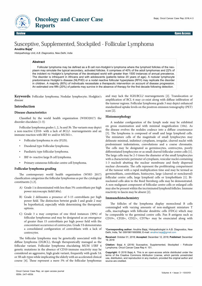
Figure 7: Follicular lymphoma with nodular centrocytes and centoblasts.
Figure 8: Follicular lymphoma with proteinaceous deposit.
Figure 9: Follicular lymphoma with consistent uniform nodules.
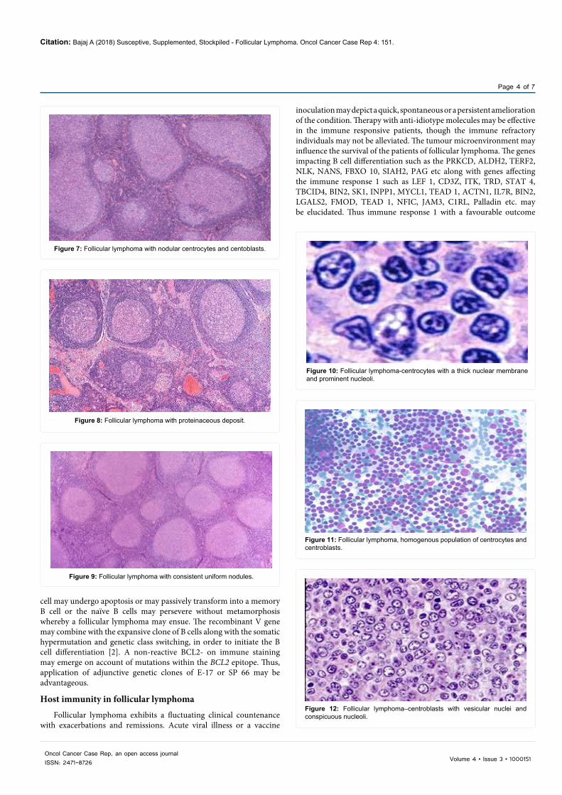
Figure 10: Follicular lymphoma-centrocytes with a thick nuclear membrane and prominent nucleoli.
Figure 11: Follicular lymphoma, homogenous population of centrocytes and centroblasts.
Figure 12: Follicular lymphoma–centroblasts with vesicular nuclei and conspicuous nucleoli.
cell may undergo apoptosis or may passively transform into a memory B cell or the naïve B cells may persevere without metamorphosis whereby a follicular lymphoma may ensue. The recombinant V gene may combine with the expansive clone of B cells along with the somatic hypermutation and genetic class switching, in order to initiate the B cell differentiation [2]. A non-reactive BCL2- on immune staining may emerge on account of mutations within the BCL2 epitope. Thus, application of adjunctive genetic clones of E-17 or SP 66 may be advantageous.
Host immunity in follicular lymphoma
Follicular lymphoma exhibits a fluctuating clinical countenance with exacerbations and remissions. Acute viral illness or a vaccine
inoculation may depict a quick, spontaneous or a persistent amelioration of the condition. Therapy with anti-idiotype molecules may be effective in the immune responsive patients, though the immune refractory individuals may not be alleviated. The tumour microenvironment may influence the survival of the patients of follicular lymphoma. The genes impacting B cell differentiation such as the PRKCD, ALDH2, TERF2, NLK, NANS, FBXO 10, SIAH2, PAG etc along with genes affecting the immune response 1 such as LEF 1, CD3Z, ITK, TRD, STAT 4, TBCID4, BIN2, SK1, INPP1, MYCL1, TEAD 1, ACTN1, IL7R, BIN2, LGALS2, FMOD, TEAD 1, NFIC, JAM3, C1RL, Palladin etc. may be elucidated. Thus immune response 1 with a favourable outcome
Page 5 of 7
Volume 4 • Issue 3 • 1000151Oncol Cancer Case Rep, an open access journalISSN: 2471-8726
Citation: Bajaj A (2018) Susceptive, Supplemented, Stockpiled - Follicular Lymphoma. Oncol Cancer Case Rep 4: 151.
Figure 13: Follicular lymphoma-centroblasts with fine well dispersed chromatin in peripheral blood.
Figure 14: Follicular lymphoma in contrast to a reactive follicular hyperplasia.
may delineate abundant genetic manifestation within the peripheral blood T lymphocytes [2]. Genetic expression associated with immune response 2 may exemplify LGMN, TLR5, C1QA, SEPT10, ME1, MITF, NDN, C4A, C4B, C1QB etc. and may further be accompanied with the specified genetic enunciation within the peripheral blood monocytes and a worse prognosis. However, the applicability of immune response signatures and relevant biomarkers may not be beneficial in routine clinical practice [2]. Follicular lymphoma may exhibit a prognosis arising of a multi-factorial aetiology. Analytical compilations of the risk proliferation index, the tumour grade, the disease stage, the FLIP1 index, the host immune reaction or the microenvironment and the mutational profile (m 7-FLIP) may be mandated [2].
Histological evolution of follicular lymphoma
Accessory modification of the follicular lymphoma may ensure a variegated tumour histology and phenotype, accompanied with the BCL2 mutation and a clone specific affiliation to follicular lymphoma.
• The most frequent emergence may be of a diffuse lymphoma(DLBCL)
• A high grade, double hit B cell (HGBCL-DH) lymphomawith MYC, BCL2 and/or BCL6 genetic mutations may bedemonstrated [2].
• Pre-B lymphoblastic lymphoma/leukaemia may be exemplified
• A classical Hodgkin’s Lymphoma may appear.
• Histiocytic and dendritic cell sarcoma may arise with aprogressive follicular lymphoma.
• TP53 mediated metamorphosis of the follicular lymphomamay depict a conversion to a diffuse (DLBCL) lymphoma withelucidation of BCL2+ and MIB 1+ reactivity [2].
Follicular lymphoma may lack the MYC – rearrangements whereas the diffuse (DLBCL) lymphomas may express the MYC+ modifications. The diffuse (DLBCL) lymphomas may exclusively react to the MYC+ protein and may elucidate a c MYC recombination. Follicular lymphoma and the diffuse (DLBCL) lymphomas may display a BCL2 rearrangement. The co-existing BCL2 and MYC rearrangements may signify a high grade, double hit (HGBL-DH) lymphoma [2].
The follicular lymphomas may signify a lymphoblastic metamorphosis with the elucidation of a precursor B cell phenotype which may immune react to TdT+. The bone marrow and the peripheral blood may be implicated with a clinical exponent resembling an acute lymphoblastic leukaemia (ALL). A clone specific analogy to follicular lymphoma may be analyzed with translocation t (14:18). The variant may exemplify a c MYC rearrangement, though it may not be a component of the “double hit” lymphomas enlisted with the world health organization (WHO- DH). Low grade follicular lymphomas may perpetuate locally within the bone marrow and lymph nodes. The variant may elucidate a µ micro-protein + and a non-reactive or a weak BCL2 – [2].
Follicular lymphoma - in situ (FLIS)
As defined by the world health organization (WHO, 2017), an in situ follicular lymphoma may be a focal and circumscribed tumour confined to the follicles with an immune reaction to BCL2+. The B cell follicular lymphoma may congregate within and respond to the germinal centre microenvironment. The variant may be determined within 2-3% of the lymph node biopsies and may occasionally coincide with accessory B cells. The genetic anomalies apart from the BCL2 rearrangement may be minimal. The probability of developing a follicular lymphoma may be less than 5%. Therapeutic management may not be required [2].
Duodenal type of follicular lymphoma
The variant manifests an immune phenotype and genetic modifications identical to the follicular lymphoma of the lymph nodes such as the BCL2 and IGH recombination, although immune reactivity to IgA + may be manifest. Apart from the aforementioned genetic anomalies, adjuvant aberrations may be minimal. The disorder frequently appears in the duodenum along with accessory locations such as the distal small bowel. Superficial polypoid mucosal lesions may erupt. The intestinal lymphocytes may depict homing receptors such as the ᾳ4β7 intgerin. Activation induced cytidine deaminase (AID) function may be lacking. The disorder of a restricted emergence may depict amelioration and localized reoccurrences with an absence of tumour dissemination. The lymphoma may be immune reactive for BCL2+ and CD10+ with an absence of CD3- [2].
Paediatric follicular lymphoma
The variant is exceptional (1-2%) and arises in children. Organs such as tonsils, nasopharynx, gastrointestinal tract, testis or lymph nodes may be implicated. The lymphoma may characteristically be high grade (3A/3B). Males are frequently involved (M>>F: 10:1), with a predominant male: female ratio. A majority (85%) of the instances may be confined with stage I and stage II disease at detection. The nodal and extra-nodal configurations of the lymphoma may not be appropriately elucidated [2]. The lymphoma categorized within the head and neck
Page 6 of 7
Volume 4 • Issue 3 • 1000151Oncol Cancer Case Rep, an open access journalISSN: 2471-8726
Citation: Bajaj A (2018) Susceptive, Supplemented, Stockpiled - Follicular Lymphoma. Oncol Cancer Case Rep 4: 151.
lymph nodes may belong to stage I. Rearrangements of BCL 2 and/or BCL6 may be absent. The lymphoma typically exemplifies reactivity for CD10+, BCL6+ with an absent immune reaction for BCL2- and MUM1-(2). Organs such as the tonsil or Waldeyer’s ring when involved, may manifest an analogous male: female (M: F) ratio. A concomitant elucidation of MUM 1, BCL 6 andCD10 may be encountered. A cleavage at IRF4 (6p25) may be frequent [2].
i) The IRF 4+ large B cell lymphoma may be categorized asa conditional entity with the contemporary WHO classification. Paediatric testicular, follicular lymphomas may exemplify a stage 1 disease with favourable prognosis and may be reactive to CD10+ and BCL 6+ with a lack of BCL2- and MUM- reaction. Infrequent cleavage within the BCL6 oncogene may be elucidated. Paediatric follicular lymphomas may display a clone specific germinal centre B cell multiplication of indeterminate malignant capability. The mean age of presentation varies from 15-18 years with infrequent lesions beyond 40 years. Clonally rearranged CD10+ B cells may be delineated with flow cytometry. Immunoglobulin recombination as detected by the polymerase chain reaction (IG PCR +) may be demonstrated. Genetic aberrations may be lacking in the BCL2, BCL6 and/or IRF 4 molecules (2). A consistent complete remission (CR) may be achieved following adequate surgical excision without extended chemo/mono therapy. Thus a conservative therapeutic option may be recommended. Immunoglobulin D (Ig D) may be elucidated at the follicular perimeter along with CD79a+ and CD10+ immune phenotypic cells.
ii) Paediatric type - Nodal follicular lymphoma may elucidatereoccurring mutations within the entire genome. Mutations within TNFRSF14 with concurring copy number neutral loss of 1p 36 may be exhibited in greater than half (>50%) the instances. KMT2D (MLL2) mutation may appear in roughly 16% individuals. These genes may also be modified with the “classic” follicular lymphoma, although with dissimilar percentages. Mutations within the MAPK pathway may be common with MAP2K 1 in half (≈50%) the instances or infrequently an RRAS: MAPK 1 mutation may appear [2].
Discussion
Therapeutic protocolsGrade 3 follicular lymphoma may manifest dubitable treatment
strategies in concordance with adjunctive follicular or diffuse large B cell lymphomas. Persistent amelioration of diffuse lymphomas (DLBCL) with varying treatment outcomes may also be analyzed. However, the ensuing follicular lymphoma international prognostic index 2 (FLIPI 2) following the employment of rituximab may not be incorporatedwith the lymphoma grade amidst the various, predominant prognosticfactors affecting survival [14]. The optimal employment of rituximabwith cyclophosphamide, doxorubicin, vincristine and prednisone (RCHOP) for grade 3 follicular lymphomas may express an objectiveresponse rate (ORR) of 100% with a 3-year progression free survival(PFS) survival benefit of 70%. The minimal requirement of a reducedprogression free survival (PFS) survival may manifest as greater than4 lymph node sites with co-existing B symptoms (fever, night sweats,weight loss >10% of the body weight) [5]. Evolution to a diffuselymphoma (DLBCL) may exemplify an increase in the internationalprognostic index (IPI) score with elevated lactic dehydrogenase (LDH). Acute Myeloid Leukaemia may appear in patients beyond 60 years atthe commencement of therapy. Rituximab with cyclophosphamide,doxorubicin, vincristine and prednisone (R CHOP) may be efficaciousin grade 3 follicular lymphoma with an enhanced progression freesurvival (PFS) and a prognosis concordant with the limited stage ofdiffuse lymphoma (DLBCL). The exclusion of a frank conversion to adiffuse lymphoma (DLBCL may be a pre-requisite prior to the initiation of treatment. The geriatric patients may necessitate the employmentof adjunctive therapeutic options. The optimal implementation forrituximab with cyclophosphamide, doxorubicin, vincristine andprednisone (R-CHOP) in grade 3 follicular lymphoma may requireconcordant favourable aspects as such [15,16] (Table 1).
• Age beyond 60 years.
• Male sex.
• Eastern Cooperative Oncology Group Performance Status(ECOG PS) greater than 1.
• Stage 3 or 4 with Ann Arbor classification.
• More than four incriminated lymph node sites.
• Bulky disease beyond 6-centimetre magnitude
• More than a solitary, implicated extra-nodal site.
Complete effacement of architecture Preservation of nodal architectureEven distribution of follicles throughout cortex and medulla Follicles more prominent in cortical portion of lymph node
Slight or moderate variation in size and shape of follicles Marked variation in size and shape of follicles with presence of elongated, angulated and Dumbbell shaped forms
Fading of follicles Sharply demarcated reaction centres
Massive infiltration of capsule and peri-capsular fat with or without formation of malignant follicles outside capsule
Nil or moderate infiltration of the capsule or peri-capsular fat with inflammatory cells that may be arranged as focal or peri-vascular aggregates(when accompanied
with lymphadenitis)Condensation of reticulin fibres at the periphery of the follicles Little or no alteration of the reticular framework
CytologyFollicles composed of malignant cells exhibiting cellular pleomorphism with nuclear
aberrations.Centres of follicles (reaction centres) composed of lymphoid cells, histiocytes and
reticulum cells, with few or no cellular/nuclear aberrations
Lack of phagocytosis Active phagocytosis in reactivegerminal centres
Relative paucity of mitotic figures usually without significant differences inside and outside the follicles, presence of atypical mitosis
Moderate to pronounced mitotic activity in reaction centres, rare or no mitosis outside reaction centres, no atypical mitosis
Analogy of cell type inside and outside the follicles Infiltration of tissue between the reaction centres with inflammatory cells (accompanied with lymphadenitis)
Table 1: Allegory of reactive/lymphoid neoplasm follicles [1].
Page 7 of 7
Volume 4 • Issue 3 • 1000151Oncol Cancer Case Rep, an open access journalISSN: 2471-8726
Citation: Bajaj A (2018) Susceptive, Supplemented, Stockpiled - Follicular Lymphoma. Oncol Cancer Case Rep 4: 151.
• Bone marrow involvement.
• The appearance of B symptoms.
• International Prognostic Index (IPI) of the range 3 or 4.
• Follicular Lymphoma International Prognostic Index (FLIPI)of the magnitude 3 to 5.
ConclusionRadiotherapy may be employed as an accessory treatment modality
particularly within the lymph nodes and the incriminated drainage areas or with the implicated organs with specified lymph node drainage. A computerized tomography (CT/PET) or a positron emission tomography in conjunction with the enunciated Ki 67 subsequent to the initiation of chemotherapy, may be evaluated at mid therapy [12]. Therapeutic outcomes may be analysed by the physical examination, complete blood counts, serum lactic dehydrogenase (LDH) values and a computerized tomography (CT) and/or a positron emission tomography (PET CT) imaging. A complete response (CR) may be investigated by performing a bone marrow aspiration and biopsy. The therapeutic response may be categorized by the foundation laid by the international working group. A type II anti CD20 monoclonal antibody obinutuzumab may be employed to enhance the efficacy of rituximab. A non-apoptotic cell demise may ensue with a diminished complement activation. Amplified direct cell dissolution, antibody dependent cell mediated cytotoxicity along with a B cell elimination from whole blood and lymphoid tissues may ensue. Relapsed or refractory follicular lymphoma may appropriately be managed by obinutuzumab with cyclophosphamide, doxorubicin, vincristine and prednisone (G CHOP) elucidating an objective response rate (ORR) of 96% or with cyclophosphamide in combination with fludarabine and obinutuzumab with a manifested objective response rate (ORR) of 93%.
References
1. Ackerman’s R (1825) Surgical pathology (10th edn) p: 1825.
2. Jaffe ES (2017) Follicular lymphoma: Histological spectrum. National CancerInstitute, Bethesda, USA.
3. Swerdlow SH, Campo E, Pileri SA, Harris NL, Stein H, et al. (2016) Revision of the World Health Organization classification of lymphoid neoplasms. Blood 127: 2375-2390.
4. Teras LR, DeSantis EC, Cerhan JR, Morton LM, Flowers CR, et al. (2016) USlymphoid malignancy statistics by World Health Organization Subtypes. CA.Cancer J Clin 66: 443-459.
5. Bierman PJ (2007) Natural history of Follicular lymphomas grade 3 non-Hodgkin’s lymphoma” Curr Opin Oncol 19: 433-437.
6. Bosga-Bouwer AG, Den Berg AV, Haralambieva E, De Jong D, Boonstra R,et al. (2006) Molecular, cytogenetic and immune phenotypic characteristic ofFollicular Lymphoma Grade 3B; A separate entity or a part of the spectrum ofdiffuse large B cell lymphoma or follicular lymphoma? Hum Pathol 37: 528-533.
7. Horn H, Schmelter C, Leich E, Salaverria I, Katzenberger T, et al. (2011)Follicular lymphoma grade 3B is a distinct neoplasm according to cytogeneticand immune histo-chemical profile. Haematologica 96: 1327-1334.
8. Borovecki A, Korac P, Nola M, Dominis M (2008) Prognostic significance of B cell differentiation genes encoding proteins in diffuse large B cell lymphomaand follicular lymphoma grade 3. Croat Med J 49: 625-635.
9. Guo Y, Karube K, Kawano R, Ohshima K (2005) Low grade follicular lymphoma with t (14:18) presents a homogenous disease entity otherwise the restcomprises as minor groups of heterogeneous disease entities with BCL2amplification, BCL6 translocation or other gene aberrances. Leukaemia 19: 1058-1063.
10. Maeshima AM, Hirokazu T, Junko N, Ken-ichi M, Suguru F, et al. (2001)Prognostic implication of histological grade and intensity of BCL2 expression in follicular lymphoma undergoing rituximab containing therapy. Hum Pathol 44: 2529 -2535.
11. Pandolfi A, Barreyro L, Steid U (2013) Concise review pre leukaemia stem cells molecular biology and clinical implications of the precursor to leukaemia stemcells. Stem Cell Transl Med 2: 143-150.
12. Wohrer S, Jaeger U, Kletter K, Becherer A, Hauswirth A, et al. (2006) 18fluoro deoxy glucose positron emission tomography (18 FDG-PET) visualizes follicular lymphoma irrespective of grading. Ann Oncol 17: 780-784.
13. Lunning M (2007) The curability of follicular lymphoma. Transfus Apher Sci 37: 31-35.
14. Federico M, Bellei M, Marcheselli L, Luminari S, Lopez-Guillermo A, et al.(2009) Follicular Lymphoma International Prognostic Index 2: A new prognostic index for Follicular Lymphoma Prognostic Factor Project. J Clin Oncol 27: 4555-4562.
15. Strati P, Fowler N, Pina-Oviedo S, Medeiros LJ, Overman MJ, et al. (2018) Long term remission of patients with follicular lymphoma grade 3 treated with R CHOP. Clin Lymphoma Myeloma Leuk 18: 103-108.
16. Salamoon M (2017) New trends for the treatment of follicular lymphoma inrelapse: The role of type II anti CD 20. Sci J Blood Disorders 1: 1-3.