*Correspondence: M.C. Valadares. Faculdade de Farmácia, Universidade Federal de Goiás. Praça Universitária esquina com a primeira avenida s/n, setor Universitário - 74605-220 - Goiânia, GO, Brasil. Phone / fax: +55 62 3209-6044 ext. 22. E-mail: [email protected]Article Brazilian Journal of Pharmaceutical Sciences vol. 48, n. 3, jul./sep., 2012 Synadenium umbellatum Pax. promotes cell cycle arrest and induces apoptosis in K-562 leukemia cells Mariana Flavia da Mota, Polyana Lopes Benfica, Marize Campos Valadares * Cellular Toxicology and Pharmacology Laboratory, Faculty of Pharmacy, Federal University of Goiás Chronic myeloid leukemia (CML) is a clonal myeloproliferative disease that shows apoptosis resistance. The introduction of imatinib mesylate has revolutionized the treatment of CML, but imatinib resistance may develop at any time and inevitably leads to disease progression. Synadenium umbellatum Pax. belongs to the Euphorbiaceae family and is popularly used in Brazil for the treatment of cancer. The cytotoxicity of Euphorbiaceae is associated with the ability of these plants and their bioactive compounds to induce apoptotic tumor cell death. Therefore, we aimed to investigate the cytotoxicity and the mechanisms of death induced by S. umbellatum extract in leukemic cells. S. umbellatum cytotoxicity was evaluated by trypan blue exclusion assay and flow cytometric analysis of the cell cycle; the mechanisms involved in K-562 cell death were investigated by light microscopy and flow cytometry. The results demonstrate that S. umbellatum is cytotoxic to leukemic cells in a concentration-dependent manner. Morphological analysis revealed that S. umbellatum treatment induced K-562 cell death by an apoptotic pathway. Furthermore, data indicate ROS overproduction, alterations in mitochondrial membrane potential, phosphatidylserine externalization and activation of caspase 9. Taken together, the results demonstrate that S. umbellatum extract arrested the cell cycle and triggered apoptosis at several levels in K-562 cells. Uniterms: Synadenium umbellatum/pharmacognosy. Synadenium umbellatum/cytotoxicity/evaluation. K-562 cells/apoptosis. K-562 cells/cycle arrest. A leucemia mielóide crônica (LMC) é uma doença mieloproliferativa clonal, que apresenta resistência à apoptose. A introdução do mesilato de imatinibe revolucionou o tratamento da LMC, porém a resistência ao imatinibe pode ser desenvolvida em qualquer tempo e, inevitavelmente, leva à progressão da doença. Synadenium umbellatum Pax. pertence à família Euphorbiaceae e é usado popularmente no Brasil para o tratamento do câncer. A citotoxicidade das Euphorbiaceae está associada com a capacidade dessas plantas e seus compostos bioativos em induzir apoptose em células tumorais. Portanto, este trabalho teve como objetivo investigar a citotoxicidade e os mecanismos de morte induzidos por S. umbellatum em células leucêmicas. A citotoxicidade de S. umbellatum foi avaliada pelo ensaio de exclusão do azul de tripano e a análise do ciclo celular foi feita por citometria de fluxo. Os mecanismos envolvidos na morte celular das células K-562 foram investigados por microscopia óptica e por citometria de fluxo. Os resultados demonstraram que S. umbellatum é citotóxico para células leucêmicas de uma maneira dependente da concentração. A análise morfológica revelou que o tratamento com S. umbellatum induziu as célula K-562 à morte por via apoptótica. Além disso, os dados indicam aumento de ERO S , alterações no potencial de membrana mitocondrial, externalização da fosfatidilserina e ativação de caspase 9. Em conjunto, os resultados demonstram que S. umbellatum promoveu retenção do ciclo celular das células K-562 e induziu estas células à morte por apoptose. Unitermos: Synadenium umbellatum/farmacognosia. Synadenium umbellatum/citotoxicidade/avaliação. Célula K-562/apoptose. Célula K-562/retenção do ciclo.
Transcript
*Correspondence: M.C. Valadares. Faculdade de Farmácia, Universidade Federal de Goiás. Praça Universitária esquina com a primeira avenida s/n, setor Universitário - 74605-220 - Goiânia, GO, Brasil. Phone / fax: +55 62 3209-6044 ext. 22. E-mail: [email protected]
Art
icleBrazilian Journal of
Pharmaceutical Sciencesvol. 48, n. 3, jul./sep., 2012
Synadenium umbellatum Pax. promotes cell cycle arrest and induces apoptosis in K-562 leukemia cells
Mariana Flavia da Mota, Polyana Lopes Benfica, Marize Campos Valadares*
Cellular Toxicology and Pharmacology Laboratory, Faculty of Pharmacy, Federal University of Goiás
Chronic myeloid leukemia (CML) is a clonal myeloproliferative disease that shows apoptosis resistance. The introduction of imatinib mesylate has revolutionized the treatment of CML, but imatinib resistance may develop at any time and inevitably leads to disease progression. Synadenium umbellatum Pax. belongs to the Euphorbiaceae family and is popularly used in Brazil for the treatment of cancer. The cytotoxicity of Euphorbiaceae is associated with the ability of these plants and their bioactive compounds to induce apoptotic tumor cell death. Therefore, we aimed to investigate the cytotoxicity and the mechanisms of death induced by S. umbellatum extract in leukemic cells. S. umbellatum cytotoxicity was evaluated by trypan blue exclusion assay and flow cytometric analysis of the cell cycle; the mechanisms involved in K-562 cell death were investigated by light microscopy and flow cytometry. The results demonstrate that S. umbellatum is cytotoxic to leukemic cells in a concentration-dependent manner. Morphological analysis revealed that S. umbellatum treatment induced K-562 cell death by an apoptotic pathway. Furthermore, data indicate ROS overproduction, alterations in mitochondrial membrane potential, phosphatidylserine externalization and activation of caspase 9. Taken together, the results demonstrate that S. umbellatum extract arrested the cell cycle and triggered apoptosis at several levels in K-562 cells.
A leucemia mielóide crônica (LMC) é uma doença mieloproliferativa clonal, que apresenta resistência à apoptose. A introdução do mesilato de imatinibe revolucionou o tratamento da LMC, porém a resistência ao imatinibe pode ser desenvolvida em qualquer tempo e, inevitavelmente, leva à progressão da doença. Synadenium umbellatum Pax. pertence à família Euphorbiaceae e é usado popularmente no Brasil para o tratamento do câncer. A citotoxicidade das Euphorbiaceae está associada com a capacidade dessas plantas e seus compostos bioativos em induzir apoptose em células tumorais. Portanto, este trabalho teve como objetivo investigar a citotoxicidade e os mecanismos de morte induzidos por S. umbellatum em células leucêmicas. A citotoxicidade de S. umbellatum foi avaliada pelo ensaio de exclusão do azul de tripano e a análise do ciclo celular foi feita por citometria de fluxo. Os mecanismos envolvidos na morte celular das células K-562 foram investigados por microscopia óptica e por citometria de fluxo. Os resultados demonstraram que S. umbellatum é citotóxico para células leucêmicas de uma maneira dependente da concentração. A análise morfológica revelou que o tratamento com S. umbellatum induziu as célula K-562 à morte por via apoptótica. Além disso, os dados indicam aumento de EROS, alterações no potencial de membrana mitocondrial, externalização da fosfatidilserina e ativação de caspase 9. Em conjunto, os resultados demonstram que S. umbellatum promoveu retenção do ciclo celular das células K-562 e induziu estas células à morte por apoptose.
Chronic myeloid leukemia (CML) is a clonal my-eloproliferative disease driven by the BCR-ABL fusion protein that arises from the translocation of chromosomes 9 and 22. This oncoprotein exhibits constitutively active kinase activity that confers growth potential and apopto-sis resistance to CML cells (Cortez, Kadlec, Pendergast, 1995). BCR-ABL promotes cell division and blocks apoptosis, leading to unregulated growth of hematopoietic stem cells. Usually, patients with no chemotherapeutic treatment progress after several years from a chronic phase to a blast crisis, which can rapidly lead to death (O’Hare, Corbin, Druker, 2006).
The introduction of imatinib mesylate (Glivec/Gleevec; Novartis Pharmaceuticals), a tyrosine kinase inhibitor, has revolutionized the treatment of CML. Ima-tinib is widely accepted as the standard of care for the first-line treatment of CML, due to its well-documented clinical activity, resulting in durable responses and pro-longed survival. Moreover, toxicities associated with this therapy are considered low, and the main toxicities are myelosuppression, edema, and myalgia/arthralgia. On the other hand, imatinib resistance may develop at any time and inevitably leads to disease progression (Fausel, 2007). In this context, new agents are required for the treatment of CML.
Synadenium umbellatum Pax., a species of Euphor-biaceae widely found in South America, is popularly used in Brazil for the treatment of cancer. Studies performed by our group demonstrated scientific evidence for the ef-fectiveness of S. umbellatum extract as an antitumor and antiangiogenic agent in vivo using the experimental model of Ehrlich ascitic tumor (EAT) (Nogueira et al., 2008). Re-cently, we also demonstrated the mechanisms of EAT cell death induced by S. umbellatum, in vitro. The cytotoxic effects found in EAT cells were mediated by activation of the intrinsic and extrinsic pathways of apoptosis. Further-more, we determined by HPLC that the main constituents of S. umbellatum crude ethanolic extract are flavonoids and terpenes (Mota et al., 2012).
It has been reported in the literature that Euphor-biaceae species are cytotoxic against different tumor cell lines, especially K-562 leukemia cells. In most cases, this activity is associated with the ability of these plants to in-duce cell death, mainly by apoptotic mechanisms (Abreu et al., 2006; Amirghofran, et al., 2006; Block et al., 2005; Fatope et al., 1996; Grymberg et al., 1999; Itokawa et al., 1989; Kupchan et al., 1976; Kielland et al., 1996; Maciel et al., 2000; Morales et al., 2005; Ngamkitidechakul et al., 2010; Uthaisang et al., 2004).
In the present work, we investigated the cytotoxic activity of S. umbellatum in K-562 and Jurkat leukemia cell lines. Since K-562 is a BCR-ABL-expressing hu-man leukemia cell line, with a drug resistance profile, the mechanisms of death induced by S. umbellatum were investigated in this cell model.
MATERIALS AND METHODS
Reagents
RPMI 1640 medium, fetal bovine serum, strep-tomycin, penicillin G, propidium iodide, Giemsa dye, RNase, DCFH-DA, EDTA, rhodamine 123, and BSA were purchased from Sigma-Aldrich (St. Louis, MO, USA). Ethanol and trypan blue were obtained from Vetec (Rio de Janeiro, RJ, Brazil). Annexin V-FITC, binding buffer (TACS Annexin V-FITC apoptosis detection kit), caspase substrate 9, DTT, and lysis buffer (protease colorimetric assay kit) were acquired from R&D Systems (Minneapo-lis, MN, USA). All reagents used were of analytical grade.
Synadenium umbellatum Pax
S. umbellatum (17 kg) aerial parts were collected in September 2005 in Goiânia, GO, Brazil (787 m altitude; 16º 40’ 15.5’’ S; 49º 14’ 13.6’’ W). Voucher specimens (No. UFG-27160) were deposited in the Herbarium of the Universidade Federal de Goiás (Instituto de Ciências Biológicas, Goiânia, GO, Brazil). Air-dried ground S. umbellatum material (1.3 kg) was exhaustively macerated in 95% ethanol (Vetec, RJ, Brazil), and after filtration, the extract was dried in a rotary evaporator (MA 120, Marconi, SP, Brazil) to dryness under low pressure and temperature to obtain 90 g of crude ethanolic extract (6.9% yield).
Cell culture
K-562 and Jurkat cell lines were obtained from the American type culture collection (ATCC, Rockville, MD, USA) and maintained in RPMI 1640 medium containing 10% heat-inactivated fetal bovine serum (FBS), 100 mg/mL streptomycin, 100 U/mL penicillin G, and 2 mM L-gluta-mine in a humidified atmosphere of 5% CO2 in air at 37 oC. The cells were passaged twice a week and used in the exponential growth phase. Cell viability was determined by trypan blue exclusion.
Cytotoxicity assay
The viabilities of K-562 and Jurkat cells were evalu-
Synadenium umbellatum Pax. promotes cell cycle arrest and induces apoptosis in K-562 leukemia cells 499
ated using the trypan blue exclusion method. The cells (1x10⁶ cells/mL) were seeded in 96-well flat microtiter plates (Corning, USA) in RPMI 1640 medium supple-mented with 10% FBS and incubated with or without eight concentrations (0.007 – 1 mg/mL) of S. umbellatum extract in six replicates for 24 h. The cell suspension and a trypan blue solution (0.2% in phosphate buffered saline - PBS) were mixed 1:10 and the viability of cells was estimated using a hemacytometer (Boeco, Germany). Stained cells were scored as dead. IC50 values (concentration that inhib-ited cell growth by 50%, compared to untreated controls) were determined. For all subsequent assays, a value near the IC50 was used.
Light microscopy
The morphological alterations between K-562 cells untreated and treated with S. umbellatum were assessed by light microscopy. Untreated cells and cells treated with 0.06 mg/mL S. umbellatum extract were incubated for 24 h at 37 oC and 5% CO2 and, after incubation, placed on glass slides by cytospin and stained with Giemsa. After stain-ing, the slides were examined by light microscopy (DM 2000, Leica microsystems, USA) and images were taken using a 40 x objective amplified with 3 x zoom (Canon Powershot S80).
Cell cycle analysis
K-562 cells (1x10⁶/mL) were incubated with or without 0.06 mg/mL S. umbellatum extract for 24 h. After incubation, the cells were washed with ice-cold PBS-EDTA, centrifuged at 296.82 g for 10 min and fixed with 2 mL 70% ice-cold ethanol at 4 oC for 24 h. After fixation, cells were washed again, resuspended in 500 µL PBS solution containing 0.2 mg/mL RNAse and 0.05 mg/mL propidium iodide (PI), and then incubated at 4 oC for 2 h. Fluorescence was measured with a FACSCanto II flow cytometer, using PE x FL2 channels. The assay was car-ried out in triplicate and 10,000 events were analyzed per experiment using the BDFACSDiva software.
Measurement of intracellular ROS
K-562 cells (1x10⁶ /mL) were incubated with or without 0.06 mg/mL S. umbellatum extract for 12 and 24 h. Afterwards, cells were washed with PBS, centrifuged at 296.82 g for 10 min and incubated with 500 µL DCFH-DA (10 µM) for 1 h, and fluorescence determined by flow cytometry using the FL1 channel.
Assessment of mitochondrial membrane potential (Δψm)
K-562 Cells (1x10⁶/mL), untreated and treated with 0.06 mg/mL S. umbellatum extract for 24 h, were washed with PBS-EDTA, centrifuged at 296.82 g for 10 min, and incubated with 1 µg/mL rhodamine 123 at 37 oC for 1 h, and the fluorescence determined by flow cytometry, using the FL1 channel.
Annexin V-FITC/PI double-staining for phosphatidylserine externalization analysis
K-562 Cells (1x10⁶/mL), untreated and treated with 0.06 mg/mL S. umbellatum extract for 24 h, were washed with PBS-BSA and centrifuged at 296.82 g for 10 min. The cells were then incubated for 15 min with 100 µL binding buffer (10 mmol/L HEPES/NaOH, 140 mmol/L, NaCl 2.5 mmol/L CaCl2) with 1 µL annexin V-FITC and 10 µL PI solution, and analyzed by flow cytometry. Fluorescence of annexin V-FITC and PI was measured using the FL1 and FL2 channels, respectively.
Caspase activity
The activity of caspase 9 was measured using a colorimetric protease assay kit, following the manu-facturer’s instructions. After 24 h of incubation with or without 0.015 mg/mL S. umbellatum extract, K-562 cells (1x10⁶/mL) were washed with ice-cold PBS, centrifuged at 296.82 g for 10 min and incubated on ice for 20 min with 25 µL of cell-lysis buffer (provided in the kit). The cell lysate was collected by centrifugation at 29,682.9 g at 4 oC for 15 min. The assay was performed in 96 well plates containing 50 µL cell lysate, 50 µL 2x reaction buf-fer, 5 µL caspase colorimetric substrate and 10 µL DTT. Duplicate wells were prepared containing blank (no cell extract), negative control (extract from untreated cells), positive controls (no cell lysate and no substrate) and S. umbellatum-treated cells. The plates were incubated at 37 oC for 2 h, and absorbance was read with a microplate reader (Stat Fax 2100, Awareness Technology, Germany) at 405 nm. Caspase 9 activity was determined by measur-ing p-nitroanaline (pNA) released during the cleavage of LEHD-pNA substrate.
Statistical analysis
Statistical analysis was performed using Graph Pad Prism 5 (version 5.00 for Windows 98, Graph Pad Soft-ware, San Diego, CA) software. The evaluation of cytotox-
M. F. Mota, P. L. Benfica, M. C. Valadares500
icity was carried out in three separate experiments. Results were converted to percentages of the control and IC50 values were obtained by non-linear regression analysis. Results are expressed as the mean ± SD of six replicates. All other assays were analysed by Student’s t-test. Results were considered statistically significant when P < 0.05.
RESULTS
Cytotoxic effects of S. umbellatum in leukemic cells
S. umbellatum cytotoxicity was evaluated in the leukemic cell lines, K-562 and Jurkat, by the trypan blue exclusion method, after 24 h of exposure to dif-ferent concentrations (0.007 - 1 mg/mL). As shown in Figure 1, both cell lines studied were highly sensitive to S. umbellatum extract, in a concentration-dependent manner. Differences in the susceptibility of the two leu-kemic cell lines to S. umbellatum were observed in the IC50 values (IC50 Jurkat = 0.041 mg/mL and IC50 K-562 = 0.073 mg/mL). Additionally, the effects of S. umbellatum on normal lymphocytes from primary culture were stud-ied; these experiments revealed a high cytotoxic activity (data not shown). Based on these findings, the K-562 cell line was chosen for subsequent mechanistic studies.
K-562 cell morphology after S. umbellatum treatment
Morphological analysis of K-562 cells stained with Giemsa revealed alterations after treatment with S. umbel-latum extract. As can be seen in Figure 2A, control cells exhibited nuclei with dispersed chromatin and organized
plasma membrane. In contrast, cells treated with S. umbel-latum for 24 h (Figure 2B) showed membrane blebbing and chromatin condensation. These microscopic changes, caused by S. umbellatum treatment, suggested an activa-tion of apoptosis.
Effects of S. umbellatum in cell cycle distribution
Flow cytometric analysis revealed changes in cell cycle progression between untreated and treated K-562 cells (Figures 3A, B). S. umbellatum extract (0.06 mg/mL) increased the number of cells in G0/G1 phases by 19.7 ± 7.9% (P <0.05), followed by a reduction of 74.0 ± 2.4% in the S phase (P <0.001) and an increase of 51.2 ± 5.7%
FIGURE 1 - Cytotoxic effects of Synadenium umbellatum extract on K-562 and Jurkat cells after 24 h of treatment (0.007–1 mg/mL). The cytotoxic activity was determined by trypan blue exclusion assay. Experiments were performed in sextuplicate. Each bar presents the mean ± SD of three independent experiments. The IC50 was 0.041 mg/mL and 0.073 mg/mL in Jurkat and K-562 cells, respectively.
FIGURE 2 - Morphological changes of Synadenium umbellatum-induced apoptosis in K-562 cells. Cells were treated with 0.06 mg/mL Synadenium umbellatum extract for 24 h and images were taken using a 40 x objective amplified with 3 x zoom. (A) Control cells stained with Giemsa, showing nuclei with dispersed chromatin and organized plasma membrane (arrow). (B) Synadenium umbellatum-treated cells displaying membrane blebbing and chromatin condensation (arrows). Bars: 20 µm.
Synadenium umbellatum Pax. promotes cell cycle arrest and induces apoptosis in K-562 leukemia cells 501
in G2 (P <0.01). These results suggest that S. umbellatum arrested K-562 cells in the G phases.
S. umbellatum-induced oxidative stress
In addition to cell cycle arrest and morphological changes induced by S. umbellatum treatment, a time-dependent increase in intracellular ROS was found after 12 and 24 h of incubation with 0.06 mg/mL S. umbel-latum extract. As shown in Figure 4, the generation of ROS dramatically increased with incubation time. Treat-ment with S. umbellatum for 12 h increased intracellular ROS levels by 36.2 ± 13.7%, reaching 111.4% at 24 h (P <0.05).
S. umbellatum-induced alterations in mitochondrial membrane potential
The most common alteration in Δψm after oxidative stress is cellular depolarization, but an early hyperpolariza-tion can occur. As shown in Figure 5, treatment with 0.06 mg/mL S. umbellatum extract for 24 h increased Δψm in K-562 cells by 117.6 ± 1.9%.
FIGURE 3 - Flow cytometry analysis of K-562 cell cycle with and without treatment with Synadenium umbellatum extract (0.06 mg/mL) for 24 h. (A) Histograms showing cells untreated and treated with 0.06 mg/mL S. umbellatum for 24 h. (B) Percentage of K-562 cells in G1, S and G2 phases with and without S. umbellatum treatment. Each bar represents the mean ± SD of two independent experiments (*P < 0.05; **P < 0.01;***P < 0.001).
Phosphatidylserine exposure, induced by S. umbellatum
To differentiate cells undergoing apoptosis by PS
F IGURE 4 - Effects of S. umbel latum t reatment of K-562 cells on ROS generation. Cells were treated with 0.06 mg/mL S. umbellatum extract for 12 and 24 h, incubated with 10 µM DCFH-DA for 1 h and analyzed by flow cytometry (* P < 0.05 compared to untreated).
M. F. Mota, P. L. Benfica, M. C. Valadares502
exposure or necrosis by rupture of plasma membrane in S. umbellatum-mediated cell death, K-562 cells were labeled with annexin V-FITC (A) and PI. As shown in Figure 6,
after 24 h of treatment with 0.06 mg/mL S. umbellatum extract, the number of live cells (A-/PI-) decreased by 16.0 ± 0.8% (P < 0.01), while the number of apoptotic cells (A+/PI+) increased by 286.1 ± 30.8% (P <0.001).
Activation of caspase 9 by S. umbellatum
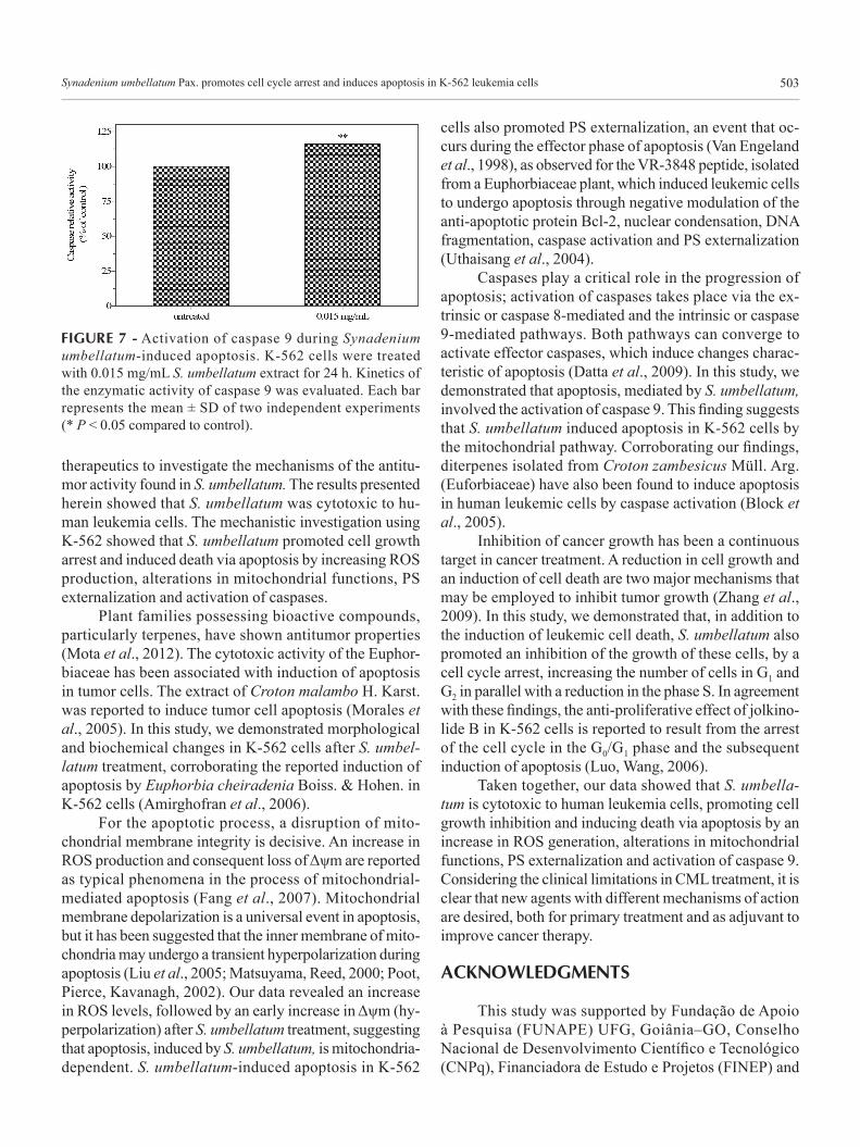
To investigate whether caspases were activated dur-ing S. umbellatum-induced cell death, a colorimetric assay was performed. As shown in Figure 7, after 24 h of expo-sure to 0.015 mg/mL S. umbellatum, there was a significant increase in caspase 9 activity (15.9± 0.4%), as detected by cleavage of LEHD-pNA substrate. Thus, induction of caspase activity may be involved in S. umbellatum-induced apoptosis.
DISCUSSION AND CONCLUSION
In the present study, a BCR-ABL-expressing human leukemic cell line, K562, was used as a model system of resistance to apoptosis induced by conventional chemo-
FIGURE 5 - Effect of Synadenium umbellatum on mitochondrial membrane potential (Δψm) of K-562 cells. The graph shows rhodamine 123 intensity of untreated cells and cells treated with 0.06 mg/mL S. umbellatum extract for 24 h (**P < 0.01).
FIGURE 6 - Flow cytometry analysis of phosphatidylserine externalization in Synadenium umbellatum-treated K-562 cells. Cells were labeled with annexin V-FITC and PI. (A) Untreated cells and cells treated with 0.06 mg/mL S. umbellatum for 24 h. Lower left quadrants (A-/PI-) = % of viable cells. Lower right quadrants (A+/PI-) = % of early apoptotic cells. Upper left quadrants (A+/PI+) = % of late apoptotic cells. Upper right quadrants (A-/PI+) = % of necrotic cells. (B) Comparison of fluorescence intensity of annexin V-FITC and PI between untreated and S. umbellatum-treated cells. The bar represents the mean ± SD of two independent experiments (**P < 0.01;***P < 0.001 compared to untreated).
Synadenium umbellatum Pax. promotes cell cycle arrest and induces apoptosis in K-562 leukemia cells 503
FIGURE 7 - Activation of caspase 9 during Synadenium umbellatum-induced apoptosis. K-562 cells were treated with 0.015 mg/mL S. umbellatum extract for 24 h. Kinetics of the enzymatic activity of caspase 9 was evaluated. Each bar represents the mean ± SD of two independent experiments (* P < 0.05 compared to control).
therapeutics to investigate the mechanisms of the antitu-mor activity found in S. umbellatum. The results presented herein showed that S. umbellatum was cytotoxic to hu-man leukemia cells. The mechanistic investigation using K-562 showed that S. umbellatum promoted cell growth arrest and induced death via apoptosis by increasing ROS production, alterations in mitochondrial functions, PS externalization and activation of caspases.
Plant families possessing bioactive compounds, particularly terpenes, have shown antitumor properties (Mota et al., 2012). The cytotoxic activity of the Euphor-biaceae has been associated with induction of apoptosis in tumor cells. The extract of Croton malambo H. Karst. was reported to induce tumor cell apoptosis (Morales et al., 2005). In this study, we demonstrated morphological and biochemical changes in K-562 cells after S. umbel-latum treatment, corroborating the reported induction of apoptosis by Euphorbia cheiradenia Boiss. & Hohen. in K-562 cells (Amirghofran et al., 2006).
For the apoptotic process, a disruption of mito-chondrial membrane integrity is decisive. An increase in ROS production and consequent loss of Δψm are reported as typical phenomena in the process of mitochondrial-mediated apoptosis (Fang et al., 2007). Mitochondrial membrane depolarization is a universal event in apoptosis, but it has been suggested that the inner membrane of mito-chondria may undergo a transient hyperpolarization during apoptosis (Liu et al., 2005; Matsuyama, Reed, 2000; Poot, Pierce, Kavanagh, 2002). Our data revealed an increase in ROS levels, followed by an early increase in Δψm (hy-perpolarization) after S. umbellatum treatment, suggesting that apoptosis, induced by S. umbellatum, is mitochondria-dependent. S. umbellatum-induced apoptosis in K-562
cells also promoted PS externalization, an event that oc-curs during the effector phase of apoptosis (Van Engeland et al., 1998), as observed for the VR-3848 peptide, isolated from a Euphorbiaceae plant, which induced leukemic cells to undergo apoptosis through negative modulation of the anti-apoptotic protein Bcl-2, nuclear condensation, DNA fragmentation, caspase activation and PS externalization (Uthaisang et al., 2004).
Caspases play a critical role in the progression of apoptosis; activation of caspases takes place via the ex-trinsic or caspase 8-mediated and the intrinsic or caspase 9-mediated pathways. Both pathways can converge to activate effector caspases, which induce changes charac-teristic of apoptosis (Datta et al., 2009). In this study, we demonstrated that apoptosis, mediated by S. umbellatum, involved the activation of caspase 9. This finding suggests that S. umbellatum induced apoptosis in K-562 cells by the mitochondrial pathway. Corroborating our findings, diterpenes isolated from Croton zambesicus Müll. Arg. (Euforbiaceae) have also been found to induce apoptosis in human leukemic cells by caspase activation (Block et al., 2005).
Inhibition of cancer growth has been a continuous target in cancer treatment. A reduction in cell growth and an induction of cell death are two major mechanisms that may be employed to inhibit tumor growth (Zhang et al., 2009). In this study, we demonstrated that, in addition to the induction of leukemic cell death, S. umbellatum also promoted an inhibition of the growth of these cells, by a cell cycle arrest, increasing the number of cells in G1 and G2 in parallel with a reduction in the phase S. In agreement with these findings, the anti-proliferative effect of jolkino-lide B in K-562 cells is reported to result from the arrest of the cell cycle in the G0/G1 phase and the subsequent induction of apoptosis (Luo, Wang, 2006).
Taken together, our data showed that S. umbella-tum is cytotoxic to human leukemia cells, promoting cell growth inhibition and inducing death via apoptosis by an increase in ROS generation, alterations in mitochondrial functions, PS externalization and activation of caspase 9. Considering the clinical limitations in CML treatment, it is clear that new agents with different mechanisms of action are desired, both for primary treatment and as adjuvant to improve cancer therapy.
ACKNOWLEDGMENTS
This study was supported by Fundação de Apoio à Pesquisa (FUNAPE) UFG, Goiânia–GO, Conselho Nacional de Desenvolvimento Científico e Tecnológico (CNPq), Financiadora de Estudo e Projetos (FINEP) and
M. F. Mota, P. L. Benfica, M. C. Valadares504
Coordenação de Aperfeiçoamento de Pessoal de Nível Superior (CAPES).
REFERENCES
ABREU, P.; MATTHEW, S.; GONZÁLEZ, T.; COSTA, D.; SEGUNDO, M.A.; FERNANDES, E. Anti-inflammatory and antioxidant activity of a medicinal tincture from Pedilanthus tithymaloides. Life Sci., v.78, p.1578-1585, 2006.
AMIRGHOFRAN, Z.; BAHMANI, M.; AZADMEHR, A.; JAVIDNIA, K. Induction of apoptosis in leukemia cell lines by Linum persicum and Euphorbia cheiradenia. J. Cancer Res. Clin. Oncol., v.132, p.427-432, 2006.
BLOCK, S.; GERKENS, P.; PEULEN, O.; JOLOIS, O.; MINGEOT-LECLERCQ, M.P.; DE PAUW-GILLET, M.C.; QUETIN-LECLERCQ, J. Induction of apoptosis in human promyelocytic leukemia cells by a natural trachylobane diterpene. Anticancer Res., v.25, p.363-368, 2005.
CORTEZ, D.; KADLEC, I.; PENDERGAST, A.M. Structural and signaling requirement for BCR-ABL-mediated transformation and inhibition of apoptosis. Mol. Cell Biol., v.15, p.5531-5541, 1995.
DATTA, S.; MAZUMDER, S.; GROSH, D.; DEY, S.; BHATTACHARYA, S. Low concentration of arsenic could induce caspase-3 mediated head kidney macrophage apoptosis with JNK-p38 activation in Clarias batrachus. Toxicol. Appl. Pharmacol., v.241, p.329-338, 2009.
FANG, L.; HE, Q.; HU, Y.; YANG, B. MZ3 induces apoptosis in human leukemia cells. Cancer Chemother. Pharmacol., v.59, p.397-405, 2007.
FATOPE, M.O.; ZENG, L.; OHAYAGA, J.E.; SHI, G.; McLAUGHLIN, J.L. Selectively cytotoxic diterpenes from Euphorbia poisonii. J. Med. Chem., v.39, p.1005-1008, 1996.
FAUSEL, C. Targeted chronic myeloid leukemia therapy: seeking a cure. J. Manag. Care Pharm., v.13, p.8-12, 2007.
GRYMBERG, N.F.; ECHEVARRIA, A.; LIMA, J.E.; PAMPLONA, S.S.R.; PINTO, A.C.; MACIEL, M.A.M. Anti-tumour activity of two 19-nor-clerodane diterpenes, trans-dehydrocrotonin and trans-crotonin, from Croton cajucara. Planta Med., v.65, p.687-689, 1999.
ITOKAWA, H.; ICHIHARA, Y.; WATANABE, K.; TAKEYA, K. An antitumor principle from Euphorbia lathyris. Planta Med., v.55, p.271-272, 1989.
KIELLAND, I.S.; DORNISH, J.M.; MALTERUD, K.E.; HVISTENDAHL, G.; ROMMING, C.; BOCKMAN, O.C.; KOLSAKER, P.; STENSTROM, Y.; NORDAL, A. Cytotoxic triterpenoids from the leaves of Euphorbia pulcherrima. Planta Med., v.62, p.322-325, 1996.
LIU, M.J.; WANG, Z.; JU, Y.; WONG, R.N.S.; WU, Q.Y. Diosgenin induces cell cycle arrest and apoptosis in human leukemia K562 cells with the disruption of Ca2+ homeostasis. Cancer Chemother. Pharmacol., v.55, p.79-90, 2005.
LUO, H.; WANG, A. Induction of apoptosis in K562 cells by jolkinolide B. Can. J. Physiol. Pharmacol., v.84, p.959-965, 2006.
MACIEL, M.A.M.; PINTO, A.C. ; ARRUDA, A.C. ; PAMPLONA, S.G.S.R.; VANDERLINDE, F.A.; LAPA, A.J.; ECHEVARRIA, A.; GRYNBERG, N.F.; CÔLUS, I.M.S.; FARIAS, R.A.F.; COSTA, A.M.L.; RAO, V.S.N. Ethnopharmacology, phytochemistry and pharmacology: a successful combination in the study of Croton cajucara. J. Ethnopharmacol., v.70, p.41-55, 2000.
MATSUYAMA, S.; REED, J.C. Mitochondria-dependent apoptosis and cellular pH regulation. Cell Death Differ., v.7, p.1155-1165, 2000.
M O R A L E S , A . ; P É R E Z , P. ; M E N D O Z A B , R . ; COMPAGNONEC, R.; SUAREZ, A.I.; ARVELOB, F.; RAMÍREZ, J.L.; GALINDO-CASTRO, I. Cytotoxic and proapoptotic activity of ent-16b-17a-dihydroxykaurane on human mammary carcinoma cell line MCF-7. Cancer Lett., v.218, p.109-116, 2005.
MOTA, M.F.; BENFICA, P.L.; BATISTA, A.C.; MARTINS, F.S.; PAULA, J.R.; VALADARES, M.C. Investigation of Ehrlich ascites tumor cell death mechanisms induced by Synadenium umbelatum Pax. J. Ethnopharmacol., v.139, p.319-329, 2012.
Synadenium umbellatum Pax. promotes cell cycle arrest and induces apoptosis in K-562 leukemia cells 505
NGAMKITIDECHAKUL, C.; JAIJOY, K.; HANSAKUL, P.; SOONTHORNCHAREONNON, N.; SIREERATAWONG, S. Antitumour effects of Phyllanthus emblica L.: induction of cancer cell apoptosis and inhibition of in vivo tumour promotion and in vitro invasion of human cancer cells. Phytother. Res., v.24, p.1405-1413, 2010.
NOGUEIRA, I.A.L.; LEÃO, A.B.B.; VIEIRA, M.S.; BENFICA, P.L.; CUNHA, L.C.; VALADARES, M.C. Antitumoral and antiangiogenic activity of Synadenium umbellatum Pax. J. Ethnopharmacol., v.120, p.474-478, 2008.
POOT, M.; PIERCE, R.H.; KAVANAGH, T.J. Flow cytometric and fluorometric methods of quantifying and characterizing apoptotic cell death. In: DAVIS, M. A. (Ed.). Apoptosis methods in pharmacology and toxicology. Totowa: Humana Press, 2002. p.11-36.
UTHAISANG, W.; REUTRAKUL, V.; KRACHANGCHAENG, C.; WILAIRAT, P.; FADEEL, B. VR-3848, a novel peptide derived from Euphobiaceae, induces mitochondria-dependent apoptosis in human leukemia cells. Cancer Lett., v.28, p.171-178, 2004.
VAN ENGELAND, M.; NIELAND, L.J.W.; RAMAEKERS, F.C.S.; SCHUTTE, B.; REUTELINGSPERGER, C.P.M. Annexin V-affinity assay: a review on an apoptosis detection system based on phosphatidylserine exposure. Cytometry, v.31, p.1-9, 1998.
ZHANG, Y.B. ; YEB, Y.P. ; WUC, X.D.; SUN, H.X. Astilbotriterpenic acid induces growth arrest and apoptosis in HeLa cells through mitochondria-related pathways and reactive oxygen species (ROS) production. Chem. Biodivers., v.6, p.218-230, 2009.
Received for publication on 06th December 2012Accepted for publication on 29th May 2012