+ All Categories
Home > Documents > TESTING A CONCEPTUAL MODEL OF VOCAL TREMOR: …

TESTING A CONCEPTUAL MODEL OF VOCAL TREMOR: …

Date post: 28-Jan-2022
Category:
Upload: others
View: 1 times
Download: 0 times
Share this document with a friend
75
TESTING A CONCEPTUAL MODEL OF VOCAL TREMOR: RESPIRATORY AND LARYNGEAL CONTRIBUTIONS TO ACOUSTIC MODULATION by Jordon LeBaron A thesis submitted to the faculty of The University of Utah in partial fulfillment of the requirements for the degree of Master of Science in Speech-Language Pathology Department of Communication Sciences and Disorders The University of Utah December 2016
Transcript

TESTING A CONCEPTUAL MODEL OF VOCAL TREMOR: RESPIRATORY

AND LARYNGEAL CONTRIBUTIONS TO ACOUSTIC MODULATION

by

Jordon LeBaron

A thesis submitted to the faculty of The University of Utah

in partial fulfillment of the requirements for the degree of

Master of Science

in

Speech-Language Pathology

Department of Communication Sciences and Disorders

The University of Utah

December 2016

Copyright © Jordon LeBaron 2016

All Rights Reserved

T h e U n i v e r s i t y o f U t a h G r a d u a t e S c h o o l

STATEMENT OF THESIS APPROVAL

The thesis of Jordon LeBaron

has been approved by the following supervisory committee members:

Julie Barkmeier-Kraemer , Chair May 4, 2016

Date Approved

Michael Blomgren , Member May 4, 2016

Date Approved

Bruce Smith , Member May 4, 2016

Date Approved

and by Michael Blomgren , Chair/Dean

Department/College/School of Communication Sciences and Disorders

and by David B. Kieda, Dean of The Graduate School.

ABSTRACT Vocal tremor is a neurogenic voice disorder characterized by rhythmic

modulation of pitch and loudness during sustained phonation and is acoustically

measured as modulation of formants, fundamental frequency (fo), and sound

pressure level (SPL). To date, links between oscillating vocal tract structures and

acoustic modulation of the first two formants were shown in those with vocal

tremor. However, laryngeal and respiratory contributions to acoustic modulation

patterns in those with vocal tremor are difficult to separate. The purpose of this

study was to compare acoustic patterns associated with volitional laryngeal

versus respiratory structure oscillations in trained singers. Laryngeal oscillation

was hypothesized to correspond with fo modulation patterns, whereas respiratory

system oscillation was hypothesized to correspond with SPL modulation. Ten

classically trained female singers with no less than 5 years’ experience and no

history or current complaints of voicing problems were recruited between 40–65

years of age. All participants underwent simultaneous recording of

nasoendoscopic views of the larynx, respiratory kinematic and acoustic signals

during three trials of sustained phonation of /i/ using either vibrato or the

Accented Method of Voicing (AMV). Normalized measures of signal modulation

rate and magnitude were completed on the acoustic (fo and SPL) and kinematic

recordings. A mixed effects logistic regression compared within subject

measurement differences between voicing conditions. The results showed

iv

significantly greater magnitude of respiratory kinematics during AMV (47.5%

(+1.2)) than for vibrato (0% (+0) (p < .001) corresponding with significantly

greater SPL modulation magnitude (AMV = 40% (+20); vibrato = (10% (+0)),

respectively (p = .026). A significant difference was also found between voicing

conditions for modulation rate of fo (p = .049) and SPL (p < .001). The rates of

modulation during AMV were slower (fo = 2.8 (+ .8) Hz; SPL = 2.1 (+ .7) Hz) than

for vibrato (fo = 5.1 (+ .7) Hz; SPL = 5 (+ .6) Hz). However, laryngeal kinematic

and acoustic fo and SPL magnitude patterns did not differ between voicing

conditions. Outcomes support predicted contributions of the respiratory system

to voicing modulation; however, the larynx appears interactive with the

respiratory and other speech structures during voicing.

TABLE OF CONTENTS

ABSTRACT ...........................................................................................................iii LIST OF FIGURES ...............................................................................................vii LIST OF TABLES ..................................................................................................ix ACKNOWLEDGEMENTS ......................................................................................x INTRODUCTION ...................................................................................................1

Background .....................................................................................................1 Statement of Purpose .....................................................................................14

METHODS ...........................................................................................................16

Subject ............................................................................................................16 Screening .......................................................................................................16 Procedures .....................................................................................................17 Respiratory Kinematic Recordings ...............................................................17

Respiratory Kinematic Equipment .............................................................17 Respiratory Kinematic Signal Calibration ..................................................19 Respiratory Kinematic Procedures ............................................................20

Acoustic Recordings .......................................................................................20 Acoustic Recording Equipment .................................................................20 Acoustic Recording Procedures ................................................................20

Laryngeal Imaging ..........................................................................................22 Laryngeal Imaging Equipment ..................................................................22 Laryngeal Imaging Procedures .................................................................22 DATA COLLECTION AND MEASURES ..............................................................24

Classification of Participant Voice Modulation Conditions by Expert Judges ............................................................................................................24 Hearing Screenings ........................................................................................24 Listening Task ................................................................................................24 Physiologic Measurement of all Recordings ...................................................26 Respiratory Kinematic Analysis ......................................................................28

Respiratory Kinematic Oscillation Rate .....................................................28

vi

Respiratory Kinematic Measure Adjustments for Slope ............................28 Respiratory Kinematic Oscillation Extent ..................................................32

Acoustic Measures .........................................................................................33 Acoustic Modulation Rate .........................................................................33 Acoustic Modulation Extent .......................................................................34

fo Extent Measures ....................................................................................35 SPL Extent Measures ...............................................................................35

Laryngeal Imaging Kinematic Analysis ...........................................................36 Laryngeal Oscillation Rate .......................................................................37 Laryngeal Oscillation Extent .....................................................................37 Referent Anatomical Distance Measures .......................................37

Laryngeal Lengthwise Extent Measures ........................................38 Laryngeal Abduction/Adduction Extent Measures ..........................38

Statistical Analysis ..........................................................................................39 Intrarater Reliability ...................................................................................41 RESULTS ............................................................................................................42

Qualitative Analysis of Results .......................................................................42 Respiratory System Contributions to Acoustic Modulation .............................42 Phonatory System Contributions to Acoustic Modulation ...............................44 Vocal Tract Movements by Condition .............................................................47

Vertical Laryngeal Movement ....................................................................47 Pharyngeal Movement ..............................................................................48

DISCUSSION .......................................................................................................49

Respiratory System Contributions to Acoustic Modulation .............................50 Laryngeal Contributions to Acoustic Modulation ............................................52 Application of Current Findings to Vocal Tremor ............................................55 Limitations ......................................................................................................56 Conclusion ......................................................................................................58

APPENDIX ...........................................................................................................59 REFERENCES ....................................................................................................61

LIST OF FIGURES

Figures

1 Barkmeier-Kraemer and Story (2010) Conceptual Model of Vocal Tremor .......7

2 Lengthwise (A) and medial/lateral (B) oscillation directions within the Phonatory System ..............................................................................................8

3 LabChart Pro display of simultaneously recorded laryngeal images and acoustic and respiratory signals .......................................................................18

4 A comparison of signals rated for each voicing condition (i.e., vibrato and AMVT). A. A comparison of signals rated for the vibrato condition from the two subjects rated with 44% expert agreement from S03 (a-c) and S04 (d-f) and signals with 100% expert agreement from S05 (g-i) versus. B A comparison of signals rated for the AMVT condition with 67% expert agreement from S10 (a-c) and 100% expert agreement from S05 (d-f) ............................................27 5 Example of determining rate for respiratory kinematic oscillation. Each arrow marks the peak of each modulation cycle displayed in the 2-s window. A total of 11 peak-to-peak cycles are shown in the 2-s window giving a 5.5 Hz respiratory kinematic rate ........................................................29

6 Example of measuring extent of respiratory kinematic oscillation. The minimum and maximum values associated with summated rib cage and abdominal movements in the figure represent the original data points. The original data points were corrected before extent was calculated. After adjustment for the sloping values, the relative %VC extent was calculated as described above ...............................................................................................29

7 Example of fo (top line) and SPL (bottom line) plot of vibrato within Praat. Arrows have been added to the signal to indicate the beginning of the

cycle .................................................................................................................32

8 Example of fo (top line) and SPL (bottom line) plot of vibrato within Praat. Arrows have been added to the signal to indicate the maximum and minimum points of cycle 3 (fo) and cycle 8 (SPL) ............................................................32

viii

9 An example of laryngeal imaging measures for the first end point of one laryngeal oscillatory cycle. Each panel displays A) the image analyzed, B) the anatomical referent line measure, C) the relative measure of vocal fold length, and D) the relative measure of interarytenoid distance associated with the first end point of one laryngeal oscillation cycle. B. Example of laryngeal imaging measures for the second end point of one laryngeal oscillatory cycle. Each panel displays A) the image analyzed, B) the anatomical referent line measure, C) the relative measure of vocal fold length, and D) the relative measure of interarytenoid distance associated with the second end point of one laryngeal oscillation cycle ........................................................................36

10 Kinematic extent comparisons between voicing conditions (i.e., AMVT and vibrato) ..........................................................................................................45 11 Kinematic extent comparisons between voicing types (i.e., AMVT and vibrato) ..........................................................................................................45

12 Acoustic extent comparisons between voicing conditions (i.e., AMVT and vibrato) ..........................................................................................................46

13 Acoustic rate comparisons between voicing conditions (i.e., AMVT and vibrato) .........................................................................................................46

LIST OF TABLES

Tables 1 Speech structures reported to exhibit tremor ....................................................4

2 Barkmeier-Kraemer and Story (2010) summary of conceptual model of vocal tremor ................................................................................................................9 3 Aims of this study ...........................................................................................15

4 Percent agreement between expert judges and intended production condition .........................................................................................................26

5 Intrarater reliability determined using intraclass correlations .........................40

6 AVMT voicing condition descriptive statistical summary for individual subjects ...........................................................................................................43

7 Vibrato voicing condition descriptive statistical summary for individual subjects ...........................................................................................................43

ACKNOWLEDGEMENTS

This investigation was supported by the University of Utah Study Design

and Biostatistics Center, with funding in part from the National Center for

Research Resources and the National Center for Advancing Translational

Sciences, National Institutes of Health, through Grant 8UL1TR000105 (formerly

UL1RR025764).

The completion of thesis would not be possible without the guidance, help,

and support from Dr. Julie Barkmeier-Kraemer, Dr. Michael Blomgren, Dr. Bruce

Smith, Dr. Ingo Titze, Dr. Angela Presson, Chong Zhang, Dan Houtz, Elena

Cervantes, Amy Collard, and Sharon Benavides. A sincere expression of

gratitude is owed to my thesis committee supervisor, Dr. Barkmeier-Kraemer, for

taking an active role in my research education and providing constant guidance

and support.

Finally, this thesis and my entire graduate education would not be possible

without the loving support of my wife, Angela, who made it possible for me to

work long hours, and to whom I owe an enormous amount.

INTRODUCTION

Background

Tremor is defined as involuntary, rhythmic, oscillatory movement produced

by either synchronous or alternating contractions of antagonistic muscles (e.g.,

the biceps and triceps) (Dalvi & Premkumar, 2011; Finnegan, Luschei,

Barkmeier, & Hoffman, 2003; Schneider & Deuschl, 2015). Tremor is considered

to occur related to disinhibition, excitation, or poor regulation of central nervous

system oscillatory neural networks associated with neurological disorders,

pharmaceutical side effects, or limbic system effects (Dalvi & Premkumar, 2011).

Tremor is typically classified by its rate, the structures affected, and conditions

under which it manifests (Dalvi & Premkumar, 2011).

The majority of the literature describing tremor focuses on neurological

disorders involving the limbs, head, and trunk of the body. However, tremor can

also involve structures associated with speaking, typically resulting in the

production of a shaky voice quality referred to as, “vocal tremor.” Chronic

occurrence or progression of vocal tremor has been shown to significantly impact

the intelligibility and quality of life of affected individuals (Louis & Machado,

2015). The resulting negative impact on quality of life may motivate them to seek

remediation through pharmaceutical (Gurey, Sinclair, Blitzer, 2013; Warrick et al.,

2000), behavioral (Barkmeier-Kraemer, Lato, & Wiley, 2011) or surgical

management (Taha, Janszen, & Favre, 1999). Current knowledge about vocal

2

tremor is related to its association with other neurological disorders and its

acoustic patterns. The most common etiology associated with vocal tremor is

Essential Tremor (ET), the most common form of movement disorder (Dalvi &

Premkumar, 2011). Interestingly, 93% of those diagnosed with Essential Vocal

Tremor are female (Sulica & Louis, 2010). Vocal tremor has also been identified

in individuals diagnosed with other neurological disorders such as spasmodic

dysphonia and Parkinson’s disease (Wolraich, Marchis-Cristan, Redding, Khella,

& Mirza, 2010); however, demographic comparisons of vocal tremor across the

latter neurological disorders have not been completed.

A small number of studies have characterized speaking patterns

associated with vocal tremor (Lundy, Roy, Xue, Casiano, & Jassir, 2004) as well

as the ability to perceive vocal tremor across different speech contexts (Brown &

Simonson, 1963; Lederle, Barkmeier-Kraemer, & Finnegan, 2012). Vocal tremor

is best perceived during sustained phonation context (Brown & Simonson, 1963),

although severe vocal tremor can also be perceived within connected speech

(Lederle et al., 2012). Individuals with vocal tremor have also been shown to

speak at a slower rate, on average, compared to normal speakers (Lundy et al.,

2004). In general, vocal tremor is perceived as a rhythmic modulation of pitch

and loudness associated with acoustic modulation of fundamental frequency (fo)

and sound pressure level (SPL), respectively.

Modulation of pitch and loudness associated with vocal tremor has been

characterized by measuring the acoustic correlates, fundamental frequency (fo)

and sound pressure level (SPL), respectively. Vocal tremor modulation rate for

both fo and SPL has been reported to occur between 3-8 Hz (Brown & Simonson,

3

1963; Dromey, Warrick, & Irish, 2002; Ramig & Shipp, 1987). The extent of fo

modulation in vocal tremor has been reported to range between 3-17% with an

extent of SPL modulation reported to range between 19-61% (Barkmeier-

Kraemer, Lato, & Wiley, 2011; Dromey et al., 2002; Ramig & Shipp, 1987).

Thus, it appears that the larger extent of acoustic modulation during vocal tremor

may be due to SPL compared to fo modulation.

The majority of literature addressing vocal tremor has characterized the

rate, extent, and conditions under which it is detected relying primarily on

associated acoustic patterns. A small number of studies also investigated

musculature or structures within the speech mechanism exhibiting tremor

associated with vocal tremor. Based on prior literature identifying tremor within

structures of the speech mechanism, the majority noted tremor within the

pharyngeal constrictors (Sulica & Louis, 2010), larynx (Ackermann & Ziegler,

1991; Adler et al., 2004; Bové et al., 2006; Finnegan et al., 2003; Gamboa et al.,

1998; Sulica & Louis, 2010; Tomoda, Shibasaki, Kuroda, & Shin, 1987), and

tongue (Gamboa et al., 1998; Jiang, Lin, & Hanson, 2000; Sulica & Louis, 2010;

Lester, Barkmeier-Kraemer, & Story, 2013) (see Table 1). The latter structures

may be most frequently associated with vocal tremor due to their ease of

observation during endoscopic evaluation. However, tremor has also been

identified in other structures such as the soft palate (Sulica & Louis, 2010) and

respiratory musculature (Tomoda et al., 1987). Although the majority of literature

addressing vocal tremor has focused on the larynx, approximately 25% of those

with vocal tremor exhibit tremor in structures outside of the larynx, or within the

vocal tract (Bové et al., 2006). Although prior studies have noted tremor affecting

4

Table 1. Speech structures reported to exhibit tremor.

Study Subjects

Methods Speech Structures Studied with Tremor

Sulica, L., & Louis, E. D. (2010). Clinical characteristics of essential voice tremor: A study of 34 cases. The Laryngoscope, 120(3), 516-528.

N=34 with ET

Vocal Tremor Scoring System (VTSS) for laryngeal and pharyngeal movement, Washington Heights Inwood Genetic Study of Essential Tremor (WHIGET) rating scale for arm tremor, Voice Handicap Index (VHI) for voice disability rating.

Larynx, Pharynx, Palate, Tongue

Finnegan, E. M., Luschei, E. S., Barkmeier, J. M., & Hoffman, H. T. (2003). Synchrony of laryngeal muscle activity in persons with vocal tremor. Archives of Otolaryngology–Head & Neck Surgery, 129(3), 313-318.

N=6 with VT

EMG and acoustic analysis from voice recordings

Laryngeal musculature: Cricothyroid, Thyroarytenoid, Sternohyoid, Thyrohyoid

Tomoda, H., Shibasaki, H., Kuroda, Y., & Shin, T. (1987). Voice tremor: Dysregulation of voluntary expiratory muscles. Neurology, 37(1), 117-122.

N=3 with Tremor in voice and hands

EMG and acoustic analysis from voice recordings

Cricothyroid (larynx), Rectus abdominis (chest wall)

Ackermann, H., & Ziegler, W. (1991). Cerebellar voice tremor: an acoustic analysis. Journal of Neurology, Neurosurgery & Psychiatry, 54(1), 74- 76.

N=1 with Cerebellar tremor

Acoustic analysis from voice recordings

Larynx only

Gamboa, J., Jiménez-Jiménez, F. J., Nieto, A., Cobeta, I., Vegas, A., Ortí-Pareja, M., García-Albea, E. (1998). Acoustic voice analysis in patients with essential tremor. Journal of Voice, 12(4), 444-452.

N=56 (28 with ET; 28 control)

Acoustic analysis from voice recordings

Larynx, Vocal Tract (articulators)

Jiang, J., Lin, E., & Hanson, D. G. (2000). Acoustic and Airflow Spectral Analysis of Voice Tremor. Journal of Speech, Language & Hearing Research, 43(1), 191.

N=10 (5M, 5F) neuro-logical disease showing signs of VT

Acoustic analysis, airflow analysis Vocal Tract (articulators)

5

Table 1. Continued

Lester, R. A., Barkmeier-Kraemer, J., & Story, B. H. (2013). Physiologic and Acoustic Patterns of Essential Vocal Tremor. Journal of Voice, 27(4), 422-432.

N=1 with EVT Rigid videostroboscopy, acoustic analysis, simulation using computer model

Larynx, Vocal Tract (articulators)

Adler, C. H., Bansberg, S. F., Hentz, J. G., Ramig, L. O., Buder, E. H., Witt, K., Edwards, B. W., Krein-Jones, K., & Caviness, J. N., (2004). Botulinum toxin type A for treating voice tremor. Archives of Neurology, 61(9), 1416-1420.

N=13 with VT Video laryngostroboscopy, acoustic analysis

Larynx

Bové, M., Daamen, N., Rosen, C., Wang, C. C., Sulica, L., & Gartner-Schmidt, J. (2006). Development and Validation of the Vocal Tremor Scoring System. The Laryngoscope, 116(9), 1662-1667.

N=10 with VT Transnasal videostroboscopy, acoustic analysis

Larynx

6

speech structures in individuals with vocal tremor, none of these studies

compared the contribution of oscillating speech structures to the final acoustic

output.

To better elucidate the impact of tremor on voice and speech, improved

understanding of the contribution of speech structures on the associated acoustic

signal needs to be prospectively studied. To date, attempts to characterize vocal

tremor by neurogenic disorder has not been successful due to the range of

acoustic patterns demonstrated using primarily fo and SPL rate patterns, in some

cases comparing measures across pitch productions. However, literature

addressing tremor in the limbs has systematically studied the rate, extent, and

conditions under which tremor occurs to classify and diagnose different forms of

tremor. The impact of tremor on functional movements during everyday activities

is considered by neurologists to be the symptoms that bring patients to the clinic.

Similarly, patients with vocal tremor complain of speech and voice problems, but

analysis of the acoustic correlates of the symptoms does not provide insight into

the underpinnings of vocal tremor physiology and its influence on the speech

mechanism. To address the physiologic underpinnings of vocal tremor, a

conceptual model was developed by Barkmeier-Kraemer and Story (2010) (see

Figure 1).

The conceptual model of vocal tremor proposes that tremor oscillation

originating from structures of the respiratory, phonatory, and articulatory systems

will contribute hypothesized patterns of acoustic modulation during voice

production (see Figure 1). For example, tremor oscillations within the respiratory

system are hypothesized to result in acoustic modulation of sound pressure level

7

Figure 1. Barkmeier-Kraemer and Story (2010) Conceptual Model of Vocal Tremor

(SPL) during phonation due to subglottal pressure changes associated with

rhythmic compression and expansion movements of the thoracic cavity due to

tremor affecting muscles of the rib cage, diaphragm, or abdomen (see Figure 1).

Oscillation of the articulatory structures is hypothesized to result in acoustic

modulation of the formant frequencies (i.e., resonant frequencies). The latter is

based upon a model of speech production developed by Brad Story (Story,

1995). This model renders the oral and pharyngeal cavities lined by associated

articulators to behave as a resonating chamber that filters the sound produced at

the level of the larynx by varying length, diameter, and shape. This resonating

chamber is referred to as the vocal tract. If an articulator associated with any

portion of the vocal tract oscillates (e.g., the base of tongue and posterior

Phonatory System Oscillation:

- Vocal fold length changes

o Results in fo

modulation

- Abductory/Adductory vocal

fold movements

o Results in SPL

modulation

Articulatory System

Oscillation:

- Diameter and length

changes in the vocal

tract

- Results in F1 and F2

modulation

Respiratory System

Oscillation:

- Subglottal

Pressure

changes

- Results in SPL

modulation

8

oropharyngeal wall region), the result is oscillation of diameter due to alternation

of constriction and dilation of the vocal tract, or length changes occurring due to

vertical oscillation of the larynx (see Figure 1). Finally, tremor causing oscillation

within the phonatory system is hypothesized to result in two different, or

combined acoustic modulations: 1) lengthwise oscillation of the vocal folds is

hypothesized to predominantly result in modulation of fundamental frequency (fo),

and 2) medial/lateral oscillations of the vocal folds (i.e., oscillation causing

abduction/adduction of the vocal folds) is hypothesized to predominantly result in

modulation of SPL (see Figure 2). Thus, the conceptual model of vocal tremor

offers specific predictions regarding acoustic patterns resulting from tremor

affecting each of the speech mechanism systems to explain the range of vocal

tremor acoustic patterns described in the literature.

The Conceptual Model of Vocal Tremor was developed to help frame

future research investigating characteristics of tremor affecting the speech

Figure 2. Lengthwise (A) and medial/lateral (B) oscillation directions within the Phonatory System

B A

9

mechanism and associated acoustic patterns. For a summary of the model, see

Table 2. To date, testing of this model has primarily been completed using case-

based studies and simulation of tremor within isolated speech mechanism

systems.

One example combined case-based testing of vocal tremor and simulation

of vocal tremor is a study by Lester and colleagues (Lester, et al., 2013). This

study evaluated acoustic patterns in an individual observed to present with

lengthwise vocal fold oscillation during sustained phonation as determined using

stroboscopic imaging. Although lengthwise vocal fold oscillation was

hypothesized within the Conceptual Model of Vocal Tremor to result in a

predominance of acoustic modulation of fo, SPL modulation extent was found to

predominate. Reevaluation of the original stroboscopic evaluation identified that

the laryngeal vestibule appeared to also oscillate in a lengthwise direction

associated with vocal fold lengthwise oscillation. Further investigation was

completed with consideration that the laryngeal vestibule may serve as part of

the vocal tract acting as a resonating chamber and contribute to the acoustic

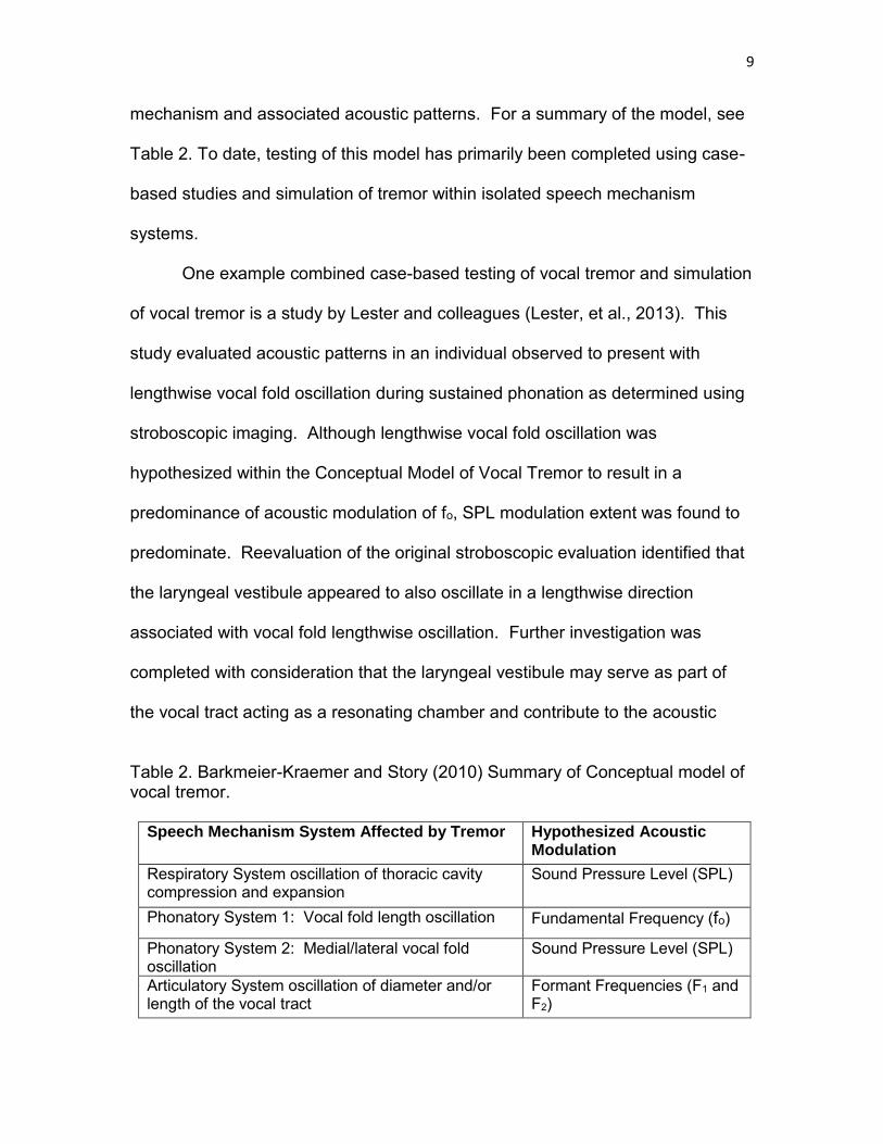
Table 2. Barkmeier-Kraemer and Story (2010) Summary of Conceptual model of vocal tremor.

Speech Mechanism System Affected by Tremor Hypothesized Acoustic Modulation

Respiratory System oscillation of thoracic cavity compression and expansion

Sound Pressure Level (SPL)

Phonatory System 1: Vocal fold length oscillation Fundamental Frequency (fo)

Phonatory System 2: Medial/lateral vocal fold oscillation

Sound Pressure Level (SPL)

Articulatory System oscillation of diameter and/or length of the vocal tract

Formant Frequencies (F1 and F2)

10

patterns predicted for the vocal tract resulting in formant modulation and

subsequent SPL modulation. Acoustic analysis of the vocal tract formants

supported that the case under study demonstrated formant modulation consistent

with the idea that the laryngeal vestibule contributed to vocal tract oscillation. To

further test the idea of the laryngeal vestibule as a component of the vocal tract

for this individual case, an analysis-by-synthesis approach was utilized. That is,

the acoustic characteristics from the case voice recordings were used to model

acoustic modulation patterns resulting from oscillation originating within the

larynx alone compared to the larynx plus the vocal tract. Systematic analysis of

varied possible conditions of laryngeal and vocal tract oscillation patterns and

associated acoustic patterns demonstrated that the Conceptual Model of Vocal

Tremor helped elucidate the location of tremor within and outside of the vocal

folds of one individual and supported that the laryngeal vestibule resonating

chamber contributed to acoustic modulation patterns as predicted for the vocal

tract (Lester et al., 2013).

Although the case example described above was helpful for testing the

Conceptual Model of Vocal Tremor, general testing on groups of individuals with

vocal tremor has not yet been completed. One reason for this arises due to

difficulty successfully identifying individuals representing tremor isolated within

each system of the speech mechanism. Thus, the question arises whether a

human demonstration of volitional oscillation within one subsystem of the speech

mechanism could be used as a surrogate approach to test the Conceptual Model

of Vocal Tremor (Barkmeier-Kraemer & Story, 2010).

One possible approach to studying vocal tremor acoustic patterns

11

associated with oscillation of speech mechanism structures is to study volitional

modulation of the voice, or vibrato. Western classical singing proponents

consider vibrato to be the “quasi automatic” result of correct singing technique,

used by singers to produce an aesthetically pleasing singing voice (Sundberg,

1994). Vibrato shares many similar acoustic features to vocal tremor such as

rate and extent of fo and SPL (Anand, Shrivastav, Wingate, & Chheda, 2012;

Anand, Wingate, Smith, & Shrivastav, 2012). Similar to vocal tremor, typical

vibrato rate is between 4-7 Hz (Anand, Widgate, et al., 2012; Guzman et al.,

2012; Howes, Callaghan, Davis, Kenny, & Thorpe, 2004; Prame, 1994; Ramig &

Shipp, 1987; Seashore, 1931; Sundberg, 1994; Titze, Story, Smith, & Long,

2002; Watson, Williams, & James, 2012) with an extent of fo modulation between

0.25-2 semitones (Anand, Windgate, et al., 2012; Guzman, et al., 2012; Howes,

et al., 2003; Prame 1994; Seashore 1931). One semitone is equivalent to a

modulating extent of about 6%. Therefore, 0.25-2 semitones would equate to an

extent of about 2-12%. As reported earlier, a typical vocal tremor rate is between

4-8 Hz and is associated with a 3-17% extent of fo.

Also similar to vocal tremor, vibrato is produced by oscillation of structures

within the speech mechanism resulting in acoustic modulation. Based on prior

literature, vibrato studied in trained singers’ results from oscillation in laryngeal

and respiratory structures. Vibrato is predominantly the result of alternating

contraction between the cricothyroid (CT) and a combination of the

thyroarytenoid (TA) and lateral cricoarytenoid (LCA) resulting in laryngeal

oscillations (Dromey & Smith, 2008; Hsiao, Solomon, Luschei, & Titze, 1994;

Sundberg 1994; Titze, et al., 2002). Given the roles of medial/lateral or

12

lengthwise change in the positioning of the vocal folds associated with LCA and

TA/CT contractions, respectively, laryngeal oscillations associated with vibrato

would be expected to result in modulation of SPL and fo acoustic components,

respectively.

Although the predominant involvement of laryngeal musculature appears

associated with TA/LCA and CT musculature, supplementary respiratory (i.e.,

sternocleidomastoid) and postural (e.g., scalenes and latissimus dorsi)

musculature also co-varied activation with production of fo modulation during

production of vibrato (Pettersen & Westgaard, 2005; Watson, et al., 2012).

However, the role of supplementary respiratory musculature was difficult to

interpret from the description in these studies. It is possible that supplementary

respiratory musculature contributes to the artistic aim to achieve balanced

participation between the respiratory and laryngeal systems. For example, as

laryngeal movements associated with production of vibrato occur, SPL changes

can occur due to oscillation in laryngeal valving patterns possibly requiring

supplementary respiratory musculature to respond in an opposite and similar

pattern to achieve stability across speech structures during vibrato performance.

Given that the involvement of the primary expiratory and inspiratory respiratory

musculature was not found, it is likely that the role of supplementary respiratory

musculature was not related to respiratory pressure generation. Therefore,

supplementary respiratory musculature likely serves an antagonistic function

during vibrato generation to maintain stable laryngeal positioning and

performance of the speech mechanism during production of vibrato during

singing.

13

Instruction on the production of vibrato involves extensive training of

techniques that aim to manipulate and balance the use of the respiratory and

laryngeal structures to facilitate modulation of the voice (Kirkpatrick, 2008).

Specifically, vibrato associated with Western classical singing is trained in

singers by teaching the techniques to facilitate oscillation of laryngeal structures

via a reflexive struggle between the CT and TA musculature (Titze et al., 2002).

According to the conceptual model, this would result in lengthwise vocal fold

oscillation resulting in predominant extent of modulation of fo in the acoustic

signal.

Although supplementary respiratory musculature has been implicated

during production of vibrato, respiratory involvement during vibrato is considered

a sign of poor vibrato technique (Kirkpatrick, 2008). However, a healthy use of

the respiratory system for modulation of the voice can be used to facilitate

improved respiratory-phonatory coordination during phonation. One method

documented as successful in achieving this goal is the Accent Method Voice

Therapy (AMVT) (Kotby & Fex, 1998). AMVT involves teaching individuals to

use rhythmic accentuated phoneme productions (Kotby & Fex, 1998) to enhance

phonatory-respiratory coordination during voice production. The rhythmic

accentuation during phonation results from a focus on expiratory rhythmic pulsing

through volitional abdomino-diaphragmatic contractions. Accordingly, these

volitional abdominal accents could be studied to determine whether individuals

can volitionally isolate respiratory oscillation to produce voice modulation

predominantly associated with SPL modulation as predicted by the Conceptual

Model of Vocal Tremor. One study reported a correlation between volitional

14

abdominal accents characteristic of AMVT and a generally associated increase in

SPL and fo (Kotby, Shiromoto, & Hirano, 1993). Although covariation between

SPL and fo was not determined in this study, it did confirm SPL linkage to

volitional abdominal accents. Thus, the AMVT approach to volitional respiratory

system accent production using a rhythmic pattern could be used to test the

Conceptual Model of Vocal Tremor hypothesis of respiratory system contribution

to voice modulation.

Statement of Purpose

Vocal tremor typically presents simultaneously across speech structures

making it difficult to examine predicted contributions of individual portions of the

speech mechanism to predicted acoustic patterns. However, vibrato and AMVT

offer two volitional methods for testing hypothesized contributions of the

laryngeal and respiratory oscillations to acoustic modulation patterns. In

addition, testing these two volitional manipulations of voice production could

elucidate whether the laryngeal and respiratory systems can be isolated from

each other during a voicing task, or are linked in movement patterns. That is,

these voluntary forms of voice modulation (i.e., vibrato and AMVT) could occur

by isolated oscillation of targeted speech structures or may require coordinated

cooscillation of speech structures. If oscillation across systems is demonstrated,

this would support the possibility of volitional motor planning linkages between

speech mechanism structures that might apply to individuals with vocal tremor in

which multiple speech structures appear to be simultaneously affected.

The purpose of this study is to investigate laryngeal and respiratory

15

system physiologic and acoustic correlates as predicted by the Conceptual

Model of Vocal Tremor using trained singers to volitionally produce vibrato and

rhythmic accented production of loudness (i.e., AMVT). The hypothesized

contribution of the respiratory and laryngeal systems to acoustic modulation

patterns are (see Table 3):

1) Rhythmic accented production of loudness using AMVT via chest wall

expiratory movements is hypothesized to be associated with greater

extent of SPL compared to fo modulation.

2) Production of vibrato via lengthwise vocal fold oscillation within the

larynx is hypothesized to be associated with greater extent of fo

compared to SPL modulation.

Table 3. Aims of this study Aims Independent

Variable Dependent Variables

Hypothesized Outcome

1. Study acoustic modulation patterns associated with respiratory system oscillation

Rhythmic accented loudness modulation achieved by AMVT production during sustained phonation

a. Chest wall oscillation rate and extent b. SPL modulation rate and extent c. fo modulation rate and extent

Chest wall movements during accented loudness production will be associated with the rate and extent of SPL > fo modulation

2. Study acoustic modulation patterns associated with laryngeal structure oscillation

Rhythmic fo modulation achieved by vibrato production during sustained phonation

a. Vocal Fold oscillation rate and extent b. fo modulation rate and extent c. SPL modulation rate and extent

Laryngeal oscillation movements during vibrato will be associated with rate and extent of fo > SPL modulation

METHODS

Subjects

This study was approved by the University of Utah Institutional Review

Board (IRB, protocol #IRB 00084972). A total of 11 singers without report of

voicing problems or singing complaints responded to IRB-approved flyers

distributed through social media, campus postings, and email listserves.

Subjects were required to be 40-65 years of age with no less than 5 years as a

trained singer and without report of voicing or singing problems prior to consent

and completion of screening procedures. All consented participants were

screened for the presence of singing and voicing problems (see the description

of the screening procedures below). One individual did not meet inclusion

criteria out of the 11 volunteers for this study. Recruitment continued until a total

of 10 singers meeting inclusion criteria were recruited.

Screening Procedures

All consented participants completed the Voice Handicap Index (VHI)

(Jacobson, et al., 1997) and the Singing Voice Handicap Index (Singing VHI)

(Cohen, et al., 2007) to determine whether voice complaints or singing problems,

respectively, were indicated by abnormal scores (>20 points total score). In

addition, participants completed a questionnaire regarding their singing training

and genre to assure that participants completed a minimum of 5 years of training

17

of the singing voice in Western Classical Singing with a self-identified skill in

producing vibrato and the capability of producing rhythmic accented production of

loudness during sustained phonation (see Appendix a). Of the 10 final

participants, 2 participants initially demonstrated total scores in the abnormal

range on the VHI; however, both individuals clarified that they misunderstood the

VHI rating descriptors and adjusted their scores into the normal range during

discussion regarding the outcomes of the screening procedures.

Procedures

Respiratory kinematic, audio, and laryngeal imaging signals were

recorded simultaneously during sustained phonation tasks. The respiratory

kinematic, audio, and laryngeal imaging signals were synchronized for analysis

of corresponding laryngeal and respiratory kinematic and acoustic patterns. An

example of the simultaneous recording for comparison of laryngeal endoscopy

with simultaneous audio and respiratory kinematic signals can be viewed in

Figure 3.

Respiratory Kinematic Recordings

Respiratory Kinematic Equipment

Two piezo respiratory belt transducers from AD Instruments (Model MLT

1132) were used to measure chest wall movement of the participants. The

transducers were connected to the AD Instruments 8-channel PowerLab (Model

PL 3516) console and LabChart Pro (version 8.1), an AD Instruments software

program. Two piezoelectric respiratory belt transducers were used to measure

respiratory chest wall oscillatory patterns. Each respiband was worn around the

18

Figure 3. LabChart Pro display of simultaneously recorded laryngeal images and acoustic and respiratory signals.

19

participant’s chest wall with one band placed over the rib cage and the

other placed over the abdominal region. The respiratory belt transducer

responsible for measuring rib cage movement was secured around the

circumference of the rib cage at the approximate level of the nipple, running

across the sternum in the front and the upper back. The respiratory belt

transducer responsible for measuring abdominal movement was secured around

the circumference of the abdomen inferior to the ribcage in the front and the

lower back. During the setup, the respiratory belt transducers were positioned to

avoid slipping or repositioning, and to reflect rib cage and abdominal movements

during inhalation, exhalation, and phonation. Also, the recorded signal was

checked to ensure the entire range of chest wall expansion and compression

could be captured within the range of

the LabChart channel.

Respiratory Kinematic Signal Calibration

To calibrate for differences in size and range of utilization of the vital

capacity during singing tasks across participants, the respiratory signal was

normalized across the maximum range of chest wall expansion to compression

during a vital capacity maneuver task. Participants were instructed to inhale air

until they could inhale no further and then to exhale until they could exhale no

further while standing upright. This task was repeated three times. The

maximum recorded value associated with the maximum inhalation maneuver was

set to represent 100% vital capacity, whereas the minimum value associated with

the maximum exhalation maneuver was set to represent 0% vital capacity. The

20

calibration was performed within LabChart and applied to the entire recording for

each participant to enable comparison of respiratory modulation measures within

and between participants.

Respiratory Kinematic Procedures

The signals from the respiratory transducers were recorded in LabChart

(AD Instruments, Version 8.1). Signals were recorded showing the degree of

expansion and compression of the rib cage and abdomen during quiet breathing

and during singing tasks. A summated signal from each portion of the chest wall

was recorded directly onto the AD Instruments LabChart at a sampling rate of 10

kHz. The oscillatory movements of the chest wall were measured for rate and

extent relative to acoustic vibrato and accent (AMVT) patterns.

Acoustic Recordings

Acoustic Recording Equipment

Acoustic recordings were obtained using an AKG head-mounted

condenser microphone (model C520) and preamplifier (Symetrix 302 Dual Mic

Pre-Amp) such that signals were recorded using the AD Instruments 8-channel

PowerLab (Model PL 3516) and LabChart Pro (version 8.1) into the software

simultaneously with laryngeal imaging and respiratory kinematic signals. Audio

signals were recorded at a sampling rate of 40 kHz.

Acoustic Recording Procedures

Once the participant verbally expressed readiness to begin recordings, the

experimental sessions began by providing instruction to the participant to sustain

21

three different vowels, /a/, /u/ and /i/, for the duration of 5 s each. These vowels

were selected to represent “corner vowels” on the English Vowel Chart, produced

using a high forward (i.e., /i/) versus high back (i.e., /u/) and low back (i.e., /a/)

tongue position. It was anticipated that views of the larynx would not be

obstructed by the tongue during production of the two high corner vowels

compared to the occluded views of the larynx during production of the low back

vowel, /a/. The differing vocal production techniques utilized across participants

produced a variety of imaging results, with visibility of the larynx ranging from

fully visible to fully obstructed.

Although 3 corner vowels were recorded, only the recordings of /i/ were

analyzed to test the aims of this thesis. The additional vowel recordings were

conducted as part of a larger data collection for future analysis outside of the

aims of this thesis.

Each participant produced three trials of each vowel with vibrato and

rhythmic accented loudness production using AMVT at comfortable pitch and

loudness. The order of vowel production was not counterbalanced across

participants to assure similar conditions of production for all participants.

However, the order of voice modulation condition for each vowel (vibrato versus

AMVT) was counterbalanced. Three trials for each vowel and voice modulation

condition were produced by each participant. The sequence for production of

each voice modulation condition for each participant was determined in advance

of the recording sessions to assure that equal representation of voice modulation

condition sequencing occurs across all participants. Thus, each participant

produced 18 stimuli (3 vowels: /a/, /u/, and /i/) x 2 voice modulation types (vibrato

22

and AMVT) x 3 trials = 18).

Laryngeal Imaging

Laryngeal Imaging Equipment

Laryngeal imaging was obtained using the Pentax Medical

Nasolaryngoscope System (KayPentax, model 9310HD) and nasoendoscope

(KayPentax, VNL-1070STK) simultaneously with audio and respiratory kinematic

signals via AD Instruments 8-channel PowerLab (Model PL 3516) and LabChart

Pro (version 8.1) hardware/software enabling simultaneous video capture (Figure

3).

Laryngeal Imaging Procedures

Laryngeal imaging was initiated once the audio microphone and

respiratory kinematic bands were placed on the participant. Laryngeal imaging

was obtained using flexible nasoendoscopy to minimize impact on vocal tract

configuration during recording of voicing tasks. The laryngeal imaging

procedures were completed by the supervising thesis advisor (i.e., Dr.

Barkmeier-Kraemer). Topical anesthesia (4% viscous lidocaine) was applied

using a Qtip placed within the left or right entry to the nasal passage and anterior

middle meatus based upon the preference of the subject. The topical anesthesia

was also applied to the scope tip portion of the nasoendoscope posterior to the

lens to minimize discomfort during endoscope placement for laryngeal imaging.

All participants tolerated the scope placement and experimental procedures

without additional need for topical anesthesia.

The nasoendoscope was passed transnasally until the larynx and vocal

23

folds were visible in entirety. Once the nasoendoscope was positioned for

optimal viewing of the larynx during voicing, the participant was instructed to

“warm up” until they felt accustomed to the scope placement during singing.

Once the participant conveyed readiness, the experimental speech tasks were

initiated as described within the audio recording procedures.

DATA COLLECTION AND MEASURES

Classification of Participant Voice Modulation Conditions

by Expert Judges

Three expert judges of the singing voice with at least five years of

experience instructing singers were recruited from the faculty of the University of

Utah School of Music and the School of Musical Theater to complete audio-

perceptual judgments of participant voice recordings and classify each

participant’s recordings as one of the voice modulation types under study.

Hearing Screenings

Each expert judge underwent a hearing screening administered by a

certified SLP to assure typical hearing. The screening tested hearing ability at

500, 1000, 2000, and 4000 Hz at 25 dB SPL in each ear using a pure-tone

audiometer and earphones while sitting in a sound treated booth. The 3 expert

judges included in this study passed the hearing screening according to the

above criteria.

Listening Task Each expert judge individually listened to each participant’s audio

recordings of vibrato and AMVT for each audio recorded trial of /i/. The 3 audio

recording trials of the /i/ vowel in each voice modulation condition were

concatenated into one file for presentation to judges for each of the voice

25

modulation conditions (AMVT vs. vibrato). Each file was presented 3 times for

intrarater reliability purposes. Therefore, 60 total audio samples were presented

to the expert judges (1 vowel x 2 modulation types x 3 presentations of each file

sample x 10 participants = 60 total samples).

These audio files were presented for classification by judges using a

randomized presentation order. Listener stimuli were presented using individual

PowerPoint slides displaying each audio file for evaluation. Judges could

proceed at their own rate through the audio files and replay each file as many

times as necessary to rate the classification of voicing method. Judges listened

to audio file presentations via a high fidelity headset (Sennheiser HD 429) at a

comfortable loudness level while sitting in a sound treated booth. Each listening

trial was classified by each judge as “vibrato,” “accented loudness,” or “unable to

classify.” Judges recorded their ratings for each listening trial onto a formatted

scoring sheet such that the classification of a condition was indicated by circling

the choice that best characterizes what the judge perceived.

Classification of each stimulus was determined by comparing judge

classification ratings to the intended production. A match between judge ratings

and the intended production was scored as an accurate production. A mismatch

between judge ratings and the intended production was scored as an inaccurate

production. Rating scores for the total proportion of participant conditions scored

as accurate were averaged across the three judges’ scores. Recordings from

participants judged as accurate > 75% of the time were used for experimental

analysis. Recordings that did not meet the accuracy criteria above were noted

and reviewed by the authors. A mismatch occurred for the vibrato condition of 2

26

participants and for the accent condition for 1 participant (see Table 4). The

acoustic and respiratory signals from these files were visually inspected and

compared to other recordings judged accurately by judges and did not appear

acoustically dissimilar (see Figure 4A and 4B). Thus, the measures from the

three files judged inconsistently were included within the final data set analyzed

for this study.

Physiologic Measurement of all Recordings

All simultaneously recorded respiratory kinematic, acoustic, and laryngeal

endoscopic signals were saved and stored for each subject recording session.

Two seconds from the midportion of each recorded experimental trial was

selected for analysis. A random number generator was used to code each file

into randomized order to assure blinding of condition, trial, and subject during

measurement of physiologic signals. Nine additional files were randomly

selected (15%) of the total number of files to be analyzed (n = 60) so that

intrarater reliability could be assessed for each physiologic signal measurement

method. Once all measures were completed across all physiologic signals, files

were decoded for statistical analysis. Table 4. Percent agreement between expert judges and intended production condition.

Condition S01 S03 S04 S05 S06 S07 S08 S09 S10 S11

Vibrato 89% *44% *44% 100% 100% 100% 100% 89% 89% 89% AMVT 100% 100% 100% 100% 100% 100% 100% 89% *67%

100%

*This production condition did not meet accuracy criteria and was subject to review by the authors.

27

A.

B.

Figure 4. A comparison of signals rated for each voicing condition (i.e., vibrato and AMVT). A. A comparison of signals rated for the vibrato condition from the two subjects rated with 44% expert agreement from S03 (a-c) and S04 (d-f) and signals with 100% expert agreement from S05 (g-i) versus. B. A comparison of signals rated for the AMVT condition with 67% expert agreement from S10 (a-c) and 100% expert agreement from S05 (d-f).

28

Respiratory Kinematic Analysis

Respiratory oscillation movements were analyzed during the same time

frame analyzed for the audio recordings to compare simultaneous respiratory

kinematic and acoustic patterns.

Respiratory Kinematic Oscillation Rate

The rate of respiratory kinematic oscillation was determined by identifying

peak-to-peak or trough-to-trough patterns of the modulating waveform per

second from the summated rib cage and abdominal voltage signal (see Figure 5).

The rate of oscillation was determined from the number of cycles recorded

per second (Hz).

Respiratory Kinematic Measure Adjustments for Slope

Given that participants sustained phonation while producing vibrato or

AMVT, the lung volume continually decreased across the recorded trials. Thus,

adjustments to the respiratory measures were needed to reduce, or eliminate the

changing lung volume effect on measures obtained over the duration of the

recording. To achieve this, the mean slope of each signal was calculated for

each 2-s segment and factored in to each maximum and minimum value

obtained to adjust for the slope (see Figure 6). The mean slope of the entire

signal was multiplied by the time point associated with the maximum or minimum

value within the 2-s signal and then added to the value measured at the peak

and valley values of the modulating respiratory kinematic signal. Note that the

time point value is based on a 0-2 s time interval, rather than the timestamp of

the entire recording. For example, the 2-s window in Figure 6 took place at

29

Figure 5. Example of determining rate for respiratory kinematic oscillation. Each arrow marks the peak of each modulation cycle displayed in the 2-s window. A total of 11 peak-to-peak cycles are shown in the 2-s window giving a 5.5 Hz respiratory kinematic rate.

Figure 6. Example of measuring extent of respiratory kinematic oscillation. The minimum and maximum values associated with summated rib cage and abdominal movements in the figure represent the original data points. The original data points were corrected before extent was calculated. After adjustment for the sloping values, the relative %VC extent was calculated as described above.

30

timestamp 48-50 s. Therefore, a timestamp of 48.783 s for the data point would

be equal to an adjusted time point value of 0.783 s. The equation used to adjust

the kinematic %vital capacity (%VC) measures to eliminate the slope

effects is shown in equation 1 below: y corrected = y original + [mean slope * time point max/min] (1) Thus, with reference to the values displayed in Figure 6, the following

calculations were completed to obtain the adjusted maximum and minimum

values of one respiratory kinematic cycle:

Cycle 2 minimum corrected = 34.73%VC + [-7.45 * 0.783 s] = 28.90%VC

Cycle 2 maximum corrected = 37.06%VC + [-7.45*0.691 s] = 30.30%VC

Respiratory Kinematic Oscillation Extent

The extent of respiratory kinematic modulation was determined by

measuring the maximum and minimum %VC from the summated rib cage and

abdominal wall voltage signal for each oscillation cycle after the slope adjustment

was applied. The maximum and minimum %VC values were identified by

selecting the peak and trough within the summated rib cage and abdominal wall

signal in LabChart. Then, using the LabChart data pad functions for calculating

maximum and minimum values within a selection, the values were automatically

populated in the data pad spreadsheet. The values were then entered into an

Microsoft Excel spreadsheet for further calculations.

After the original data points were adjusted for the sloping signal, the

%VC extent was calculated by dividing the difference between the minimum

31

%VC value and the maximum %VC value for one cycle by the sum of the

maximum and minimum %VC values for that same cycle and multiplying by 100

to obtain a percentage value. All oscillation cycle relative %VC extent values

were averaged for each trial. Figure 6 can be used again for an example of

respiratory kinematic measurement. Using the adjusted values previously

calculated, the following method was used to determine the relative extent of

respiratory kinematic modulation in equation 2: Extent = (Cycle Max - Cycle Min) / (Cycle Max + Cycle Min) * 100 (2)

Extent of Cycle 2 = (34.15 - 27.85) / (34.15 + 27.85) * 100 = 10.2%

Acoustic Measures

Acoustic modulation patterns of fo and SPL were measured from the

middle 2-s portion of each recorded trial. The fo and SPL values within each

selected acoustic segment for analysis were displayed in Praat (Boersma &

Weenink, 2015; v 5.4.09) for analysis of rate and extent of modulation.

Acoustic Modulation Rate

The 2-s segments of fo and SPL modulation were analyzed for rate by

counting the number of peak-to-peak or trough-to-trough fo and SPL modulation

cycles displayed and dividing by 2-s to record rate of modulation in Hertz

(cycles/s) (see Figure 7). The number of fo and SPL modulation cycles per

s was determined for the signals displayed in Figure 7 with the following equation 3:

32

Figure 7. Example of fo (top line) and SPL (bottom line) plot of vibrato within Praat. Arrows have been added to the signal to indicate the beginning of the cycle. Rate (Hz) = (Number of cycles) / (time (s)) (3)

Rate for fo (top signal) is 9.5 cycles / 2-s = 4.8 Hz

Rate for SPL (bottom signal) is 9.5 cycles / 2-s = 4.8 Hz

Acoustic Modulation Extent

The extent of fo and SPL modulation for each cycle was determined by

measuring the maximum and the minimum values from the peak and valley

portions of each cycle (see Figure 8). The maximum and minimum values were

identified by importing the 2-s portion of the acoustic files being analyzed into

Praat. The modulation cycles were identified using the peak to peak or trough to

trough analysis (see Figure 8). Each cycle was then highlighted by dragging the

cursor across one cycle. Then, functions were completed within Praat to

calculate the maximum and minimum values of fo and SPL (i.e., get minimum

pitch, get maximum pitch, get minimum intensity, and get maximum intensity).

The respective values were then entered into an MS Excel spreadsheet for

calculation of the extent values.

33

Figure 8. Example of fo (top line) and SPL (bottom line) plot of vibrato within Praat. Arrows have been added to the signal to indicate the maximum and minimum points of cycle 3 (fo) and cycle 8 (SPL). fo Extent Measures

Calculation of fo extent was completed by subtracting the minimum fo value

from the maximum fo value and dividing the resulting value by the sum of the

maximum and minimum fo values of that cycle and multiplying by 100 to

determine a percentage value. This was repeated for all fo modulation cycles

within the selected segment of each trial as shown in the equation 4 below for Figure 8: Extent = (fo Max - fo Min) / (fo Max + fo Min) * 100 (4)

Figure 8 Extent fo = (246 Hz - 237 Hz) / (246 Hz + 237 Hz) * 100 = 1.9%

SPL Extent Measures

The extent of SPL modulation was determined from the maximum and

minimum dB SPL values associated with SPL modulation (see Figure 8). SPL

values were converted to a linear scale, Pascals, so that the same calculation

method used for fo extent could be performed for SPL extent. Using the values

shown in Figure 8 below, the following equation 5 offers an example of one cycle

of SPL modulation extent calculation:

34

SPL Extent = (SPL Max – SPL Min) / (SPL Max + SPL Min) * 100 (5)

Figure 8 Extent SPL = (.017 Pa - .016 Pa) / (.017 Pa + .016 Pa) * 100 = 3.03%

Laryngeal Imaging Kinematic Analysis

Frame by frame analysis of laryngeal oscillatory movements was

completed to determine the predominant pattern of laryngeal movement

associated with each voice modulation condition (vibrato vs. AMVT). Laryngeal

oscillation movements were analyzed from a similar or the same 2-s duration

segment as the respiratory kinematic and acoustic signals for each experimental

trial. Adjustments in the portion of the endoscopic recording were made if

pharyngeal or laryngeal postures occluded views of the vocal folds. During the

latter situations, the 2-s segment analyzed was shifted earlier or later as needed

to assure continuous views of the vocal folds for kinematic analysis. Given the

subjective impression that laryngeal movements remained continuous during all

trials, it was not expected that analysis from a shifted time segment would impact

measurement outcomes. The maximum and minimum range of vocal fold

movement in the anterior/posterior (i.e., lengthwise) and medial/lateral (i.e.,

abductor/adductor) directions was measured to determine laryngeal oscillation

rate and extent for comparison to acoustic and respiratory kinematic modulation

patterns. The endoscopic video recordings for each subject were imported into

QuickTime to select each of the mid-portion 2-s segments analyzed for the

acoustic and respiratory kinematic signals. The maximum and minimum

movement frames associated with each laryngeal movement cycle within the

analyzed segment were copied and imported into ImageJ (October, 2015) for

35

measurement.

Laryngeal Oscillation Rate

Laryngeal oscillation rate was determined by recording the number of

laryngeal oscillatory cycles obtained during the 2-s segment analyzed for

each trial divided by 2 s to determine the rate in Hertz (cycles/s).

Laryngeal Oscillation Extent

Extent of laryngeal oscillation was measured by identifying image frames

displaying the minimum and maximum displacement of laryngeal structures/vocal

folds during the predominant direction of oscillation (i.e., lengthwise versus

abduction/adduction). The lens to larynx distance varied during recordings,

requiring image measures to be normalized by creating a ratio of distance (unit =

pixels) between laryngeal movement measures and an anatomical measure

observed to remain constant (e.g., width of the epiglottis apex). Referent Anatomical Distance Measures

Individual frames judged to exhibit the maximum displacement end points

of laryngeal oscillation were selected and saved for analysis within ImageJ

software (Rasband, 2015). The referent anatomical distance between two

constant and clearly identifiable locations measuring the interarytenoid distance

was measured for each maximum and minimum cyclic displacement image (see

Figure 9, Part A and B). This was accomplished by drawing a line between the

two referent image points using the ImageJ line tool and recording the length of

the line in pixels.

36

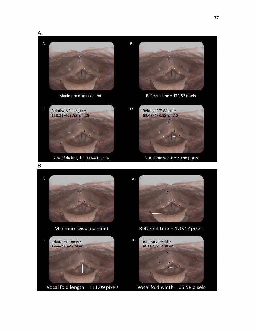
Figure 9. An example of laryngeal imaging measures for the first end point of one laryngeal oscillatory cycle. Each panel displays A) the image analyzed, B) the anatomical referent line measure, C) the relative measure of vocal fold length, and D) the relative measure of interarytenoid distance associated with the first end point of one laryngeal oscillation cycle. B. Example of laryngeal imaging measures for the second end point of one laryngeal oscillatory cycle. Each panel displays A) the image analyzed, B) the anatomical referent line measure, C) the relative measure of vocal fold length, and D) the relative measure of interarytenoid distance associated with the second end point of one laryngeal oscillation cycle.

37

A.

B.

38

Laryngeal Lengthwise Extent Measures

The length of the vocal folds during maximum and minimum laryngeal

cyclic movements was measured by drawing a line from the anterior commissure

(or most anterior visible portion of the vocal folds) and posterior commissure (or

most visible posterior point on the vocal folds) and recording the number of pixels

associated with the length. The magnitude, or extent, of lengthwise vocal fold

movement during the laryngeal movement cycle associated with vibrato or AMVT

was determined by dividing the vocal fold length measure (in pixels) by the

standard referent distance (in pixels) (see Figure 9). The extent of lengthwise

change was determined using the following equation (6) for

each cycle of maximum and minimum movements:

% vocal fold lengthwise extent = (vocal fold maximum length – vocal fold

minimum length) / (vocal fold maximum length + vocal fold minimum

length) * 100 (6)

In the laryngeal oscillation cycle shown in Figure 9, the extent of laryngeal abduction/adduction extent would be: Relative VF Lengthwise Extent = (.25 - .24) / (.25+.24) * 100 = 2.0% Laryngeal Abduction/Adduction Extent Measures

The interarytenoid distance was measured during maximum and minimum

laryngeal cyclic movements to determine changes in abduction and adduction of

the vocal folds. Due to the absence, or small number of pixels associated with

the glottal width between vocal processes, the interarytenoid distance was used

39

as a surrogate measure to assure adequate sampling distance for analysis. In

the absence of a glottis during full approximation of the vocal folds, the measure

would yield 0 pixels and would cause errors in the calculation of extent. Thus,

interarytenoid distance was determined to reflect changes in glottal width for the

purposes of this study.

The interarytenoid distance was measured by drawing a line between the

most lateral visible point on the superior surface of the left vocal process and the

opposite lateral visible point on the right vocal process (see Figure 9, Part A and

B, Image D). The extent of abduction/adduction change was determined using

the following equation (7) for each cycle of maximum and minimum movements:

% vocal fold ABDuction / ADDuction extent = (vocal fold maximum

abduction – vocal fold minimum abduction) / (vocal fold maximum

abduction + vocal fold minimum abduction) * 100 (7)

In the laryngeal oscillation cycle shown in Figure 9, the extent of laryngeal abduction/adduction extent would be:

Relative Vocal Fold Abductor/Adductor Extent =

(.14 - .13) / (.14 + .13) * 100 = 3.7%

Statistical Analysis

The dependent variables for investigation are the average rate and extent

of fo and SPL acquired from acoustic recordings; the average rate and extent of

respiratory kinematic signals acquired from movements of the chest wall; and the

average rate and extent of laryngeal kinematic images acquired from lengthwise

40

laryngeal movements and abductor/adductor laryngeal movements. Each

variable was compared within and across voice modulation conditions, vibrato

and AMVT (see Table 5).

Statistical analysis was completed to determine whether hypothesized

differences between the extent of SPL and fo for each location of oscillation

(respiratory system versus laryngeal system) occurred. To accomplish this, a

mixed effects logistic regression was completed to compare dependent variable

outcomes predicted between respiratory kinematic measures and associated

acoustic modulation measures (%SPL extent > % fo extent) and laryngeal

imaging kinematic measures and associated acoustic modulation measures

(lengthwise oscillation = % fo extent > %SPL extent; abductor/adductor oscillation

= %SPL extent > % fo extent).

Intrarater reliability was also evaluated using an Intra Class Correlation

(ICC) approach including calculation of 95% confidence intervals on the 9 files

Table 5. Intrarater reliability determined using intraclass correlations.

Dependent Variable AMVT (N=3) Vibrato (N = 6)

Avg fo Rate (Hz) 0.882 (-0.001 ~0.997) 1 (NaN ~ NaN)

Avg fo Extent (%) 0.994 (0.913 ~1) 0.999 (0.996 ~ 1)

Avg SPL Rate (Hz) 0.857 (-0.105 ~0.996) 0.878 (0,44 ~0.982)

Avg SPL Extent (%) 0.807 (-0.262 ~0.995) 0.986 (0.922 ~0.998)

Avg Respiratory Kinematic Rate (Hz) 1 (NaN ~NaN) 1 (.61 ~ 1)

Avg Respiratory Extent (%) 1 (NaN ~NaN) 1 (.61 ~ 1)

Avg Laryngeal Rate (Hz) 0.977 (0.683 ~ 0.999) 0.723 (0.02 ~ 0.955)

Avg vocal fold length Extent (%) 0.934 (.293 ~0.998) 0.925 (0.622 ~ 0.989)

Avg vocal fold abduction/adduction extent (%) .489 (-0.692 ~0.983) 0.723 (0.02 ~0.955)

41

randomly selected for repeated measures. The 9 measures were only used for reliability measures and were not included in the final analysis.

Intrarater Reliability

Intraclass Correlation (ICC) results demonstrated high levels of intrarater

reliability levels for all dependent variables except for the average vocal fold

abduction/adduction extent (%) measure under the AMVT condition which

achieved moderate reliability (i.e., 0.5 – 0.6) (see Table 5). The lower reliability

levels for vocal fold abduction/adduction measures under the AMVT condition

may relate to the lower number of files analyzed (n = 3), or may reflect the

greater difficulty determining laryngeal oscillation patterns given significantly

slower rates of oscillation that included vertical laryngeal movements.

RESULTS

Qualitative Analysis of Results

Tables 6 and 7 provide a summary of the average measures and their

standard deviations for each subject under the AVMT and vibrato conditions,

respectively. As can be seen, individual singers generally exhibited similar rates

of acoustic modulation within each of the singing conditions (AMVT versus

vibrato). That is, the AMVT condition was associated with slower acoustic

modulation rates for both fo and SPL (i.e., approximately 2-4 Hz) than for the

vibrato condition (i.e., approximately 5-7 Hz). Similarly, laryngeal kinematic rates

for vocal fold length change and abduction and adduction of the vocal folds were

slower under the AMVT voicing condition than for the vibrato condition.

Respiratory kinematic rates were absent under the vibrato condition and present

at a similar rate to laryngeal kinematic rates for AMVT.

Respiratory System Contributions to Acoustic Modulation

It was hypothesized that the AMVT condition would be associated with a

predominant oscillation of the chest wall contributing to a greater extent of SPL

acoustic modulation compared to the vibrato condition. As shown in Tables 6

and 7, the average respiratory kinematic extent for all subjects during the AVMT

condition was measured at 47.5% (SD = 1.2%). For the vibrato condition,

respiratory kinematic extent was measured at 0% (SD = 0%). The respiratory

43

Table 6. AVMT voicing condition descriptive statistical summary for individual subjects.

*Signifies measures that are significantly different between voicing conditions. Table 7. Vibrato voicing condition descriptive statistical summary for individual subjects.

*Signifies measures that are significantly different between voicing conditions.

1 3 4 5 6 7 8 9 10 11

Avg fo Rate (Hz)

(SD) 4.5 (0) 5 (0) 6.2 (0.3) 5 (0) 5.2 (0.3) 5.3 (0.3) 6 (0) 4.5 (0) 5.3 (0.3) 4 (0) 5.1 (0.7)*

Avg fo Extent (%)

(SD) 3.5 (0.2) 4.8 (0.4) 4.1 (0.7) 4.4 (0.3) 2.2 (0.2) 3.7 (0.4) 1.9 (0.1) 4.7 (0.9) 3.4 (0.3) 2.0 (0.4) 3.5 (1.1)

Avg SPL Rate (Hz)

(SD) 4.5 (0) 4.8 (0.3) 6.2 (0.3) 5 (0) 5 (0.5) 5.2 (0.3) 5.8 (0.3) 4.5 (0) 5.2 (0.3) 4 (0) 5 (0.6)*

Avg SPL Extent

(%) (SD) 4.5 (1.6) 8.9 (0.4) 19.6 (5.7) 7.2 (1.0) 2.8 (0.5) 6.9 (1.3) 6.7 (0.3) 9.1 (3.5) 12.1 (0.7) 4.4 (1.4) 10.0 (0)*

Avg Respiratory

Kinematic Rate

(Hz) (SD)

0 (0) 0 (0) 0 (0) 0 (0) 0 (0) 0 (0) 0 (0) 0 (0) 0 (0) 0 (0) 0 (0)*

Avg Respiratory

Kinematic Extent

(%) (SD)

0 (0) 0 (0) 0 (0) 0 (0) 0 (0) 0 (0) 0 (0) 0 (0) 0 (0) 0 (0) 0 (0)*

Avg Laryngeal

Kinematic Rate

(Hz) (SD)

4.2 (1.9) 6 (1.3) 10.2 (2) 6.3 (0.8) 6.5 (3) 7.5 (0) 10.3 (1.6) 6.5 (0.5) 6.3 (1.6) 4.8 (2.5) 6.9 (2)*

Avg Vocal Fold

Lengthwise

Extent (%) (SD)

1.8 (1.2) 2 (0.2) 3.3 (1.8) 6.3 (1.3) 0.9 (0.1) 5.4 (1.9) 1.5 (0.4) 3.8 (0.5) 3.7 (0.7) 2.9 (1.1) 3.2 (1.7)

Avg Vocal Fold

Abduction/Adduct

ion Extent (%) (SD)

3.9 (0.8) 1.5 (0.2) 4.7 (0.3) 4.8 (1.7) 2.2 (0.9) 5.9 (1.9) 3 (0.6) 6.6 (1.2) 4.1 (0.1) 2.8 (0.7) 3.9 (1.6)

SUBJECTSMEASURES

ACOUSTIC

RESPIRATORY KINEMATICS

LARYNGEAL KINEMATICS

ALL

SUBJECTS

1 3 4 5 6 7 8 9 10 11

Avg fo Rate (Hz)

(SD) 3.8 (0.8) 2.5 (0) 2.2 (0.3) 3.7 (1) 3.5 (1.3) 2 (0.9) 2.2 (0.3) 3.5 (0.9) 2.5 (0.5) 1.7 (1.2) 2.8 (0.8)*

Avg fo Extent (%)

(SD) 6.1 (3.2) 8.9 (0.2) 2.8 (1.0) 1.6 (0.3) 2.7 (0.9) 2.8 (0.2) 1.7 (0.2) 4.2 (0.4) 2.6 (0.5) 2.0 (0.5) 3.5 (2.3)

Avg SPL Rate (Hz)

(SD) 3.5 (0.5) 2.5 (0) 2.3 (0.3) 1.8 (0.3) 1.5 (0) 1.5 (0) 2 (0) 3 (0) 2 (0) 1 (0) 2.1 (0.7)*

Avg SPL Extent

(%) (SD)

28.4

(13.1)

49.5

(5.4)35 (8.9)

12.6

(6.7)

32.6

(10.6)

68.2

(08.7)23 (1.3)

49.2

(1.9)

28.3

(1.6)

37.7

(9.9)40 (20)*

Avg Respiratory

Kinematic Rate

(Hz) (SD)

3.7 (0.3) 2.5 (0.5) 2.7 (0.8) 2 (0) 1.7 (0.3) 1.7 (0.3) 2 (0) 3 (0) 2.2 (0.3) 1.8 (0.6) 2.3 (0.6)*

Avg Respiratory

Kinematic Extent

(%) (SD)

46.2

(1.2)

48.5

(0.4)45.7 (1.4)

48.5

(1.3)47 (3)

48.6

(0.1)47.6 (1.1)

48.4

(1.1)

48.6

(0.5)

46.1

(4.7)47.5 (1.2)*

Avg Laryngeal

Kinematic Rate

(Hz) (SD)

6.7 (1.8) 4.2 (0.3) 2.5 (1) 3.7 (3.2) 2.3 (1.3) 2.7 (0.3) 1.8 (1.6) 5.2 (1) 3.3 (0.8) 1.5 (1.5) 3.4 (1.6)*

Avg Vocal Fold

Lengthwise

Extent (%) (SD)

1.8 (0.4) 2.8 (0.5) 2.4 (0.5) 3.1 (2.7) 2.2 (1.5) 4.3 (1.5) 0.9 (1.6) 3 (1.7)10.7

(3.2)0.7 (0.8) 3.2 (2.8)

Avg Vocal Fold

Abduction/Adduct

ion Extent (%) (SD)

3.4 (1.5) 3.3 (1.3) 4.9 (1.7) 3 (2.6) 3 (1.3) 3.3 (0.7) 1.4 (2.5) 7.6 (1.5) 3.7 (1.6) 3 (3) 3.7 (1.6)

LARYNGEAL KINEMATICS

MEASURESSUBJECTS ALL

SUBJECTS

ACOUSTIC

RESPIRATORY KINEMATICS

44

kinematic extent was demonstrated to be significantly greater, on average, under

the AMVT condition than for vibrato (p < .001). Refer to Figure 10 for a graphical

comparison of respiratory kinematic extent across the voicing conditions (i.e.,

AMVT and vibrato).

The average SPL modulation extent for the AVMT condition was

measured at 40% (SD=20%), whereas the average SPL extent for the vibrato

condition was 10% (SD=0%). The acoustic measure of SPL extent was also

significantly greater under the AMVT condition, on average, than for vibrato (p =

.026) supporting the hypothesized contribution of the respiratory system to voice

modulation. Interestingly, the rate of SPL modulation was also found to

significantly differ between voicing conditions. Respiratory oscillation under the

AMVT condition was associated with significantly slower rate of SPL modulation

than for the vibrato condition (p < .001). Refer to Figures 11, 12, and 13 for a

graphical comparison of kinematic oscillation rates, SPL modulation extent, and

SPL modulation rate, respectively, across voicing conditions (i.e., AMVT and

vibrato).

Phonatory System Contributions to Acoustic Modulation

It was hypothesized that the vibrato condition would be associated with a

predominant laryngeal kinematic oscillation, vocal fold lengthwise oscillation,

resulting in greater fo modulation extent compared to the AVMT condition. As

shown in Tables 6 and 7, The average vocal fold length extent for the vibrato

condition was measured at 3.2% (SD = 1.7%). The average vocal fold length

extent for the AVMT condition was measured at 3.6% (SD = 3%). Statistical

45

Figure 10. Kinematic extent comparisons between voicing conditions (i.e., AMVT

and vibrato)

Figure 11. Kinematic extent comparisons between voicing types (i.e., AMVT and vibrato)

46

Figure 12. Acoustic extent comparison between voicing conditions (i.e., AMVT and vibrato)

Figure 13. Acoustic rate comparison between voicing conditions (i.e., AMVT and vibrato)

47

evaluation showed that the laryngeal kinematic extent was not significantly

greater under the vibrato condition. Rather, the laryngeal kinematic extent did not

significantly differ between the two voice modulation conditions (p = .95). Refer

to Figure 13 for a graphical comparison of laryngeal kinematic extent across the

voicing conditions (i.e., AMVT and vibrato).

The average fo modulation extent for the vibrato condition was measured

at 3.5% (SD = 1.1%). The average fo modulation extent for the AVMT was 3.5%

(SD = 2.3%). The acoustic measure of fo extent also did not differ significantly

under the two voice modulation conditions (p = .92) as was hypothesized.

However, the rate of fo modulation was found to significantly differ between

voicing conditions demonstrating that laryngeal oscillation under the vibrato

condition was a significantly faster rate than for the AVMT condition (p < .001).

Furthermore, the average laryngeal kinematic rate was significantly higher for the

vibrato condition compared to the AVMT condition (p < .001). Refer to Figures 12

and 13 for a graphical comparison of fo modulation extent and rate, respectively,

across voicing conditions (i.e., AMVT and vibrato).

Vocal Tract Movements by Condition

Vertical Laryngeal Movement The presence or absence of vertical laryngeal movement was recorded

during laryngeal kinematic analysis and observed to demonstrate predominant

patterns associated with each voicing condition. During the AVMT condition, 70%

of participants demonstrated vertical laryngeal movement in at least 2 of 3 trials

or more (67%). In contrast, only 40% of participants demonstrated vertical

48

laryngeal movement in at least 2 of 3 trials in the vibrato condition.

Pharyngeal Movement

The presence or absence of pharyngeal constriction was recorded during

laryngeal kinematic analysis for comparison between voicing conditions. During

the AVMT condition, 30% of participants demonstrated pharyngeal movement in

at least 2 of 3 trials compared to 60% of participants during the vibrato condition.

DISCUSSION

The purpose of this study was to test the contribution of respiratory and

laryngeal oscillation patterns to acoustic modulation patterns. The conceptual

model of vocal tremor developed by Barkmeier-Kraemer and Story (2010)

proposed that oscillation of the respiratory system would be reflected by SPL

modulation patterns in the acoustic signal. They also proposed that laryngeal

oscillation patterns would be associated with acoustic patterns specific to the

laryngeal kinematic patterns exhibited. Vocal fold length oscillation was

hypothesized to cause fo extent modulation whereas vocal fold abduction/

adduction oscillation would affect interarytenoid distance and be reflected in

acoustic SPL extent modulation. The current study investigated voluntary

manipulation of laryngeal and respiratory oscillation within trained singers to

study associated patterns of acoustic modulation. This was achieved by

comparing AVMT and vibrato voicing methods during sustained phonation.

AVMT modulates the voice using accented contraction of the respiratory system

(Kotby & Fex, 1998). In contrast, vibrato utilizes vocal fold lengthwise oscillation

to modulate fo (Dromey & Smith, 2008; Hsiao, Solomon, Luschei, & Titze, 1994;

Sundberg 1994; Titze, et al., 2002).

50

Respiratory System Contributions to Acoustic Modulation

The results of this study supported the hypothesized contribution of

respiratory oscillation to acoustic modulation. The AVMT voicing condition was

used to provide respiratory system oscillation during voicing modulation and was

shown to predominantly be associated with greater respiratory kinematic

oscillation extent and SPL modulation extent. Kotby, Shiromoto, & Hirano (1993),

reported that significant SPL modulation occurred associated with abdomino-

diaphragmatic contraction during production of the AVMT method of voicing. The

current study’s outcomes lend support to physiologic models of phonation

proposing modulation of SPL associated with respiratory system compression

and expansion movements during voicing (Barkmeier-Kraemer & Story, 2010;

Farinella, Hixon, Hoit, Story, Jones, 2006; Story, 1995;). The distinct difference

between respiratory kinematics and acoustic modulation patterns during the

AMVT and vibrato conditions supports prior literature showing that the chest wall

portion of the respiratory system does not appear to contribute toward natural

vibrato production in trained singers (Pettersen & Westgaard, 2005; Watson, et

al., 2012).

The slow respiratory oscillation rate measured during the AVMT condition

distinguished respiratory oscillation from laryngeal oscillation in this study. The

slower rate of oscillation and acoustic SPL modulation during the AMVT condition

compared to the vibrato condition may relate to the larger mass of the respiratory

structures and musculature such as the diaphragm, abdominal muscles, and

intercostal muscles. In contrast, the laryngeal skeletal framework, structures,

and musculature are significantly less massive likely enabling the faster

51

oscillation rates measured for the larynx (Dalvi & Premkumar, 2011).

The findings revolving around the AVMT voicing condition have important

clinical implications for clinical evaluation and management of vocal tremor.

Individuals with symptoms of vocal tremor exhibiting greater acoustic SPL

modulation extent than fo modulation extent at a rate closer to 3 Hz should be

evaluated for respiratory contributions to their vocal tremor. In cases where the

respiratory system is the predominant contributor to vocal tremor, consideration

regarding optimal medical or behavioral management would be required. For

example, vocal tremor is most commonly medically treated by injecting Botox®

into the intrinsic laryngeal musculature (i.e., thyroarytenoid, interarytenoid, or

posterior cricoarytenoid muscles) (Kendall & Leonard, 1995; Schneider &

Deuschl, 2015). This is warranted if the vocal tremor is predominantly caused by

tremor within the intrinsic laryngeal muscles. However, vocal tremor caused by

the respiratory system and not the laryngeal musculature may not provide

optimal results using Botox® injections into the laryngeal musculature (Bove et

al., 2006). In addition, behavioral clinical treatment typically involves increased

recruitment of respiratory contraction during phonation to offload laryngeal and

throat muscle tension. Such a speech treatment approach may be more difficult

for individuals to perform if the source of their vocal tremor is from the respiratory

system.

52

Laryngeal Contributions to Acoustic Modulation

Laryngeal oscillation contributions to vocal tremor were hypothesized to

result in greater fo modulation extent acoustically. This was tested in the current

study by comparing the correspondence between vocal fold lengthwise and

abduction/adduction oscillations during vibrato to modulation of fo extent. The

hypothesized differences between acoustic measures of fo extent and vocal fold

kinematics during AVMT and vibrato were not supported. In contrast, vibrato and

AVMT voicing conditions were shown to equally contribute to fo extent modulation

and vocal fold lengthwise and abduction/adduction laryngeal oscillation. Although

laryngeal kinematic patterns appeared similar between the AMVT and vibrato

conditions, the kinematic rates were significantly different between voicing

conditions and closely corresponded with the rate of acoustic modulation for

each. That is, the rate of laryngeal kinematic patterns was significantly faster

during the vibrato condition than for AMVT. These findings suggest that the

laryngeal kinematic patterns measured during the AMVT condition were largely

influenced by respiratory system kinematics. This is not entirely surprising given

that the larynx is considered to be part of the respiratory system. The utilization

of the larynx for voice production cannot be disassociated from its reliance on

respiratory pressure and flow patterns for which laryngeal configuration may

adjust associated with lung volume levels (Lowell, Barkmeier-Kraemer, Hoit,

Story, 2008).

The determination that laryngeal kinematics are similar, but slower during

AMVT compared to vibrato demonstrates the reliance of laryngeal configurations

on respiratory functions during voice production. In addition, the finding that

53

abduction/adduction vocal fold movements were a component of vibrato is in

contrast to prior literature reporting vibrato production associated with the

antagonist relationship of the TA and CT muscles (Dromey & Smith, 2008; Hsiao,

Solomon, Luschei, & Titze, 1994; Sundberg 1994; and Titze, et al., 2002). The

findings in this study suggest that vibrato is produced using a complex interaction

between the phonatory, respiratory, and articulatory (i.e., vocal tract) systems.

Another interesting finding of this study was the observation of inferior

constrictor muscle activation associated with laryngeal movements during

vibrato. This was also not entirely surprising to observe given the suspension of

the larynx within the throat by anterior and posterior throat musculature. Thus,

observation of counter-contraction of the thyropharyngeus musculature opposite

laryngeal vibrato movements is speculated to demonstrate postural stabilization

via pharyngeal constrictor muscle activation.

The influence of the respiratory condition in this study (i.e., AVMT) on

laryngeal kinematic patterns suggests that laryngeal behaviors are not

independent from other speech mechanism structures. This is consistent with

prior findings in individuals diagnosed with vocal tremor. For example, a prior

study by Lester et al. (2013) tested the contribution of lengthwise vocal fold

oscillation to acoustic modulation patterns and did not find support for the

predicted contribution of laryngeal oscillation to fo extent modulation. However,

upon expanded acoustic analysis, implication of the vocal tract was found as

indicated by formant modulation. Upon further review of the laryngeal imaging

recordings, it was discovered that the epilarynx, or laryngeal vestibule oscillatory

patterns, were likely affecting formant locations within the vocal tract and affected

54

SPL extent modulation as predicted by the conceptual framework model. In this

study, systematic analysis of laryngeal vocal fold movements in addition to

presence/absence of pharyngeal constriction movements documented that

laryngeal movements during vibrato (i.e., predominantly laryngeal involvement)

were also frequently associated with pharyngeal constriction movements in

addition to laryngeal vestibule compression and expansion. These findings

suggest that laryngeal oscillations are likely to contribute to alteration of vocal

tract configuration requiring co-contraction of vocal tract musculature involved in

laryngeal posturing functions. Additional investigation implementing

electromyography (EMG) to improve upon prior literature investigating the

involvement of supplementary respiratory musculature would help resolve the

timing and role of observed vocal tract structure involvement during vibrato

production (Finnegan et al., 2003; Tomoda et al., 1987).

In general, the outcomes of this study demonstrate that the vibrato

condition shows close correspondence between laryngeal kinematic patterns and

respiratory system behaviors. These findings support that speech treatment for

vocal tremor that trains improved utilization of the respiratory system may

influence laryngeal configurations and muscle contractions associated with

laryngeal-based vocal tremor. Importantly, the findings of this study demonstrate

significant differences in the rate of vocal tremor associated with laryngeal versus

respiratory sources of oscillation as well as acoustic SLP extent modulation

patterns. These findings support the proposal of the conceptual model of vocal

tremor (Barkmeier-Kraemer and Story, 2010) that the speech structures affected

by tremor may be identified by their contributions to the acoustic modulation

55

patterns. However, hypothesized contributions of the larynx to acoustic fo extent

associated with vocal fold lengthwise oscillation was not successfully tested due

to linkage between respiratory and laryngeal behaviors during vibrato and AVMT

voicing.

Application of Current Findings to Vocal Tremor

The results of this study offer further support for the respiratory

contributions to voice modulation as hypothesized by the conceptual model of

vocal tremor (Barkmeier-Kraemer & Story, 2010). The current findings showed

correspondence between respiratory kinematic patterns and SPL rate and extent

of modulation within the AMVT condition. Although respiratory contributions to

vocal tremor have not been directly studied, the presence of respiratory structure

oscillation in individuals with vocal tremor has been reported in the literature

(Tomoda et al., 1987). Future work will need to address similar patterns in those

with vocal tremor to confirm similar association of respiratory kinematics and

voice modulation.

One problem that has impeded testing of hypothesized laryngeal

contributions to voice modulation in vocal tremor is the cooccurrence of

oscillation of vocal tract structures with laryngeal oscillations. The current study

aimed to isolate laryngeal oscillation through the implementation of vibrato

compared to AMVT in trained singers. We hypothesized that laryngeal oscillation

would be absent during production of AMVT compared to vibrato. However, the

trained singers in this study did not demonstrate differences in vocal fold

lengthwise and abduction/adduction kinematic patterns between the AMVT and

56

vibrato conditions as hypothesized. The primary distinction between laryngeal

kinematic oscillation patterns in the AMVT and vibrato conditions was the rate of

movement. The rate of laryngeal kinematic patterns was significantly slower

during the AMVT condition than during the vibrato condition. That is, kinematic

pattern rate distinguished between laryngeal oscillation conditions rather than

specific SPL or fo extent of modulation. As such, future work investigating

laryngeal kinematic patterns associated with acoustic modulation in those with

vocal tremor may benefit from a comparison of acoustic rate patterns to

determine the source of oscillation within the speech mechanism. Thus, future

research needs to compare and contrast all three sources of tremor (i.e.,

respiratory, laryngeal, and vocal tract) to further test and refine the conceptual

model of vocal tremor.

Limitations

The current study offered important contributions toward understanding

the contributions of laryngeal and respiratory oscillation to SPL and fo acoustic

patterns. Future research on this topic could improve upon the current findings

by consideration of methodology limitations of this study.

Laryngeal kinematic measures were limited by the use of nasoendoscopy

to analyze the dynamic larynx without a calibrated light grid, or other

measurement calibration methods that would have improved upon the accuracy

of this study’s kinematic measures. That said, the reliability of the laryngeal

kinematic measures was highly reliable for all but the abduction/adduction

measures specific to the AMVT condition. However, the range of findings for the

57

latter was likely due to the smaller randomly chosen files for analysis that were at

least moderately reliable. Future research could be improved by the use of

electromyography to study musculature associations with observed movements

and incorporation of calibration lighting as this technology improves.

The within-subjects design of this study did not require calibration of SPL

for comparison within individuals between two conditions of voicing. SPL

calibration signals were recorded; however, the calibration factors caused peak

clipping of recorded signals due to the large amplitude signals recorded by

singers in this study. Thus, the decision was made to utilize relative SPL. Thus,

relative SPL values were recorded and compared within subjects between the

two conditions. However, reporting of SPL values in this study requires caution

as averaged measures were relative SPL. In future work, calibrated SPL values

would enable comparison of SPL between individuals.

Another future consideration would be to include aerodynamic measures

of SPL and airflow. Similarly, consideration of laryngeal behaviors associated

with lung volume levels would help interpret linked laryngeal and respiratory

behaviors during vibrato and AVMT conditions. It is possible that laryngeal

oscillation patterns vary between voicing initiation at higher lung volumes

compared to voicing toward the end of voicing (Lowell et al., 2008). Such factors

may be important to study associated with structural kinematic behaviors to

elucidate further the role of the larynx relative to respiratory and vocal tract voice

production conditions.

Finally, generalization of the findings of this study to the larger population

of singers will require increased numbers of participants in future work. However,

58

the robust outcomes of this study would be expected to be supported in future

larger studies.

Conclusion

The purpose of this study was to test the conceptual model of vocal tremor

developed by Barkmeier-Kraemer and Story, 2010 by linking respiratory and

laryngeal kinematic oscillations to acoustic modulation patterns during AVMT and

vibrato conditions. Specifically, respiratory oscillation during AVMT was

hypothesized to correspond with the acoustic extent of SPL modulation whereas

lengthwise oscillation of the vocal folds during vibrato voicing was hypothesized

to correspond with the acoustic extent of fo modulation. The hypothesized

contributions of the respiratory system to SPL modulation were confirmed.

However, the hypothesized contributions of the phonatory system to fo

modulation were not supported. Laryngeal kinematic patterns during vibrato and

AVMT appeared similar although vibrato oscillations occurred at a significantly

higher rate than measured during AVMT. The linkage between the larynx and

respiratory kinematic patterns suggest that the laryngeal movements were

unable to be voluntarily isolated. These findings offer important information

about laryngeal and respiratory physiology that may be further investigated for

utility in the clinical evaluation and treatment of individuals with vocal tremor.

APPENDIX

QUESTIONNAIRE FOR SINGING PARTICIPANTS

60

Questionnaire for Singing Participants

Please answer the questionnaire completely.

Age: _________ Gender: _____________

1) How many years of vocal training have you had?

___________________________________________________________

2) In what genre(s) do you feel most comfortable singing?

___________________________________________________________

3) How would you describe your current singing voice condition (e.g., in-

shape, sing often, been a while since I’ve sung, etc.)?

___________________________________________________________

4) Do you use vibrato while you sing?

___________________________________________________________

a. If you answered yes to #4, how often do you use vibrato while

singing (e.g., all the time, at the end of phrases, etc.)?

_____________________________________________________

b. If you answered yes to #4, have you experienced or are you

currently experiencing any problems with your vibrato? ______

c. If you answered yes to #4, do you vary your vibrato production

across genres? ______

i. If so, how?

________________________________________________

________________________________________________

5) Are you currently experiencing any voice problems? If so, describe:

___________________________________________________________

___________________________________________________________

REFERENCES

Ackermann, H., & Ziegler, W. (1991). Cerebellar voice tremor: an acoustic analysis. Journal of Neurology, Neurosurgery & Psychiatry, 54(1), 74-76. Adler, C. H., Bansberg, S. F., Hentz, J. G., Ramig, L. O., Buder, E. H., Witt, K.,

Edwards, B. W., Krein-Jones, K., Caviness, J. N., (2004). Botulinum toxin type A for treating voice tremor. Archives of Neurology, 61(9), 1416-1420.

Anand, S., Shrivastav, R., Wingate, J. M., & Chheda, N. N. (2012). An acoustic-

perceptual study of vocal tremor. Journal of Voice, 26(6), 811-817. Anand, S., Wingate, J. M., Smith, B., & Shrivastav, R. (2012). Acoustic

parameters critical for an appropriate vibrato. Journal of Voice, 26(6), 819-825.

Barkmeier-Kraemer, J., Lato, K., & Wiley, K. (2011). Development of a speech treatment program for a client with essential vocal tremor. Seminars in Speech and Language, 32(1), 43-57.

Barkmeier-Kraemer, J., & Story, B. (2010). Conceptual and clinical updates on

vocal tremor. ASHA Leader, 15(14), 16-19. Boersma, Paul & Weenink, David (2015). Praat: doing phonetics by computer

[Computer program]. Version 5.4.09, Retrieved 2 June 2015 from http://www.praat.org/

Bové, M., Daamen, N., Rosen, C., Wang, C.-C., Sulica, L., & Gartner-Schmidt, J.

(2006). Development and validation of the vocal tremor scoring system. The Laryngoscope, 116(9), 1662-1667.

Brown, JR & Simonson, J (1963). Organic voice tremor. A tremor of phonation.

Neurology, 13, 520-525. Cohen, S. M., Jacobson, B. H., Garrett, C. G., Noordzij, J. P., Stewart, M. G.,

Attia, A., Cleveland, T. F. (2007). Creation and validation of the singing voice handicap index. Annals of Otology, Rhinology & Laryngology, 116(6), 402-406.

Dalvi, A., & Premkumar, A. (2011). Tremor: etiology, phenomenology, and

clinical features. Disease-a-Month, 57(3), 109-126.

62

Dromey, C., & Smith, M. E. (2008). Vocal tremor and vibrato in the same person: acoustic and electromyographic differences. Journal of Voice, 22(5), 541-545.

Dromey, C., Warrick, P., & Irish, J. (2002). The influence of pitch and loudness

changes on the acoustics of vocal tremor. Journal of Speech, Language & Hearing Research, 45(5), 879.

Farinella K. A., Hixon T. J., Hoit J. D., Story B. H., Jones P. A. (2006). Listener

perception of respiratory-induced voice tremor. American Journal of Speech Language Pathology, 15(1):72-84.

Finnegan, E. M., Luschei, E. S., Barkmeier, J. M., & Hoffman, H. T. (2003).

Synchrony of laryngeal muscle activity in persons with vocal tremor. Archives of Otolaryngology–Head & Neck Surgery, 129(3), 313-318.

Gamboa, J., Jiménez-Jiménez, F. J., Nieto, A., Cobeta, I., Vegas, A., Ortí-Pareja,

M., García-Albea, E. (1998). Acoustic voice analysis in patients with essential tremor. Journal of Voice, 12(4), 444-452.

Gurey, L., Sinclair C., & Blitzer, A. (2013). A new paradigm for the management

of essential vocal tremor with botulinum toxin. Laryngoscope, 123(10), 2497-2501.

Guzman, M. A., Dowdall, J., Rubin, A. D., Maki, A., Levin, S., Mayerhoff, R., &

Jackson-Menaldi, M. C. (2012). Influence of emotional expression, loudness, and gender on the acoustic parameters of vibrato in classical singers. Journal of Voice, 26(5), 675.

Howes, P., Callaghan, J., Davis, P., Kenny, D., & Thorpe, W. (2004). The

relationship between measured vibrato characteristics and perception in Western operatic singing. Journal of Voice, 18(2), 216-230.

Hsiao, T.-Y., Solomon, N. P., Luschei, E. S., & Titze, I. R. (1994). Modulation of

fundamental frequency by laryngeal muscles during vibrato. Journal of Voice, 8(3), 224-229.

Jacobson, B. H., Johnson, A., Grywalski, C., Silbergleit, A., Jacobson, G.,

Benninger, M. S., & Newman, C. W. (1997). The voice handicap index (vhi) development and validation. American Journal of Speech- Language Pathology, 6(3), 66-70.

Jiang, J., Lin, E., & Hanson, D. G. (2000). Acoustic and Airflow Spectral Analysis

of Voice Tremor. Journal of Speech, Language & Hearing Research, 43(1), 191.

63

Kendall, K. A., & Leonard, R. J. (2011). Interarytenoid muscle botox injection for treatment of adductor spasmodic dysphonia with vocal tremor. Journal of Voice, 25(1), 114-119.

Kirkpatrick, A. (2008). Teaching methods for correcting problematic vibratos:

using sustained dynamic exercises to discover and foster healthy vibrato. Journal of Singing, 64(5), 551-556.

Kotby, M., & Fex, B. (1998). the accent method: behavior readjustment voice

therapy. Logopedics Phoniatrics Vocology, 23(1), 39-43. Kotby, M., Shiromoto, O., & Hirano, M. (1993). The accent method of voice

therapy: Effect of accentuations on FO, SPL, and airflow. Journal of Voice, 7(4), 319-325.

Lederle, A., Barkmeier-Kraemer, J., & Finnegan, E. (2012). Perception of vocal

tremor during sustained phonation compared with sentence context. Journal of Voice, 26(5), 668-669.

Lester, R. A., Barkmeier-Kraemer, J., & Story, B. H. (2013). Physiologic and

acoustic patterns of essential vocal tremor. Journal of Voice, 27(4), 422-432.

Louis, E. D., & Machado, D. G. (2015). Tremor-related quality of life: A

comparison of essential tremor vs. Parkinson's disease patients. Parkinsonism & Related Disorders, 21(7), 729-735.

Lowell S. Y., Barkmeier-Kraemer J. M., Hoit J. D., Story B. H. (2008).

Respiratory and laryngeal function during spontaneous speaking in teachers with voice disorders. Journal of Speech, Language, and Hearing Research, 51(2):333-49.

Lundy, D. S., Roy, S., Xue, J. W., Casiano, R. R., & Jassir, D. (2004).

Spastic/spasmodic vs. tremulous vocal quality: motor speech profile analysis. Journal of Voice, 18(1), 146-152.

Pettersen, V., & Westgaard, R. H. (2005). The activity patterns of neck muscles

in professional classical singing. Journal of Voice, 19(2), 238-251. Prame, E. (1994). Measurements of the vibrato rate of ten singers. The Journal

of the Acoustical Society of America, 96(4), 1979-1984. Ramig, L. A., & Shipp, T. (1987). Comparative measures of vocal tremor and

vocal vibrato. Journal of Voice, 1(2), 162-167. Rasband, W.S., ImageJ, U. S. National Institutes of Health, Bethesda, Maryland,

USA, Retrieved from http://imagej.nih.gov/ij/, 2015

64

Schneider, S. A., & Deuschl, G. (2015). Medical and surgical treatment of tremors. Neurologic Clinics, 33, 57-75.

Seashore, C. E. (1931). The natural history of the vibrato. Proceedings of the

National Academy of Sciences of the United States of America, 17(12), 623-626.

Story, B. H. (1995). Physiologically-based speech simulation using and

enhanced wave-reflection model of the vocal tract. (Unpublished doctoral dissertation). University of Iowa, Iowa City, IA.

Sulica, L., & Louis, E. D. (2010). Clinical characteristics of essential voice tremor:

A study of 34 cases. The Laryngoscope, 120(3), 516-528. Sundberg, J. (1994). Acoustic and psychoacoustic aspects of vocal vibrato. STL-

QPSR, 35(2-3), 045-068. Taha, J. M., Janszen, M. A., & Favre, J. (1999). Thalamic deep brain stimulation

for the treatment of head, voice, and bilateral limb tremor. Journal of Neurosurgery, 91(1), 68-72.

Titze, I. R., Story, B., Smith, M., & Long, R. (2002). A reflex resonance model of

vocal vibrato. The Journal of the Acoustical Society of America, 111(5), 2272-2282.

Tomoda, H., Shibasaki, H., Kuroda, Y., & Shin, T. (1987). Voice tremor:

dysregulation of voluntary expiratory muscles. Neurology, 37(1), 117-122. Warrick, P., Dromey, C., Irish, J., Durkin, L., Pakiam, A., Lang, A. (2000).

Botulinum toxin for essential tremor of the voice with multiple anatomical sites of tremor: a crossover design study of unilateral versus bilateral injection. Laryngoscope, 110(8), 1366-74.

Watson, A. H. D., Williams, C., & James, B. V. (2012). Activity patterns in

latissimus dorsi and sternocleidomastoid in classical singers. Journal of Voice, 26(3), 95-105.

Wolraich, D., Marchis-Crisan, C.V., Redding, N., Khella, S.L., & Mirza, N. (2010).

Laryngeal tremor: co-occurrence with other movement disorders. Otorhinolaryngology, 72, 291–294.


Recommended