24
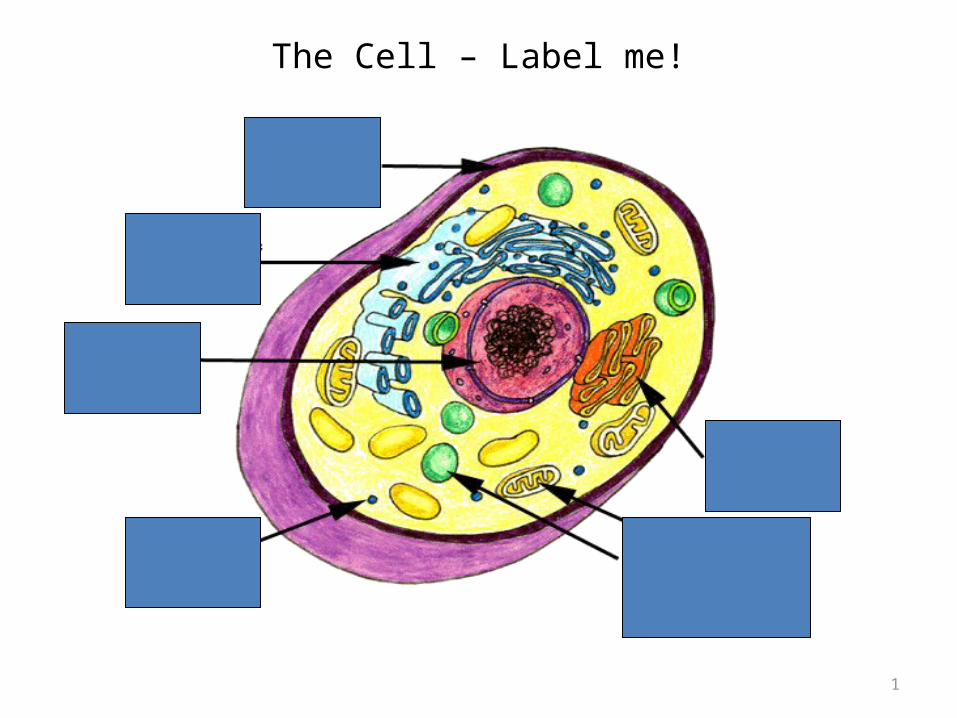
The Cell – Label me! 1
| Date post: | 22-Dec-2015 |
| Category: |
Documents |
| Upload: | catherine-webb |
| View: | 215 times |
| Download: | 0 times |
1
The Cell – Label me!

Learning Objectives1. Describe the fluid mosaic model of membrane structure and
explain the underlying reasons for this structure.
2. Outline the roles of phospholipids, cholesterol, glycolipids, proteins and glycoproteins in membranes.
3. Outline the roles of the plasma membrane, and the roles of membranes within cells.

Key words you should know
• Phospholipids
• Polar
• Hydrophilic
• Hydrophobic
• Micelles
• Phospholipid bilayer
• Fluid mosaic model
Glycoproteins
Glycolipids
Cholesterol
Proteins
Transport proteins
Receptor molecules

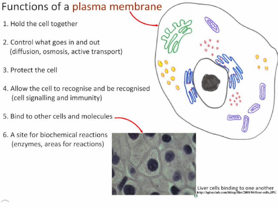
Cell membrane• All living things are surrounded by a membrane.A cell membrane is also known as plasma membrane.
Controls exchange of materials such as nutrients and waste between cells and their environment.
Has other important functions for example to enable cells to receive hormones.
To understand the function of anything in biology, you must study the structure first!

Cell Membranes from Opposing Neurons (TEM x436,740).
} cell membrane
7nm wide
Cell membrane {
Nerve cell
Nerve cell
Gap between cells


• HYDROPHILIC heads (water liking)-Attracted to the water
• called POLAR
• HYDROPHOBIC tails (water fearing)-Not attracted to the water
• called NON-POLAR
Cell membranes are made of PHOSPHOLIPIDs
A Phospholipid

A Phospholipid BilayerPhospholipids can form:
BILAYERS
-2 layers of phospholipids with
hydrophobic tails protected inside by the hydrophilic heads.
The PHOSPHOLIPIDBILAYER is the basicstructure of membranes.


Structure of the cell membranePhospholipidsCell membranes are made mainly of
phospholipids. They have:
HYDROPHILIC heads (water liking)-Attracted to the water POLARHYDROPHOBIC tails (water fearing)-Not attracted to the water NON-POLAR
Phospholipids can form BILAYERS-2 layers of phospholipids with hydrophobic tails protected inside by the hydrophilic heads.
The PHOSPHOLIPID BILAYER is the basic structure of membranes.

Diagram representing the cell membraneRemember the membrane is 7nm wide
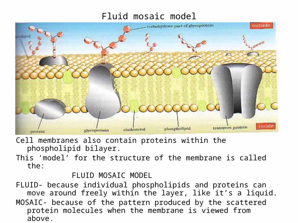
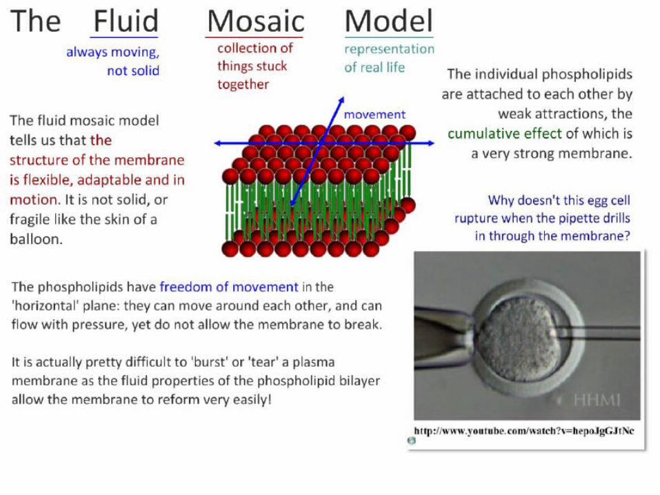
Fluid mosaic model
Cell membranes also contain proteins within the phospholipid bilayer.This ‘model’ for the structure of the membrane is called the:
FLUID MOSAIC MODELFLUID- because individual phospholipids and proteins can move around freely within
the layer, like it’s a liquid.MOSAIC- because of the pattern produced by the scattered protein molecules when
the membrane is viewed from above.
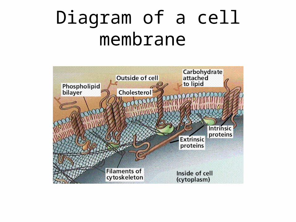
Diagram of a cell membrane


Cell Membranes from Opposing Neurons (TEM x436,740).
} Phospholipid Bilayer
7nm wide
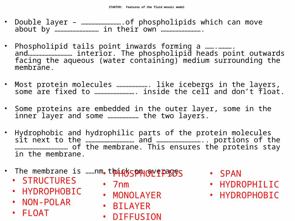
STARTER: Features of the fluid mosaic model
• Double layer – ……………………….of phospholipids which can move about by ………………………… in their own ……………………….
• Phospholipid tails point inwards forming a …….………. and………………………… interior. The phospholipid heads point outwards facing the aqueous (water containing) medium surrounding the membrane.
• Most protein molecules …………………. like icebergs in the layers, some are fixed to ………………………. inside the cell and don’t float.
• Some proteins are embedded in the outer layer, some in the inner layer and some ………………… the two layers.
• Hydrophobic and hydrophilic parts of the protein molecules sit next to the …………………………… and ………………………….. portions of the ……………………………… of the membrane. This ensures the proteins stay in the membrane.
• The membrane is ……nm thick on average.
• STRUCTURES • HYDROPHOBIC • NON-POLAR • FLOAT
• SPAN • HYDROPHILIC • HYDROPHOBIC
• PHOSPHOLIPIDS • 7nm • MONOLAYER• BILAYER • DIFFUSION

Features of the fluid mosaic model
• Double layer – BILAYER of phospholipids which can move about by DIFFUSION in their own MONOLAYER
• Phospholipid tails point inwards forming a NON-POLAR HYDROPHOBIC interior. The phospholipid heads point outwards facing the aqueous (water containing) medium surrounding the membrane.
• Most protein molecules float like icebergs in the layers, some are fixed to STRUCTURES inside the cell and don’t float.
• Some proteins are embedded in the outer layer, some in the inner layer and some SPAN the two layers.
• Hydrophobic and Hydrophilic parts of the protein molecules sit next to the HYDROPHOBIC AND HYDROPHILIC portions of the PHOSPHOLIPIDS of the membrane. This ensures the proteins stay in the membrane.
• The membrane is 7nm thick on average.
23
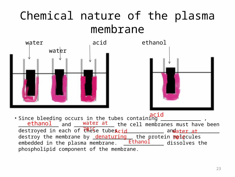
Chemical nature of the plasma membranewater acid ethanol water
• Since bleeding occurs in the tubes containing _____________ , _____________ and _____________ the cell membranes must have been destroyed in each of these tubes. _____________ and _____________ destroy the membrane by _____________ the protein molecules embedded in the plasma membrane. _____________ dissolves the phospholipid component of the membrane.
acid
ethanol water at 70°C Acid water at
70°CdenaturingEthanol