66


The CENTRE OF ADVANCED STUDIES IN MARICULTURE was
started in 1979 at the Central Marine Fisheries Research Institute, Cochin. This is one of the Sub-projects of the ICAR/UNDP project on 'Post-graduate Agricultural Education and Research'. The main objective of the CAS in Mariculture is to catalyse research and education in mariculture which forms a definite means and prospective sector to augment fish production of the country. The main functions of the Centre are to:
— provide adequate facilities to carry out research of excellence in mariculture/coastal aquaculture;
— improve the quality of post-graduate education in mariculture;
— make available the modern facilities, equipments and the literature;
— enhance the competence of professional staff;
— develop linkages between the Centre and other Institutions in the country and overseas;
— undertake collaboration programmes; and
— organise seminars and workshops.
Under the programmes of the Centre, Post-graduate courses leading to M.Sc. (Mariculture) and Ph.D. are offered in collaboration with the University of Cochin since 1980.

HANDBOOK ON DIAGNOSIS AND CONTROL
OF BACTERIAL DISEASES IN FINFISH AND SHELLFISH CULTURE
C. THANKAPPAN PILLAI
Central Marine Fisheries Research Institute, Cochin - 682 018
A. «#
CMFRI SPECIAL PUBLICATION
Number 17
CENTRE OF ADVANCED STUDIES IN MARICULTURE,
CENTRAL MARINE FISHERIES RESEARCH INSTITUTE
INDIAN COUNCIL OF AGRICULTURAL RESEARCH P. B. No. 1912, COCHIN-682 018, INDIA

(LIMITED DISTRIBUTION)
Published by : E. G. Silas Director, Central Marine Fisheries Research Institute, Cochin - 682 018.
Edited by K. Rengarajan Scientist, Central Marine Fisheries Research Institute, Cochin - 682 018.
PRINTED IN INDIA
AT PAICO PRINTING DIVISION, ERNAKULAM, COCHIN 682 031

PREFACE
A national priority has been set for the development of coastal and inland aquaculture in India. Besides seed and feed, one of the major constraints that has been identified towards the development of extensive as well as intensive aquaculture is the problem of diseases. This could affect any stage from hatchery production to the grow-out system. The importance of fish diseases in aquaculture system is also evident from the large volume of literature that is appearing each year in print.
At the Central Marine Fisheries Research Institute, we have identified fish pathology as an important area for research, as specific disease problems are encountered in the culture of some of the species; often resulting in the heavy loss of or mortality of the stocked species. While a beginning has been made in the study of marine fish pathology, a lot has to be investigated. Dr. C. Thankappan Pillai who has been working in this field for the past few. years, has prepared this "Handbook on Diagnosis and Control of Bacterial Diseases in Finfish and Shellfish Culture" as a guide to those involved with aquaculture particularly coastal aquaculture. Besides describing the various types of diseases encountered and those to be met with in brackishwatar and marine environment, he has discussed matters relating to defenses against infection, prophylaxis and disease check-up, sampling techniques for disease diagnosis, classifiication of pathogens, screening of bacteria for identification and methods to be adopted for despatch of diseased specimens for study. It is hoped that this hand book will be a ready guide to our aquaculturists in upgrading the environmental quality and for the well being of the organisms cultivated.
I wish to express my thanks to Dr. C. Thankappan Pillai for the effort he has put in for the preparation of the hand book based on his own experience and from the active guidance and advice he received from Dr. A. L. S. Munro, Principal Scientific Officer, Marine Laboratory, Department of Agriculture and Fisheries, Aberdeen, Scotland, who visited the Centre of Advanced Studies in Mariculture at the Central Marine Fisheries Research

Institute as an expert consultant in 1983. I wish to record my sincere thanks to Dr. Munro for the consultancy afforded and the keen interest shown towards the development of research infrastructure in fish pathology at the Central Marine Fisheries Research Institute, Cochin. Dr. Munro also took great pains to critically go through this Manual and suggested changes which has greatly helped for the improvements of the contents. I am thankful to Dr. P. Vedavyasa Rao, Head, Physiology, Nutrition and Pathology Division and Shri C. Mukundan, Head, Demersal Fisheries Division, Central Marine Fisheries Research Institute, Cochin who have read and commented on the Handbook. I am also very thankful to Shri K. Rengarajan for editing this Handbook.
E. G. SILAS Director,
Cochin-682 018, Central Marine Fisheries December 1984. Research Institute

CONTENTS
CONCEPT OF DISEASE AND PATHOGEN . . . . . . 1
HETEROTROPHIC BACTERIA, INFECTION AND DISEASE . . . 5
DEFENSES AGAINST INFECTION . . . . . . . . . 7
BACTERIAL DISEASES . . . . . . . . . . . . 11
PROPHYLAXIS AND DISEASE CHECK UP . . . . . . 20
THE NEED FOR ANAESTHETICS . . . . . . . . . 25
SAMPLING TECHNIQUES FOR DISEASE DIAGNOSIS . . . 29
CLASSIFICATION OF PATHOGEN(S) . . . . . . . . . 34
SCREENING OF BACTERIA FOR IDENTIFICATION . . . 36
THE DESPATCH OF DISEASED SPECIMENS . . . . . . 41
PATHOLOGY-AN OUTLOOK . . . . . . . . . 43
SUGGESTED READINGS . . . . . . . . . 47
GLOSSARY... . . . . . . . . . . . . 49

1 CONCEPT OF DISEASE AND PATHOGEN
Just as in the husbandry of land animals or in the production of agricultural crops, disease occurs in the culture of fishes and shellfishes. The problem of diseases has assumed considerable significance with the rapid developments that are now occurring in the culture of aquatic species. New diseases are occurring in intensive culture systems, whether they are under experimental conditions or in grow out systems, and unless we have a sufficient knowledge of the nature of these diseases and their control, aqua-culture practices can hardly become successful. An attempt is made here to define disease and pathogen, and different categories of diseases.
The term 'disease' by its popular acceptance denotes a condition or a state of the body in which there is departure from normal function as a result of one or more of the following: defect of heredity, infection by disease producing organisms, insufficiency or excess in diet, or inadequate environment. Every disease, generally speaking, has a cause or causes and likewise an effect or a set of effects. Let us take, as an example, amoebiasis, a parasitic disease of man. Amoebiasis is characterised by intestinal infection and consequent pathological changes. It is caused by a protozoan parasite Entamoeba histolytica which causes different disorders, illness and suffering in the afflicted, of which the most important is an acute dysentery phase. The infection leads to a succession of malfunctions and subsequent disorders of which the manifested ones are called signs. Signs are medically important in as much as they are signposts - indicative of the disease. They must be systematically recorded and evaluated in the process of investigation leading to diagnosis of the cause of a disease condition.

In human medicine symptoms are described by the patient to the doctor. What the doctor sees from this study of the sick patient are called signs. It follows that in the study of morbid or sick aquatic animals, the doctor or in this situation the pathologist, will only see the signs of disease as his patients cannot describe their symptoms to him.
In a diseased aquatic animal, the pathologist will look for gross signs on the exterior of the animal as well as in the internal organs. He will also use other means of looking for signs of disease. For example, by histological methods, he will examine with the light microscope chemically fixed and stained sections of tissues for changes in the architecture of organs and cells and with the electron microscope even finer changes in the architecture of individual cells of particular organ. He will also use the methods of clinical chemistry to look for signs of change indicating pathology. For example, changes in the numbers of RBC and WBC in blood, the levels of proteins, lipids, minerals and enzymes of blood.
Now, we may define disease as any state or condition, resulting from an abnormal change in the anatomy and/or physiology of cell(s) or organ(s) which harms the normal life of a living organism. Diseases may be classified as:
i. Communicable diseases: Involving infection by viruses, bacteria, algae, fungi or attack by parasites.
ii. Abiotic diseases: Due to malfunctioning of cell(s)/ organ(s) and/or associated changes due to unfavourable ecological or other accidental conditions, e.g. pollution or poisoning.
iii. Nutritional diseases: Owing to lack of suitable balanced diet, e.g. deficiency of vitamin, minerals, amino acid or essential fatty acids.
iv. Genetic diseases: Due to lack of a gene or failure to express it, resulting in inherent susceptibility to disease, e.g. melanoma in fish.
Of these, communicable diseases are very important and merit considerable attention in research. It has to be emphasized that to prevent a disease, the cause has to be identified and at best removed or at least controlled. Signs of the disease generally help to diagnose the case to a certain extent. In the case of vibriosis of fish,
2

detection of the aetiological agent Vibrio anguillamm, confirms the disease. Such causative agents are called pathogens. However, it should be borne in mind that every organism which causes disease is not necessarily a pathogen. Sometimes, certain phyto-flagellates, for example, Prymnesium parvum, when present in blooms, may cause fish mortality which is known as 'fish kill'. Here, the causative factor, the algal toxin, is lethal and is an abiotic factor, but not a pathogen. A pathogen is an organism which survives and flourishes in a host causing signs which result in a diseased condition. Moreover, in order to be termed the aetiological agent of a disease, the organism must satisfy certain stringent stipulations known as 'Koch's postulates' which are:
i. The suspected organism should be associated regularly with all cases of the suspected disease, in logical pathological relationship to the disease and its signs.
ii. The suspected organism should be isolated in pure culture from the suspected case.
iii. When such pure culture is inoculated into suitable animals, the disease should be reproduced.
iv. The same organism should be reisolated in pure culture from the inoculated animals.
Only when an organism satisfies these four requisites, it can be calimed as the pathogen causing a particular disease. Viral diseases such as lymphocystis, infectious pancreatic necrosis, and infectious haematopoietic necrosis, and bacterial diseases such as furunculosis, mycobacteriosis are typical communicable diseases. Tests to see whether these postulates are fulfilled become necessary only when dealing with a new disease of unknown case. In cases of regular occurrence, the signs and isolation of the pathogen will be sufficient identification.
Abiotic diseases which are often common in the fish culture systems, are hypoxia, acidosis, alkalosis and intoxication (Hydrogen sulphide, ammonia).
Another aspect of prime importance for successfully managing culture systems is the provision of adequately balanced diets. Imbalances in the diet of cultured animals may result in mass mortality.
3

Therefore, care should be taken in formulating balanced diets in accordance with available knowledge to avoid nutritional diseases such as avitaminosis, hypervitaminosis, nephrocalcmosis, lipoid liver disease and sekoke disease.
Diseases of genetic origin such as certain kinds of tumours and deformities may not be of serious nature as they occur infrequently in culture systems.
4
2 HETEROTROPHIC BACTERIA, INFECTION AND DISEASE
Diseases by bacterial infection, are amongst the more frequent and harmful in fish and shellfish culture systems.
Bacteria, procaryotic microorganisms of the PROCARYOTAE, have always been an object of study by researchers, because of their activities, both harmful and beneficial. Heterotrophic bacterial species (those which obtain their energy by breaking down organic nutrients) are widely distributed in nature and live in many diversified situations. This dynamic group of organisms can be tentatively classified into saprophytes and parasites based on their mode of living. Saprophytes subsist on dead and decomposing organic matter, whereas parasites depend on live hosts for their existence.
The flora of live hosts may be divided into commensals and pathogens. The commensal species live in harmony with the healthy host while those organisms that harm the host and provoke disease are called pathogens. Host animals in poor health may be compromised with respect to their defence against microbial attack. In such circumstances, opportunistic pathogens may cause disease and they can be either commensals or saprophytes. The agents which can only live by causing disease as a result of infection in the host are called obligate pathogens e.g. Aeromonas sabnonicida the aetiological agent of the fish disease called furunculosis.
Infection is the lodging and multiplication of a parasite in or on a host. A material contaminated with a microbe(s) is designated as infective. In the case of the bacterial flora of fish, the mucus, skin, gills, stomach or intestine will be infected and the bacterial load commonly ranges between 10M06 per gram weight of these materials. However, the flesh and body fluids of newly
5

caught healthy fish are found to be sterile. Disease can be recognised when infection results in significant host responses and damage to the host. Disease is normally dependent on various factors such as the virulence of the attacking agent, the state of the host's defensive system, inherent genetically determined resistance factors and the environmental circumstances in which the host is located.
Other definitions in disease processes can be classified based on the time sequence and place of infection. The initial infection in a host with a parasite is called Primary infection and subsequent infection by the same parasite is termed as reinfection.
When a host's resistance is lowered by a pre-existing disease or parasite infection, an additional disease is called secondary infection. If the infection is only in a localised area in a host (e.g. gill rot), it is known as local infection or local sepsis, but if the microbes multiply internally in the vital organ, then it is described as a systemic infection (e.g. furunculosis).
In the host, if signs of infection are not apparent, but the disease gaent can be detected, then it may be denoted as inapparent or latent infection. Continuous infection with a potential pathogen is known as persistent infection, e.g. most fish surviving infection with infectious pancreatic necrosis virus (IPNV) carry virus in other tissues for life.
The presence of a known or potential bacterial pathogen in the blood is called bacteremia. Previously in the older tests this condition was called as septicaemia.
A condition in which the organism remains localised in a host and releases its toxin in the blood stream is called toxemia.
An ailment is said to be contagious when it is transmitted by direct contact whereas infectious implies all (means of sources) including direct contact, e.g. via water, food or by other animals.
Epidemiology is the study of the distribution and spread of diseases in man and animals. When animal diseases are discussed, some changes in epidemiological terminology are sometimes made to denote that diseases of animals are being discussed. Thus, enzootic replaces endemic and epiozotic epidemic. Enzootic means when a disease is widespread in a disease in a population. It may be in tank or pond population or all such population in an aqua-culture farm or all the farms in an area or even a country.
6

3 DEFENSES AGAINST INFECTION
In the previous chapter infection and disease have been discussed. Now, let us examine how infection may be overcome by host's defenses. The protective measures of the host against infection^) are called defense mechanisms. They may be classified as the nonspecific which include structural, humoral and cellular defenses and the specific or adaptive defenses. Higher veitebrates such as mammals including man and birds possess all the defenses and almost certainly most fish do as well. Molluscs and crustaceans probably possess most or all of the nonspecific defenses, but much less is known of their ability to mount adaptive responses.
STRUCTURAL DEFENSES
The main body surfaces of finfish and shellfish are skin, shell, gills and alimentary canal through which invading microbes must pass to get access to the host's tissue. Moreover, many of these surfaces are covered by a layer of mucus which is continually shed making microbial attachment and penetration difficult. If the mucus is breached, the invading microbes may get attached and subsequently gain entry into the epithelium and deeper tissues and in consequence, finally cause disease.
HUMORAL DEFENSES
The various humoral defenses mostly contained in the blood of fish, crustaceans and molluscs are as follows.
a. Natural non-specific antibodies
These natural antibodies are considered to have antipathogen properties. They attach to the surface of pathogens and as a consequence increase the efficiency of complement and phagocytosis.
7

b. Complement
This system is a complex of enzymes which may be activated by the products produced by pathogens or by antibody attaching to them or the pathogen itself. It has effective antipathogen (killing) properties mainly as a result of damage to pathogen cell membranes.
c. C-reactive protein (CRP)
It is considered to be normally present in the serum of fish and functions as a defence factor against pathogens.
d. Interferon
This is an important antiviral agent probably produced by all fish and it is likely to be produced by crustaceans and molluscs, but no proof yet exists.
e. Lysozyme Lysozyme, an enzyme, is an important weapon in the host's
defense and functions against various pathogens. It is present in the mucus, skin and seium and in phagocytic cells.
/ . Natural haemolysins
These haemolysins are in fact the complement system recognizing foreign red cells as foreign and lysing them.
CELLULAR DEFENSES
When the pathogen gains entry and begins dividing and causing damage to host cells, defense systems become altered. The cellular defense system has a series of components which collectively are called the inflammatory reaction. It is well defined in finfishes, less in crustacean and least in molluscs. A characteristic feature of inflammatory reaction is invasion of the damaged area with leucocytic cells of different functions. Now, there are several alternative fates. As the infection progresses, vasodilation causing swelling and fluid accumulation takes place. Various leucocytic cell types including phagocytes are attracted to the site of infection by the release of specific chemicals from damaged host cells. This is called positive chemotaxis. Moreover, infectious agents are phagocytosed often with the help of humoral factors in the blood, known as complement and lysozyme.
8

If the invasive microbes are not fully destroyed at the site of attack, they may be carried by the blood stream to the various organs of the body. In fish, the spleen and kidney are the main blood filtering organs and they have a primary role in dealing with microbes in the blood stream, rather like the lymph node in mammals. In fish the work of destroying the pathogens is carried out mainly by wandering phagocytes. There are also fixed phagocytic cells scattered throughout the body of fish and shellfish.
In fish, the blood contains many monocytes which are the precursors of the macrophage. They will differentiate into macrophages, the principal phagocytic cell in fish, when stimulated by injury and invasive microbes. Some notable features are:
i. The atrium in fish and sehllfish has got significant phagocytic populations.
ii. There are no lymph nodes in fishes, but organs with lymph node activity such as spleen and kidney are present in fish. Shellfish appear not to have any organ with such activity.
In fish the neutrophile is the dominant granulocyte and it is found circulating in the blood. Unlike mammalian granulocytes it is not phagocytic, but it is chemotactic invading areas of inflammation where it bursts discharging cell contents which are lethal for pathogens. The other granulocyte of fish, the eosinophil, does not have phagocytic activity either. Large populations are present in gut and gill in healthy fish, but these may migrate to the sites of infection in any organ where they behave like neutrophiles. There is speculation that they perform the role of tissue mast cells in higher animals.
In shellfish, it is not established that as many populations of leucocytes with specialised function exist, rather it seems a few populations of less specialised cells exist, performing all or most of the functions of the many specialised leucocytes in higher animals.
In fish, the thrombocyte leucocytes promote the clotting mechanism of the blood. Clotting seals areas of injury preventing escape of body fluids. However, in crustaceans only acellular clotting mechanisms are found, nevertheless they act efficiently to seal any gaps in the vascular system. Clotting in molluscs does occur, but by a very different mechanism.
9
INDUCED OR ADAPTIVE IMMUNITY
Another defense system possessed by higher animals and fish, recognises foreign material if it penetrates the structural defenses. There are both cellular and humoral responses to foreign material. They are brought about by leucocytic cells called lymphocytes. In fish the lymphocytes carry receptor sites capable of recognizing foreign materials which are called antigens. There are two types of lymphocytes called T and B lymphocytes. T lymphocytes are produced in the thymus and spleen and they are responsible for cell mediated immunity a mechanism whereby pathogens are killed by the T lymphocytes making physical and chemical content. B lymphocytes are responsible for antibody production.
While in mammals five distinct classes of immunoglobulins (Ig) are identified, in teleost fishes only one class of Ig is detected. The antibodies can act in a variety of ways. Based on the nature of the foreign material, they may agglutinate particulate antigens, such as whole bacteria. They also precipitate soluble antigens and thus neutralise toxins. Antigens which are coated with antibody are much more readily phagocytosed.
The structural, humoral and cellular defenses are the first line of defense against invaders. They are non-specific in their activity. A significant difference between the defense system of fishes and shellfishes is that acquired immunity detected in fishes is limited to a primitive antibody-like substance in some crustaceans, but no immune response is found in molluscs.
Pathogenic microbes entering the host's body may escape from phagocytes by producing aggressins which kill the phagocytes, by not eliciting host cells to release biologically active chemicals, i.e., negative chemotaxis thus not attracting leucocytes to their point of entry, by producing capsules which prevent phagocytosis or are motile and escape from the phagocyte. In these situations, the host becomes susceptible and the pathogens enter directly into the blood stream or through the lymphatics.
10

4 BACTERIAL DISEASES
All living beings, in their natural surroundings, are exposed and periodically saccumb to diseases. Fishes and shellfishes are no exception. In their natural conditions, finfishes and shellfishes become diseased, but most of the cases pass unnoticed possibly due to lack of interest and/or closer observation. Occurrence of diseases of fish and shellfish in culture systems is more obvious and will adversely affect production. Therefore, a thorough study of disease and its control is essential, especially in the present context of the objective of enhancing food production through aquaculture.
Considerable work has been carried out in several countries in this field and substantial literature is available (Conroy and Herman, 1970; Sindermann, 1970, 1977; Snieszko, 1970; Bullock, 1971; Bullock et al., 1971; Mawdesley Thomas, 1972; Reichen-bach Klinke, 1973; van Duijan, 1973; Roberts and Shepherd, 1974; Ribelin and Migaki, 1975; Dulin, 1976; Hoffman, 1977; Roberts, 1978, 1982; Ahne, 1980). However, few investigations have been undertaken in this discipline in our country and it is imperative that concerted efforts should be made to study and understand the various important fish and shellfish diseases affecting our resources, preferably before our emerging aquaculture industry is faced with significant disease problems.
Because fish and shellfishes are mute and live in a dynamic environment, detection of their diseases and also diagnosis presents many problems.
The signs produced by a disease may be of considerable help to provisionally diagnose a case. However, detection of the aetio-logical agent of the disease will be necessary to confirm diagnosis. Additionally, the latter may be especially important in order to ensure correct treatment, e. g. by antibiotic therapy or by adopting
11

TABLE 1. Some important finfish and shellfish diseases (common in India) of bacterial aetiology, host species, gross clinical signs, recommended drugs and treatments. The figure references are given in parentheses.
Disease
1
Aetiological Agent(s)
2
Host species
3
Gross clinical signs
4
Drug(s)
5
Administration
6
Hemorrhagic Septicemia (PL. I A)
Pseudomonas aeruginosa
Pseudomonas aeruginosa
Epinephilus pantherinus,
Lutuvms johni, Platax teira, Trachinotus
ovatus
Etroplus suratensis,
Mugil cephalus, Chanos chanos
Body reddening, skin lesions with fluid and or blood, swollen belly, protruding scales, sunken or protruded eyes, inflamed anus, discharge from the vent, pale liver, swollen spleen, inflamed swim bladder and anaemia
Terramycin, Chloromycin
Gentamycin
Alongwith feed, at a rate of 75 mg of either of the drug per kilogram body weight of the fish for 7-21 days or mass injection (intraperitoneal of either of the drugs at a level of 20-40 mg per kilogram body weight of the fish.
As stated above.

TABLE 1 (Contd.)
1
Furunculosis* (PL. I B)
Skin lesion (PL. I CD
2
Aeromonas salmonicida
Vibrio cholerae
Aeromonas hydrophila sub. sp. hydrophila
3
Mugil cutmesius
Gerres spp.
Tilapia mossambica
4
Boils and/or uecer may be present, skin lesion with fluid and/or blood, pale gills, sluggish movements, fraying of the fins, inflamed intestine, myocardial necrosis, liquefaction of the kidney and spleen and bleeding from the gills.
Skin lesion clearly visible in water, blood in the lesion, sluggishness and anorexia.
As above
5
Terramycin, Chloromycin
Nifurpirinol
Terramycin, Chloromycin
6
Alongwith feed 75 mg of either of the drug per kilogram body weight of the fish for 7-21 days.
As bath for one hour in 1-10 mg of the drug per one litre of habitat water for 7-21 days.
Alongwith feed at a rate of 75 mg of either of the drug per kilogram body weight of one fish for 7-21 days.
•Almeida, L. J. 1962.

TABLE 1 (Contd.)
1
Vibriosis (PL. I D)
Skin spottiness (PL. II A)
Gill rot (PL. II B)
2
Vibrio anguillurum
Vibrio fischeri
Klesiella pneumoniae
3
Etrolpus suratensis
Etroplus suratensis
Etroplus suratensis.
Tilapia mossambica
4
Erythemia of the skin and fins, petechiae in the mouth, swimming at water surface, body darkening, abdominal distension, corneal opacity, anorexia, pale gills, enlarge
5
Terramycin, Chloromycin
ment or liquefaction of the spleen and kidney and myocardial lesions.
Shallow to deep sores with fluid and/or blood and weak movements.
Isolated movements anorexia, restlessness, floating at surface, orientation against the current, gill tissue decay or proliferation and weakness.
Terramycin
Nifurpirinol
6
As above stated
As above stated
1-10 mg of the drug in a litre of habitat water as one hour bath for 7-21 days.

TABLE 1 (Contd.)
1
Tail rot (PL. UC)
Fin rot ( P L . H O
Eye disease (PL. H D)
2
Proteus vulgaris
Pseudomonas stutzeri
Vibrio parahaemolyticus
3
Etroplus suratensis,
Tilapia mossambica
Chanos chanos, Etroplus
suratensis, Tilapia
mossambica
Genes spp., Therapon jarbua, Lutianus jokni, Drepane punctata
4
Loss of natural colour beginning from the outer margin of tail/fin, fraying of the tail/fin, progressive disintegration of the tail/fin tissue, swimming near water surface. Ecchymosis may be noticed.
As above
Eye opacity with tissue proliferation weak movements, pale gills and
5
Acriflavine
*
Terramycin, Chloromycin
6
Bath in 1-5 mg of the drug in a litre of habitat water for 7-21 days or Nifurpirinol may be used.
As stated above.
As already stated.

1
Enteritis (PL. HI A)
Black spot disease
(PL. Ill B)
Scale disease
(PL. m Q
Skin ulcer (PL. IH D)
2
Escherichia coli
Alcaligenes eutrophus
Bacillus thuringiensis
TABLE 1
3
Etroplus suratensis
Mugil cephalus
Vibrio Chanos chanos parahaemolyticus
Haemophilus sp. Drepane punctata
(Contd.)
4
Enteritis, sluggish movements, body lesions, body discolouration, reddening of the anus, cyclic movements, swimming near water surface. Kidney may be infected.
Depressed black spots in the skin and pale gills.
Scale protrusion with liquid and/ or blood and weakness.
Shallow open sores mostly with white rim, presence of liquid and/or blood eroded fins and mouth.
5
Sulphadiazine, Sulphisoxazole
Nifurpirinol
Nifurpirinol
Terramycin, Chloromycin
6
Alongwith feed, 75 mg of either of the drug per kilogram body weight of the fish for 7-10 days.
As already stated.
As already stated.
Alongwith feed, 75 mg of either of the drug per kilogram body weight of the fish for 7-21 days.
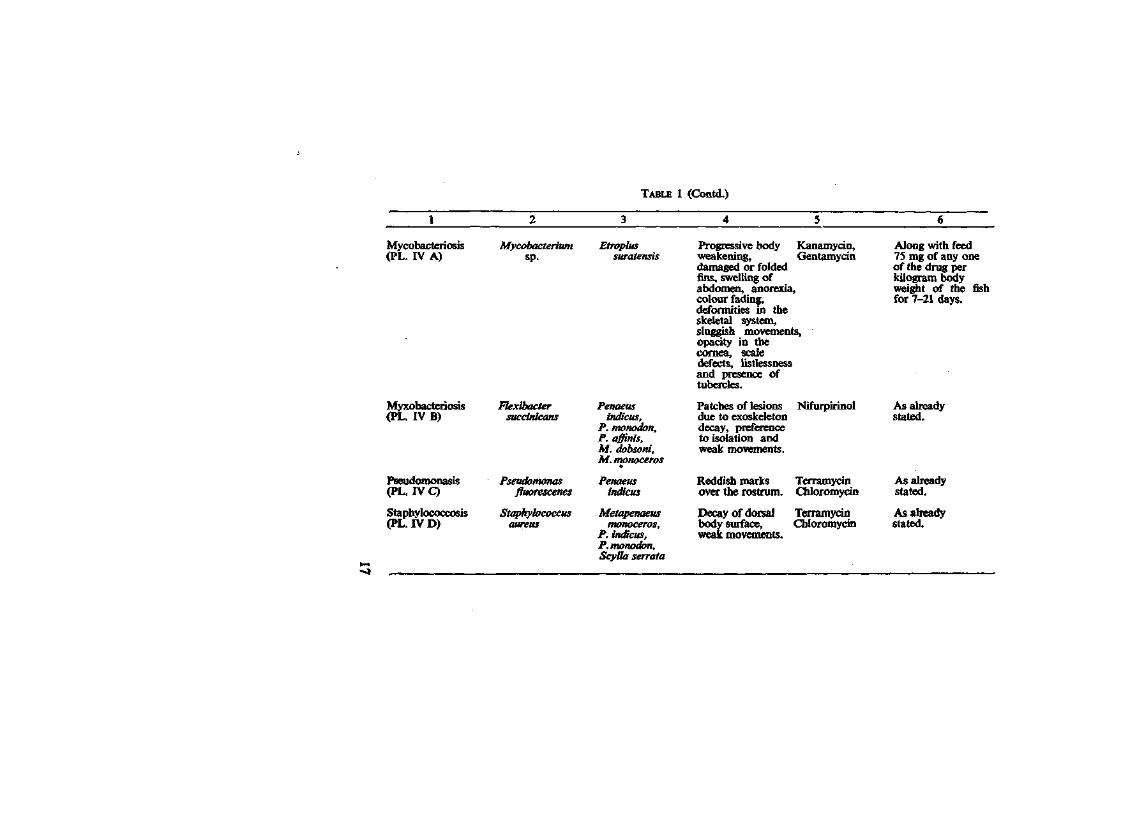
TABLE 1 (Contd.)
Mycobacteriosis Mycobacterium Etroplus Progressive body Kanamycin, (PL. IV A) sp. suratensis weakening, Gentamycin
damaged or folded fins, swelling of abdomen, anorexia, colour fading, deformities in the skeletal system, sluggish movements, opacity in the cornea, scale defects, listlessness and presence of tubercles.
Along with feed 75 mg of any one of the drug per kilogram body weight of the fish for 7-21 days.
Myxobacteriosis (PL. IV B)
Pseudomonasis (PL. IV Q
Staphylococcosis (PL. IV D)
Flexibacter succimcans
Pseudomonas fluorescenes
Staphylococcus aureus
Penaeus indicus,
P. monodon, P. affinis, M. dobsoni, M. monoceros
Penaeus indicus
Metapenaeus monoceros,
P. indicus, P. monodon, ScyUa serrata
Patches of lesions due to exoskeleton decay, preference to isolation and weak movements.
Reddish marks over the rostrum.
Decay of dorsal body surface, weak movements.
Nifurpirinol
Terramycin Chloromycin
Terramycin Chloromycin
As already stated.
As already stated.
As already stated.

00 TABLE 1 (Contd.)
Shell disease (PL. V A)
Vibrio fischeri P.indicus, Depressed black Terramycin P. monodon spots with margins Chloromycin
in the exoskeleton with or without underlying tissue necrosis.
As already stated.
Flavobacteriosis (PL. V B)
Streptococcosis (PL. V C)
Muscle necrosis (PL. VI A)
Flavobacterium ulginosum
Streptococcus pyogenes
Vibrio fischeri
Penaeus monodon
Panulirus homarus
P. indicus, Crassostrea
madrassensis, P. indicus
Irregular decay of tail region, weak movements.
Shell decay with adjacent tissue neucrosis, sluggishness, presence of blisters and anorexia.
Focal necrosis of tissue with varied colour.
Terramycin, Chloromycin
Terramycin, Chloromycin
Terramycin, Chloromycin
As already stated.
Alongwith feed at a rate of 75 mg of either of the drug per kilogram body weight of the shellfish for 7-21 days or intramuscular injection of a dmg at a dose of 20-40 mg/kg body weight of the host.
As already stated.

PLATE I. A. Haemorrhagic septicemia caused by Pseudomonas alcaligenes in Epinephelus pantherinus-B. Furunculosis caused byAeromonas salmonicida in Cichalosoma meeki (diagrammatic)' L f n | n
hJ e S ' T C a U S e d „ b y Pseudomonas fluoresce™ in Etroplus suratansis and D. Vibrios s
caused by Vibrio anguillarum in Etroplus suratensis. B

PLATE II: A. Skin spottiness caused by Vibrio fischeri in E. suratensis; B. Gill rot caused by Klebsiella pneumoniae in Tilapia mossambica; C. Tail rot caused by Photobacterium phosphoreum in E. suratensis and D. Eye disease caused by Vibrio parahaemolyticus in Cerres sp.

PLATE III. A. Entente caused by Escherichia coli in E. suratensis; B. Black spot disease caused by V,bno fisher, mPenaeus indicus; C. Scale disease caused by Vibrio parah^Tm^us in E. suratensis and D. ulcer caused by Haemophilus sp. in Trachinotus ovatus

0 PLATE IV: A. Mycobacteriosis caused by Mycobacterium sp. in E. suratensis; B. Myobacteriosis caused by
Flexibacler succinicans in P. indicus; C. Pseudomonasis caused by Pseudomonas fluorescens in P. indicus and D. Staphylococcosis caused by Staphylococcus aureus in P. indicus.

PLATE V: A. Shell disease caused by Vibrio fisheri in Panulirus sp; B Flavo-bacteriosis caused by Flavobacterium ulginosum in P. monodon and C. streptococcosis caused by Streptococcus pyogenes in Panulirus

TS^aaU^-
PLATE VI: A. Muscle necrosis caused by Vibrio fisheri in P. indicus; B. The anaesthetised fish E. suratensis and the anaesthetic MSS 222 (Tricaine Methane Sulphonate); C. Instruments for post-mortem examination and D. Inoculated (left) and uninoculated (right) culture plates.

PLATE VII: A. Fish infusion nutrient agar in conical flasks and B. Culture petridishes — Monoculture (left) and Polyculture (right).
W* JSfe. *" ^ * > i
wt
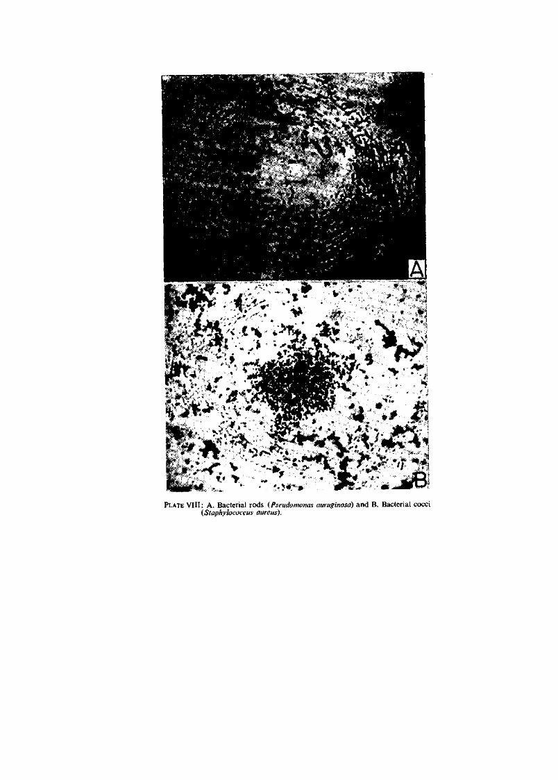
PLATE VIII: A. Bacterial rods (Pseudomonas auruginosa) and B. Bacterial cocci (Staphylococcus aureus).


5 PROPHYLAXIS AND DISEASE CHECK UP
In all animals the occurrence of infectious disease is unpredictable. It is often considered that the occurrence of disease in the culture system is unavoidable. However, this need not be so, for many diseases can be prevented by adopting prophylactic measures or certain management techniques.
It is essential that finfishes and shellfishes are safeguarded against diseases as they damage the resources in the natural and culture systems, which result in death, reduced growth and bodily damages resulting in non-marketability. Above all, the possibility of certain types of infections being transmitted through aquaculture products to man cannot be ruled out. Further, the occurrence of diseases in cultivable organisms may pose serious problems to the management of wild resources.
Culture systems use a variety of water sources which are classified in Table 2.
TABLE 2. Classification of culture ecosystem
CULTURE ECOSYSTEM
V SALT WATER FRESH WATER
Open Saline Estuaries Brackish Rivers Ponds Tanks Lakes sea lagoons water (Canals) (Reser
voirs) Each water source has its own ecological and biological chara
cteristics. The applicability of the following guidelines has certain limitations. However, the following prophylactic measures would be helpful.
20

METHODS OF PROPHYLAXIS
1. Selection of certified disease-free seeds for culture
As for agricultural seed crops, producing finflsh and shellfish seeds free of specified disease agents would be highly useful. This is in practice in some foreign countries such as U.S.A., Canada, U.K. and Denmark and might be adopted in India 'for some species where brood animals can be maintained in a specified pathogen-free status.
2. Development of disease-resistant seeds
This may be carried out by selective breeding from strain resistant to specific pathogens. Although genetic selection using desirable traits could be important, finding resistant strains may be lengthy process and there is no guarantee of success.
3. Suitable site selection
Sites for culture should be chosen according to the criteria known to be required for the cultivar. There should be adequate supplies of good quality water for the volume of production planned. Seasonal variation in quality may occur and therefore it will be important to gain knowledge of the physical properties and biological populations present. Possible sources of pollution (e.g. industrial wastes, agricultural sewage and pesticide residues) in the vicinity may necessitate rejection of potential sites.
4. Disinfection of culture site
It may be desirable to drain off culture ponds and treat them with a disinfectant (e.g. Quick lime/Calcium cyanamide/Sodium hydroxide). When such chemical treatment is carried out, stocking should be done only after a period of time to allow for dilution by flushing. The pH and other parameters such as temperature and salinity will have to be checked before stocking.
5. Installation of proper bund and sluice
This is applicable in the case of culture systems with tidal water exchange. Suitable bunds which are seepage proof are essential. Sluices should be installed in the bunds to control the inflow and outflow of water and permit good circulation.The mesh size of the screens of the sluices should be suitable to retain the
21

stocked animals and the same time prevent the entry of the free swimming larval parasites and predators from outside. Care should be taken for the proper maintenance of the screens and sluices.
6. Stock density
The field should be stocked with the recommended number of seeds for the species being cultured and the nature of the site. Because of the considerable variation in the productive capacity of sites, trial evaluations to determine optimum stocking densities may be necessary.
7. Prevention of environmental stress
Sudden change (e.g. stress in animals caused by occurrence of poor environmental condition) can impose severe imbalance in homeostasis to the stocked animals. For example, presence of excess of decaying organic matter may result in drastic decrease in the dissolved oxygen content of the water body. Here, the oxygen deficiency acts as an acute stress factor to the standing stock. Every effort should be taken to minimise or avoid any kind of stress of this kind to the animals.
8. Knowledge of life history of the pathogenic parasites
A knowledge of the life history of opportunistic pathogens may help us in preventing disease. This can be achieved by killing other host organisms necessary to complete the life cycle or preventing their entry into the culture system.
9. Food
Where food is provided, supply of a well-balanced diet free of pathogens should be maintained. Compounded feeds are usally pasteurised by the pelleting process. In the case of natural food collected for feeding, such as phytoplankton and zooplankton, they may be treated with ultraviolet radiation and/or deep-frozen over 24 hours (kills many parasites) for stock, both process reducing the numbers of any potential pathogens which may be present.
10. Maintenance of general cleanliness
Hygiene is very important in culture practices. Ultensils, nets and other instruments should be clean and regularly disinfected using materials such as iodophores or hypoclorite. In the absence
22

of sufficient facilities, the hatchery instruments may be scrubbed clean with detergent and water and well sun-dried before re-using. The actions will help to eliminate cross infection.
11. Pathogens and recirculating water systems
When an infectious disease occurs in animals in a recirculation system the biofilter will always have to be suspected as a source of infection. Therefore, it is desirable to operate a single filter until the disease has been cured/or the stock killed and then to replace or clean the filter bed.
12. Limitation of handling
Frequent handling of the animals is to be avoided, especially by inexperienced staff. Where appropriate, anaesthetise the animal before handling. Moreover, handling should be as gentle as possible, as injuries may cause disease often of epizootic proportions.
13. Exclusion of animals and birds
As far as possible, no opportunity should be given for animals and birds to get into the culture systems. They may act as disease vectors through their laeces. They may also be predators of the culture. 14. Seed immunisation
Mass immunisation against vibriosis is often successful by an immersion in the liquid vaccine. Oral vaccination is not so effective on the commercial scale. However, use of vaccines in aquaculture is as yet in the early stages of development.
15. Disease monitoring
Where certain infectious diseases occur regularly, e.g. in a hatchery, one method of control is to monitor their presence and destroy the infected population(s). This can only be contemplated when many non-infected groups are reared {e.g. in oyster and prawn hatcheries).
16. Prevention of lateral transmission of disease
Hatcheries and similar establishments which produce young or seed stock for grow-out elsewhere have a crucial role to play in the continuous supply of young stock. Widespread disease occurr-
23

ence in such a unit would disrupt production in many grow oul farms. It is therefore important to design hatcheries so that il disease does occur, it does not spread to the surrounding hatchery populations. Adequate physical separation of stocks, separate utensils for their maintenance and even separate staff for their husbandry may be necessary to achieve this.
17. Growth and survival
Fish and shellfish farmers are strongly advised to measure growth rates and survival in grow-out populations. If poor survival is indicated, growth is poor or the sampled animals look unhealthy they should seek expert advice.
24

6 THE NEED FOR ANAESTHETICS
In general, anaesthetics are central nervous system (CNS) depressants. Because certain manipulative actions are necessary in culture activities, they are used to render the animal unconscious. Their use is important to minimise stress from fright reactions and possible self injury or from handling. Excess exposure to the drug causes paralysis of the respiratory and vasomotor centres which finally leads to death.
The use of anaesthetics in fish culture is often necessary to perform injections, branding, tagging and weight and length measurements.
The methods of inducing anaesthesia in fishes are by inhalation, hypothermia and injection.
Inhalation can be successfully done by adding the anaesthetic in the gill irrigating water. Here, the anaesthetic is absorbed into the blood stream through the gills. Inhalation by bathing is considered the easiest and most effective method of inducing anaesthesia. This is the commonest method in use in aquaculture.
Hypothermia is a physical method of inducing anaesthesia (cooling of the body for 10-15 minutes, inordesr to reduce oxygen requirements of the cells). This method has its limitations.
Injection can be intramuscular, intraperitoneal, intravenous and it is the least preferred route for the opsration.
Chloretone and benzocaine are good anaesthetics for shallow (short-term) anaesthesia. However, MS 222 (Tricaine methane
25

0\ TABLE 3. Anaesthetics and their use
Common name
Chemical name
Dose Time (in minutes) for
Immobi- Recovery lisation
Effect on fish
Effect on human beings
Remarks
Chloretone/ Chlorobutanol Trichloro-
tert-butyl alcohol
0-1% 2-5 3-7 Short term Irritant When the fish is immers-anaesthesia ed in a solution of anaes
thetic, an initial phase of excitement takes place followed by erratic swimming. Then, the fish become inactive and sink to the bottom of the water body. Anaesthetised fish should be removed immediately for the 'purpose.' Time for immobilisation and recovery vary with species. So behaviour of the fish should be observed carefully while they are being anaesthetised to avoid cardiac arrest due to excess anaesthesia. It is not desirable to use the anaesthetised fish for food within the following 21 days of anaesthetisa-tion. The anaesthetic is cheap and locally available.

MS 222 Tricaine 0.01 % Methane
Sulphonate
-a
Short and Negli-deep gible
anaesthesia toxic effect
When the fish is immersed in a solution of anaesthetic, an initial phase of excitement takes place followed by erratic swimming. Then, the fish become inactive and sink to the bottom of the water body. Anaesthetised fish should be removed immediately for the 'purpose.' Time for immobilisation and recovery vary with species. So behaviour of the fish should be observed carefully while they are being anaesthetised to avoid cardiac arrest due to excess anaesthesia. It is not desirable to use the anaesthetised fish for food within the following 21 days of anaesthetisa-tion. The anaesthetic is expensive and not easily available locally.

sulphonate) is a better anaesthetic for deep (long-term) anaesthesia, for 10-30 minutes (PL. VI. B). Adequate care has to be taken in administering the correct dosages. Appropriate concentrations are listed in Table 3.
The other fish anaesthetics are Carbon dioxide, Chloral hydrate, Ether, Methylpentynol, Novacaine, 2-Phenoxyethanol, Quinal-dine, Sodium amytal, Tribromoethanol and Tertiary-amyl alcohol.
Fish should be held in the anaesthetic for the minimum time possible and returned to fresh anaesthetic-free water as soon as the manipulation to be performed, has been done. Before an anaesthetic is used, a trial on a small number of animals is advised.
The recommended general anaesthetics for use in aquaculture are:
i. Benzocaine or chlorobutanol for fishes. However, great care must be taken to ensure benzocaine is not used in direct sunlight. In sunlight it decomposes to produce chlorine which is acutely toxic.
ii. For Crustacea (shrimps) quinaldine may be used as an anaesthetic at a level of 25 ppm.
iii. To anaesthetise molluscs, menthol is used.
28

7 SAMPLING TECHNIQUES FOR DISEASE DIAGNOSIS
Collection and examination of moribund specimens before death is very important for diagnostic purposes because post-mortem (PM) changes may obscure or confuse diagnosis.
Moribund specimens should be collected and sent alive as soon as possible to the Pathology Laboratory furnished with the necessary PM equipment.
The essential items required for the post-mortom examination (PL. VI C) are Surgical knife/scalpel, Scissors, Tissue forceps, Dissection needles and pins, Hand lens, Dissection board, Bacteriological wire loop, Slides and cover slips, Pipettes, test tubes, Petri dishes, Tissue paper, Gloves, Microscope, Bacteriological medium e.g. Nutrient agar/Fish infusion agar plates and Histological fixative e.g. 10% buffered formol saline.
Once the moribund specimen collection is over, the minimum procedure for further study is as follows:
Procedure for Post mortem examination
a. Species : Record species, age, sex, length and weight.
b. Disease signs : Compare at least 6-20 moribund animals to confirm the signs of disease and study the commonest signs both external and internal.
c. : Record external and internal pathologies.
29

d. : Extract blood for study of haema-tological parameters e.g., haema-tocrit red and white cell counts, differential counts, etc.
e. : Make smears/scrapingsfrom lesions, body fluids (blood) and tissue imprints for subsequent staining.
f. : Take bacteriological samples on loop and plate out.
g. : Cut out pieces of each of the major organs for histology e.g. liver, kidney, pancreas, spleen, gills, etc. in fish; affected tissue and haemolymph in crustaceans and affected tissue and haemolymph in molluscs.
A case study sheet is appended below to serve as a guide for data aquisition on disease and the concerned biological and environmental factors.
The quality of water is an index for disease investigation. Water samples may be collected in clean, dry and sterile glass containers taking precautions to minimise external contamination. Every effort should be made to avoid delay and post-collection contamination of the samples.
CASE STUDY SHEET
GENERAL
1. Farm name 2. Owner
3. Address
A. CULTIVAR HISTORY
Species diseased Age Sex Length Weight Kind of feed
30

Method of feeding : Stocking density : Standing stock : Recent history of treatments : Any recent handling
of population : Estimate of loss (Number) :
Water chemistry (if relevant)
Habitat water - colour : smell : turbidity : oxygen : salinity : temperature : pH : phytoplankton : zooplankton :
B. PRESENT COMPLAINTS
C. PHYSICAL EXAMINATION
C-l. Reflex activities
Defensive reflex : Escape reflex : Ocular reflex : Tail reflex :
C-2. System history (external)
Visual nature of - slime scale skin/body cephalothorax abdominal segments mouth antenna antennules rostrum pharynx carapace oral cavity

nostril eyes operculum gills tail/fins postrostral carina dorsal carina belly nostril pereiopods pleopods urogenital carina vent thelycum/petasma
C-3. System history (internal)
Visual nature of - body tissue brain heart liver pancreas spleen gall bladder swim bladder kidney stomach intestines hepatopancreas pyloric caecae urinary bladder gonads body cavity spine
D. PREVIOUS CASE HISTORY (if any)
E. WET MOUNT MICROSCOPICAL EXAMINATION
LABORATORY DATA
F. HAEMATOLOGY
Total counts of RBC WBC
Haematocrit
32

Differential white cell counts
Neutrophils Eosinophils Basophils Lymphocytes Monocytes Haemoglobin ESR
G. VIROLOGY
Tissue culture (fish only) Transmission Transplantation Electron microscopy
H. HISTOPATHOLOGY
I. RADIOGRAPHY
J. CULTURE AND ANTIBIOTIC SENSITIVITY
Locally affected cells Blood (cardiac) Kidney Hepatopancreas
K. SATISFACTION OF KOCH'S/RIVERS' POSTULATES
(Optional, only required if a new disease is being studied and the results to be written up for scientific publication).
L. CASE DIAGNOSIS
M. CONCLUDING REPORT
N. DRUG(S) PRESCRIPTION
Laboratory Place Date
Signature Name of the Scientist Designation

8 CLASSIFICATION OF PATHOGEN(S)
In investigations of mortality and morbidity, consideration of an infectious cause is an essential aspect. The signs of dead and dying animals aid in the provisional diagnosis. To prove such a hypothesis microscopical examination of blood (stained smears) and tissues (wet mounts of scrapings, stained histological sections and tissue imprints)must be taken for the cultivation and isolation of probable microbial pathogens, e.g. bacteria or fungi. Animal parasites can generally be identified from scrapings and/or histological sections. If a bacterial cause is suspected from the examination (microscopic) aseptically removed sample(s), by bacteriological loop, should be directly inoculated on to the culture medium (PL. VID) There is no single medium in use tor culture and isolation of all the pathogens which may be present in a sample. However, a suitable all-purpose culture medium for bacterial pathogens is fish infusion nutrient agar (Fina).
FISH INFUSION NUTRIENT AGAR (PL. VII A)
Peptone : 1.0 gm Agar agar : 1.5 „ Beef extract : 0.1 „ Fish infusion* : 100 ml.
(pH adjusted to 7.2 and sterilized at 121 °C for 15 minutes).
454 gm of fresh fish meat is minced and mixed into one litre of water**. The mixture is kept overnight in a refrigerator at 4°C. Then, the mixture is boiled using a water bath for about 30
Preparation of fish infusion. In the case of water, aged and filtered sea water is preferred for isolation of bacterial pathogens from marine fish and distilled water with 0.5 % sodium chloride for fresh water fish.
34

minutes. The precipitated and coagulable protein in the mixture can be removed by filtering through a lint and filter paper.
Shrimp infusion nutrient agar, crab infusion nutrient agar and mussel infusion nutrient agar are prepared in similar manner, replacing the fish muscle by shrimp, crab or mussel meat for isolating bacterial pathogens from the respective species. Fresh media may always be used as the cost of these media is low and the best results are obtained.
The inoculated culture media may be incubated aerobically at room temperature, e.g. 28±2°C for 24-72 hours.
The predominant bacterial species in the sample will usually be the most numerous of the colonies growing on the plate (PL. VII B). Select a representative of the dominant or, if more than one all the dominating colonial types. Study their colonial characteristics such as sizes, shape, margin, elevation, consistency, opacity and colour. Transfer by loop the organisms into a fish infusion nutrient broth (a broth tube for each colony type). The isolate has to be checked for its purity (i.e. consistent colony morphology) by streaking over a fish infusion nutrient agar. The purified isolate is then subjected to identification procedures.
Generally based on the experience of the pathology team investigating the disease, the signs presented by the morbid animals and the isolation of known pathogens will be sufficient to allow case diagnosis. Only where a new disease involving a pathogen not previously characterised or not characterised as causing that sign of that disease, there is need to resort to Koch's postulates.
35

9 SCREENING OF BACTERIA FOR IDENTIFICATION
Identification of bacterial isolates is the next step in the process of disease diagnosis. The problems here are greater for the aquatic microbiologist compared to those of a medical microbiologist, because of the larger number of studies which have been made in medical microbiology.
For identifying bacteria, various systems of classification exist (Scholes and Shewan, 1964). A very detailed and useful key is provided by Buchanan and Gibbons (1974). However, problems still exist in system of classification. The most detailed system of classification described in Bergey's Determinative Bacteriology (Buchanan and Gibbons, 1974) can be handled only by specialists.
In order to aid beginners in microbial taxonomy, a simple and rapid system is called for and this has led to the formation of the keys given in Tables 4 and 5 for screening bacteria: rods and cocci (PL. VIII A, B)
These keys require the following tests for categorising an organism.
1. Cell morphology and Gram's staining according to Hucker's modification (Bullock, 1971). The staining method is given below.
2. Motility in hanging drop preparations from cultures following inoculation for 6-18 hours in peptone watei or sea water peptone (1% peptone in aged and filtered sea water, pH 7.2-7.5).
3. Sensitivity to 2.5 I.U. of pencillin and '0/129'* (Shewan et al, 1954). (1 mg of pencillin is 1667 I.U.)
* '0/129'=2,4—Diamino—6, 7 diisopropyl pteridine
36

4. Breakdown of glucose (Hugh and Leifson, 1953).
5. Oxidase according to Kovacs (1956).
6. Haemolysis in blood agar (cooled sterile nutrient agar plus 5% sterile defibrinated blood).
7. Oxygen relationship (Mackie and McCartney, 1962).
8. Coagulase (Mackie and McCartney, 1962).
9. Cellulose digestion (Skerman, 1959).
10. Acid and gas production in peptone water including respective sugar with Andrade's indicator (Cruickshank et ah, 1975).
11. Pigmentation in milk agar.
If the disease is suspected to be caused by a mycobacterial species, inoculate the sample in glycerol agar (Nutrient agar plus 1% glycerol) and incubate at room temperature for a few days upto 2 weeks. Colony characters should be noted if acid-fast and Ziehl Neelson-positive organism is observed. The organism may be tentatively classified, based on the pigment, as Mycobacterium marinum if the pigment is photochromogenic and M. fortuitum if there is no pigment.
STAINING METHODS IN BACTERIAL IDENTIFICATION
Gram's staining
Step I a. Crystal violet (BDH, England) : 20.0 gm Absolute alcohol : 200 ml
b. Ammonium oxalate (BDH, AR) : 8.0 gm Distilled water : 800 ml
Mix solutions a and b
Step II Iodine (BDH, AR) Potassium iodide (BDH, AR) Distilled water (Dissolve the solutes well and then add 147.5 ml of distilled water)
0.5 gm 1.0 „ 2.5 ml
37

Step III Safranin 0(BDH, AR) : 2.5 gm Absolute alcohol : 10 ml Distilled water : 100 ml
Procedure
1. Prepare the slide (Dry the smear well) (Using a sterile inoculating loop, bacteria are picked off the media and mixed with a few drops of sterile physiological saline on a slide. A thin smear is made and allowed to air dry and fixed by passing the slide through a Bunsen flame).
2. Flood the smear well enough with ammonium oxalate-crystal violet solution for one minute.
3. Wash the smear in tap water for a few seconds.
4. Cover the smear with iodine solution and allow to stay for one minute.
5. Wash the smear in tap water for a few seconds.
6. Decolourise the smear using absolute alcohol for one minute.
7. Wash the smear again, in tap water, for a few seconds.
8. Counter-stain with Safranin for 30 seconds.
9. Wash the smear in tap water for a few seconds.
10. Dry the smear for examination.
(Bacteria appearing as violet-blue in colour are Gram-positive and pink-red are Gram negative).
38

TABLE 4. Screening tests of microbes
BACTERIA (RODS)
Spores
Bacillus sp. <BAClLLOSIS)
GRAM'S STAINING
Corvnebacterium sp. (KIDNEY DISEASE)
1 Short rods
> I Long thin rods
Flexibacter/Cytophaga sp.
(MYXOBACTERIOSIS)
I T
Breakdown of glucose
I
Sensitivity to penicillin (2.5JJ.U.)
I Pigment
Flavobacterium sp. (FLAVOBACTERIOSIS)
J_ No pigment
Alcaligenes sp. (ALCALIGENOSIS)
Fermentative -I -
With gas but in sensitive to '0/129*
Motility
Motile Aeromonas sp.
without gas but sensitive to '0/129' Vibrio sp.
(VIBRIOSIS)
1 Non-fermentative
1 Kovacs' oxidase
Non-motile Aeromonas saimonicida
V +
Pseudomonas sp. Enterobacteriaceae (HEMORRHAGIC SEPTICEMIA) (FURUNCULOSIS) (HEMORRHAGIC SEPTICEMIA) (ENTERIC BACTERIOSIS)
"0/129* = 2, 4 Diamino 6, 7 diisopropyl pteridine

TABLE 5. Screening tests of microbes'
(GRANT, COCCI)
Motility _ _ J
Motile Planococcus sp.
j - i Non-inotile
Breakdown of g
u lucose
Haemolytic cells in chains
Streptococcaceae
• T May or may not be
haemolytic, cells in irregular clusters
Fermentative J
May or may not be haemolytic, cells
in packets of eight
Coagulase
1
Sarcina sp. ~\ Aerobic
Micrococcus sp. (MICROCOCCOSIS)
- i
Staphylococcus aureus Staphylococcus epidermidis OR
Staphylococcus saprophyticus
Ruminococcus sp.
In chain Peptostreptcoccus sp.
Non-fermentative
Oxygen relation
7 Anaerobic
Peptococcaceae (except the genus
Sarcina)
Cellulose digestion
' 1 Cell arrangement u
Not in chain Peptococcus sp.
Haemolytic cells in chains
Streptococcus sp. (STREPTOCOCCOSIS)
Non-haemolytic lenticular cells
Lcuconostoc sp.
cells in pair or in tetrads
Pediococcus sp.
cell in tetrads
Aerococcus viridans (AEROCOCCOSIS)
Haemolytic cells of flattened
adjacent sides
Gemella sp.

10 THE DESPATCH OF DISEASED SPECIMENS
In some countries where aquaculture is established, fish disease laboratories exist to help the fish and shellfish farmers. In India there is at present little expertise on this subject. However, research organisations such as Central Marine Fisheries Research Institute, Cochin; Central Inland Fisheries Research Institute, Barackpore and the College of Fisheries of Bangalore Agricultural University are centres where the expertise is being gained.
If diseases among the cultivated fishes are detected, live specimens or preserved samples should be sent to the nearest fish pathology laboratory for disease diagnosis. However, there are, certain observations to be made before concluding that the fish stock is suffering from an infectious disease. If possible it should be verified whether the stock is getting poisoned (due to consequence of) poor water quality. Poisoning may be suspected if there is lack of macro- or microscopical signs of disease and/or a rapid depletion of the standing stock (often the sudden onset of death of the entire population is suggestive of poisoning). In such cases, it will be important to consult scientists with experience of the ecology of aquatic ecosystems and the chemistry of the waters.
If the case is suspected to be due to infection, the specimens may be despatched in accordance with the following suggestions.
1. Forward specimens of the diseased species alive, say, 6 (representative specimens) in a clean container (polythene bags) with 1/3 of the volume filled with water. Protect the bag by placing it in a cardboard box or container. Fill with oxygen, if possible.
2. Dead animals should not be sent. If live animals cannot be sent, send preserved tissues in 10% buffered formalin.
41

3. Never deep-freeze the specimens for bacteriological analysis.
4. All the available information, regarding the case, may be furnished in the case study sheet (vide specimen copy in the earlier chapter).
5. Regarding water samples deep - freeze upto 3 litres immediately and store until expert advice is available. This will be suitable for certain chemical analysis. Fix some 100-200 ml of water with Lugol's iodine to preserve phytoplankton.
42

11 PATHOLOGY —AN OUTLOOK
In the field of pathology, there has been considerable progress in the human and veterinary clinical medicine. In aquatic animal pathology, finfish and shellfish disease investigation and associated research remains a virgin field. This is because of a lack of awareness and the absence of trained personnel. Such conditions are now changing in India and elsewhere because of the global awareness of the importance of the culture of finfishes and shellfishes. Large scale developmental programmes now being contemplated will require a better knowledge of the diseases and their control in the cultivable species.
Aquatic animal pathology (aquatic veterinary pathology) is concerned with six main areas of study.
1. Aetiology: This means the causes of disease and is a subject for study by itself. Moreover, it includes the study of infections and other causes of disease.
2. Pathogenesis: This is the progressive development of the course of a disease in the affected host from the time of infection to the final stage of recovery or death.
3. Pathophysiology: It comprises the changes occurring in the biochemical components of the body tissue and fluids of the diseased host (e.g. changes in red and white cells, etc.).
4. Histopathology: Study of the changes in tissue architecture as a consequence of disease e.g. inflammatory responses and necrosis are two of the many possible changes which may occur.
43

5. Epizootiohgy: Systematic study of the origin, distribution and spread of diseases. It involves the recognition of species, sex and age-group affected, percentage of mortality, including the disease distribution.
6. Control measures: Control measures mean prevention of disease and treatment of diseased animals. Prevention means to avoid the occurrence of the disease. This can be achieved by several means. In the case of vibriosis, there are several measures, such as immunization, prophylactic chemotherapy, genetic control (breeding resistant species). Several sanitary measures are significant. Physical control by timely removal of the infected population is also useful.
7. Treatment: This is to treat and cure or control the disease. There are several methods of chemotherapy. Antibiotics may be administered by way of mass injection, feeding or through mixing in water. For treatment of vibriosis, oxytetracycline is found an effective control by oral administration usually with the animal food.
44
REFERENCES
AHNE, W. (Ed.) 1980. Fish diseases. Springer-Verlag, New York. 252 pp.
ALMEIDA, L. J. 1962. M.Sc. Thesis. University of Bombay, Bombay.
BUCHANAN, R. A. AND N. E. GIBBONS (Eds.) 1974. Bergey's Manual of Determinative Bacteriology. Williams and Wilkins Co., Baltimore, Md. 8th edn. 1268 pp.
BULLOCK, G. L. 1971. Bacterial diseases of fishes. Book 2 B, TFH Publication, Jersey City NJ. 41 pp.
, D. A. CONROY AND S. F. SNIESZKO 1971. Bacterial diseases of fishes. Book 2 A. Ibid., 151 pp.
CONROY, D. A. AND R. L. HERMAN 1970. Text book offish diseases. TFH Publications, Jersey City USA. 302 pp.
CRUICKSHANK, R. (Ed). 1962. Mackie and McCartney's Hand book of Bacteriology. Edinburgh and London. 980 pp.
, J. P. DUGUID, B. P. MARMION AND R.H.A. SWAIN 1975. Medical microbiology. Edinburgh: Churchill Livingstone. 12th edn. 587 pp.
DULIN, M. P. 1976. Diseases of marine aquarium fishes. TFH Publications. 128 p.
HOFFMAN, G. L. (Ed.) 1977. Methods for the diagnosis of fish diseases. Amerind Publishing Co. Pvt. Ltd., New Delhi, 140 pp.
HUGH, R. AND E. LEIFSON 1953. The taxonomic significance of fermentative versus oxidative metabolism of carbohydrates by various gram negative bacteria. Jour. Bad., 66: 24-26.
KOVACS, N. 1956. Identification of Pseudomonas pyocyanea by the oxidase reaction. Nature (London), 178: 703.
MAWDESLEY-THOMAS, L. E. (Ed.) 1972. Diseases of fishes. Symposia of the Zoological Society of London. No. 30. Academic Press Publication, 380 pp.
MIMS, C. A. 1976. The pathogenesis of infectious disease. Academic Press Inc. (Lodon) Ltd., 246 pp.
PILLAI, C. THANKAPPAN 1978. Studies on microorganisms associated with some fish diseases. M.Sc. Thesis, Univ. of Bombay.
1982. Studies on finfish and Shellfish diseases, Ph.D. Thesis, Univ. of Cochin.
REICHENBACH-KLINKE, H. H. 1973. Fish Pathology. TFH Publication.
RIBELIN, W. E. AND G. MIGAKI (Ed.) 1975. The pathology of fishes. The University of Wisconsis Press Publication, 1004 pp.
45

ROBERTS, R. J. AND C. J. SHEPHERD 1974. Handbook of trout and salmon diseases. Fishing News (Books) Ltd. 168 pp.
(Ed.) 1978. Fish Pathology. Bailliere-Tindall Publication, London. 318 pp.
(Ed.) 1982. Macrobial diseases of fish. Academic Press Inc. (London) Ltd., 305 pp.
SCHOLES, R. B. AND J. M. SHEWAN 1964. The present status of some aspects of marine microbiology. Adv. mar. Biol, 2: 133-170.
SHEWAN, J. M., W. HODGKISS AND J. LISTON 1954. A method for the rapid differentiation of certain nonpathogenic asperogenous bacillus. Nature (London), 173: 208.
SINDERMANN, C. J. 1970 a. Diseases of marine fishes. TFH Publications. 89 pp.
1970 b. Principal diseases of marine fish and shellfish. Academic Press Publication, NY. 369 pp.
1977. Disease diagnosis and control in North American marine aquaculture. An Elsevier Publication. 329 pp.
SKERMAN, V. B. D. 1959. A guide to the identification to the general of bacteria. Baltimore, Williams and Wilkins Co.
VAN DUUN JR. C. 1973. Diseases of fishes. Iliffe Books Ltd., London. 372 pp.
46

SUGGESTED READINGS
AHNE, W. (Ed.) 1980. Fish diseases. Springer-Verlag, New York. 252 pp.
CONROY, D. A. AND R. L. HERMAN 1970. Text book offish diseases (Translation of Taschenbuch der Fischkrankheiten by Amlachcr, E.). TFH Publication.
DAVIS, H. S. 1953. Culture and diseases of game fishes. University of California Press Publication.
DOGIEL, V. A., G. K. PETRUSHEVSKI AND YU. I. POLYANSKI 1966. Parasitology of fishes (English translation by Kabata, Z., in 1970) TFH Publication.
DULIN, M. P. 1966. Diseases of marine aquarium fishes. TFH Publication.
ELKAN, E. AND H.H. REICHENBACH-KLINKE 1974. Colour atlas of the diseases of fishes, amphibians and reptiles. TFH Publication.
GOLDSTEIN, R. 1971. Diseases of aquarium fishes. TFH Publication.
HOFFMAN, G. L. AND F. P. MEYER 1974. Parasites of fresh water fishes. TFH Publication.
HOFFMAN, G. L. (Ed.) 1977. Methods for the diagnosis of fish diseases (Translation of Metodicke Novody K Diagnostic* Nemoci Ryb by Lucky, Z) Amerind Publishing Co. Pvt. Ltd.
MAWDESLEY-THOMAS, L. E. (Ed.) 1972. Diseases of fishes. Symposia of the Zoological Society of London, No. 30. Academic Press Publication.
REICHENBACH-KLINKE, H. H., H. HERMAN AND E. ELKEN 1965. Principal diseases of lower vertebrates. Academic Press Publication.
REICHENBACH-KLINKE, H. H. 1973. Fish. Pathology. TFH Publication.
RIBELIN, W. E. AND G. MIGAKI (Eds.) 1975. The Pathology of fishes. The University of Wisconsin Press Publication.
ROBERTS, G. 1971. Diseases of aquarium fishes. TFH Publication.
ROBERTS, R. J. AND C. J. SHEPHERED 1974. Handbook of trout and salmon diseases. Fishing News (Books) Ltd.
ROBERTS, R. J. (Ed.) 1978. Fish. Pathology. Bailliere-Tindall Publication.
SCHUBERT, G. 1974. Cure and recognize aquarium fish diseases. TFH Publication.
SINDERMANN, C. J. 1970 a. Diseases of marine fishes. TFH Publication.
SINDERMANN, C. J. 1970 b. Principal diseases of marine fish and shellfish. Academic Press Publication.
47

SINDERMANN, C. J. 1977, Disease diagnosis and control in North American marine aquaculture. Elsevier Publication.
SNIESZKO, S. F. (Ed.) 1970. Symposium on diseases of fishes and shellfishes. Special Publication 5. American Fisheries Society Publication.
SNIESZKO, S. F. AND H. R. AXELROD (Eds.) 1971. In: Diseases of Fishes.
Book 1 Crustacea as enemies of fishes by Z. Kabata. TFH Publication.
Book 2 A Bacterial diseases of fishes by G. L. Bullock. D. A. Conroy and S.F. Snieszko. TFH Publication.
Book 2 B Identification of fish pathogenic bacteria, by G. L. Bullock. TFH Publication.
Book 3. Prevention and treatment of diseases of warm water fishes under subtropical conditions with special emphasis on intensive fish farming, by S. Sarig, TFH Publication.
Book 4. Fish immunology by D. F. Anderson. TFH Publication.
VAN DUIJN, JR. C. 1973. Diseases of fishes. Iliffe Books Ltd.
48

GLOSSARY
Aetiology — Study of the cause(s) of a disease.
Anaemia — Lack of red cells in blood.
Anorexia — Not feeding.
Anoxia — Absence of oxygen.
Asphyxiation — Suffocation.
Biopsy — Study, usually be histological technique, of
tissues removed from a live host.
Diagnosis — Examination of an infected host.
Ecchymosis — An effusion of blood beneath the skin.
Enopthalmia — A condition of retracted eyes.
Erythemia — Superficial redness in the skin/body.
Exopthalmia — Protruding eyes.
Health — A state of complete well being of the host. Haemorrhage — Loss of blood from any part of the vascular
system.
Hyperemia — Excessive amount of blood in any part of the
body.
Hypoxia — Reduced amount of oxygen.
Ischemia — Deficiency of arterial blood in any part or all of an organ.
Lesion — Microscopic or macroscopic alteration in the tissues as a result of infection, insult or injury.
Macrophage — Group of cells (both free and fixed) in the body having the phagocytic power.
49

Necropsy — An examination of the diseased host after death. This is also known as autopsy or post-mortem examination.
Necrosis — Death of cells of a living host.
Pathogenesis — Course and progression of a disease.
Pathognomonic — Typical sign of a disease; may be used to diagnose the cause of a disease.
Phagocyte — A defensive cell in the body having the power to engulf any foreign cell or foreign material.
Prognosis — An opinion of the course, progression and outcome of a disease or treatment of a disease.
Sign of disease — It is a clinical expression, manifested by a living host, as a result of tissue changes of a diseased condition.
50

Manuals of Research Methods issued under the Centre of Advanced Studies in Mariculture, Central Marine Fisheries Research Institute, Cochin.
1. Manual of Research Methods for Crustacean Biochemistry and Physiology. CMFRI Special Publication No. 7, 1981, 172 pp.
*2. Manual of Research Methods for Fish and Shellfish Nutrition. CMFRI Special Publication No. 8, 1981, 125 pp.
3. Manual of Research Methods for Marine Invertebrate Reproduction. CMFRI Special Publication No. 9, 1982, 214 pp.
*4. Approaches to Finfish and Shellfish Pathology Investigations. CMFRI Special Publication No. 11, 1983, 43 pp.
5. Application of Genetics in Aquaculture. CMFRI Special Publication No. 13, 1983, 90 pp.
6. Manual of Research Methods for Invertebrate Endocrinology. CMFRI Special Publication No. 14, 1983, 114 pp.
7. Production and Use of Artemia in Aquaculture. CMFRI Special Publication No. 15, 1984, 74 pp.
8. Manual on Marine Toxins in Bivalve Molluscs and General consideration of Shellfish Sanitation. CMFRI Special Publication No. 16, 1984, 100 pp.
9 Handbook on Diagnosis and Control of Bacterial Diseases in Finfish and Shellfish Culture. CMFRI Special Publication No. 17, 1984, 50 pp.
10. Mariculture Research under the Centre of Advanced Studies in Maricultue. CMFRI Special Publication No. 19, 1984, 109 pp.
11. Water Quality Management in Aquaculture. CMFRI Special Publication No. 22, 1985 (in press).
* Out of print.


