Page 1
Universität Trier – Fachbereich I – Psychobiologie
Dissertation zur Erlangung des
Doktorgrades der Naturwissenschaften (Dr. rer. nat.)
The Cold Pressor Stress Test: From basic psychophysiology to
application
Autor
Mauro Larrá y Ramírez, Dipl.-Psych.
Eingereicht am 5. Mai 2015
Gutachter
Prof. Dr. med. Hartmut Schächinger
Dr. rer. nat. Ewald Naumann
Page 2
Dissertationsort
Trier
Page 3
Index
iii
This dissertation thesis and the presented research were performed at the
Division of Clinical Psychophysiology
Institute of Psychobiology – University of Trier
Trier, Germany
Affiliation of Supervisors
Prof. Dr. med. Hartmut Schächinger
Clinical Psychophysiology Division – Institute of Psychobiology – University of Trier
Dr. rer. nat. Ewald Naumann
General Psychology and Methods Division – Department of Psychology – University of Trier
Part of the here presented research was supported by the
International Research Training Group “Psychoneuroendocrinology of
Stress: From Molecules and Genes to Affect and Cognition”
Funded by the German Research Foundation (Deutsche Forschungsgemeinschaft: DFG),
project GRK 1389/1
Page 4
Index
iv
Acknowledgements
I would like to thank all people that directly or indirectly contributed to the creation of this
thesis.
Prof. Hartmut Schächinger, for being the best supervisor one could wish for. I am deeply
grateful for all the support, encouragement, patience and trust over the last years.
Dr. Ewald Naumann for introducing me into the IRTG, for his supervision and revision of this
work and for being an inexhaustible source of methodological advice.
Dr. Immo Curio for his invaluable technical advice and help and most enjoyable company on
many occasions.
All my colleagues, especially Dr. André Schulz, Bart Kozik, Dr. Christian Deuter, Dr. Corinna
Peifer, Daniel Best, Dr. Diana Ferreira de Sá, Johannes Finke, Lisa Pramme, Dr. Thomas
Schilling, Xenia Hengesch and Xinwei Zhang, for the nice work environment and successful
collaboration, for the fun times, great conversations and lots of help in many ways.
All our research students for helping with the experiments and data analyses.
The participants that took part in the experiments.
My parents and family for a stimulating and warm environment, the freedom to make my own
choices and lots of support throughout the years.
Alma for giving me purpose and making me smile.
Kati for just everything.
Page 5
Index
v
General Abstract
The last decades of stress research have yielded substantial advancements highlighting the
importance of the phenomenon for basic psychological functions as well as physical health and
well-being. Progress in stress research heavily relies on the availability of suitable and well
validated laboratory stressors. Appropriate laboratory stressors need to be able to reliably
provoke a response in the relevant parameters and be applicable in different research settings
or experimental designs. This thesis focuses on the Cold Pressor Test (CPT) as a stress induction
technique. Three published experiments are presented that show how the advantages of the CPT
can be used to test stress effects on memory processes and how some of its disadvantages can
be met by a simple modification that retains its feasibility and validity.
The first experiment applies the CPT in a substantial sample to investigate the consolidation
effects of post-learning sympathetic arousal. Stressed participants with high increases in heart
rate during the CPT showed enhanced memory performance one day after learning compared
to both the warm water control group and low heart rate responders. This finding suggests that
beta-adrenergic activation elicited shortly after learning enhances memory consolidation and
that the CPT induced heart rate response is a predictor for this effect. Moreover, the CPT proved
to be an appropriate stressor to test hypothesis about endogenous adrenergic effects on memory
processes.
The second experiment addresses known practical limitations of the standard dominant hand
CPT protocol. A bilateral feet CPT modification is presented, the elicited neuroendocrine stress
response assessed and validated against the standard CPT in a within-subjects design. The
bilateral feet CPT elicited a substantial neuroendocrine stress response. Moreover, with the
exception of blood pressure responses, all stress parameters were enhanced compared to the
standard CPT. This shows that the bilateral feet CPT is a valid alternative to the standard CPT.
The third experiment further validates the bilateral feet CPT and its corresponding control
procedure by employing it in a typical application scenario. Specifically, the bilateral feet CPT
was used to modulate retrieval of event files in a distractor-response binding paradigm that
required lateralized bimanual responses. Again, the bilateral feet CPT induced significant
increases in heart rate, blood pressure and cortisol, no such increases could be observed in the
Page 6
Index
vi
warm water control condition. Moreover, stressed participants showed diminished retrieval
compared to controls. These results provide further evidence for the feasibility and validity of
the bilateral feet CPT and its warm water control procedure.
Together the experiments presented here highlight the usefulness of the CPT as a tool in
psychophysiological stress research. It is especially well suited to test hypothesis concerning
stress effects on memory processes and its applicability can be further increased by the bilateral
feet modification.
Page 7
Index
vii
Table of Contents
Acknowledgements .................................................................................................................. iv
General Abstract ...................................................................................................................... v
Table of Contents ................................................................................................................... vii
Index of Figures ....................................................................................................................... xi
Index of Tables ...................................................................................................................... xiii
Index of Publications ............................................................................................................. xiv
Index of Abbreviations........................................................................................................... xv
Chapter I: General Background ............................................................................................. 1
1.1 Introduction and Outline .................................................................................................. 1
1.2 Stress ................................................................................................................................ 2
1.2.1 The stress response .................................................................................................... 2
1.2.1.1 Sympathetic Nervous System ............................................................................. 3
1.2.1.2 Hypothalamic-Pituitary-Adrenal Axis ............................................................... 4
1.2.2 Stress effects on the brain .......................................................................................... 5
1.2.3 Eliciting stress in the laboratory ................................................................................ 7
1.3 The Cold Pressor Test ...................................................................................................... 8
1.3.1 Physiological mechanism and responses ................................................................... 9
1.3.2 The Cold Pressor Test in psychophysiological research ........................................... 9
1.3.3 Advantages and Disadvantages of the Cold Pressor Test ....................................... 10
1.4 Experimental Investigations ........................................................................................... 11
1.4.1 Heart rate response to post-learning stress predicts memory consolidation ........... 12
1.4.2 Enhanced neuroendocrine stress response by a bilateral feet compared to a unilateral
hand Cold Pressor Test ..................................................................................................... 13
1.4.3 Stress disrupts distractor-based retrieval of SR episodes ........................................ 14
Page 8
Index
viii
References Chapter I ............................................................................................................ 15
Chapter II: Heart rate response to post-learning stress predicts memory consolidation
.................................................................................................................................................. 24
2.0 Abstract .......................................................................................................................... 24
2.1 Introduction .................................................................................................................... 25
2.2 Materials and Methods ................................................................................................... 27
2.2.1 Sample ..................................................................................................................... 27
2.2.2 Procedure ................................................................................................................. 28
2.2.2.1 General Procedure ............................................................................................ 28
2.2.2.2 Acquisition ....................................................................................................... 28
2.2.2.3 Memory Testing ............................................................................................... 28
2.2.2.4 Cold Pressor Test ............................................................................................. 29
2.2.2.5 Physiological measurements ............................................................................ 29
2.2.3 Stimuli and Apparatus ............................................................................................. 30
2.2.3.1 Stimuli .............................................................................................................. 30
2.2.3.2 Heart Rate and Blood Pressure ......................................................................... 30
2.2.3.3 Cortisol ............................................................................................................. 30
2.2.3.4 Stress and Arousal Ratings ............................................................................... 30
2.2.4 Data Preparation and Statistical Analysis ............................................................... 31
2.3. Results ........................................................................................................................... 32
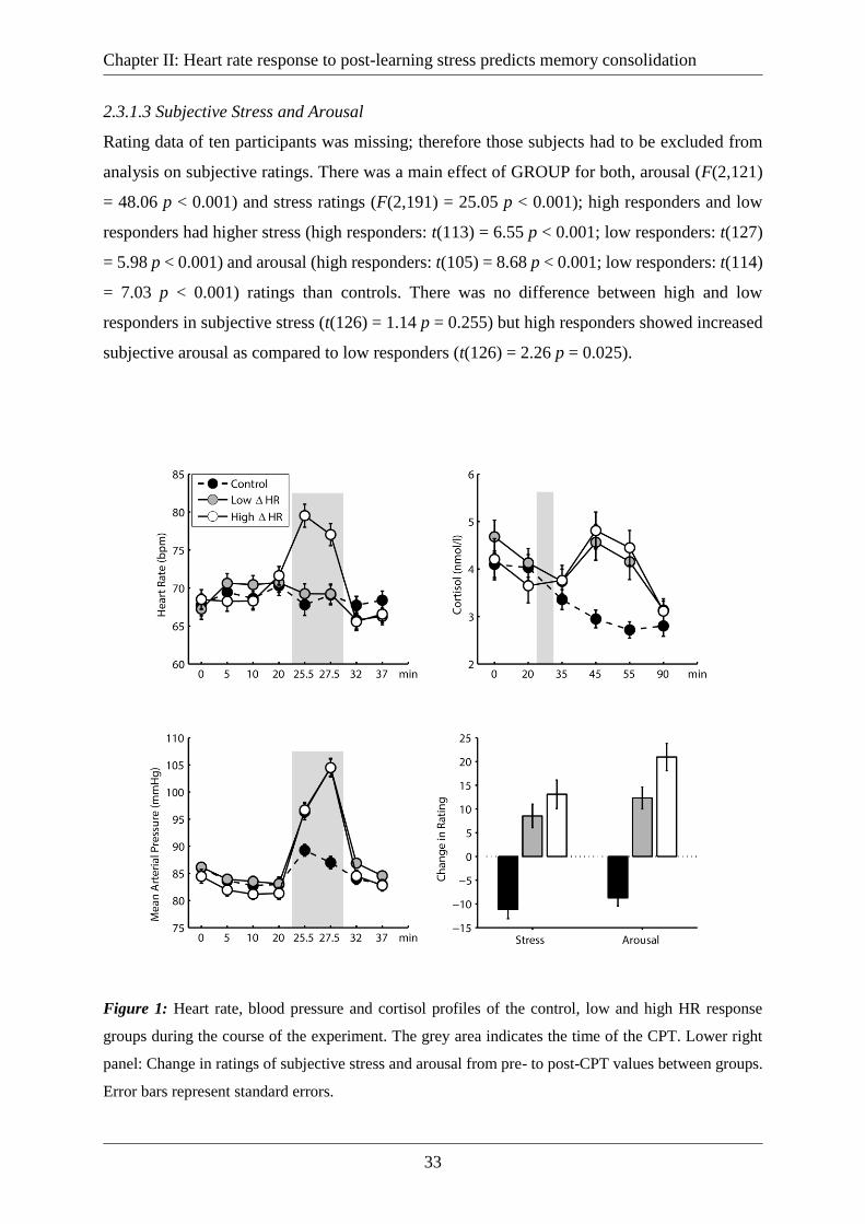
2.3.1 Response to the CPT ............................................................................................... 32
2.3.1.1 Heart Rate and Blood Pressure ......................................................................... 32
2.3.1.2 Cortisol ............................................................................................................. 32
2.3.1.3 Subjective Stress and Arousal .......................................................................... 33
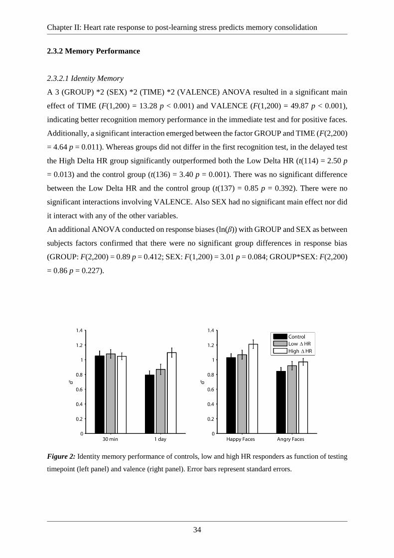
2.3.2 Memory Performance .............................................................................................. 34
2.3.2.1 Identity Memory ............................................................................................... 34
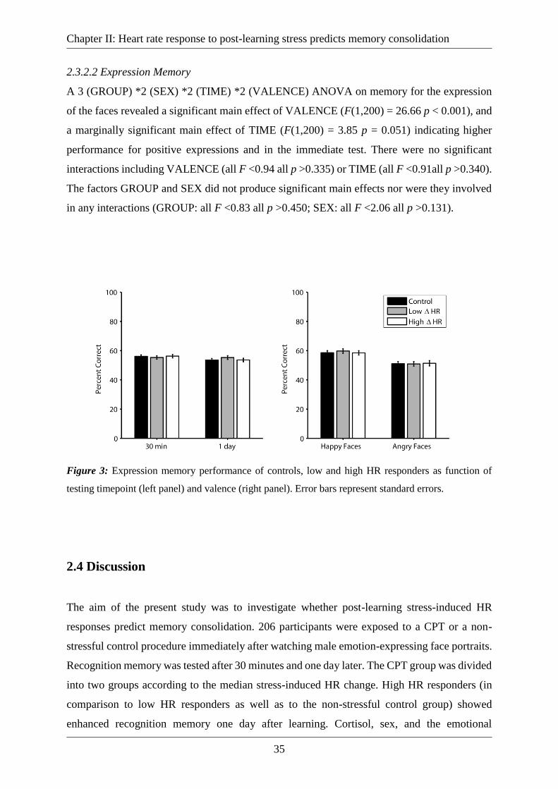
2.3.2.2 Expression Memory ......................................................................................... 35
Page 9
Index
ix
2.4 Discussion ...................................................................................................................... 35
References Chapter II ........................................................................................................... 40
2.i Author Notes ................................................................................................................... 48
Chapter III: Enhanced stress response by a bilateral feet compared to a unilateral hand
Cold Pressor Test ................................................................................................................... 49
3.0 Abstract .......................................................................................................................... 49
3.1 Introduction .................................................................................................................... 50
3.2 Methods .......................................................................................................................... 51
3.2.1 Sample ..................................................................................................................... 51
3.2.2 General procedure ................................................................................................... 51
3.2.3 Cold Pressor Test .................................................................................................... 52
3.2.4 Physiological measurements ................................................................................... 53
3.2.4.1 Cortisol ............................................................................................................. 53
3.2.4.2 Salivary alpha-amylase (sAA) ......................................................................... 53
3.2.4.3 Heart rate and blood pressure ........................................................................... 54
3.2.5 Subjective ratings .................................................................................................... 54
3.2.6 Data preparation and statistical analysis ................................................................. 54
3.3 Results ............................................................................................................................ 55
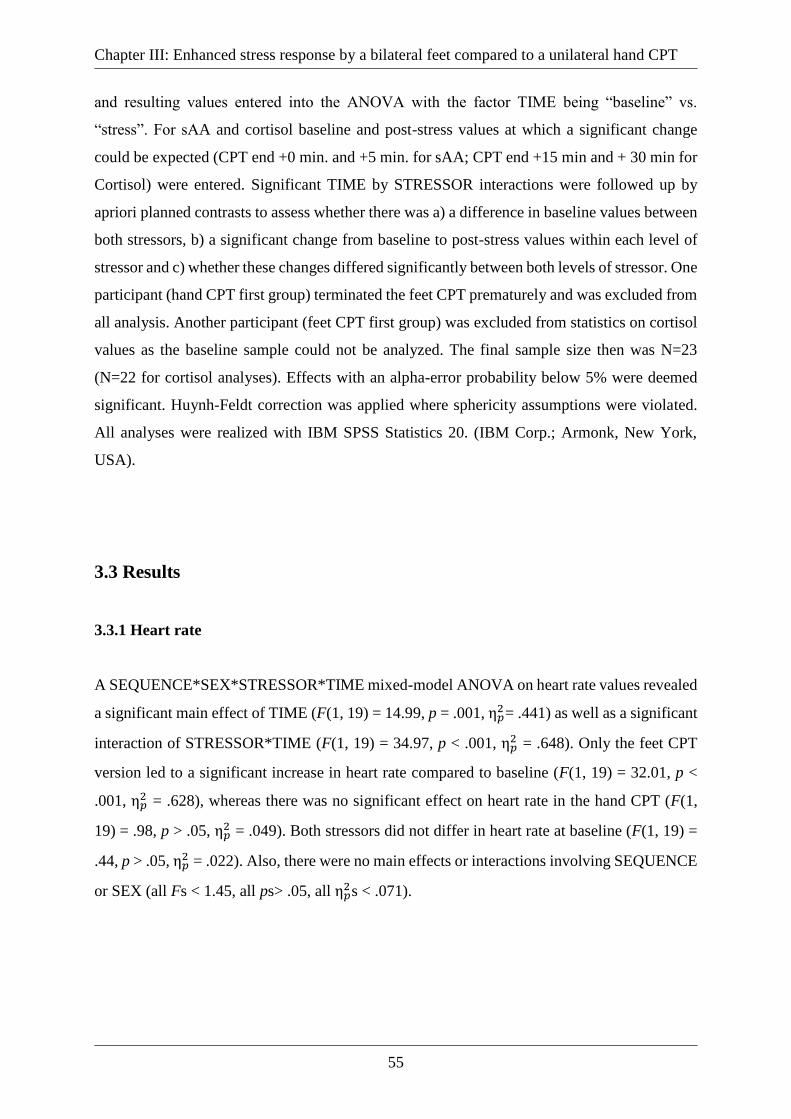
3.3.1 Heart rate ................................................................................................................. 55
3.3.2 Blood pressure ......................................................................................................... 56
3.3.3 Cortisol .................................................................................................................... 57
3.3.4 Salivary alpha-amylase ........................................................................................... 57
3.3.5 Subjective Ratings ................................................................................................... 59
3.3.5.1 Stress ................................................................................................................ 59
3.3.5.2 Pain ................................................................................................................... 59
3.3.6 Correlations between hand and feet CPT responses ............................................... 60
3.4 Discussion ...................................................................................................................... 60
Page 10
Index
x
References Chapter III ......................................................................................................... 65
3.i Author Notes ................................................................................................................... 69
Chapter IV: Stress disrupts distractor-based retrieval of SR episodes ............................ 70
4.0 Abstract .......................................................................................................................... 70
4.1 Introduction .................................................................................................................... 71
4.2 Methods .......................................................................................................................... 75
4.2.1 Participants. ............................................................................................................. 75
4.2.2 Stress test ................................................................................................................. 76
4.2.3 Physiological measurements ................................................................................... 77
4.2.4 Materials and Apparatus .......................................................................................... 77
4.2.5 Procedure ................................................................................................................. 77
4.2.6 Design ...................................................................................................................... 78
4.3 Results ............................................................................................................................ 79
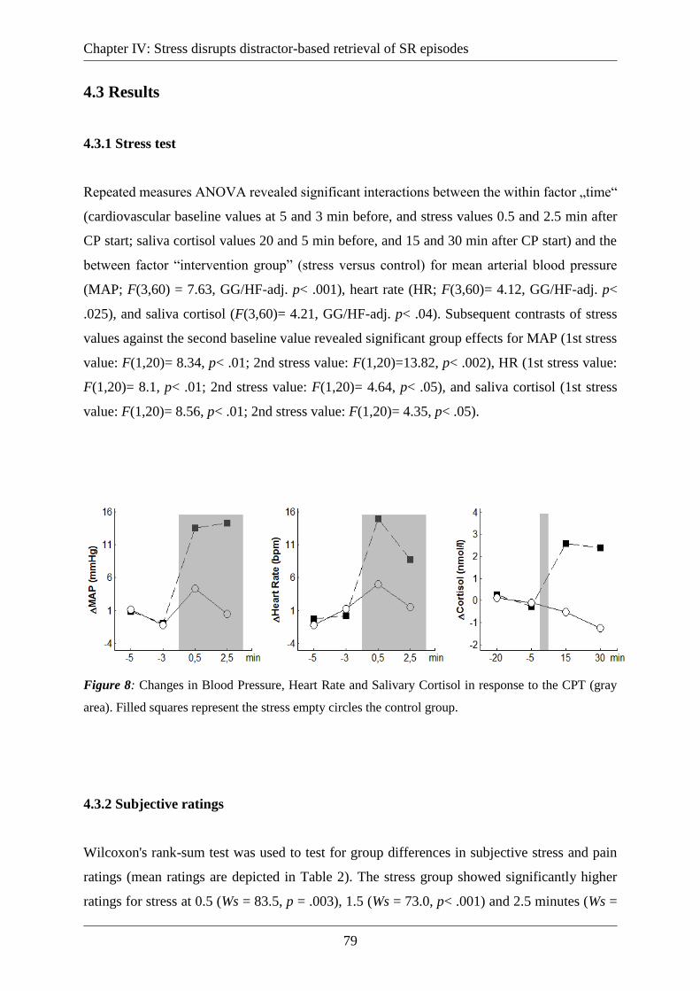
4.3.1 Stress test ................................................................................................................. 79
4.3.2 Subjective ratings .................................................................................................... 79
4.3.3 Binding effects ........................................................................................................ 80
4.4 Discussion ...................................................................................................................... 84
References Chapter IV ......................................................................................................... 86
4.i Author Notes ................................................................................................................... 91
Page 11
Index
xi
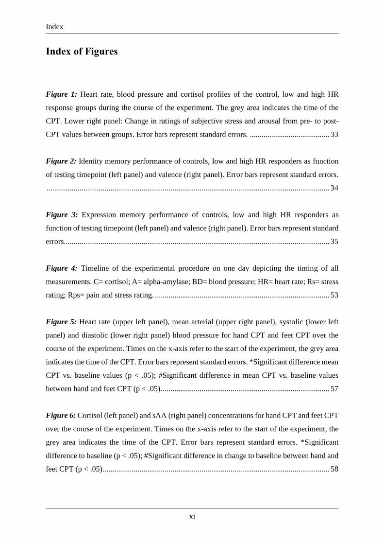
Index of Figures
Figure 1: Heart rate, blood pressure and cortisol profiles of the control, low and high HR
response groups during the course of the experiment. The grey area indicates the time of the
CPT. Lower right panel: Change in ratings of subjective stress and arousal from pre- to post-
CPT values between groups. Error bars represent standard errors. ......................................... 33
Figure 2: Identity memory performance of controls, low and high HR responders as function
of testing timepoint (left panel) and valence (right panel). Error bars represent standard errors.
.................................................................................................................................................. 34
Figure 3: Expression memory performance of controls, low and high HR responders as
function of testing timepoint (left panel) and valence (right panel). Error bars represent standard
errors. ........................................................................................................................................ 35
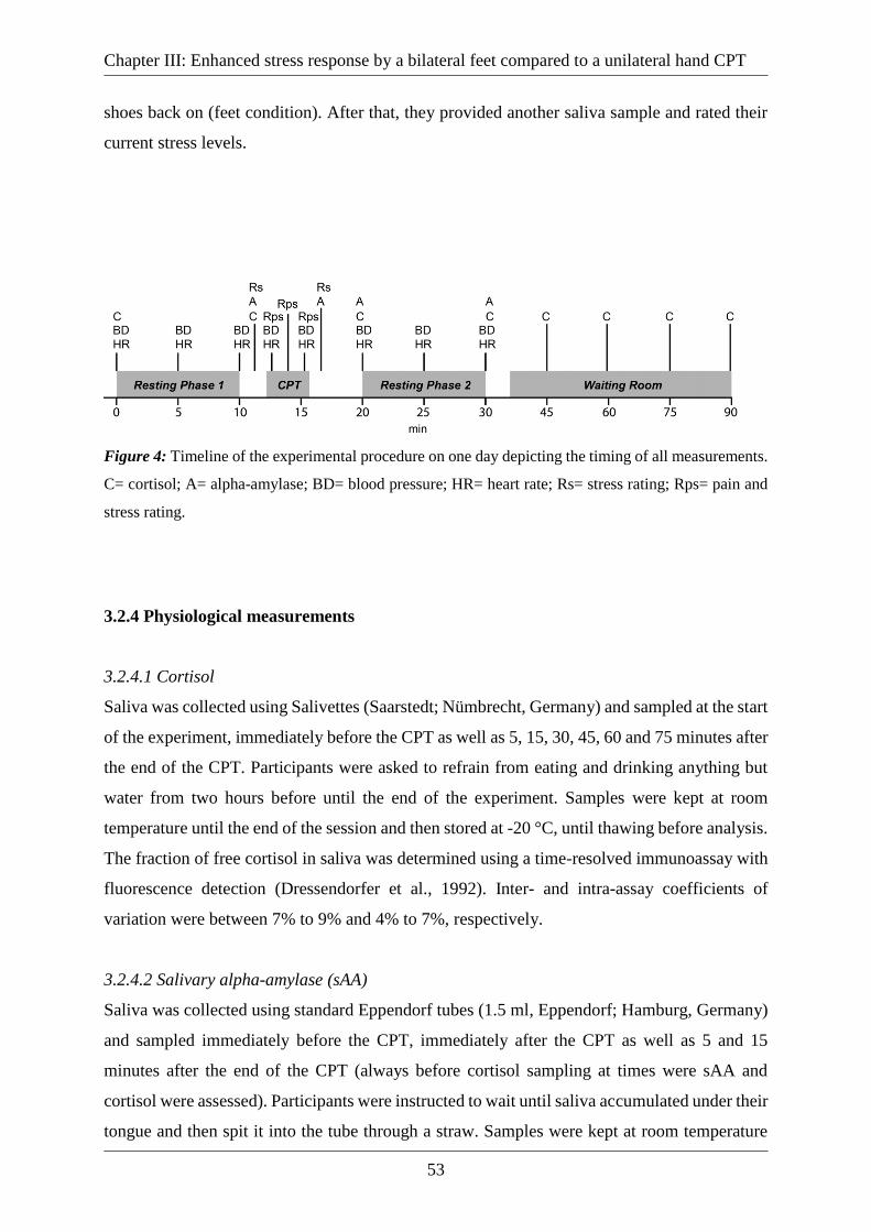
Figure 4: Timeline of the experimental procedure on one day depicting the timing of all
measurements. C= cortisol; A= alpha-amylase; BD= blood pressure; HR= heart rate; Rs= stress
rating; Rps= pain and stress rating. .......................................................................................... 53
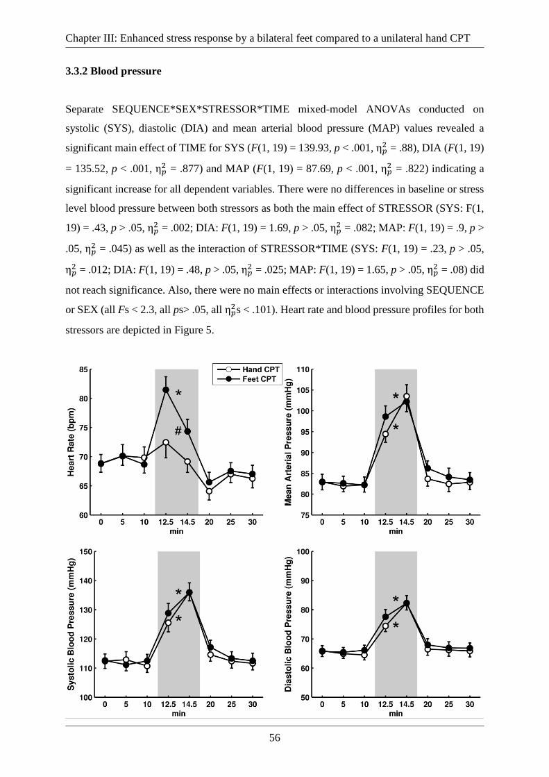
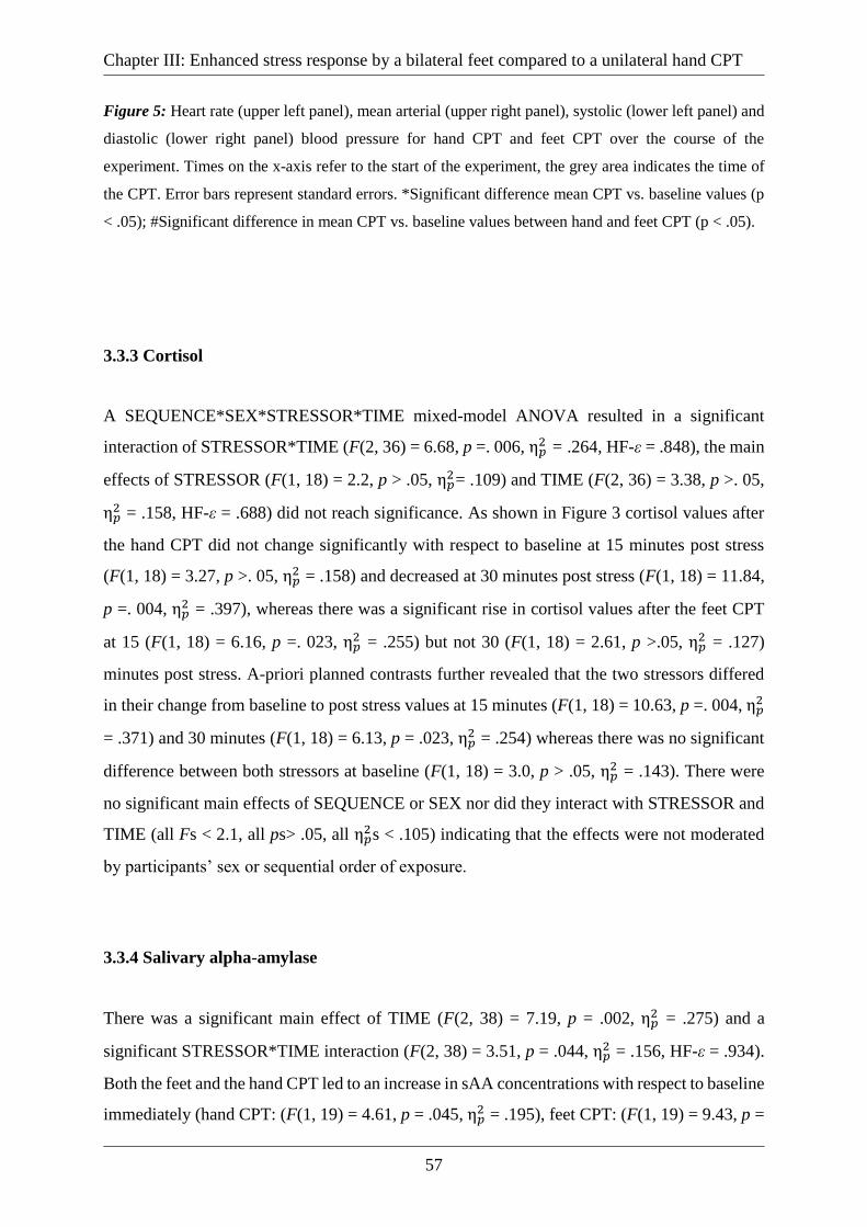
Figure 5: Heart rate (upper left panel), mean arterial (upper right panel), systolic (lower left
panel) and diastolic (lower right panel) blood pressure for hand CPT and feet CPT over the
course of the experiment. Times on the x-axis refer to the start of the experiment, the grey area
indicates the time of the CPT. Error bars represent standard errors. *Significant difference mean
CPT vs. baseline values (p < .05); #Significant difference in mean CPT vs. baseline values
between hand and feet CPT (p < .05). ...................................................................................... 57
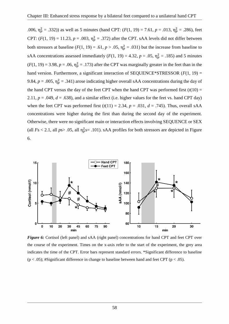
Figure 6: Cortisol (left panel) and sAA (right panel) concentrations for hand CPT and feet CPT
over the course of the experiment. Times on the x-axis refer to the start of the experiment, the
grey area indicates the time of the CPT. Error bars represent standard errors. *Significant
difference to baseline (p < .05); #Significant difference in change to baseline between hand and
feet CPT (p < .05). .................................................................................................................... 58
Page 12
Index
xii
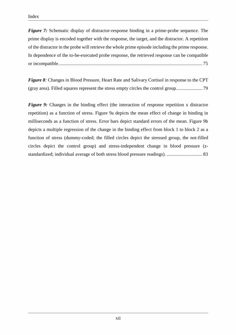
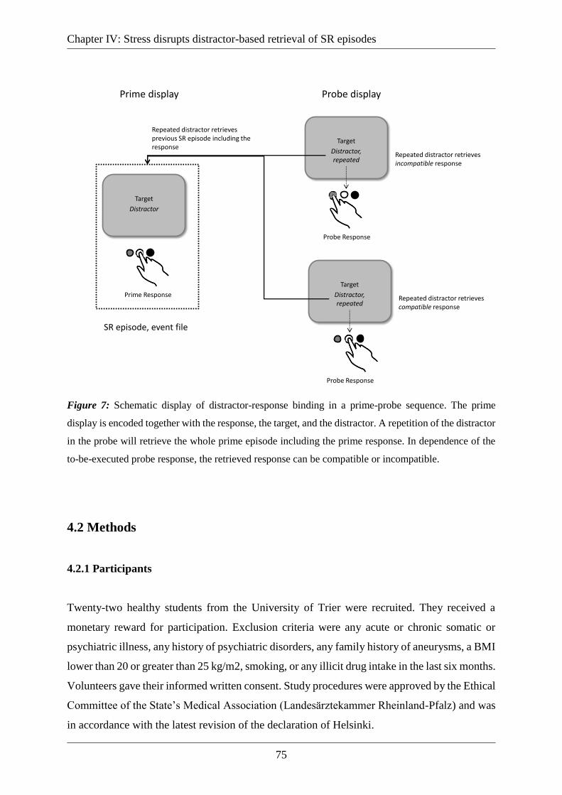
Figure 7: Schematic display of distractor-response binding in a prime-probe sequence. The
prime display is encoded together with the response, the target, and the distractor. A repetition
of the distractor in the probe will retrieve the whole prime episode including the prime response.
In dependence of the to-be-executed probe response, the retrieved response can be compatible
or incompatible. ........................................................................................................................ 75
Figure 8: Changes in Blood Pressure, Heart Rate and Salivary Cortisol in response to the CPT
(gray area). Filled squares represent the stress empty circles the control group. ..................... 79
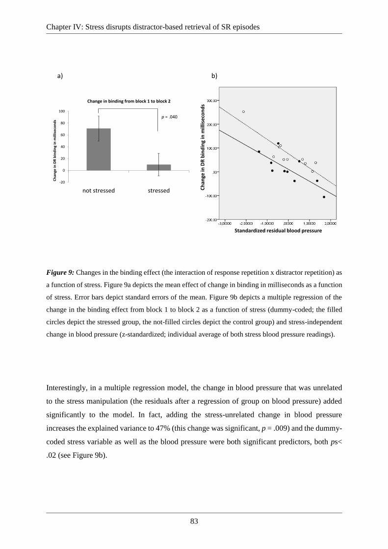
Figure 9: Changes in the binding effect (the interaction of response repetition x distractor
repetition) as a function of stress. Figure 9a depicts the mean effect of change in binding in
milliseconds as a function of stress. Error bars depict standard errors of the mean. Figure 9b
depicts a multiple regression of the change in the binding effect from block 1 to block 2 as a
function of stress (dummy-coded; the filled circles depict the stressed group, the not-filled
circles depict the control group) and stress-independent change in blood pressure (z-
standardized; individual average of both stress blood pressure readings). .............................. 83
Page 13
Index
xiii
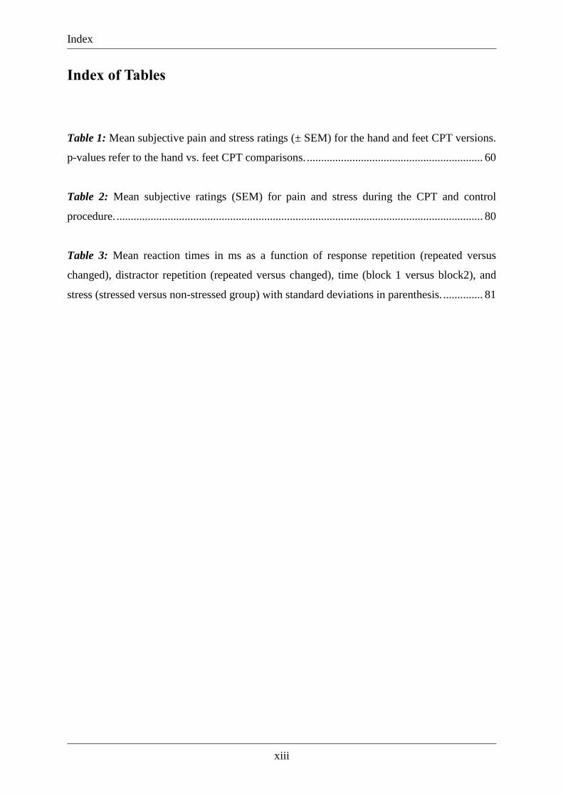
Index of Tables
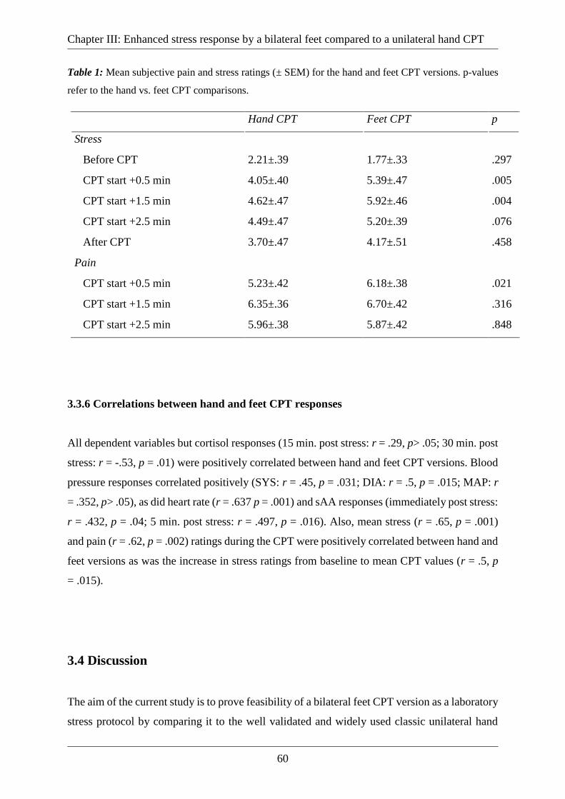
Table 1: Mean subjective pain and stress ratings (± SEM) for the hand and feet CPT versions.
p-values refer to the hand vs. feet CPT comparisons. .............................................................. 60
Table 2: Mean subjective ratings (SEM) for pain and stress during the CPT and control
procedure. ................................................................................................................................. 80
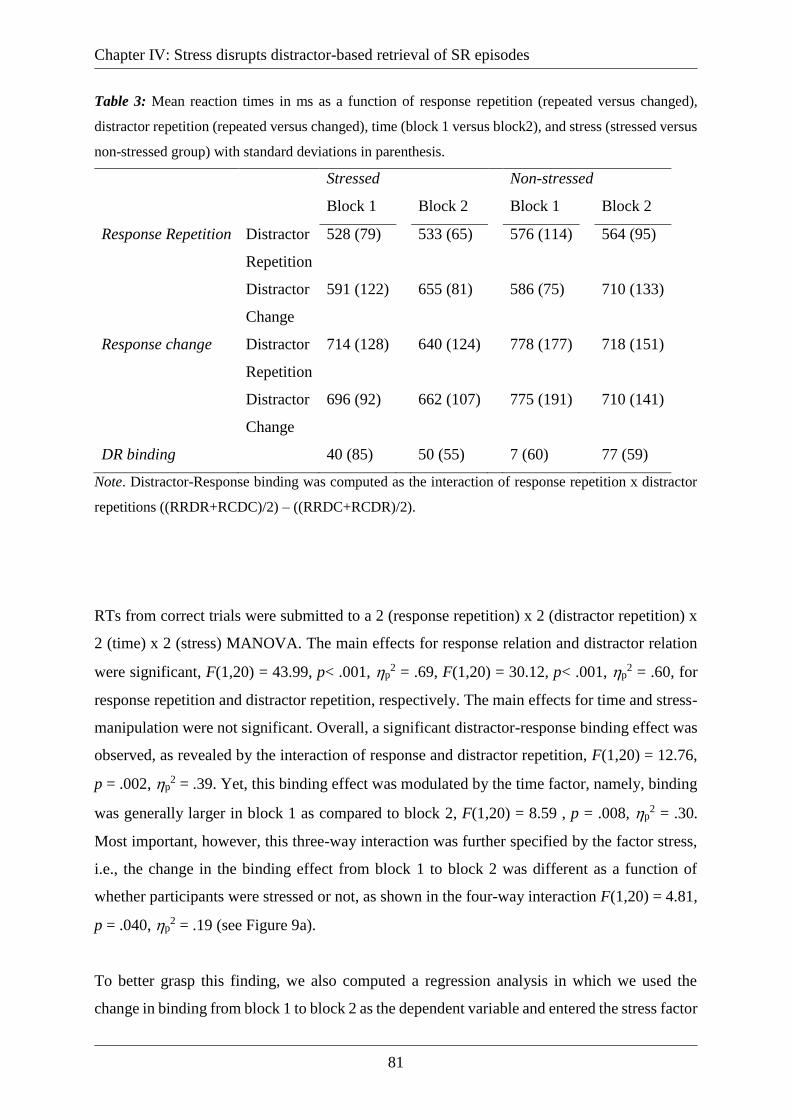
Table 3: Mean reaction times in ms as a function of response repetition (repeated versus
changed), distractor repetition (repeated versus changed), time (block 1 versus block2), and
stress (stressed versus non-stressed group) with standard deviations in parenthesis. .............. 81
Page 14
Index
xiv
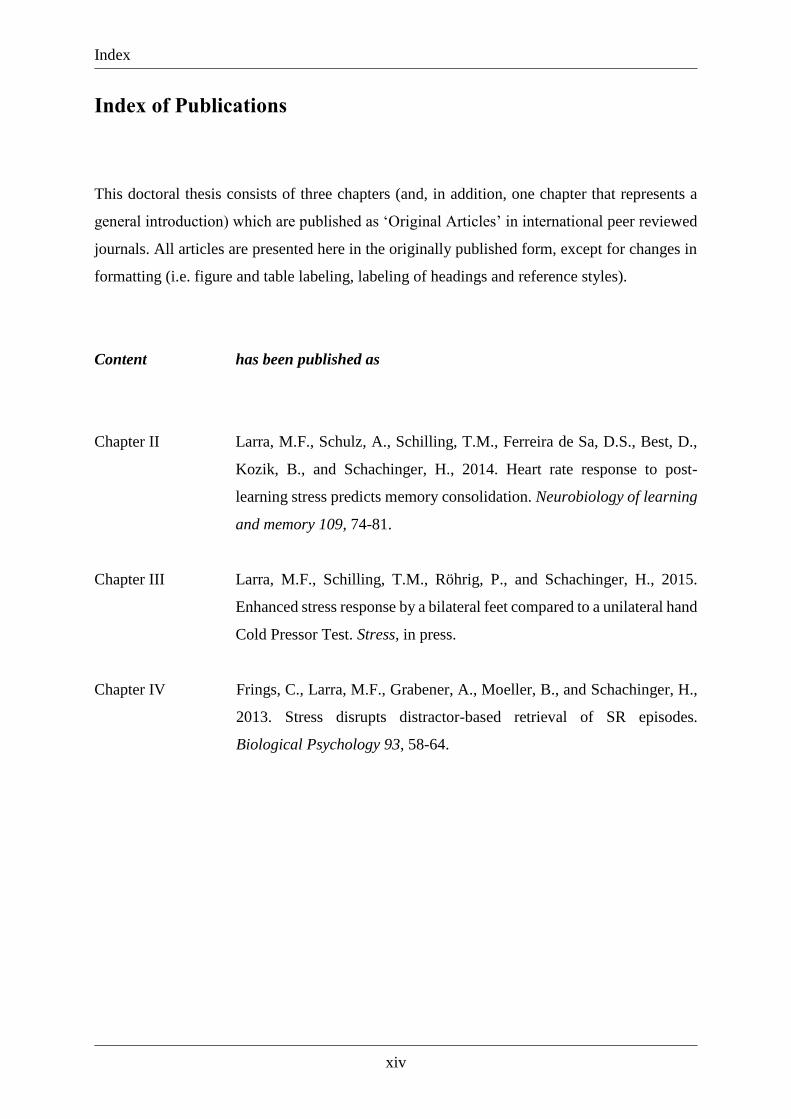
Index of Publications
This doctoral thesis consists of three chapters (and, in addition, one chapter that represents a
general introduction) which are published as ‘Original Articles’ in international peer reviewed
journals. All articles are presented here in the originally published form, except for changes in
formatting (i.e. figure and table labeling, labeling of headings and reference styles).
Content has been published as
Chapter II Larra, M.F., Schulz, A., Schilling, T.M., Ferreira de Sa, D.S., Best, D.,
Kozik, B., and Schachinger, H., 2014. Heart rate response to post-
learning stress predicts memory consolidation. Neurobiology of learning
and memory 109, 74-81.
Chapter III Larra, M.F., Schilling, T.M., Röhrig, P., and Schachinger, H., 2015.
Enhanced stress response by a bilateral feet compared to a unilateral hand
Cold Pressor Test. Stress, in press.
Chapter IV Frings, C., Larra, M.F., Grabener, A., Moeller, B., and Schachinger, H.,
2013. Stress disrupts distractor-based retrieval of SR episodes.
Biological Psychology 93, 58-64.
Page 15
Index
xv
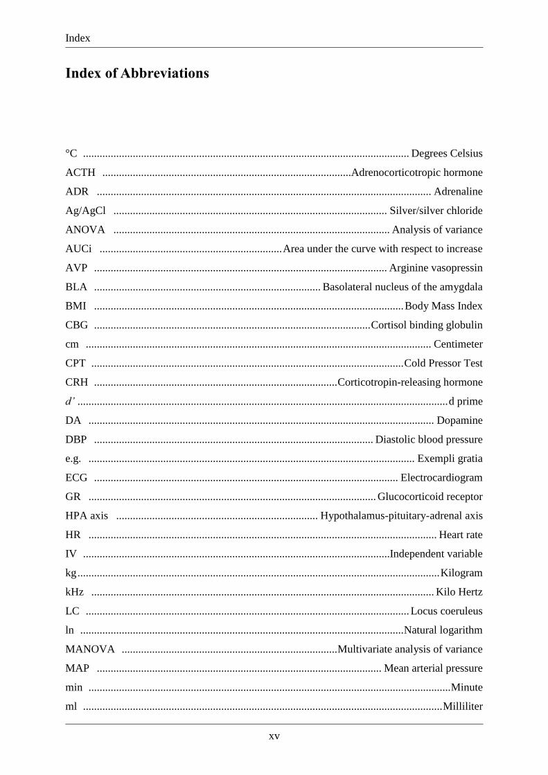
Index of Abbreviations
°C ...................................................................................................................... Degrees Celsius
ACTH ..........................................................................................Adrenocorticotropic hormone
ADR ......................................................................................................................... Adrenaline
Ag/AgCl ................................................................................................... Silver/silver chloride
ANOVA .................................................................................................... Analysis of variance
AUCi .................................................................. Area under the curve with respect to increase
AVP .......................................................................................................... Arginine vasopressin
BLA .................................................................................. Basolateral nucleus of the amygdala
BMI ................................................................................................................ Body Mass Index
CBG .................................................................................................... Cortisol binding globulin
cm ............................................................................................................................. Centimeter
CPT ................................................................................................................. Cold Pressor Test
CRH ........................................................................................ Corticotropin-releasing hormone
d’ ...................................................................................................................................... d prime
DA ............................................................................................................................. Dopamine
DBP ..................................................................................................... Diastolic blood pressure
e.g. ...................................................................................................................... Exempli gratia
ECG .............................................................................................................. Electrocardiogram
GR ........................................................................................................ Glucocorticoid receptor
HPA axis ......................................................................... Hypothalamus-pituitary-adrenal axis
HR .............................................................................................................................. Heart rate
IV ...............................................................................................................Independent variable
kg ................................................................................................................................... Kilogram
kHz ............................................................................................................................ Kilo Hertz
LC ..................................................................................................................... Locus coeruleus
ln ..................................................................................................................... Natural logarithm
MANOVA .............................................................................. Multivariate analysis of variance
MAP ....................................................................................................... Mean arterial pressure
min ................................................................................................................................... Minute
ml .................................................................................................................................. Milliliter
Page 16
Index
xvi
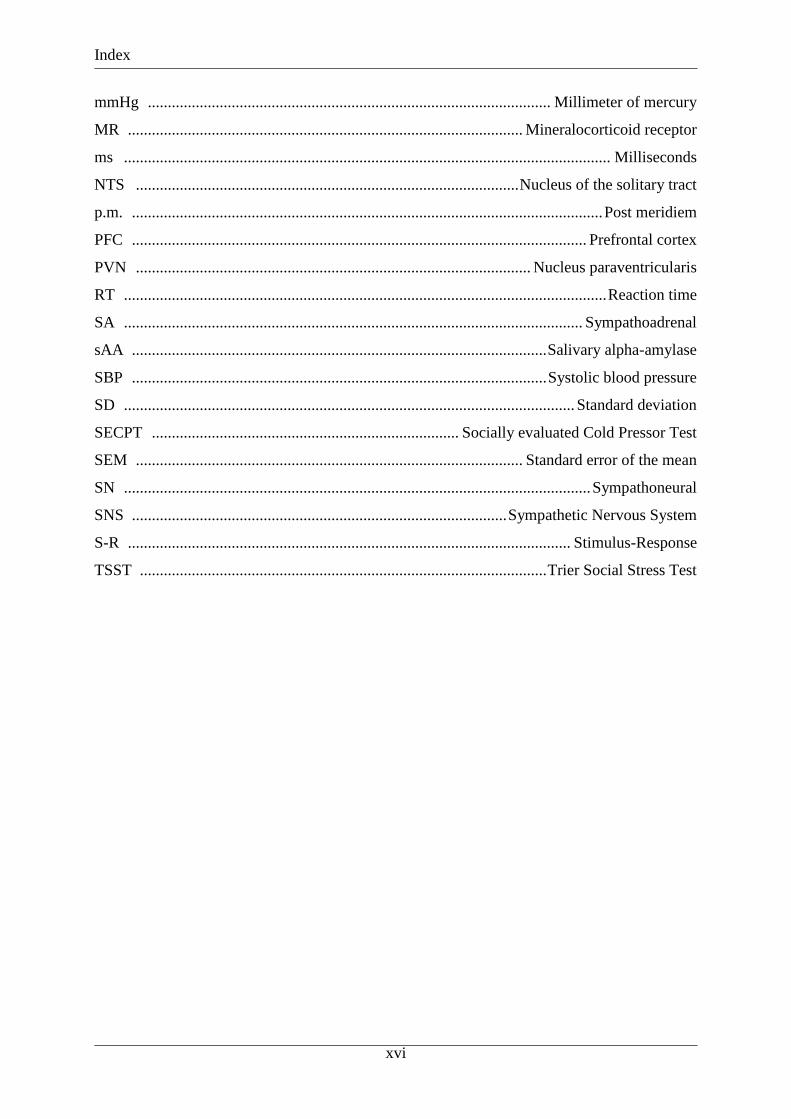
mmHg ..................................................................................................... Millimeter of mercury
MR ................................................................................................... Mineralocorticoid receptor
ms .......................................................................................................................... Milliseconds
NTS ................................................................................................ Nucleus of the solitary tract
p.m. ...................................................................................................................... Post meridiem
PFC .................................................................................................................. Prefrontal cortex
PVN ................................................................................................... Nucleus paraventricularis
RT ......................................................................................................................... Reaction time
SA ................................................................................................................... Sympathoadrenal
sAA ........................................................................................................ Salivary alpha-amylase
SBP ........................................................................................................ Systolic blood pressure
SD ................................................................................................................. Standard deviation
SECPT ............................................................................. Socially evaluated Cold Pressor Test
SEM ................................................................................................. Standard error of the mean
SN ..................................................................................................................... Sympathoneural
SNS .............................................................................................. Sympathetic Nervous System
S-R ............................................................................................................... Stimulus-Response
TSST ...................................................................................................... Trier Social Stress Test
Page 17
Chapter I: General Background
1
Chapter I: General Background
1.1 Introduction and Outline
The study of stress has essentially contributed to our understanding of the ways in which
adverse events are causally linked to physical health and well-being. The last decades have seen
fundamental progress in research on the topic of stress. Stress has been shown to be involved
in the genesis of a variety of pathological conditions (Chrousos and Kino, 2007; Marin et al.,
2011) and to affect diverse psychological processes (Campeau et al., 2011) while recent
advancements allowed to trace some of these stress effects to specific actions that stress
hormones exert on the brain (Erickson et al., 2003; Lupien et al., 2007; Roozendaal and
McGaugh, 2011).
Progress in stress research heavily relies on the availability of suitable and well validated
laboratory stressors. Appropriate laboratory stressors need to be able to reliably provoke a
response in the relevant parameters and be applicable in different research settings or
experimental designs. However, stress responses have been shown to differ according to the
type of stressors employed (Dickerson and Kemeny, 2004; Pacak and Palkovits, 2001) and
experimental designs often pose restrictions that render an otherwise appropriate stressor
unfeasible. The present work focuses on the Cold Pressor Test (CPT) as a stress induction
technique. Three published experiments are presented that show how the advantages of the CPT
can be used to test stress effects on memory processes and how restrictions of certain
experimental designs can be met by a simple modification that retains its feasibility and validity.
This thesis consists of four chapters. In the following chapter I will describe the scientific
background to the experimental investigations presented in chapters II to IV. First, I will give
a general introduction into the topic of stress in which the basic physiological mechanisms of
the stress response, stress effects on the brain and forms of its operationalization in
psychobiological experiments are addressed. The second section focuses on the CPT as such a
laboratory model of stress. I will briefly describe its physiological mechanisms and effects in
different fields of study and discuss problems as well as advantages in its application. Finally,
the three experimental investigations are outlined, briefly summarizing their main aims, design,
results and final conclusions. The following chapters II to IV contain the published reports on
Page 18
Chapter I: General Background
2
the three experiments.
1.2 Stress
Stress is a phenomenon referring to the elicitation of a specific response pattern, the “stress
response”, by a certain class of stimuli termed “stressors”. Stressors have been very generally
defined as being any perceived or sensed threat to homeostasis or well-being (Ulrich-Lai and
Herman, 2009), a mismatch between expectation and perception that elicits a patterned
compensatory response (Goldstein and Kopin, 2007) or as any demand on the body that causes
a stress response (Selye, 1976). They may be differentiated on the basis of their origin and the
kind of threat they pose. Accordingly, four classes of stressors have been suggested (Pacak and
Palkovits, 2001): Physical stressors that are directly sensed as pain, cold, noise or chemical
agents: Psychological stressors that require evaluation by higher brain areas to be perceived as
threat. Social stressors that arise from interactions with other individuals and bodily stressors
that pose a demand on cardiovascular or metabolic homeostasis.
While psychological theories on stress focus on the interpretation and evaluation of stressors
with respect to available resources (Lazarus, 1999; Lazarus and Folkman, 1984), the
physiological response pattern, its mediators and their effects lie at the core of psychobiological
stress research. Those will be addressed in the following sections.
1.2.1 The stress response
The stress response is a complex phenomenon comprised of reactions and interactions in
behavioral, autonomic, endocrine, and immune systems. Today’s recognition of the stress
response as a fundamental physiological mechanism was mainly primed by the influential
works of Walter Cannon and Hans Selye. In the first half of the 20th century they popularized
the topic and lay the foundation for our understanding of the basic principles of the stress
response.
Cannon (1939) introduced the concept of homeostasis meaning the maintenance of
physiological parameters within an acceptable range. He discovered that a wide variety of
Page 19
Chapter I: General Background
3
threats to homeostasis including psychosocial factors would lead to an activation of the
sympathetic nervous system (SNS) and release of adrenaline (ADR) from the adrenal medulla.
Cannon thought these two effectors to act as a unit, the “sympathoadrenal system”, that upon
activation would produce compensatory and anticipatory adjustments (the “fight or flight
response”) to restore homeostasis and promote survival.
Selye, who popularized the scientific term stress, defined stress as a nonspecific response
pattern to diverse noxious stimuli mainly characterized by an activation of the hypothalamus-
pituitary-adrenal (HPA) axis and its effects (Selye, 1950; Selye, 1976). Although Selye(1950)
acknowledged that there are also stressor specific responses he did not consider them to be part
of the stress response. This doctrine of non-specifity has been challenged and it is now widely
acknowledged that the stress response is to some extent specific depending on the type of
stressor. Signaling pathways that lead to HPA axis and SNS activation differ according to the
type of stressor triggering responses that are commensurate with the nature of the stimulus
(Goldstein, 2010; Pacak and Palkovits, 2001).
Modern accounts of the stress response see the SNS and HPA axis as main components of a
physiological stress system which is largely controlled by the hypothalamus (Chrousos, 1998;
Johnson et al., 1992). The hypothalamus is the principal integrator of stress signals. Brainstem
centers that sense systemic stressors as blood loss as well as limbic regions that process
psychological stressors project to the nucleus paraventricularis (PVN) of the hypothalamus
(McEwen, 2007). The PVN mainly orchestrates the SNS and HPA axis response to stress
(Ulrich-Lai and Herman, 2009), these two main components of the stress response will we
explained in detail below.
1.2.1.1 Sympathetic Nervous System
The SNS provides a fast physiological response to stressors through neural innervation of its
target organs taking effect within seconds. It may be divided into two branches, the
sympathoneural (SN) and the sympathoadrenal (SA) arm (Kvetnansky et al., 2009) and there is
some evidence that these two branches act partially independent giving rise to specific reactions
depending on the type of stressor (Goldstein and Kopin, 2007; Pacak and Palkovits, 2001).
Sympathetic preganglionic neurons in both branches are controlled by catecholaminergic and
noncatecholaminergic sympathetic premotor neurons located mainly in the hypothalamus and
Page 20
Chapter I: General Background
4
brainstem. Some systemic stressors that signal major threats to the organism, as blood loss, pain
or inflammation, may activate preganglionic neurons without hypothalamic involvement
through reflex arcs at the intermediolateral cell column (Pacak and Palkovits, 2001; Ulrich-Lai
and Herman, 2009). The SN arm is organized in a two neuron chain consisting of pregranglionic
and postganglionic sympathetic neurons. Preganglionic neurons activate postganglionic
neurons by release of acetylcholine. Upon activation varicosities of the postganglionic fibers
release noradrenaline (NA) at their target organs. They do not form a synaptic junction with
cells in their target organs but NA is released via exocytosis over a broad area of the target
tissue. In the SA arm preganglionic neurons of the SNS innervate chromaffin cells in the adrenal
medulla. Chromaffin cells store mainly ADR but also NA. After excitatory signals arrive from
preganglionic neurons, the chromaffin cells secrete ADR and NA into the general circulation
via exocytosis causing widespread effects at multiple target sites (for a detailed overview of the
SNS see Goldstein, 2009; Palkovits, 2009).
Sympathetic activation thus results in a rise in levels of circulating ADR and NA and leads to
an increase in heart rate and force of contraction, peripheral vasoconstriction, increased blood
flow to skeletal muscles and energy mobilization (Chrousos and Gold, 1992) giving rise to a
general state of arousal that Cannon referred to as fight-or-flight response. However, this
response is shortlived and rapidly counteracted by reflex parasympathetic activation (Ulrich-
Lai and Herman, 2009).
1.2.1.2 Hypothalamic-Pituitary-Adrenal Axis
The HPA axis acts as interface between the central nervous system (CNS) and the endocrine
system mediating the endocrine response to centrally processed stressors. It consists of three
core structures, the PVN, the pituitary and the adrenal glands that communicate with each other
through specific neurohormones and hormones. The PVN regulates the HPA axis response to
stress (Ziegler and Herman, 2002). It receives signals from brainstem centers as well as the
limbic system and prefrontal cortex (PFC) allowing for an activation through systemic and
directly sensed as well as psychological or anticipated stressors (Herman et al., 2005; Ulrich-
Lai and Herman, 2009). During stress the parvocellular neurons of the PVN release regulatory
neurohormones, mainly corticotropin-releasing hormone (CRH) and arginine vasopressin
(AVP), into the hypophysial portal vasculature. Through the portal vessels these
neurohormones rapidly reach the anterior pituitary gland where they act synergistically to
Page 21
Chapter I: General Background
5
stimulate the secretion of adrenocorticotropic hormone (ACTH) into the bloodstream. After its
release from the anterior pituitary gland circulating ACTH moves to the cortex of the adrenal
glands. Here, it stimulates synthesis and secretion of glucocorticoids mainly in the zona
fasciculata. Glucocorticoids, cortisol in humans, are the ultimate endproduct of the HPA axis
response to stress and can be expected to increase about ten minutes after stressor onset (for a
detailed overview of the HPA axis see Fulford and Harbuz, 2005; Herman, 2009).
To prevent glucocorticoid levels from overshooting HPA axis activity is downregulated by
complex negative feedback mechanisms exerted at multiple sites and timescales. Negative
feedback is exerted on both the hypothalamus and the pituitary as well as on brain sites
projecting to the PVN as limbic structures and the PFC (Dallman, 2007; Herman et al., 2005).
An initial rapid feedback develops within seconds by a nongenomic mechanism mediated
through membrane receptors (Di et al., 2003). It is short in duration (approximately ten minutes)
and sensitive to changes in glucocorticoid concentrations rather than absolute levels (Herman,
2009). Delayed feedback sets in about half an hour after an acute elevation of glucocorticoid
levels and may last for hours (Dallman, 2007). It is mediated through genomic mechanisms
initialized by nuclear mineralocorticoid and glucocorticoid receptors (Dallman et al., 1992).
Together fast and slow negative feedback on HPA axis activity enable the termination of the
stress response and ensure that glucocorticoid levels stay within tolerable limits.
1.2.2 Stress effects on the brain
Investigations on stress would probably not play such a prominent role in psychobiological
research if not for its profound effects on multiple psychological functions as well as physical
and mental health. Everybody has experienced how stress can affect us in our normal
functioning and in the last decades substantial advances have been made in our understanding
how stress influences brain functions and thereby our experience and behavior.
Stress effects on the brain are mainly mediated by the central actions of the two main
endproducts of the stress response, ADR/NA and cortisol (Erickson et al., 2003; Lupien et al.,
2007; Roozendaal and McGaugh, 2011). Cortisol traverses the cell membrane and binds to
nuclear mineralocorticoid (MR) and glucocorticoid receptors (GR) that then translocate to the
cell nucleus to alter gene transcription (de Kloet et al., 1993). Besides these genomic effects
Page 22
Chapter I: General Background
6
cortisol may also exert faster non-genomic effects through receptors residing in the cell
membrane (Falkenstein et al., 2000; Orchinik et al., 1991). Most of the circulating cortisol in
the blood is bound to a carrier protein (cortisol binding globulin, CBG) and albumin which
renders it physiologically inactive. Unbound cortisol by contrast can cross the blood-brain
barrier and readily enters the brain (Mason et al., 2010; Murphy et al., 1967) to act on membrane
and nuclear MRs and GRs. Brain structures as the hippocampus, the amygdala and the
prefrontal cortex (PFC) express a high density of MRs and GRs allowing for modulations by
cortisol during stress (Patel et al., 2000; Sanchez et al., 2000). Indeed, cognitive functions that
are associated with these structures as memory (Roozendaal and McGaugh, 2011), attentional
(Sanger et al., 2014) and affective processes (Campeau et al., 2011) have been shown to be
modulated by cortisol and stress.
Peripheral catecholamines released during stress cannot directly enter the brain (Weil-Malherbe
et al., 1959). However, they might affect central processes via vagal afferents projecting to the
nucleus of the solitary tract) (NTS; Williams et al., 2000). The NTS heavily projects to the
amygdala which in turn sends widespread connections throughout the brain (McGaugh, 2004).
In addition, central catecholaminergic pathways mainly originating from the locus coeruleus
(LC) and projecting to the limbic system and PFC may contribute to modulatory stress effects
on the brain (Berridge and Waterhouse, 2003).
Among the cognitive functions influenced by stress memory processes are probably the most
extensively studied within stress research. A prominent model of how ADR/NA and cortisol
released during stress may interact in mediating stress effects on memory has been proposed by
Roozendaal and McGaugh (2011). Based on a series of rodent experiments they argue that
interactions of NA and cortisol at the basolateral nucleus of the amygdala (BLA) cause the
retention enhancement of stressful and emotional memories. Specifically, circulating ADR acts
on peripheral beta-adrenoreceptors from vagal afferents projecting to the NTS which in turn
directly and indirectly (via the LC) changes noradrenergic activation within the BLA (Williams
et al., 2000). The amygdala then modulates memory processes through its widespread
connections to brain structures mediating memory functions, particularly the hippocampus and
caudate nucleus (Ferry et al., 1999; McIntyre et al., 2012). An activation of GRs in the BLA
and memory processing areas is required for this effect (Roozendaal et al., 1996). Similarly,
cortisol effects on memory require concurrent noradrenergic activation within the BLA
(Roozendaal et al., 2006; Setlow et al., 2000).
Page 23
Chapter I: General Background
7
Animal experiments involving infusions of peripherally and centrally acting adrenoreceptor
agonists and antagonists as well as manipulation of GRs and cortisol levels provide compelling
evidence for this model (for review see McIntyre et al., 2012; Roozendaal and McGaugh, 2011).
The role of endogenously elicited (via Cold Pressor stress) sympathetic arousal in modulating
memory consolidation in humans is assessed in one of the experimental investigations
presented in this thesis (Chapter II).
1.2.3 Eliciting stress in the laboratory
Establishing ethically acceptable paradigms to elicit stress in humans under laboratory
conditions is a continuing challenge. The specifity of stress reactions as a result of the type of
stressor applied (Goldstein, 2010) as well as interindividual differences (Gerra et al., 2001;
Kajantie and Phillips, 2006) pose the main problem in research with laboratory stressors. Not
all of them are qualified to produce a full neuroendocrine stress response in every individual,
especially substantial cortisol increases are often lacking (Dickerson and Kemeny, 2004). As
has been pointed out above stress effects on the brain rely on rather specific actions of cortisol
and catecholamines released during stress, therefore, the choice of an appropriate stressor is
crucial. Additionally, depending on the research question further limitations need to be taken
into account as certain stressors may be unfeasible e.g. due to their application duration,
interference with other experimental paradigms or the lack of a valid control procedure.
Typically, stress experiments follow a sequence beginning with a baseline, or initial rest, period
that allows to examine within-subject changes between baseline and task. The stressor is then
administered usually followed by a recovery period. A vast variety of stimulations have been
used as stressors. Commonly used stress elicitation paradigms are mental arithmetic (e.g. paced
subtraction or addition tasks; McCann et al., 1993), exercise (e.g. hand-grip task; Nielsen and
Mather, 2015), orthostatic tasks (e.g. head-up tilt; Shoemaker et al., 2001) and psychosocial
stressors (e.g. public speaking; Gerra et al., 2001).
Mental arithmetic tasks require effortful control of attention and exercise causes an energy
demand both going along with sympathetic activation (Nielsen and Mather, 2015; Peters et al.,
1998). Orthostatic changes mainly disrupt cardiovascular homeostasis triggering a fast
Page 24
Chapter I: General Background
8
autonomic response (Fu et al., 2005). Depending on intensity and duration of exposure increases
in cortisol may be observed (Al'Absi et al., 1997; McCann et al., 1993), however, these tasks
are primarily employed to study the SNS component of the stress response. Stressors that
incorporate a social evaluative component, on the other hand, are particularly well suited to
elicit robust HPA axis responses (Dickerson and Kemeny, 2004; Schwabe et al., 2008b).
Typical examples of such social stressors are public speaking tasks and the Trier Social Stress
Test (TSST), a 20-minute paradigm specifically designed to trigger the HPA axis by a
combination of a public speech and mental arithmetic task conducted in front of a panel of
judges (Kirschbaum et al., 1993). Although these stressors are best suited to produce a full
neuroendocrine stress response including an activation of the HPA axis, they may not always
be feasible as they are time consuming, require a cognitive engagement and do not offer a
simple control procedure. Also, they rely on the performance and properties of the
experimenter, which need to be controlled. Another widely used stress protocol is the CPT
which is central to this thesis and will be portrayed in the following sections.
1.3 The Cold Pressor Test
In its core the CPT consists of a procedure in which a limb (usually the dominant hand) is
immersed into ice-water for a short period of time (usually 2 to 3 minutes). It was first
introduced in the 1930s by Hines and Brown (1932). They employed a routine that started with
a resting period during which multiple blood pressure readings were taken. After that the CPT
was carried out and a recovery resting period followed both accompanied by blood pressure
readings. This format of the reactivity study during rest, stress, and recovery lay the foundation
and still is the common adopted procedure in stress studies. Although originally intended as
means to experimentally increase blood pressure in studies on hypertension, the CPT has now
become a widely used tool in experimental research of different areas and is frequently
employed as a laboratory stress protocol.
In the following sections I will briefly summarize what is currently known about the
physiological mechanisms and responses triggered by CPT stimulation, its use in different
fields of study and finally discuss its main advantages and disadvantages as a laboratory stress
protocol.
Page 25
Chapter I: General Background
9
1.3.1 Physiological mechanism and responses
Exposure to the CPT leads to a stimulation of peripheral thermo- and nociceptors located
throughout the skin. Pain and temperature fibers enter the spinal cord in the dorsal roots and
cross contralateral to form the spinolateral tract which travels to the thalamus and sends
collaterals to the reticular formation. At the medulla level these collaterals may stimulate the
rostral ventrolateral medullary pressor area resulting in a reflexive sympathetic discharge
towards the heart and the vessels (Nakamura et al., 2008; Velasco et al., 1997). Via brainstem
projections CPT stimulation may also affect cortical and subcortical structures as the
hypothalamus that further modulate neuroendocrine reactions creating a multifaceted
physiological and subjective stress response (Lovallo, 1975; McEwen, 2007; Ulrich-Lai and
Herman, 2009).
CPT exposure leads to profound changes in cardiovascular parameters most notably a rise in
blood pressure through peripheral vasoconstriction and to a lesser extent cardiac output
resulting from an increase in both vascular alpha-adrenergic and cardiac beta-adrenergic drive
(Greene et al., 1965; Lovallo, 1975; Yamamoto et al., 1992). However, CPT effects are not
restricted to the cardiovascular system. Increases in multiple markers of sympathetic nervous
system activity as skin conductance level (Buchanan et al., 2006), plasma chatecholamines
(Goldstein et al., 1994; Pascualy et al., 1999; Ward et al., 1983), muscle sympathetic nerve
activity (Victor et al., 1987; Yamamoto et al., 1992) and more recently salivary alpha-amylase
(sAA) have been reported (Smeets et al., 2008). In addition to the effects on the sympathetic
nervous system the CPT has been shown to be capable of activating the HPA axis. McRae et
al. (2006) found elevated plasma ACTH concentration after CPT exposition. Also salivary
cortisol concentrations have shown to be elevated about 15 minutes after the CPT (al'Absi et
al., 2002; Felmingham et al., 2012; Hupbach and Fieman, 2012). On the subjective level
participants experience the CPT as painful and report heightened levels of perceived stress and
arousal during and immediately after the waterbath (al'Absi et al., 2002; Zoladz et al., 2014).
1.3.2 The Cold Pressor Test in psychophysiological research
The CPT has been employed in a wide range of psychophysiological studies. Originally, it was
designed as a standard stimulus to increase blood pressure under laboratory settings and
Page 26
Chapter I: General Background
10
primarily used in studies on the etiology of hypertension. Blood pressure responses of about 10
to 20 mmHg have been shown to be reliably elicited (Velasco et al., 1997). Furthermore,
hyperreactivity in blood pressure responses to CPT stimulation has been reported to be
predictive for the development and the severity of essential hypertension (Flaa et al., 2008;
Treiber et al., 2003), although conflicting results exist (Lambert and Schlaich, 2004). The CPT
has also been used to study sympathetic integrity and basic cardiovascular functioning in
healthy and clinical populations as to assess the severity of autonomic dysfunction in diabetes
mellitus (Sayinalp et al., 1994) and spinal cord injury (Previnaire et al., 2012). Other studies
apply the CPT as pain evoking stimulus to evaluate the analgesic effect of pharmacological and
psychological treatments (Abbott et al., 1992; Edwards and Fillingim, 2005).
As the CPT is capable of inducing increases in cortisol and catecholamines, both of which are
of major interest in psychophysiological stress research, it is also frequently employed as
laboratory stressor. In many studies on stress on behavioral and cognitive processes the CPT
has been shown to modulate a range of psychophysiological phenomena. For instance,
autonomic startle responses (Deuter et al., 2012) as well as the cardiac modulation of the startle
response (Schulz et al., 2011) are affected immediately after CPT exposure. Also, CPT stress
impairs the top-down control of attention as reflected in behavioral and electrophysiological
indices (Sanger et al., 2014). Finally, a plethora of experiments use the CPT to study stress
effects on memory processes. Here, CPT stress has been found to impair retrieval processes
while enhancing consolidation of diverse classes of stimuli (Cahill et al., 2003; Duncko et
al.,2009; Felmingham et al., 2012; Schwabe et al., 2008a; Schwabe and Wolf, 2010; Smeets et
al., 2008).
1.3.3 Advantages and Disadvantages of the Cold Pressor Test
The CPTs frequent use across diverse fields of study and experimental designs is both indicative
for its many strengths and also by itself one of its major advantages. Many studies have assessed
a multitude of different outcome variables ranging from plasma and salivary concentrations of
(neuro-)hormones (Pascualy et al., 2000; Smeets et al., 2008) over electrophysiological
parameters (Buchanan et al., 2006; Yamamoto et al., 1992) to subjective reports (al'Absi et al.,
2002; Zoladz et al., 2014). A plethora of research has contributed to knowledge about
interindividual differences that influence CPT reactions (Flaa et al., 2007; Wu et al., 2010).
Page 27
Chapter I: General Background
11
This ample level of validation and standardization allows the researcher to quite precisely
estimate what outcomes can be expected and what factors need to be taken into account when
employing the CPT. Furthermore, unlike other stressors as mental arithmetic or public speaking
tasks, the CPT is a passive task in that it does not impose any form of cognitive load on the
participant. This helps to reduce conflicts with other experimental measures. For instance,
retroactive and proactive interference due to the stressor itself can be avoided when stress
effects on memory are to be investigated. Also, the CPT requires only little time in preparation
and application which makes it an economic laboratory stressor and also allows for an accurate
timing of the intervention. Finally, with the corresponding warm water test a well validated
non-stressful control procedure to the CPT is available.
On the other hand, the CPT has been criticized for not being capable of inducing a substantial
HPA axis activation (McRae et al., 2006). Indeed, many studies fail to confirm significant
increases in cortisol after CPT exposure (Duncko et al., 2009; McRae et al., 2006; Schwabe et
al., 2008b). However, this weakness has been addressed by adding a social evaluative
component to the CPT (socially evaluated CPT, SECPT) which was found to significantly
enhance cortisol responses (Schwabe et al., 2008b). Another objection concerns practical
limitations due to the dominant hand immersion that hinders the collection of other
measurements also requiring hands during and shortly after the CPT. Moreover, depending on
the research question laterality effects due to unilateral hand immersion (Harper et al., 2000;
McGinley and Friedman, 2014) may need to be avoided. Taken together, these shortcomings
may render the CPT unfeasible with many experimental paradigms.
Thus, whereas the CPT is an advantageous laboratory stressor in many respects, some
disadvantages reduce its value within psychophysiological stress research. In Chapters III and
IV of this thesis experiments are presented that assess the validity of a modification to the
classic CPT addressing these issues.
1.4 Experimental Investigations
In the following section I will summarize the main aims, methods, results and final conclusions
of the three experiments presented in Chapters II to IV. The first experiment uses the CPT to
evaluate adrenergic influences on memory consolidation. The second experiment introduces a
Page 28
Chapter I: General Background
12
bilateral feet modification of the standard CPT aimed at solving some of its limitations. The
third experiment employs this new CPT version and its corresponding warm water control
procedure in a typical application scenario.
1.4.1 Heart rate response to post-learning stress predicts memory consolidation
Stress has been shown to enhance memory consolidation in both humans and animals. This
effect is assumed to be based on an interaction of stress induced noradrenergic activation and
cortisol within the BLA and hippocampus (see Chapter 1.2.2). Studies employing
pharmacological manipulations provide human evidence for this model. However, evidence
from human experiments assessing the impact of endogenous sympathetic arousal induced by
laboratory stressors is mixed.
This study employs the CPT to investigate the consolidation effects of post-learning
sympathetic arousal as indexed by the stress induced heart rate (HR) response. Specifically, we
hypothesized that the magnitude of the stress induced HR response would predict memory
performance one day after learning. 206 male and female participants saw a set of 52 happy
and angry faces immediately before being exposed to the CPT (N = 135) or a control procedure
(warm water, N = 71). Memory for the faces and their respective expression was tested twice,
after 30 minutes and on the next day. To prevent loss of statistical power when assessing the
influence of the HR response within the stress group, we doubled its size with respect to controls
thereby enabling us to compare equally sized groups of high HR responders, low HR responders
and controls. High HR responders (in comparison to low HR responders as well as to the non-
stressful control group) showed enhanced recognition memory one day after learning, whereas
there were no group differences in the 30 minute test.
These results show that beta-adrenergic activation elicited shortly after learning enhances
memory consolidation and that the stress induced HR response is a predictor for this effect.
Moreover, this experiment demonstrates how to make use of the advantages of the CPT
procedure and meet its potential limitations. As such, knowledge about its predominantly
adrenergic effects allowed for an informed decision on the suitability of the CPT as stressor to
test our hypothesis. Furthermore, its short application duration enabled a precise timing after
the learning epoch while minimizing carry-over effects on the first memory test. The absence
Page 29
Chapter I: General Background
13
of cognitive load during the CPT made it possible to avoid effects of retro- and proactive
interference when testing memory performance and thus to isolate the pure influence of stress.
Finally, with the availability of a control procedure we could follow an experimental between-
subjects design while we accounted for the known interindividual variability in heart rate
responses to the CPT by doubling the size of the experimental group.
1.4.2 Enhanced neuroendocrine stress response by a bilateral feet compared to a
unilateral hand Cold Pressor Test
There are some major practical problems inherent to the classical one hand CPT procedure. As
such, the typical unilateral hand immersion produces laterality specific effects (Harper et al.,
2000; McGinley and Friedman, 2014) that may create unwanted interference in all studies that
require unilateral stimulus presentation or responses in some form. Furthermore, the amount of
parameters that can be derived during and shortly after the CPT is limited due to the blocking
of one hand. For example measurement of electrodermal activity and beat-to-beat blood
pressure both require the placement of sensors on the hand or fingers. Also, local cold of the
hands may affect the speed of manual button pushes critical to studies where reaction time is of
interest.
Addressing these issues, in this study a simple modification of the classic CPT in which both
feet are immersed into ice-water is presented. We assessed feasibility and validity of the
bilateral feet CPT version by comparing the elicited neuroendocrine stress response to that of
the classical dominant hand CPT in a within-subjects design. 24 participants were exposed to
each of both CPT versions on two subsequent days and the sequential order was varied between
subjects. Heart rate, blood pressure, sAA and saliva cortisol were measured at baseline and
during or after CPT exposition, respectively, along with subjective ratings of pain and stress
assessed during the CPT. The change in all of these parameters was evaluated within each
stressor version and subsequently compared between both stressors. The feet CPT induced
marked increases in heart rate, blood pressure, sAA and cortisol. With the exception of blood
pressure, all of these measures were significantly enhanced compared to the hand CPT, which
did not lead to significant increases in heart rate or cortisol. Also, subjective stress ratings were
higher in the feet than in the hand CPT, however, only during the first two minutes.
Page 30
Chapter I: General Background
14
This study demonstrates that some of the limitations of the CPT procedure can be met by a
simple modification. The bilateral feet CPT induces a substantial neuroendocrine stress
response and is thus a valid and feasible alternative to the classic dominant hand CPT.
Furthermore, the finding that both cortisol and heart rate responses are enhanced compared to
the classic CPT makes the bilateral feet CPT a highly valuable tool in psychophysiological
research as these indicators are of crucial interest in most stress studies.
1.4.3 Stress disrupts distractor-based retrieval of SR episodes
In this study the bilateral feet CPT was put to action in a typical application scenario further
validating the bilateral feet CPT and its corresponding warm water control procedure.
Specifically, we explored the effects of Cold Pressor stress on the phenomenon of distractor-
based retrieval of stimulus-response episodes with a sequential priming paradigm, in which the
distractor stimuli of the prime trial are sometimes repeated as distractors in the probe trial and
the according difference in reaction times is assessed. This paradigm represents a typical
application scenario for the bilateral feet CPT as lateral bimanual responses are required and
manual response time is the dependent variable, rendering the unilateral hand CPT unfeasible.
22 participants worked through two blocks of the sequential priming paradigm. Immediately
before the second block, the bilateral feet CPT or the warm water control procedure was applied
and cardiovascular as well as cortisol responses and subjective ratings were assessed. The
bilateral feet CPT led to significant increases in blood pressure, heart rate and salivary cortisol.
No such increases could be observed in the warm water group which also reported low levels
of stress and arousal compared to the cold water group. Furthermore, distractor-response
binding was diminished in the second (post-stress) block in the feet CPT but not in the control
group which showed enhanced binding.
This study demonstrates that the bilateral feet CPT may be employed to successfully impair
retrieval processes paralleling earlier findings obtained with the classic CPT. Moreover, this
study replicates our previous results in that both substantial heart rate and cortisol increases can
be achieved with the bilateral feet CPT. Finally, the corresponding warm water test is shown to
be an appropriate control procedure also for the bilateral feet CPT. In conclusion, these results
further confirm the validity of the bilateral feet CPT as a laboratory stress protocol.
Page 31
Chapter I: General Background
15
References Chapter I
Abbott, F. V., Etienne, P., Franklin, K. B., Morgan, M. J., Sewitch, M. J., and Young, S. N.
(1992). Acute tryptophan depletion blocks morphine analgesia in the cold-pressor test
in humans. Psychopharmacology, 108(1-2), 60-66.
Al'Absi, M., Bongard, S., Buchanan, T., Pincomb, G. A., Licinio, J., and Lovallo, W. R. (1997).
Cardiovascular and neuroendocrine adjustment to public speaking and mental
arithmetic stressors. Psychophysiology, 34(3), 266-275.
al'Absi, M., Petersen, K. L., and Wittmers, L. E. (2002). Adrenocortical and hemodynamic
predictors of pain perception in men and women. Pain, 96(1-2), 197-204.
Berridge, C. W., and Waterhouse, B. D. (2003). The locus coeruleus-noradrenergic system:
modulation of behavioral state and state-dependent cognitive processes. Brain research.
Brain research reviews, 42(1), 33-84.
Buchanan, T. W., Tranel, D., and Adolphs, R. (2006). Impaired memory retrieval correlates
with individual differences in cortisol response but not autonomic response. Learn Mem,
13(3), 382-387.
Cahill, L., Gorski, L., and Le, K. (2003). Enhanced human memory consolidation with post-
learning stress: interaction with the degree of arousal at encoding. Learn Mem, 10(4),
270-274.
Campeau, S., Liberzon, I., Morilak, D., and Ressler, K. (2011). Stress modulation of cognitive
and affective processes. Stress, 14(5), 503-519.
Cannon, W. B. (1939). The wisdom of the body. New York,: W.W. Norton and Company.
Chrousos, G. P. (1998). Stressors, Stress, and Neuroendocrine Integration of the Adaptive
Response: The 1997 Hans Selye Memorial Lecture. Annals of the New York Academy
of Sciences, 851(1), 311-335.
Chrousos, G. P., and Gold, P. W. (1992). The concepts of stress and stress system disorders.
Overview of physical and behavioral homeostasis. JAMA : the journal of the American
Medical Association, 267(9), 1244-1252.
Page 32
Chapter I: General Background
16
Chrousos, G. P., and Kino, T. (2007). Glucocorticoid action networks and complex psychiatric
and/or somatic disorders. Stress, 10(2), 213-219.
Dallman, M. F. (2007). Glucocorticoid Negative Feedback. In G. Fink (Ed.), Encyclopedia of
Stress (pp. 172-176). New York: Academic Press.
Dallman, M. F., Akana, S. F., Scribner, K. A., Bradbury, M. J., Walker, C. D., Strack, A. M.,
and Cascio, C. S. (1992). Stress, feedback and facilitation in the hypothalamo-pituitary-
adrenal axis. Journal of neuroendocrinology, 4(5), 517-526.
de Kloet, E. R., Oitzl, M. S., and Joels, M. (1993). Functional implications of brain
corticosteroid receptor diversity. Cellular and molecular neurobiology, 13(4), 433-455.
Deuter, C. E., Kuehl, L. K., Blumenthal, T. D., Schulz, A., Oitzl, M. S., and Schachinger, H.
(2012). Effects of cold pressor stress on the human startle response. PloS one, 7(11),
e49866.
Di, S., Malcher-Lopes, R., Halmos, K. C., and Tasker, J. G. (2003). Nongenomic glucocorticoid
inhibition via endocannabinoid release in the hypothalamus: a fast feedback mechanism.
The Journal of neuroscience : the official journal of the Society for Neuroscience,
23(12), 4850-4857.
Dickerson, S. S., and Kemeny, M. E. (2004). Acute Stressors and Cortisol Responses: A
Theoretical Integration and Synthesis of Laboratory Research. Psychological Bulletin,
130(3), 355-391.
Duncko, R., Johnson, L., Merikangas, K., and Grillon, C. (2009). Working memory
performance after acute exposure to the cold pressor stress in healthy volunteers.
Neurobiol Learn Mem, 91(4), 377-381.
Edwards, R. R., and Fillingim, R. B. (2005). Styles of pain coping predict cardiovascular
function following a cold pressor test. Pain research and management : the journal of
the Canadian Pain Society = journal de la societe canadienne pour le traitement de la
douleur, 10(4), 219-222.
Erickson, K., Drevets, W., and Schulkin, J. (2003). Glucocorticoid regulation of diverse
cognitive functions in normal and pathological emotional states. Neuroscience and
biobehavioral reviews, 27(3), 233-246.
Page 33
Chapter I: General Background
17
Falkenstein, E., Tillmann, H. C., Christ, M., Feuring, M., and Wehling, M. (2000). Multiple
actions of steroid hormones--a focus on rapid, nongenomic effects. Pharmacological
reviews, 52(4), 513-556.
Felmingham, K. L., Tran, T. P., Fong, W. C., and Bryant, R. A. (2012). Sex differences in
emotional memory consolidation: the effect of stress-induced salivary alpha-amylase
and cortisol. Biological psychology, 89(3), 539-544.
Ferry, B., Roozendaal, B., and McGaugh, J. L. (1999). Role of norepinephrine in mediating
stress hormone regulation of long-term memory storage: a critical involvement of the
amygdala. Biol Psychiatry, 46(9), 1140-1152.
Flaa, A., Eide, I. K., Kjeldsen, S. E., and Rostrup, M. (2008). Sympathoadrenal Stress
Reactivity Is a Predictor of Future Blood Pressure: An 18-Year Follow-Up Study.
Hypertension, 52(2), 336-341.
Flaa, A., Ekeberg, O., Kjeldsen, S. E., and Rostrup, M. (2007). Personality may influence
reactivity to stress. BioPsychoSocial medicine, 1, 5.
Fu, Q., Witkowski, S., Okazaki, K., and Levine, B. D. (2005). Effects of gender and
hypovolemia on sympathetic neural responses to orthostatic stress. American journal of
physiology. Regulatory, integrative and comparative physiology, 289(1), R109-116.
Fulford, A. J., and Harbuz, M. S. (2005). An introduction to the HPA axis. In T. Steckler, N.
H. Kalin, and J. M. H. M. Reul (Eds.), Handbook of Stress and the Brain Part 1: The
Neurobiology of Stress (pp. 43-65). Amsterdam: Elsevier.
Gerra, G., Zaimovic, A., Mascetti, G. G., Gardini, S., Zambelli, U., Timpano, M., Raggi, M.
A., and Brambilla, F. (2001). Neuroendocrine responses to experimentally-induced
psychological stress in healthy humans. Psychoneuroendocrinology, 26(1), 91-107.
Goldstein, B., Woolf, P. D., DeKing, D., DeLong, D. J., Cox, C., and Kempski, M. H. (1994).
Heart rate power spectrum and plasma catecholamine levels after postural change and
cold pressor test. Pediatric research, 36(3), 358-363.
Goldstein, D. S. (2009). Sympathetic nervous system. In G. Fink (Ed.), Stress Science
Neuroendocrinology (pp. 290-296). Boston: Elsevier.
Page 34
Chapter I: General Background
18
Goldstein, D. S. (2010). Adrenal responses to stress. Cellular and molecular neurobiology,
30(8), 1433-1440.
Goldstein, D. S., and Kopin, I. J. (2007). Evolution of concepts of stress. Stress, 10(2), 109-
120.
Greene, M. A., Boltax, A. J., Lustig, G. A., and Rogow, E. (1965). Circulatory Dynamics during
the Cold Pressor Test. The American journal of cardiology, 16, 54-60.
Harper, R. M., Bandler, R., Spriggs, D., and Alger, J. R. (2000). Lateralized and widespread
brain activation during transient blood pressure elevation revealed by magnetic
resonance imaging. The Journal of comparative neurology, 417(2), 195-204.
Herman, J. P. (2009). Stress response: Neural and feedback regulation of the HPA axis. In G.
Fink (Ed.), Stress Science Neuroendocrinology (pp. 75-80). Boston: Elsevier.
Herman, J. P., Ostrander, M. M., Mueller, N. K., and Figueiredo, H. (2005). Limbic system
mechanisms of stress regulation: hypothalamo-pituitary-adrenocortical axis. Progress
in neuro-psychopharmacology and biological psychiatry, 29(8), 1201-1213.
Hupbach, A., and Fieman, R. (2012). Moderate stress enhances immediate and delayed retrieval
of educationally relevant material in healthy young men. Behavioral neuroscience,
126(6), 819-825.
Johnson, E. O., Kamilaris, T. C., Chrousos, G. P., and Gold, P. W. (1992). Mechanisms of
stress: A dynamic overview of hormonal and behavioral homeostasis. Neuroscience and
Biobehavioral Reviews, 16(2), 115-130.
Kajantie, E., and Phillips, D. I. (2006). The effects of sex and hormonal status on the
physiological response to acute psychosocial stress. Psychoneuroendocrinology, 31(2),
151-178.
Kirschbaum, C., Pirke, K. M., and Hellhammer, D. H. (1993). The 'Trier Social Stress Test'--a
tool for investigating psychobiological stress responses in a laboratory setting.
Neuropsychobiology, 28(1-2), 76-81.
Kvetnansky, R., Sabban, E. L., and Palkovits, M. (2009). Catecholaminergic systems in stress:
structural and molecular genetic approaches. Physiological reviews, 89(2), 535-606.
Page 35
Chapter I: General Background
19
Lambert, E. A., and Schlaich, M. P. (2004). Reduced sympathoneural responses to the cold
pressor test in individuals with essential hypertension and in those genetically
predisposed to hypertension. No support for the "pressor reactor" hypothesis of
hypertension development. American journal of hypertension, 17(10), 863-868.
Lazarus, R. S. (1999). Stress and emotion : a new synthesis. New York: Springer Pub. Co.
Lazarus, R. S., and Folkman, S. (1984). Stress, appraisal, and coping. New York: Springer
Pub. Co.
Lovallo, W. (1975). The cold pressor test and autonomic function: a review and integration.
Psychophysiology, 12(3), 268-282.
Lupien, S. J., Maheu, F., Tu, M., Fiocco, A., and Schramek, T. E. (2007). The effects of stress
and stress hormones on human cognition: Implications for the field of brain and
cognition. Brain and cognition, 65(3), 209-237.
Marin, M. F., Lord, C., Andrews, J., Juster, R. P., Sindi, S., Arsenault-Lapierre, G., Fiocco, A.
J., and Lupien, S. J. (2011). Chronic stress, cognitive functioning and mental health.
Neurobiology of Learning and Memory, 96(4), 583-595.
Mason, B. L., Pariante, C. M., Jamel, S., and Thomas, S. A. (2010). Central nervous system
(CNS) delivery of glucocorticoids is fine-tuned by saturable transporters at the blood-
CNS barriers and nonbarrier regions. Endocrinology, 151(11), 5294-5305.
McCann, B. S., Carter, J., Vaughan, M., Raskind, M., Wilkinson, C. W., and Veith, R. C.
(1993). Cardiovascular and neuroendocrine responses to extended laboratory challenge.
Psychosomatic medicine, 55(6), 497-504.
McEwen, B. S. (2007). Physiology and neurobiology of stress and adaptation: central role of
the brain. Physiological reviews, 87(3), 873-904.
McGaugh, J. L. (2004). The Amygdala Modulates the Consolidation of Memories of
Emotionally Arousing Experiences. Annual Review of Neuroscience, 27(1), 1-28.
McGinley, J. J., and Friedman, B. H. (2014). Autonomic responses to lateralized cold pressor
and facial cooling tasks. Psychophysiology.
Page 36
Chapter I: General Background
20
McIntyre, C. K., McGaugh, J. L., and Williams, C. L. (2012). Interacting brain systems
modulate memory consolidation. Neuroscience and biobehavioral reviews, 36(7), 1750-
1762.
McRae, A. L., Saladin, M. E., Brady, K. T., Upadhyaya, H., Back, S. E., and Timmerman, M.
A. (2006). Stress reactivity: biological and subjective responses to the cold pressor and
Trier Social stressors. Hum Psychopharmacol, 21(6), 377-385.
Murphy, B. E., Cosgrove, J. B., McIlquham, M. C., and Pattee, C. J. (1967). Adrenal corticoid
levels in human cerebrospinal fluid. Canadian Medical Association journal, 97(1), 13-
17.
Nakamura, T., Kawabe, K., and Sapru, H. N. (2008). Cold pressor test in the rat: medullary and
spinal pathways and neurotransmitters. American journal of physiology. Heart and
circulatory physiology, 295(4), H1780-1787.
Nielsen, S. E., and Mather, M. (2015). Comparison of two isometric handgrip protocols on
sympathetic arousal in women. Physiology and behavior, 142, 5-13.
Orchinik, M., Murray, T. F., and Moore, F. L. (1991). A corticosteroid receptor in neuronal
membranes. Science, 252(5014), 1848-1851.
Pacak, K., and Palkovits, M. (2001). Stressor specificity of central neuroendocrine responses:
implications for stress-related disorders. Endocrine reviews, 22(4), 502-548.
Palkovits, M. (2009). Sympathoadrenal system: Neural arm of the stress response. In G. Fink
(Ed.), Stress Science Neuroendocrinology (pp. 284-290). Boston: Elsevier.
Pascualy, M., Petrie, E. C., Brodkin, K., Peskind, E. R., Veith, R. C., and Raskind, M. A. (1999).
Effects of advanced aging on plasma catecholamine responses to the cold pressor test.
Neurobiology of aging, 20(6), 637-642.
Pascualy, M., Petrie, E. C., Brodkin, K., Peskind, E. R., Wilkinson, C. W., and Raskind, M. A.
(2000). Hypothalamic pituitary adrenocortical and sympathetic nervous system
responses to the cold pressor test in Alzheimer's disease. Biological psychiatry, 48(3),
247-254.
Page 37
Chapter I: General Background
21
Patel, P. D., Lopez, J. F., Lyons, D. M., Burke, S., Wallace, M., and Schatzberg, A. F. (2000).
Glucocorticoid and mineralocorticoid receptor mRNA expression in squirrel monkey
brain. Journal of psychiatric research, 34(6), 383-392.
Peters, M. L., Godaert, G. L., Ballieux, R. E., van Vliet, M., Willemsen, J. J., Sweep, F. C., and
Heijnen, C. J. (1998). Cardiovascular and endocrine responses to experimental stress:
effects of mental effort and controllability. Psychoneuroendocrinology, 23(1), 1-17.
Previnaire, J. G., Soler, J. M., Leclercq, V., and Denys, P. (2012). Severity of autonomic
dysfunction in patients with complete spinal cord injury. Clinical autonomic research:
official journal of the Clinical Autonomic Research Society, 22(1), 9-15.
Roozendaal, B., Carmi, O., and McGaugh, J. L. (1996). Adrenocortical suppression blocks the
memory-enhancing effects of amphetamine and epinephrine. Proceedings of the
National Academy of Sciences of the United States of America, 93(4), 1429-1433.
Roozendaal, B., and McGaugh, J. L. (2011). Memory modulation. Behavioral neuroscience,
125(6), 797-824.
Roozendaal, B., Okuda, S., Van der Zee, E. A., and McGaugh, J. L. (2006). Glucocorticoid
enhancement of memory requires arousal-induced noradrenergic activation in the
basolateral amygdala. Proceedings of the National Academy of Sciences of the United
States of America, 103(17), 6741-6746.
Sanchez, M. M., Young, L. J., Plotsky, P. M., and Insel, T. R. (2000). Distribution of
corticosteroid receptors in the rhesus brain: relative absence of glucocorticoid receptors
in the hippocampal formation. The Journal of neuroscience : the official journal of the
Society for Neuroscience, 20(12), 4657-4668.
Sanger, J., Bechtold, L., Schoofs, D., Blaszkewicz, M., and Wascher, E. (2014). The influence
of acute stress on attention mechanisms and its electrophysiological correlates.
Frontiers in behavioral neuroscience, 8, 353.
Sayinalp, S., Sozen, T., and Ozdogan, M. (1994). Cold pressor test in diabetic autonomic
neuropathy. Diabetes research and clinical practice, 26(1), 21-28.
Schulz, A., Plein, D. E., Richter, S., Blumenthal, T. D., and Schachinger, H. (2011). Cold
pressor stress affects cardiac attenuation of startle. International journal of
Page 38
Chapter I: General Background
22
psychophysiology : official journal of the International Organization of
Psychophysiology, 79(3), 385-391.
Schwabe, L., Bohringer, A., Chatterjee, M., and Schachinger, H. (2008a). Effects of pre-
learning stress on memory for neutral, positive and negative words: Different roles of
cortisol and autonomic arousal. Neurobiology of Learning and Memory, 90(1), 44-53.
Schwabe, L., Haddad, L., and Schachinger, H. (2008b). HPA axis activation by a socially
evaluated cold-pressor test. Psychoneuroendocrinology, 33(6), 890-895.
Schwabe, L., and Wolf, O. T. (2010). Stress impairs the reconsolidation of autobiographical
memories. Neurobiology of Learning and Memory, 94(2), 153-157.
Selye, H. (1950). Stress and the general adaptation syndrome. British medical journal, 1(4667),
1383-1392.
Selye, H. (1976). Stress without Distress. In G. Serban (Ed.), Psychopathology of Human
Adaptation (pp. 137-146): Springer US.
Setlow, B., Roozendaal, B., and McGaugh, J. L. (2000). Involvement of a basolateral amygdala
complex-nucleus accumbens pathway in glucocorticoid-induced modulation of memory
consolidation. Eur J Neurosci, 12(1), 367-375.
Shoemaker, J. K., Hogeman, C. S., Khan, M., Kimmerly, D. S., and Sinoway, L. I. (2001).
Gender affects sympathetic and hemodynamic response to postural stress. American
journal of physiology. Heart and circulatory physiology, 281(5), H2028-2035.
Smeets, T., Otgaar, H., Candel, I., and Wolf, O. T. (2008). True or false? Memory is
differentially affected by stress-induced cortisol elevations and sympathetic activity at
consolidation and retrieval. Psychoneuroendocrinology, 33(10), 1378-1386.
Treiber, F. A., Kamarck, T., Schneiderman, N., Sheffield, D., Kapuku, G., and Taylor, T.
(2003). Cardiovascular Reactivity and Development of Preclinical and Clinical Disease
States. Psychosomatic medicine, 65(1), 46-62.
Ulrich-Lai, Y. M., and Herman, J. P. (2009). Neural regulation of endocrine and autonomic
stress responses. Nature reviews. Neuroscience, 10(6), 397-409.
Page 39
Chapter I: General Background
23
Velasco, M., Gomez, J., Blanco, M., and Rodriguez, I. (1997). The cold pressor test:
pharmacological and therapeutic aspects. American journal of therapeutics, 4(1), 34-38.
Victor, R. G., Leimbach, W. N., Jr., Seals, D. R., Wallin, B. G., and Mark, A. L. (1987). Effects
of the cold pressor test on muscle sympathetic nerve activity in humans. Hypertension,
9(5), 429-436.
Ward, M. M., Mefford, I. N., Parker, S. D., Chesney, M. A., Taylor, C. B., Keegan, D. L., and
Barchas, J. D. (1983). Epinephrine and norepinephrine responses in continuously
collected human plasma to a series of stressors. Psychosom Med, 45(6), 471-486.
Weil-Malherbe, H., Axelrod, J., and Tomchick, R. (1959). Blood-brain barrier for adrenaline.
Science, 129(3357), 1226-1227.
Williams, C. L., Men, D., and Clayton, E. C. (2000). The effects of noradrenergic activation of
the nucleus tractus solitarius on memory and in potentiating norepinephrine release in
the amygdala. Behavioral neuroscience, 114(6), 1131-1144.
Wu, T., Snieder, H., and de Geus, E. (2010). Genetic influences on cardiovascular stress
reactivity. Neuroscience and biobehavioral reviews, 35(1), 58-68.
Yamamoto, K., Iwase, S., and Mano, T. (1992). Responses of muscle sympathetic nerve activity
and cardiac output to the cold pressor test. The Japanese journal of physiology, 42(2),
239-252.
Ziegler, D. R., and Herman, J. P. (2002). Neurocircuitry of stress integration: anatomical
pathways regulating the hypothalamo-pituitary-adrenocortical axis of the rat.
Integrative and comparative biology, 42(3), 541-551.
Zoladz, P. R., Kalchik, A. E., Hoffman, M. M., Aufdenkampe, R. L., Lyle, S. M., Peters, D.
M., Brown, C. M., Cadle, C. E., Scharf, A. R., Dailey, A. M., Wolters, N. E., Talbot, J.
N., and Rorabaugh, B. R. (2014). ADRA2B deletion variant selectively predicts stress-
induced enhancement of long-term memory in females. Psychoneuroendocrinology, 48,
111-122.
Page 40
Chapter II: Heart rate response to post-learning stress predicts memory consolidation
24
Chapter II: Heart rate response to post-learning stress predicts
memory consolidation
Authors: Mauro F. Larra, André Schulz, Thomas M. Schilling, Diana S. Ferreira de Sá, Daniel
Best, Bartlomiej Kozik, Hartmut Schächinger
2.0 Abstract
Stressful experiences are often well remembered, an effect that has been explained by beta-
adrenergic influences on memory consolidation. Here, we studied the impact of stress induced
heart rate (HR) responses on memory consolidation in a post-learning stress paradigm. 206
male and female participants saw 52 happy and angry faces immediately before being exposed
to the Cold Pressor Test or a non-stressful control procedure. Memory for the faces and their
respective expression was tested twice, after 30 minutes and on the next day. High HR
responders (in comparison to low HR responders as well as to the non-stressful control group)
showed enhanced recognition memory one day after learning. Our results show that beta-
adrenergic activation elicited shortly after learning enhances memory consolidation and that the
stress induced HR response is a predictor for this effect.
Keywords: cold pressor stress test, heart rate, memory consolidation, identity memory,
recognition memory
Page 41
Chapter II: Heart rate response to post-learning stress predicts memory consolidation
25
2.1 Introduction
Stressful situations often create long lasting memories. Abundant evidence indicates that the
high memorability of stressful and arousing events results from an enhancement of
consolidation processes (Roozendaal, 2002; Roozendaal and McGaugh, 2011). During stress,
activation of the sympathetic nervous system will lead to a state of arousal through beta-
adrenergic stimulation of peripheral (i.e. the heart) and central (i.e. the amygdala) target tissues
(Chrousos, 1998; Chrousos and Gold, 1992; Johnson et al., 1992). Depending on the type and
severity of the stressor (Dickerson and Kemeny, 2004; McRae et al., 2006), activation of the
HPA axis will result in a release of cortisol, a steroid hormone that readily passes the blood-
brain-barrier (Mason et al., 2010; Murphy et al., 1967; Pardridge and Mietus, 1979). Animal
experiments could demonstrate that stress effects on consolidation are driven by beta-
adrenergic mechanisms and corticosteroid hormones (McGaugh, 2000; Roozendaal et al.,
2009). Specifically, stress leads to beta-adrenoreceptor activation within the basolateral
amygdala, and it has been shown that such amygdala activation strengthens memory
consolidation via its widespread network of efferent projections to other brain regions
(McGaugh, 2004; Roozendaal and McGaugh, 2011).
In line with the animal model, considerable evidence suggests an involvement of the
adrenergic/noradrenergic system in human memory regulation (Chamberlain et al., 2006;
Lonergan et al., 2012; van Stegeren, 2008). A well replicated finding is that administration of
the nonspecific beta-blocker propanolol before learning leads to impaired emotional memory
(Cahill et al., 1994; Maheu et al., 2004; O'Carroll et al., 1999a; Strange and Dolan, 2004; van
Stegeren et al., 1998). Conversely, enhancing noradrenergic turnover potentiates emotional
memories (O'Carroll et al., 1999b). However, these results remain somehow equivocal with
respect to the postulated actions on consolidation since the observed effects could theoretically
also be explained by influences on encoding. To overcome this problem, a paradigm has been
introduced in which adrenergic manipulations are administered post-learning as this allows for
a clear attribution to consolidation. Applied after learning, exogenous triggering of beta-
adrenergic transmission via administration of adrenaline or yohimbine also led to enhanced
memory performance (Cahill and Alkire, 2003; Southwick et al., 2002).
However, albeit the evidence for beta-adrenergic modulation of memory consolidation from
studies administering exogenous adrenergic agents, studies seeking to establish a relationship
Page 42
Chapter II: Heart rate response to post-learning stress predicts memory consolidation
26
between endogenous markers of post-learning beta-adrenergic activation and memory
consolidation have been less conclusive. These studies have typically measured concentrations
of salivary alpha-amylase (sAA), an enzyme thought to reflect sympathetic activation via an
adrenergic mechanism (Dantzer and Kalin, 2009; Nater et al., 2005; Strahler et al., 2010). An
association between memory consolidation and sAA was first reported by Smeets and
colleagues (2008). The authors applied the Cold Pressor Test (CPT) immediately after learning
of emotional and neutral words. sAA and cortisol concentrations rose significantly after the
stress intervention and were both positively correlated to cued recall performance assessed 24
hours later. In contrast, other studies (Bryant et al., 2013; Felmingham et al., 2012) measuring
sAA after post-learning administration of CPT could not find an effect of sAA levels on delayed
free recall of neutral and emotional pictures. Similarly, two studies applying the Trier Social
Stress Test after learning of emotional words (Smeets et al., 2009) or pictures (Preuss and Wolf,
2009) failed to detect any influence of stress induced sAA rise and delayed free recall
performance. Nevertheless, endogenously elicited post-learning arousal per se does enhance
memory consolidation as has been frequently demonstrated in the above mentioned as well as
other studies that unfortunately did not provide any physiological indicator of beta-adrenergic
activation (Anderson et al., 2006; Beckner et al., 2006; Cahill et al., 2003; Liu et al., 2007;
Nielson and Powless, 2007).
Collecting sAA is a comparatively young approach to the assessment of beta-adrenergic
activation and until now there is no consensus on the appropriateness of its use (Bosch et al.,
2011). Conversely, there is a long standing tradition in using cardiovascular parameters to
quantify beta-adrenergic activation and its impact on multiple aspects of cognition. Most
surprisingly, the predictive value of cardiovascular indicators went widely unnoticed in
research of stress effects on consolidation. Within this context, the stress induced heart rate
(HR) response seems to be an especially promising indicator. Pharmacological agents that have
been successfully employed to modify memory show commensurate alterations in HR (Cahill
and Alkire, 2003; O'Carroll et al., 1999a) and also change the HR response to stress (Houben
et al., 1982; Victor et al., 1987). Furthermore, both tonic and phasic HR responses during
encoding have repeatedly been shown to be involved in emotional memory enhancement
(Abercrombie et al., 2008; Buchanan et al., 2006; Jennings and Hall, 1980).
Thus, in the current study we attempted to assess the impact of the stress induced heart rate
response on memory consolidation in a paradigm of post-learning stress. Using a substantial
Page 43
Chapter II: Heart rate response to post-learning stress predicts memory consolidation
27
sample and the CPT as predominantly adrenergic stressor (Pascualy et al., 2000; Ward et al.,
1983) we hypothesized that the magnitude of the stress induced heart rate response would
predict memory performance on the next day. 206 male and female participants saw a set of 52
happy and angry faces immediately before being exposed to the CPT or a control procedure
(warm water). Memory for the faces and their respective expression was tested twice, after 30
minutes and on the next day. To prevent loss of statistical power when assessing the influence
of the heart rate response within the stress group, we doubled its size with respect to controls
thereby enabling us to compare equally sized groups of high HR responders, low HR responders
and controls.
2.2 Materials and Methods
2.2.1 Sample
206 healthy right-handed men (N = 100) and women (N = 106) (mean age: 23 years, SD: 2.9
years) participated in the experiment. They were randomly assigned to either the stress group
(CPT, N = 135, 70 female) or a control condition (warm water bath, N = 71, 36 female). Sex
was balanced in the whole sample and across experimental conditions. Subjects were mostly
students from the University of Trier, recruited via Email Digest and placard. Participation was
limited to right handed, healthy Caucasians with normal weight (Body Mass Index between 19
and 25) and age between 18 and 35 years. Applicants were not included if they showed any
evidence of acute or chronic diseases of the circulatory system (deviations from sine rhythm,
glaucoma, Raynaud's disease, history of fainting, resting blood pressure above 140/90 mmHg),
history of psychiatric disease or family history of arterial hypertension, and cerebral or aortic
aneurisms. Blood pressure was measured and normal sine rhythm confirmed during a 10
minutes resting period. Furthermore, the following exclusion criteria were applied: smoking of
more than five cigarettes per day, drug intake or current use of medication, increased objective
or subjective sensitivity to cold.
A personal screening interview determined if all criteria for inclusion in the study were met.
All participants were informed about their right to stop the experiment at any time and gave
written informed consent. They were compensated with 30.00 € after completion of the whole
experiment.
Page 44
Chapter II: Heart rate response to post-learning stress predicts memory consolidation
28
2.2.2 Procedure
2.2.2.1 General Procedure
The study was conducted over two subsequent days. On the first day, the study protocol started
with a ten minute resting period during which baseline measurements for heart rate and blood
pressure were taken. Hereafter, the acquisition phase began in which participants were
presented with the to-be-remembered stimuli. Immediately following acquisition, the CPT or a
control procedure with warm water was carried out. A five minute resting period followed
during which heart rate and blood pressure were measured. To prevent any stress effects on
memory retrieval, a simple reaction time task was performed before the first memory test took
place. The task lasted about 15 min. Thus, about 20 minutes following the stress procedure and
30 minutes after acquisition the first recognition memory test was conducted. The memory test
concluded the experimental session for that day.
On the next day, participants returned to the lab for a second memory testing. After completion
of the task they were compensated with 30.00 € and dismissed. All experimental sessions were
carried out between 13:30 and 18:00 to control for diurnal variations in individual cortisol
levels. All procedures were approved by the ethical committee of the state’s medical association
(Landesärztekammer Rheinland-Pfalz).
2.2.2.2 Acquisition
During acquisition participants saw a set of 52 male faces, half of them with an angry and the
other half with a happy expression. Additionally, three faces were shown before and after the
actual stimulus set to control for primacy and recency effects. These were not included in any
memory tests. Each face was presented on screen for 3 seconds during which participants were
instructed to watch it attentively. After presentation of each face they were asked to indicate
the expression of the face, to ensure that this element had been encoded correctly.
2.2.2.3 Memory Testing
Recognition memory for the faces was tested at two time points, 30 minutes after acquisition
and on the next day. In each test 26 of the old faces were presented together with 26 new lures,
so different faces were used in each test. Half of the old faces had been shown with an angry
and the other half with a happy expression during acquisition. Contrary to acquisition, during
the memory tests all faces were presented with a neutral expression. Participants were required
Page 45
Chapter II: Heart rate response to post-learning stress predicts memory consolidation
29
to not only indicate whether the face was old or new but also state which expression it had when
presented the first time. There was no time limit for making a choice; the face was presented
until the decision was placed.
2.2.2.4 Cold Pressor Test
The CPT consisted of a procedure in which participants had to immerse their right hand for 3
minutes into ice water (2-3 °C) or warm water (36 -37 °C) as control procedure. Moreover, in
the cold water condition a camera was being directed to the participant to add a social evaluative
element. Previous research has shown that the addition of a social evaluative component can
enhance the stress response to the CPT (Schwabe et al., 2008). Participants were sitting
comfortably in a chair. Before the start of the CPT, they provided a saliva sample and rated
their current subjective arousal and stress levels. When they had finished an experimenter came
in, informed them that the cold water procedure was now about to start and then set the water
bath to the right side of the test person. The participants were instructed to put their right hand
including the wrist into the water and take it out when the experimenter told so. During the
stress procedure there was no interaction between investigator and participant, they were not
informed about the time left. After the end of the stress procedure, participants were given a
towel to dry themselves. After that, they provided another rating of their subjective stress and
arousal levels.
A total of seven participants terminated the CPT procedure before 3 minutes had passed. Those
were excluded from all further analysis to ensure standardization of the intervention.
2.2.2.5 Physiological measurements
Stress values for heart rate and blood pressure during the CPT were measured at 0.5 and 2.5
minutes after hand immersion. Baseline values were obtained from three measurements taken
in 5 minute intervals during a ten minute resting period before the start of the experiment as
well as a five minute resting period after the CPT. Saliva samples were collected after the first
resting period, before the CPT as well as 10, 20 and 35 minutes after the CPT.
Page 46
Chapter II: Heart rate response to post-learning stress predicts memory consolidation
30
2.2.3 Stimuli and Apparatus
2.2.3.1 Stimuli
Stimuli consisted of 104 male faces half of which served as lures (neutral expression) in the
memory tests. The remaining 52 faces composed the learning lists and were each available with
neutral, happy and angry expressions. The order as well as the expression in which participants
saw a specific face was pseudorandomized. There were six such pseudorandomized learning
lists consisting of 26 happy and 26 angry faces each. Participants were randomly assigned to
one of the six learning lists.
Every learning list had two corresponding test lists. Test lists consisted of 52 neutral faces half
of them were presented before the others were new.
2.2.3.2 Heart Rate and Blood Pressure
Heart rate and blood pressure were assessed using ECG electrodes (Tyco Healthcare H34SG
Ag/AgCl electrodes) placed in lead II configuration and the Dinamap system (Critikon; Tampa,
Florida, USA). The cuff was placed on the right upper arm. The ECG signal was stored to disk
with a sampling rate of 1 kHz at 16 bit resolution. Beat detection was performed offline by
WinCPRS (Absolute Aliens Oy, Turku, Finland) as was artifact control.
2.2.3.3 Cortisol
Saliva was collected using Salivettes (Saarstedt, Germany). Samples were kept at room
temperature until the end of the session and then stored at -20 °C, until analysis. The fraction
of free cortisol in saliva was determined using a time-resolved immunoassay with fluorescence
detection, as described in detail elsewhere (Dressendorfer et al., 1992).
2.2.3.4 Stress and Arousal Ratings
Subjective stress and arousal were assessed before and after the CPT. Participants were asked
to rate how stressed and how aroused they felt on visual analog scales ranging from 0 to 100.
Page 47
Chapter II: Heart rate response to post-learning stress predicts memory consolidation
31
2.2.4 Data Preparation and Statistical Analysis
Baseline and stress values for heart rate and blood pressure were averaged separately and then
subtracted (mean stress – mean baseline) yielding a difference score for each participant.
Cortisol measurements were integrated by calculating the area under the curve with respect to
increase (AUCi) as described by Pruessner (Pruessner et al., 2003). To quantify the increase in
experienced stress and arousal a difference score was calculated subtracting pre CPT values
from post CPT values.
Subjects in the stress condition were divided into equal groups by median-split over their heart
rate difference score (median ∆ HR: 3.5 bpm), resulting in the factor GROUP (High HR
Responders N = 67, 37 female; Low HR Responders N = 68, 33 female and Controls N = 71,
36 female). Separate univariate analyses of variance were used to assess whether the three
groups differed in heart rate, blood pressure, cortisol and subjective ratings on stress and
arousal. Welch’s correction (Welch, 1951) was applied if the assumption of homogeneity of
variances was violated.
Memory performance was analyzed applying Signal Detection Theory. The discriminability
index d’=𝜑−1(𝐻𝑅) − 𝜑−1(𝐹𝐴𝑅) and ln(β) =[𝜑−1(𝐹𝐴𝑅)]²−[𝜑−1(𝐻𝑅)]²
2 were calculated as measure
of recognition memory performance for facial identity and response bias, probabilities of 0 or
1 were replaced by 0.5/n or (n-0.5)/n, respectively (Wickens, 2002). Expression memory
performance was quantified as percentage correct according to a two alternative forced choice
model (Stanislaw and Todorov, 1999).
Hypotheses were tested with Analysis of Variance; two separate 3 (GROUP) *2 (SEX) *2
(TIME) *2 (VALENCE) ANOVAs with TIME and VALENCE as within and GROUP and
SEX as between subjects factors were used to assess the influence of stress on identity and
expression memory. All statistical analyses were done with IBM SPSS 20 Statistics (IBM
Corp.; Armonk, New York, USA).
Page 48
Chapter II: Heart rate response to post-learning stress predicts memory consolidation
32
2.3. Results
2.3.1 Response to the CPT
2.3.1.1 Heart Rate and Blood Pressure
Separate univariate Analyses of Variance conducted on the deltas of heart rate, systolic,
diastolic blood pressure and mean arterial pressure with the between subjects factors GROUP
and SEX revealed a significant main effect of GROUP for all dependent variables (∆ HR:
F(2,131) = 90.31 p < 0.001; ∆ SBP: F(2,133) = 72,88 p < 0.001; ∆ DBP: F(2,133) = 72,81 p <
0.001; ∆ MAP: F(2,128) = 86,88 p < 0.001). Heart rate differed between high and low
responders (t(114) = 12.93 p < 0.001) as well as high responders and controls (t(136) = 11.68
p < 0.001) but not between low responders and controls (t(137) = 1.06 p = 0.293). There was
no difference between high and low responders in blood pressure (∆ SBP t(133) = 1.1 p = 0.264;
∆ DBP t(133) = 1.2 p = 0.244; ∆ MAP t(133) = 1.5 p = 0.14), but both groups differed
significantly from controls (low responders: ∆ SBP t(126) = 8.9 p < 0.001; ∆ DBP t(131) = 9.8
p < 0.001; ∆ MAP t(114) = 9.4 p < 0.001; high responders: ∆ SBP t(136) = 11.1 p < 0.001; ∆
DBP t(136) = 11.1 p < 0.001; ∆ MAP t(118) = 11.7 p < 0.001). SEX did not show any significant
main effects nor was it involved in any interactions with GROUP. Heart rate and blood pressure
profiles for the different groups are shown in Figure 1.
2.3.1.2 Cortisol
Cortisol data from two subjects, one from the control and one from the stress group, got lost
and therefore those had to be excluded from analysis on cortisol values.
Analysis of Variance with cortisol AUCi as dependent and GROUP and SEX as between
subject factors resulted in a main effect of GROUP (F(2,123) = 18.79 p < 0.001). Cortisol was
elevated with respect to control in both high (t(104) = 5.77 p < 0.001) and low responders
(t(111) = 3.74 p = 0.003) but there was no significant difference in cortisol rise between high
and low responders (t(132) = 1.81 p = 0.255). The factor SEX did not produce a significant
main effect nor did it interact with GROUP. See Figure 1 for a depiction of cortisol profiles of
the different groups.
Page 49
Chapter II: Heart rate response to post-learning stress predicts memory consolidation
33
2.3.1.3 Subjective Stress and Arousal
Rating data of ten participants was missing; therefore those subjects had to be excluded from
analysis on subjective ratings. There was a main effect of GROUP for both, arousal (F(2,121)
= 48.06 p < 0.001) and stress ratings (F(2,191) = 25.05 p < 0.001); high responders and low
responders had higher stress (high responders: t(113) = 6.55 p < 0.001; low responders: t(127)
= 5.98 p < 0.001) and arousal (high responders: t(105) = 8.68 p < 0.001; low responders: t(114)
= 7.03 p < 0.001) ratings than controls. There was no difference between high and low
responders in subjective stress (t(126) = 1.14 p = 0.255) but high responders showed increased
subjective arousal as compared to low responders (t(126) = 2.26 p = 0.025).
Figure 1: Heart rate, blood pressure and cortisol profiles of the control, low and high HR response
groups during the course of the experiment. The grey area indicates the time of the CPT. Lower right
panel: Change in ratings of subjective stress and arousal from pre- to post-CPT values between groups.
Error bars represent standard errors.
Page 50
Chapter II: Heart rate response to post-learning stress predicts memory consolidation
34
2.3.2 Memory Performance
2.3.2.1 Identity Memory
A 3 (GROUP) *2 (SEX) *2 (TIME) *2 (VALENCE) ANOVA resulted in a significant main
effect of TIME (F(1,200) = 13.28 p < 0.001) and VALENCE (F(1,200) = 49.87 p < 0.001),
indicating better recognition memory performance in the immediate test and for positive faces.
Additionally, a significant interaction emerged between the factor GROUP and TIME (F(2,200)
= 4.64 p = 0.011). Whereas groups did not differ in the first recognition test, in the delayed test
the High Delta HR group significantly outperformed both the Low Delta HR (t(114) = 2.50 p
= 0.013) and the control group (t(136) = 3.40 p = 0.001). There was no significant difference
between the Low Delta HR and the control group (t(137) = 0.85 p = 0.392). There were no
significant interactions involving VALENCE. Also SEX had no significant main effect nor did
it interact with any of the other variables.
An additional ANOVA conducted on response biases (ln(β)) with GROUP and SEX as between
subjects factors confirmed that there were no significant group differences in response bias
(GROUP: F(2,200) = 0.89 p = 0.412; SEX: F(1,200) = 3.01 p = 0.084; GROUP*SEX: F(2,200)
= 0.86 p = 0.227).
Figure 2: Identity memory performance of controls, low and high HR responders as function of testing
timepoint (left panel) and valence (right panel). Error bars represent standard errors.
Page 51
Chapter II: Heart rate response to post-learning stress predicts memory consolidation
35
2.3.2.2 Expression Memory
A 3 (GROUP) *2 (SEX) *2 (TIME) *2 (VALENCE) ANOVA on memory for the expression
of the faces revealed a significant main effect of VALENCE (F(1,200) = 26.66 p < 0.001), and
a marginally significant main effect of TIME (F(1,200) = 3.85 p = 0.051) indicating higher
performance for positive expressions and in the immediate test. There were no significant
interactions including VALENCE (all F <0.94 all p >0.335) or TIME (all F <0.91all p >0.340).
The factors GROUP and SEX did not produce significant main effects nor were they involved
in any interactions (GROUP: all F <0.83 all p >0.450; SEX: all F <2.06 all p >0.131).
Figure 3: Expression memory performance of controls, low and high HR responders as function of
testing timepoint (left panel) and valence (right panel). Error bars represent standard errors.
2.4 Discussion
The aim of the present study was to investigate whether post-learning stress-induced HR
responses predict memory consolidation. 206 participants were exposed to a CPT or a non-
stressful control procedure immediately after watching male emotion-expressing face portraits.
Recognition memory was tested after 30 minutes and one day later. The CPT group was divided
into two groups according to the median stress-induced HR change. High HR responders (in
comparison to low HR responders as well as to the non-stressful control group) showed
enhanced recognition memory one day after learning. Cortisol, sex, and the emotional
Page 52
Chapter II: Heart rate response to post-learning stress predicts memory consolidation
36
expression of the face portraits did not play a role in this effect.
The CPT is a predominantly adrenergic stressor (Pascualy et al., 2000; Ward et al., 1983)
inducing alpha- and beta-adrenergic activation. It is often employed in psychophysiological
stress research. This test also induces HPA axis activation, but this effect is less pronounced
(Dickerson and Kemeny, 2004; McRae et al., 2006). Our findings are in line with these
previously published results. We observed strong stress-induced increases in blood pressure, a
consequence of alpha-adrenergic activation, but only mild (0.4 nmol/l on average) albeit
significant increases in cortisol. The heart rate response to the CPT showed a much higher
variability between subjects. This is a common finding in CPT studies (Glenn, 2003; Jauregui-
Renaud et al., 2001; Mourot et al., 2009) suggesting individual differences in stress-induced
beta-adrenergic arousal. Beta-adrenergic activation induces symptoms (e.g. palpitations) which
may easily be perceived. Indeed, our results show that while having comparable increases in
blood pressure, HR high and low responders significantly differed in their reported levels of
subjective arousal.
Importantly, all subjects who terminated the CPT prematurely were excluded from final
statistical analyses. In other studies such participants were often included in the analysis
(Buchanan et al., 2006; Cahill et al., 2003; Schwabe and Wolf, 2010), leading to variable stress
exposure times which might contribute to the variability in individual stress responses. Here,
we assured a constant exposure time of three minutes, thus the observed differences cannot be
attributed to unstandardized conditions in the intervention protocol. The ECG signal was
manually controlled for artifacts, thus assuring that a normal sine rhythm was present in all
participants. Hence, the observed changes in HR were solely driven by autonomic nervous
system regulation. Still, an increase in HR can theoretically be induced by both, vagal
withdrawal and sympathetic activation. Nonetheless, it was previously shown that
administration of the beta-blocker propanolol completely blocks the CPT stress induced HR
response (Houben et al., 1982; Victor et al., 1987), indicating that during the CPT HR is under
predominantly beta-adrenergic control.
Earlier studies enhanced post-learning beta-adrenergic signal transmission pharmacologically
by administration of epinephrine or yohimbine (Cahill and Alkire, 2003; Southwick et al.,
2002). These studies have tested memory after an interval of 7 days. Our results corroborate
their findings and show that (endogenous) beta-adrenergic stimulation may affect memory
Page 53
Chapter II: Heart rate response to post-learning stress predicts memory consolidation
37
consolidation already after a considerably shorter interval of only one day. However, such an
effect was not detectable on the first test, 30 minutes after learning. This difference suggests
that only long term consolidation is affected by beta-adrenergic activation, probably depending
on processes initiated during sleep. It is well established that sleep has a critical function in the
consolidation of recently acquired procedural and declarative memories of different types
(Diekelmann and Born, 2010; Marshall and Born, 2007; Stickgold, 2005) extending to
recognition memory for emotional faces as well (Wagner et al 2007). Furthermore, emotional
memories, which are characterized by sympathetic arousal during and shortly after their initial
formation, seem to be particularly sensitive to the effects of sleep (Groch et al., 2011; Payne et
al., 2008; Wagner et al., 2001; Wagner et al., 2006). Thus, the temporal pattern in our results
might reflect the necessity of a sleeping period for the effects of beta-adrenergic activation on
consolidation to become apparent. Nevertheless, we did not employ neutral stimuli or a no sleep
control group and are thus not able to conclude on this issue. Also, it should be noted that
although we allowed for a minimum time window of 20 minutes between stress exposure and
memory testing, retrieval might have been impaired during the immediate post-stress period.
Elevated cortisol levels are known to impair memory retrieval (Buchanan et al., 2006).
Moreover, beta-blockade has been shown to abolish impairing stress effects on memory
retrieval (Schwabe et al., 2009), although in another study endogenous autonomic arousal
indexed by heart rate did not affect retrieval performance (Buchanan et al., 2006). However,
we cannot fully exclude that a rapid consolidation effect compensated by stress induced
retrieval inhibition had been present already in the first test.
Cortisol has been shown to impact on human memory consolidation (Andreano and Cahill,
2006; Kuhlmann and Wolf, 2006). Cortisol crosses the blood brain barrier to act on
glucocorticoid and mineralocorticoid receptors located in brain structures responsible for
memory regulation i.e. the amygdala, hippocampus and prefrontal cortex (Roozendaal, 2002;
Roozendaal and McGaugh, 2011). However, high and low HR responders did not differ in
baseline and stress-induced cortisol levels indicating that the observed memory effects cannot
be attributed to cortisol.
A rise in blood pressure leads to activation of peripheral baroreceptors and it could be shown
that such baroafferent stimulation facilitates memory processes (Moor et al., 2005). However,
high and low HR responders did not differ in stress-induced blood pressure increases, and thus
the observed memory effects cannot be attributed to memory modulation through changes in
Page 54
Chapter II: Heart rate response to post-learning stress predicts memory consolidation
38
blood pressure.
In the present study we did not observe any sex effects. This is surprising given the prominence
of sexually dimorphic results in the literature on stress (Bangasser and Valentino, 2012; Ordaz
and Luna, 2012; Regitz-Zagrosek et al., 2013) and stress effects on memory (Andreano and
Cahill, 2009; ter Horst et al., 2012). The comparatively high sample size in our study with
sexually balanced experimental groups makes it unlikely that a lack of statistical power is
responsible for this negative finding. Nevertheless, although the sample was balanced for sex
we did not take the use of hormonal contraceptives or the current phase of the menstrual cycle
into account. Previous research could demonstrate that the presence of sex differences in stress
responses crucially depends on these factors (Kajantie and Phillips, 2006). Therefore, potential
sex effects might have been cancelled out by differences in menstrual cycle.
Our results seem contrary to reports on the missing of an association between the non-invasive,
salivary marker of sympathetic activity, sAA, and memory consolidation (Bryant et al., 2013;
Felmingham et al., 2012; Preuss and Wolf, 2009; Smeets et al., 2009). A possible explanation
for this discrepancy is power differences due to the relatively small sample sizes in studies of
sAA. Here, we used a substantial sample size and avoided loss of power by doubling the size
of the experimental group. However, since sAA levels were not assessed, we are not able
conclude on this issue.
The memory paradigm employed in this study used pictures of happy and angry faces as stimuli.
Importantly, different faces were used in each of the two tests. We thereby excluded carry-over
effects in retrieval performance from the first to the second test. Moreover, all faces were
presented with a neutral expression at test. This allowed us to not only assess memory for the
faces but also for their respective expression. Additionally, this test composition (presentation
of neutral stimuli during recognition testing) ensured that stimulus induced arousal and valence
effects were isolated from the retrieval episode. Previous studies reporting beta-adrenergic
modulation of memory consolidation used exclusively free (Cahill and Alkire, 2003; Southwick
et al., 2002) or cued (Smeets et al., 2008) recall paradigms to assess memory performance.
Although in our experiment expression of the faces varied between acquisition and testing it
should be considered a test of recognition memory since recognition of facial identity does not
depend on variant features as perspective, gaze or expression of a specific face (Bruce and
Young, 1986). Measuring recognition memory requires assessment of discrimination
Page 55
Chapter II: Heart rate response to post-learning stress predicts memory consolidation
39
performance and response bias as both might differentially reflect experimental manipulations.
However, our results show that for the case of facial identity recognition, beta-adrenergic
activation enhances consolidation without affecting response bias.
We found a strong effect of stimulus valence. Happy faces were generally better remembered
than angry ones. This valence effect is frequently observed in studies concerning memory for
faces (D'Argembeau and Van der Linden, 2007; 2011; D'Argembeau et al., 2003; Putman et al.,
2004; Verde et al., 2010) and probably due to attention processes during encoding
(D'Argembeau and Van der Linden, 2007). Importantly, valence did not interact with time of
testing nor HR response, suggesting that both positive and negative stimuli benefitted equally
from an enhancement of consolidation by beta-adrenergic activation. This is in line with
previous studies showing that stress and arousal effects on memory depend on the arousal
properties of the to-be-remembered stimuli rather than their valence (Kuhlmann and Wolf,
2006; Nielson and Lorber, 2009). Yet, not all studies observe such an independence of arousal
effects from stimulus valence (Wang, 2012). The design of the present study allows us to further
elaborate on this topic. Since we presented all faces with a neutral expression in the memory
tests, we can exclude valence effects on retrieval processes that might overshadow valence
specific stress effects on consolidation. Furthermore, we found that not only were the stress
effects on identity memory independent from valence, also the memory for the valence a
specific face had previously been presented in (i.e. expression memory) was unaffected by
stress. Therefore, our results add further evidence to the notion that stimulus valence is not a
modulating factor concerning stress effects on memory consolidation.
In summary, we conclude that beta-adrenergic activation elicited endogenously after learning
enhances memory consolidation irrespective of stimulus valence, and that the stress induced
heart rate response might be an adequate predictor for this effect.
Page 56
Chapter II: Heart rate response to post-learning stress predicts memory consolidation
40
References Chapter II
Abercrombie, H. C., Chambers, A. S., Greischar, L., and Monticelli, R. M. (2008). Orienting,
emotion, and memory: phasic and tonic variation in heart rate predicts memory for
emotional pictures in men. Neurobiology of Learning and Memory, 90(4), 644-650.
Anderson, A. K., Wais, P. E., and Gabrieli, J. D. (2006). Emotion enhances remembrance of
neutral events past. Proceedings of the National Academy of Sciences of the United
States of America, 103(5), 1599-1604.
Andreano, J. M., and Cahill, L. (2006). Glucocorticoid release and memory consolidation in
men and women. Psychological science, 17(6), 466-470.
Andreano, J. M., and Cahill, L. (2009). Sex influences on the neurobiology of learning and
memory. Learning and memory, 16(4), 248-266.
Bangasser, D. A., and Valentino, R. J. (2012). Sex differences in molecular and cellular
substrates of stress. Cellular and molecular neurobiology, 32(5), 709-723.
Beckner, V. E., Tucker, D. M., Delville, Y., and Mohr, D. C. (2006). Stress facilitates
consolidation of verbal memory for a film but does not affect retrieval. Behavioral
neuroscience, 120(3), 518-527.
Bosch, J. A., Veerman, E. C., de Geus, E. J., and Proctor, G. B. (2011). alpha-Amylase as a
reliable and convenient measure of sympathetic activity: don't start salivating just yet!
Psychoneuroendocrinology, 36(4), 449-453.
Bruce, V., and Young, A. (1986). Understanding face recognition. British journal of
psychology, 77 ( Pt 3), 305-327.
Bryant, R. A., McGrath, C., and Felmingham, K. L. (2013). The roles of noradrenergic and
glucocorticoid activation in the development of intrusive memories. PloS one, 8(4),
e62675.
Buchanan, T. W., Tranel, D., and Adolphs, R. (2006). Impaired memory retrieval correlates
with individual differences in cortisol response but not autonomic response. Learn Mem,
13(3), 382-387.
Page 57
Chapter II: Heart rate response to post-learning stress predicts memory consolidation
41
Cahill, L., and Alkire, M. T. (2003). Epinephrine enhancement of human memory
consolidation: interaction with arousal at encoding. Neurobiol Learn Mem, 79(2), 194-
198.
Cahill, L., Gorski, L., and Le, K. (2003). Enhanced human memory consolidation with post-
learning stress: interaction with the degree of arousal at encoding. Learn Mem, 10(4),
270-274.
Cahill, L., Prins, B., Weber, M., and McGaugh, J. L. (1994). Beta-adrenergic activation and
memory for emotional events. Nature, 371(6499), 702-704.
Chamberlain, S. R., Muller, U., Blackwell, A. D., Robbins, T. W., and Sahakian, B. J. (2006).
Noradrenergic modulation of working memory and emotional memory in humans.
Psychopharmacology, 188(4), 397-407.
Chrousos, G. P. (1998). Stressors, stress, and neuroendocrine integration of the adaptive
response. The 1997 Hans Selye Memorial Lecture. Annals of the New York Academy of
Sciences, 851, 311-335.
Chrousos, G. P., and Gold, P. W. (1992). The concepts of stress and stress system disorders.
Overview of physical and behavioral homeostasis. JAMA : the journal of the American
Medical Association, 267(9), 1244-1252.
D'Argembeau, A., and Van der Linden, M. (2007). Facial expressions of emotion influence
memory for facial identity in an automatic way. Emotion, 7(3), 507-515.
D'Argembeau, A., and Van der Linden, M. (2011). Influence of facial expression on memory
for facial identity: effects of visual features or emotional meaning? Emotion, 11(1), 199-
202.
D'Argembeau, A., Van der Linden, M., Comblain, C., and Etienne, A.-M. (2003). The effects
of happy and angry expressions on identity and expression memory for unfamiliar faces.
Cognition and emotion, 17(4), 609-622.
Dantzer, R., and Kalin, N. (2009). Salivary biomarkers of stress: cortisol and alpha-amylase.
Psychoneuroendocrinology, 34(1), 1.
Page 58
Chapter II: Heart rate response to post-learning stress predicts memory consolidation
42
Dickerson, S. S., and Kemeny, M. E. (2004). Acute stressors and cortisol responses: a
theoretical integration and synthesis of laboratory research. Psychological bulletin,
130(3), 355-391.
Diekelmann, S., and Born, J. (2010). The memory function of sleep. Nature reviews.
Neuroscience, 11(2), 114-126.
Dressendorfer, R. A., Kirschbaum, C., Rohde, W., Stahl, F., and Strasburger, C. J. (1992).
Synthesis of a cortisol-biotin conjugate and evaluation as a tracer in an immunoassay
for salivary cortisol measurement. The Journal of steroid biochemistry and molecular
biology, 43(7), 683-692.
Felmingham, K. L., Tran, T. P., Fong, W. C., and Bryant, R. A. (2012). Sex differences in
emotional memory consolidation: the effect of stress-induced salivary alpha-amylase
and cortisol. Biological psychology, 89(3), 539-544.
Glenn, D. (2003). Vagal tone biofeedback: Respiratory and non-respiratory mediated
modulations of vagal tone challenged by cold pressor test. McGill journal of medicine,
7(2), 143.
Groch, S., Wilhelm, I., Diekelmann, S., Sayk, F., Gais, S., and Born, J. (2011). Contribution of
norepinephrine to emotional memory consolidation during sleep.
Psychoneuroendocrinology, 36(9), 1342-1350.
Houben, H., Thien, T., Wijnands, G., and Van't Laar, A. (1982). Effects of cold exposure on
blood pressure, heart rate and forearm blood flow in normotensives during selective and
non-selective beta-adrenoceptor blockade. British journal of clinical pharmacology,
14(6), 867-870.
Jauregui-Renaud, K., Hermosillo, A. G., Marquez, M. F., Ramos-Aguilar, F., Hernandez-
Goribar, M., and Cardenas, M. (2001). Repeatability of heart rate variability during
simple cardiovascular reflex tests on healthy subjects. Archives of medical research,
32(1), 21-26.
Jennings, J. R., and Hall, S. W., Jr. (1980). Recall, recognition, and rate: memory and the heart.
Psychophysiology, 17(1), 37-46.
Page 59
Chapter II: Heart rate response to post-learning stress predicts memory consolidation
43
Johnson, E. O., Kamilaris, T. C., Chrousos, G. P., and Gold, P. W. (1992). Mechanisms of
stress: a dynamic overview of hormonal and behavioral homeostasis. Neuroscience and
biobehavioral reviews, 16(2), 115-130.
Kajantie, E., and Phillips, D. I. (2006). The effects of sex and hormonal status on the
physiological response to acute psychosocial stress. Psychoneuroendocrinology, 31(2),
151-178.
Kuhlmann, S., and Wolf, O. T. (2006). Arousal and cortisol interact in modulating memory
consolidation in healthy young men. Behavioral neuroscience, 120(1), 217-223.
Liu, D., Graham, S., and Zorawski, M. (2007). Enhanced selective memory consolidation
following post-learning pleasant and aversive arousal. Neurobiology of Learning and
Memory.
Lonergan, M. H., Olivera-Figueroa, L. A., Pitman, R. K., and Brunet, A. (2012). Propranolol's
effects on the consolidation and reconsolidation of long-term emotional memory in
healthy participants: a meta-analysis. Journal of psychiatry and neuroscience : JPN,
37(6), 120111.
Maheu, F. S., Joober, R., Beaulieu, S., and Lupien, S. J. (2004). Differential effects of
adrenergic and corticosteroid hormonal systems on human short- and long-term
declarative memory for emotionally arousing material. Behavioral neuroscience,
118(2), 420-428.
Marshall, L., and Born, J. (2007). The contribution of sleep to hippocampus-dependent memory
consolidation. Trends in cognitive sciences, 11(10), 442-450.
Mason, B. L., Pariante, C. M., Jamel, S., and Thomas, S. A. (2010). Central nervous system
(CNS) delivery of glucocorticoids is fine-tuned by saturable transporters at the blood-
CNS barriers and nonbarrier regions. Endocrinology, 151(11), 5294-5305
McGaugh, J. L. (2000). Memory--a century of consolidation. Science, 287(5451), 248-251.
McGaugh, J. L. (2004). The Amygdala Modulates the Consolidation of Memories of
Emotionally Arousing Experiences. Annual Review of Neuroscience, 27(1), 1-28.
Page 60
Chapter II: Heart rate response to post-learning stress predicts memory consolidation
44
McRae, A. L., Saladin, M. E., Brady, K. T., Upadhyaya, H., Back, S. E., and Timmerman, M.
A. (2006). Stress reactivity: biological and subjective responses to the cold pressor and
Trier Social stressors. Human psychopharmacology, 21(6), 377-385.
Moor, T., Mundorff, L., Bohringer, A., Philippsen, C., Langewitz, W., Reino, S. T., and
Schachinger, H. (2005). Evidence that baroreflex feedback influences long-term
incidental visual memory in men. Neurobiology of Learning and Memory, 84(3), 168-
174.
Mourot, L., Bouhaddi, M., and Regnard, J. (2009). Effects of the cold pressor test on cardiac
autonomic control in normal subjects. Physiological research / Academia Scientiarum
Bohemoslovaca, 58(1), 83-91.
Murphy, B. E., Cosgrove, J. B., McIlquham, M. C., and Pattee, C. J. (1967). Adrenal corticoid
levels in human cerebrospinal fluid. Canadian Medical Association journal, 97(1), 13-
17.
Nater, U. M., Rohleder, N., Gaab, J., Berger, S., Jud, A., Kirschbaum, C., and Ehlert, U. (2005).
Human salivary alpha-amylase reactivity in a psychosocial stress paradigm.
International journal of psychophysiology : official journal of the International
Organization of Psychophysiology, 55(3), 333-342.
Nielson, K. A., and Lorber, W. (2009). Enhanced post-learning memory consolidation is
influenced by arousal predisposition and emotion regulation but not by stimulus valence
or arousal. Neurobiology of Learning and Memory, 92(1), 70-79.
Nielson, K. A., and Powless, M. (2007). Positive and negative sources of emotional arousal
enhance long-term word-list retention when induced as long as 30 min after learning.
Neurobiology of Learning and Memory, 88(1), 40-47.
O'Carroll, R. E., Drysdale, E., Cahill, L., Shajahan, P., and Ebmeier, K. P. (1999a). Memory
for emotional material: a comparison of central versus peripheral beta blockade. Journal
of psychopharmacology, 13(1), 32-39.
O'Carroll, R. E., Drysdale, E., Cahill, L., Shajahan, P., and Ebmeier, K. P. (1999b). Stimulation
of the noradrenergic system enhances and blockade reduces memory for emotional
material in man. Psychol Med, 29(5), 1083-1088.
Page 61
Chapter II: Heart rate response to post-learning stress predicts memory consolidation
45
Ordaz, S., and Luna, B. (2012). Sex differences in physiological reactivity to acute psychosocial
stress in adolescence. Psychoneuroendocrinology, 37(8), 1135-1157.
Pardridge, W. M., and Mietus, L. J. (1979). Regional blood-brain barrier transport of the steroid
hormones. Journal of neurochemistry, 33(2), 579-581.
Pascualy, M., Petrie, E. C., Brodkin, K., Peskind, E. R., Wilkinson, C. W., and Raskind, M. A.
(2000). Hypothalamic pituitary adrenocortical and sympathetic nervous system
responses to the cold pressor test in Alzheimer's disease. Biological psychiatry, 48(3),
247-254.
Payne, J. D., Stickgold, R., Swanberg, K., and Kensinger, E. A. (2008). Sleep preferentially
enhances memory for emotional components of scenes. Psychological science, 19(8),
781-788.
Preuss, D., and Wolf, O. T. (2009). Post-learning psychosocial stress enhances consolidation
of neutral stimuli. Neurobiology of Learning and Memory, 92(3), 318-326.
Pruessner, J. C., Kirschbaum, C., Meinlschmid, G., and Hellhammer, D. H. (2003). Two
formulas for computation of the area under the curve represent measures of total
hormone concentration versus time-dependent change. Psychoneuroendocrinology,
28(7), 916-931.
Putman, P., Van Honk, J., Kessels, R. P., Mulder, M., and Koppeschaar, H. P. (2004). Salivary
cortisol and short and long-term memory for emotional faces in healthy young women.
Psychoneuroendocrinology, 29(7), 953-960.
Regitz-Zagrosek, V., Dworatzek, E., Kintscher, U., and Dragun, D. (2013). Sex and sex
hormone-dependent cardiovascular stress responses. Hypertension, 61(2), 270-277.
Roozendaal, B. (2002). Stress and memory: opposing effects of glucocorticoids on memory
consolidation and memory retrieval. Neurobiol Learn Mem, 78(3), 578-595.
Roozendaal, B., McEwen, B. S., and Chattarji, S. (2009). Stress, memory and the amygdala.
Nature Reviews Neuroscience, 10(6), 423-433.
Roozendaal, B., and McGaugh, J. L. (2011). Memory modulation. Behavioral neuroscience,
125(6), 797-824.
Page 62
Chapter II: Heart rate response to post-learning stress predicts memory consolidation
46
Schwabe, L., Haddad, L., and Schachinger, H. (2008). HPA axis activation by a socially
evaluated cold-pressor test. Psychoneuroendocrinology, 33(6), 890-895.
Schwabe, L., Romer, S., Richter, S., Dockendorf, S., Bilak, B., and Schachinger, H. (2009).
Stress effects on declarative memory retrieval are blocked by a beta-adrenoceptor
antagonist in humans. Psychoneuroendocrinology, 34(3), 446-454.
Schwabe, L., and Wolf, O. T. (2010). Stress impairs the reconsolidation of autobiographical
memories. Neurobiology of Learning and Memory, 94(2), 153-157.
Smeets, T., Otgaar, H., Candel, I., and Wolf, O. T. (2008). True or false? Memory is
differentially affected by stress-induced cortisol elevations and sympathetic activity at
consolidation and retrieval. Psychoneuroendocrinology, 33(10), 1378-1386.
Smeets, T., Wolf, O. T., Giesbrecht, T., Sijstermans, K., Telgen, S., and Joëls, M. (2009). Stress
selectively and lastingly promotes learning of context-related high arousing
information. Psychoneuroendocrinology, 34(8), 1152-1161.
Southwick, S. M., Davis, M., Horner, B., Cahill, L., Morgan, C. A., 3rd, Gold, P. E., Bremner,
J. D., and Charney, D. C. (2002). Relationship of enhanced norepinephrine activity
during memory consolidation to enhanced long-term memory in humans. The American
journal of psychiatry, 159(8), 1420-1422.
Stanislaw, H., and Todorov, N. (1999). Calculation of signal detection theory measures.
Behavior research methods, instruments, and computers : a journal of the Psychonomic
Society, Inc, 31(1), 137-149.
Stickgold, R. (2005). Sleep-dependent memory consolidation. Nature, 437(7063), 1272-1278.
Strahler, J., Mueller, A., Rosenloecher, F., Kirschbaum, C., and Rohleder, N. (2010). Salivary
alpha-amylase stress reactivity across different age groups. Psychophysiology, 47(3),
587-595.
Strange, B. A., and Dolan, R. J. (2004). Beta-adrenergic modulation of emotional memory-
evoked human amygdala and hippocampal responses. Proceedings of the National
Academy of Sciences of the United States of America, 101(31), 11454-11458.
Page 63
Chapter II: Heart rate response to post-learning stress predicts memory consolidation
47
ter Horst, J. P., de Kloet, E. R., Schachinger, H., and Oitzl, M. S. (2012). Relevance of stress
and female sex hormones for emotion and cognition. Cellular and molecular
neurobiology, 32(5), 725-735.
van Stegeren, A. H. (2008). The role of the noradrenergic system in emotional memory. Acta
psychologica, 127(3), 532-541.
van Stegeren, A. H., Everaerd, W., Cahill, L., McGaugh, J. L., and Gooren, L. J. (1998).
Memory for emotional events: differential effects of centrally versus peripherally acting
beta-blocking agents. Psychopharmacology, 138(3-4), 305-310.
Verde, M. F., Stone, L. K., Hatch, H. S., and Schnall, S. (2010). Distinguishing between
attributional and mnemonic sources of familiarity: the case of positive emotion bias.
Memory and cognition, 38(2), 142-153.
Victor, R. G., Leimbach, W. N., Jr., Seals, D. R., Wallin, B. G., and Mark, A. L. (1987). Effects
of the cold pressor test on muscle sympathetic nerve activity in humans. Hypertension,
9(5), 429-436.
Wagner, U., Gais, S., and Born, J. (2001). Emotional memory formation is enhanced across
sleep intervals with high amounts of rapid eye movement sleep. Learning and memory,
8(2), 112-119.
Wagner, U., Hallschmid, M., Rasch, B., and Born, J. (2006). Brief sleep after learning keeps
emotional memories alive for years. Biological psychiatry, 60(7), 788-790.
Wang, B. (2012). Effect of positive emotion on consolidation of memory for faces: The
modulation of facial valence and facial gender. Memory.
Ward, M. M., Mefford, I. N., Parker, S. D., Chesney, M. A., Taylor, C. B., Keegan, D. L., and
Barchas, J. D. (1983). Epinephrine and norepinephrine responses in continuously
collected human plasma to a series of stressors. Psychosomatic medicine, 45(6), 471-
486.
Welch, B. L. (1951). On the Comparison of Several Mean Values: An Alternative Approach.
Biometrika, 38(3/4), 330-336.
Wickens, T. D. (2002). Elementary signal detection theory. Oxford [u.a.]: Oxford Univ. Press.
Page 64
Chapter II: Heart rate response to post-learning stress predicts memory consolidation
48
2.i Author Notes
This study was funded by the German Research Foundation (Deutsche
Forschungsgemeinschaft: DFG), grant GRK 1389/1.
Page 65
Chapter III: Enhanced stress response by a bilateral feet compared to a unilateral hand CPT
49
Chapter III: Enhanced stress response by a bilateral feet compared
to a unilateral hand Cold Pressor Test
Authors: Mauro F. Larra, Thomas M. Schilling, Philipp Röhrig and Hartmut Schächinger
3.0 Abstract
The Cold Pressor Test (CPT) is a frequently employed laboratory stress protocol. However,
with many experimental designs the application in its classic form (immersion of the dominant
hand into ice-water) is problematic as unilateral stimulation may need to be avoided and/or
hands are required for further measurements. Here, we describe a simple modification of the
classic CPT in which both feet are immersed into ice-water and compare the evoked
neuroendocrine stress response to the classic CPT in a within-subjects design. Twenty-four
healthy participants were exposed to each of both CPT versions on two subsequent days in
randomized order. Heart rate, blood pressure, salivary alpha-amylase and cortisol were
measured at baseline and during or after CPT exposition, respectively, along with subjective
ratings of pain and stress. The bilateral feet CPT induced marked increases in all measured
stress parameters. Moreover, with the exception of blood pressure, autonomic and endocrine
responses were enhanced compared to the classic CPT. The bilateral feet CPT thus is a valid
and simple modification and may be useful when application of the classic CPT is unfeasible
or a stronger neuroendocrine stress response is of interest.
Keywords: cold pressor test modification, feet, hand, salivary alpha-amylase, cortisol, heart
rate, blood pressure
Page 66
Chapter III: Enhanced stress response by a bilateral feet compared to a unilateral hand CPT
50
3.1 Introduction
The Cold Pressor Test (CPT) has become a widely used tool in experimental research of
different areas. First described by Hines and Brown (1932) it consists of a procedure in which
the dominant hand is immersed into ice-water for a short period of time. The test was originally
designed as a standard stimulus to increase blood pressure under laboratory settings. As
participants experience the CPT as a stressful procedure it is also frequently employed as a
physical laboratory stressor. Its ease of use, the possibility of exact timing, and the short
application duration are advantages that distinguish the CPT from other popular stress protocols
as the Trier Social Stress Test (Kirschbaum et al., 1993). However, these advantages are
opposed by some practical disadvantages due to the typical unilateral hand immersion. As such,
unilateral CPT’s induce laterality specific effects (Harper et al., 2000; McGinley and Friedman,
2014) that may create unwanted interference in all studies that require unilateral stimulus
presentation or lateralized responses (e.g. somatic motor activation) in some form. Unwanted
laterality effects may be avoided by bilateral instead of unilateral hand immersion (Suter et al.,
2007). However, this further hampers the assessments of stress response parameters during the
CPT (e.g. Finapres-type beat-to-beat blood pressure, manual button pushes, or manual report).
The necessity of rendering both hands free during CPT exposure may be addressed by changing
the stimulation site from hand to forehead (Saab et al., 1993) or foot (Previnaire et al., 2012).
Indeed, a bilateral CPT feet immersion procedure was shown (Frings et al., 2013) to elicit a
neuroendocrine stress response (e.g. salivary cortisol and heart rate increases). So far, this new
CPT version has only been compared to a control condition with warm water (Frings et al.,
2013), but not to the classic unilateral hand immersion procedure, which may represent a
weaker stressor than the bilateral feet CPT. The current study was conducted to compare the
neuroendocrine stress responses elicited by bilateral feet CPT and classical dominant hand CPT
versions. Avoiding confounding effects of interindividual response heterogeneity we exposed
participants to both stressors in randomized order and assessed responses in heart rate, blood
pressure, salivary alpha-amylase and cortisol along with subjective ratings of pain and stress.
Page 67
Chapter III: Enhanced stress response by a bilateral feet compared to a unilateral hand CPT
51
3.2 Methods
3.2.1 Sample
Twenty-four healthy male (N=12) and female (N=12) students (mean age: 22.5 years, SD: 2.5
years, mean BMI: 22.6, SD: 2.2) participated in the study. Participation was limited to right
handed, healthy Caucasians with normal weight (Body Mass Index between 19 and 25) and age
between 18 and 35 years. Six of the female participants were using oral contraceptives.
Applicants were not included if they showed any evidence of acute or chronic diseases of the
circulatory system (deviations from sine rhythm, glaucoma, Raynaud's disease, history of
fainting, resting blood pressure above 140/90 mmHg), history of psychiatric disease or family
history of arterial hypertension, and cerebral or aortic aneurisms. Furthermore, the following
exclusion criteria were applied: smoking of more than five cigarettes per day, drug intake or
current use of medication, increased objective or subjective sensitivity to cold.
A personal screening interview determined if all criteria for inclusion in the study were met.
Blood pressure was measured and normal sine rhythm confirmed during a 10 minute resting
period. All participants were informed about their right to stop the experiment at any time and
gave written informed consent. They were compensated with 50.00 € after completion of the
study. All procedures were approved by the ethical committee of the state’s medical association
(Landesärztekammer Rheinland-Pfalz) and were in accordance with the Declaration of
Helsinki.
3.2.2 General procedure
Experiments were carried out in the afternoon between 2 p.m. and 6 p.m. Participants reported
to the lab on two subsequent days at exactly the same time of day. The study protocol was the
same for both days but differed in the type of CPT being employed (hand CPT or feet CPT).
Participants were informed beforehand that the experiment on both days would consist of
multiple resting phases and a hand or feet cold water bath and that cardiovascular and saliva
measurements would be taken. They were not aware which CPT version they would be
subjected to nor that it would be alternated between sessions. Both experimental sessions were
carried out in the same room. Upon arrival on the first day, participants were randomly assigned
to one of two groups determining the sequential order in which they were exposed to feet and
Page 68
Chapter III: Enhanced stress response by a bilateral feet compared to a unilateral hand CPT
52
hand CPTs. They were sitting comfortably in a chair and after electrodes and cuffs were placed
provided a first saliva sample. The protocol then started with a ten minute resting period during
which heart rate and blood pressure were assessed. Then, the participants provided a saliva
sample and a rating of their current stress level. After that, they were exposed to either the hand
or feet CPT. Hereafter, participants again rated their current stress levels and provided two
saliva samples. The stress procedure was then followed by a 10 minute resting period during
which heart rate and blood pressure were assessed. After the resting period participants gave
another saliva sample before electrodes and cuffs were removed. They were then led into a
separate room and stayed there alone for another hour during which an experimenter came in
every 15 minutes and asked for a saliva sample. Some magazines were provided. A timeline of
the experiment is shown in Figure 4.
3.2.3 Cold Pressor Test
The CPT consisted of a procedure in which participants had to immerse their right hand or both
feet into ice-water (water temperature 2-3°C) for 3 minutes. The waterbath was prepared in a
18×30×13 cm (40×30×25 cm for the feet CPT) sized rectangular tub filled with 2.5 liters (10
liters for the feet CPT) of water. Ice was added and the waterbath stirred until 2 °C were reached.
The procedure for both feet and hand CPT followed the same protocol. Participants were sitting
comfortably in a chair and after having provided a saliva sample and a rating of their current
stress level were first asked to take off their shoes and socks (feet condition). When they had
finished a same sex experimenter came in, informed them that the cold water procedure was
now about to start and then set the water bath to the right side of or to the ground in front (feet
condition) of the test person. The participants were instructed to put their right hand including
the wrist or both feet including the ankles, respectively, into the water and take it out when the
experimenter told so. They were informed beforehand that during the experiment they would
have to immerse their hand or feet into icewater for three minutes but that they might terminate
it at their discretion. During the CPT they were not informed about the time left. The
experimenter stayed in the room and asked participants to orally rate the strength of pain and
stress experienced in one minute intervals, noting down the results. Otherwise, there was no
interaction between investigator and participant. Blood pressure and heart rate were measured
at 0.5 and 2.5 minutes after hand or feet immersion. After the end of the stress procedure,
participants were given a towel to dry themselves and asked to put their socks but not their
Page 69
Chapter III: Enhanced stress response by a bilateral feet compared to a unilateral hand CPT
53
shoes back on (feet condition). After that, they provided another saliva sample and rated their
current stress levels.
Figure 4: Timeline of the experimental procedure on one day depicting the timing of all measurements.
C= cortisol; A= alpha-amylase; BD= blood pressure; HR= heart rate; Rs= stress rating; Rps= pain and
stress rating.
3.2.4 Physiological measurements
3.2.4.1 Cortisol
Saliva was collected using Salivettes (Saarstedt; Nümbrecht, Germany) and sampled at the start
of the experiment, immediately before the CPT as well as 5, 15, 30, 45, 60 and 75 minutes after
the end of the CPT. Participants were asked to refrain from eating and drinking anything but
water from two hours before until the end of the experiment. Samples were kept at room
temperature until the end of the session and then stored at -20 °C, until thawing before analysis.
The fraction of free cortisol in saliva was determined using a time-resolved immunoassay with
fluorescence detection (Dressendorfer et al., 1992). Inter- and intra-assay coefficients of
variation were between 7% to 9% and 4% to 7%, respectively.
3.2.4.2 Salivary alpha-amylase (sAA)
Saliva was collected using standard Eppendorf tubes (1.5 ml, Eppendorf; Hamburg, Germany)
and sampled immediately before the CPT, immediately after the CPT as well as 5 and 15
minutes after the end of the CPT (always before cortisol sampling at times were sAA and
cortisol were assessed). Participants were instructed to wait until saliva accumulated under their
tongue and then spit it into the tube through a straw. Samples were kept at room temperature
Page 70
Chapter III: Enhanced stress response by a bilateral feet compared to a unilateral hand CPT
54
until the end of the session and then stored at -20 °C, until thawing before analysis. sAA
concentrations were determined using a quantitative enzyme kinetic method (Lorentz et al.,
1999). Inter- and intra-assay coefficients of variation were between 6% to 8% and 3% to 6%,
respectively. Cortisol and alpha-amylase were analyzed in the Biochemical Laboratory
(University of Trier, Trier, Germany).
3.2.4.3 Heart rate and blood pressure
Heart rate and blood pressure were assessed using the Dinamap system (Critikon; Tampa,
Florida, USA). ECG data was recorded in parallel and manually checked for artifacts and
extrasystoles with the software WinCPRS (Absolute Aliens Oy; Turku, Finland). ECG
electrodes (Tyco Healthcare H34SG Ag/AgCl electrodes) were placed in lead II configuration.
The ECG signal was stored to disk with a sampling rate of 1 kHz at 16 bit resolution. Stress
values for heart rate and blood pressure during the CPT were measured at 0.5 and 2.5 minutes
after hand or feet immersion. Baseline values were obtained from three measurements taken in
5 minute intervals during the ten minute resting period before and after the CPT.
3.2.5 Subjective ratings
Subjective stress levels were assessed before, during and after the CPT along with pain ratings
assessed during the CPT and Likert data are reported. During the CPT participants were asked
in 1 minute intervals to orally rate how intense they experienced pain and stress on a scale from
one (“not at all stressed/painful”) to ten (“extremely stressed/painful”) at 0.5, 1.5 and 2.5
minutes after hand or feet immersion.
3.2.6 Data preparation and statistical analysis
Data was analyzed with mixed-model ANOVAs conducted on the respective dependent
variables and incorporating the between subjects factors SEX (“male” vs. “female”) and
SEQUENCE (“hand CPT first” vs. “feet CPT first”) and the within subject factors STRESSOR
(“hand CPT” vs. “feet CPT”) and TIME (measurement timepoint, depending on the respective
measure). Baseline and stress values for heart rate and blood pressure were averaged separately
Page 71
Chapter III: Enhanced stress response by a bilateral feet compared to a unilateral hand CPT
55
and resulting values entered into the ANOVA with the factor TIME being “baseline” vs.
“stress”. For sAA and cortisol baseline and post-stress values at which a significant change
could be expected (CPT end +0 min. and +5 min. for sAA; CPT end +15 min and + 30 min for
Cortisol) were entered. Significant TIME by STRESSOR interactions were followed up by
apriori planned contrasts to assess whether there was a) a difference in baseline values between
both stressors, b) a significant change from baseline to post-stress values within each level of
stressor and c) whether these changes differed significantly between both levels of stressor. One
participant (hand CPT first group) terminated the feet CPT prematurely and was excluded from
all analysis. Another participant (feet CPT first group) was excluded from statistics on cortisol
values as the baseline sample could not be analyzed. The final sample size then was N=23
(N=22 for cortisol analyses). Effects with an alpha-error probability below 5% were deemed
significant. Huynh-Feldt correction was applied where sphericity assumptions were violated.
All analyses were realized with IBM SPSS Statistics 20. (IBM Corp.; Armonk, New York,
USA).
3.3 Results
3.3.1 Heart rate
A SEQUENCE*SEX*STRESSOR*TIME mixed-model ANOVA on heart rate values revealed
a significant main effect of TIME (F(1, 19) = 14.99, p = .001, η𝑝2= .441) as well as a significant
interaction of STRESSOR*TIME (F(1, 19) = 34.97, p < .001, η𝑝2 = .648). Only the feet CPT
version led to a significant increase in heart rate compared to baseline (F(1, 19) = 32.01, p <
.001, η𝑝2 = .628), whereas there was no significant effect on heart rate in the hand CPT (F(1,
19) = .98, p > .05, η𝑝2 = .049). Both stressors did not differ in heart rate at baseline (F(1, 19) =
.44, p > .05, η𝑝2 = .022). Also, there were no main effects or interactions involving SEQUENCE
or SEX (all Fs < 1.45, all ps> .05, all η𝑝2s < .071).
Page 72
Chapter III: Enhanced stress response by a bilateral feet compared to a unilateral hand CPT
56
3.3.2 Blood pressure
Separate SEQUENCE*SEX*STRESSOR*TIME mixed-model ANOVAs conducted on
systolic (SYS), diastolic (DIA) and mean arterial blood pressure (MAP) values revealed a
significant main effect of TIME for SYS (F(1, 19) = 139.93, p < .001, η𝑝2 = .88), DIA (F(1, 19)
= 135.52, p < .001, η𝑝2 = .877) and MAP (F(1, 19) = 87.69, p < .001, η𝑝
2 = .822) indicating a
significant increase for all dependent variables. There were no differences in baseline or stress
level blood pressure between both stressors as both the main effect of STRESSOR (SYS: F(1,
19) = .43, p > .05, η𝑝2 = .002; DIA: F(1, 19) = 1.69, p > .05, η𝑝
2 = .082; MAP: F(1, 19) = .9, p >
.05, η𝑝2 = .045) as well as the interaction of STRESSOR*TIME (SYS: F(1, 19) = .23, p > .05,
η𝑝2 = .012; DIA: F(1, 19) = .48, p > .05, η𝑝
2 = .025; MAP: F(1, 19) = 1.65, p > .05, η𝑝2 = .08) did
not reach significance. Also, there were no main effects or interactions involving SEQUENCE
or SEX (all Fs < 2.3, all ps> .05, all η𝑝2s < .101). Heart rate and blood pressure profiles for both
stressors are depicted in Figure 5.
Page 73
Chapter III: Enhanced stress response by a bilateral feet compared to a unilateral hand CPT
57
Figure 5: Heart rate (upper left panel), mean arterial (upper right panel), systolic (lower left panel) and
diastolic (lower right panel) blood pressure for hand CPT and feet CPT over the course of the
experiment. Times on the x-axis refer to the start of the experiment, the grey area indicates the time of
the CPT. Error bars represent standard errors. *Significant difference mean CPT vs. baseline values (p
< .05); #Significant difference in mean CPT vs. baseline values between hand and feet CPT (p < .05).
3.3.3 Cortisol
A SEQUENCE*SEX*STRESSOR*TIME mixed-model ANOVA resulted in a significant
interaction of STRESSOR*TIME (F(2, 36) = 6.68, p =. 006, η𝑝2 = .264, HF-ε = .848), the main
effects of STRESSOR (F(1, 18) = 2.2, p > .05, η𝑝2= .109) and TIME (F(2, 36) = 3.38, p >. 05,
η𝑝2 = .158, HF-ε = .688) did not reach significance. As shown in Figure 3 cortisol values after
the hand CPT did not change significantly with respect to baseline at 15 minutes post stress
(F(1, 18) = 3.27, p >. 05, η𝑝2 = .158) and decreased at 30 minutes post stress (F(1, 18) = 11.84,
p =. 004, η𝑝2 = .397), whereas there was a significant rise in cortisol values after the feet CPT
at 15 (F(1, 18) = 6.16, p =. 023, η𝑝2 = .255) but not 30 (F(1, 18) = 2.61, p >.05, η𝑝
2 = .127)
minutes post stress. A-priori planned contrasts further revealed that the two stressors differed
in their change from baseline to post stress values at 15 minutes (F(1, 18) = 10.63, p =. 004, η𝑝2
= .371) and 30 minutes (F(1, 18) = 6.13, p = .023, η𝑝2 = .254) whereas there was no significant
difference between both stressors at baseline (F(1, 18) = 3.0, p > .05, η𝑝2 = .143). There were
no significant main effects of SEQUENCE or SEX nor did they interact with STRESSOR and
TIME (all Fs < 2.1, all ps> .05, all η𝑝2s < .105) indicating that the effects were not moderated
by participants’ sex or sequential order of exposure.
3.3.4 Salivary alpha-amylase
There was a significant main effect of TIME (F(2, 38) = 7.19, p = .002, η𝑝2 = .275) and a
significant STRESSOR*TIME interaction (F(2, 38) = 3.51, p = .044, η𝑝2 = .156, HF-ε = .934).
Both the feet and the hand CPT led to an increase in sAA concentrations with respect to baseline
immediately (hand CPT: (F(1, 19) = 4.61, p = .045, η𝑝2 = .195), feet CPT: (F(1, 19) = 9.43, p =
Page 74
Chapter III: Enhanced stress response by a bilateral feet compared to a unilateral hand CPT
58
.006, η𝑝2 = .332)) as well as 5 minutes (hand CPT: (F(1, 19) = 7.61, p = .013, η𝑝
2 = .286), feet
CPT: (F(1, 19) = 11.23, p = .003, η𝑝2 = .372) after the CPT. sAA levels did not differ between
both stressors at baseline (F(1, 19) = .61, p > .05, η𝑝2 = .031) but the increase from baseline to
sAA concentrations assessed immediately (F(1, 19) = 4.32, p = .05, η𝑝2 = .185) and 5 minutes
(F(1, 19) = 3.98, p = .06, η𝑝2 = .173) after the CPT was marginally greater in the feet than in the
hand version. Furthermore, a significant interaction of SEQUENCE*STRESSOR (F(1, 19) =
9.84, p = .005, η𝑝2 = .341) arose indicating higher overall sAA concentrations during the day of
the hand CPT versus the day of the feet CPT when the hand CPT was performed first (t(10) =
2.11, p = .049, d = .638), and a similar effect (i.e. higher values for the feet vs. hand CPT day)
when the feet CPT was performed first (t(11) = 2.34, p = .031, d = .745). Thus, overall sAA
concentrations were higher during the first than during the second day of the experiment.
Otherwise, there were no significant main or interaction effects involving SEQUENCE or SEX
(all Fs < 2.1, all ps> .05, all η𝑝2s= .101). sAA profiles for both stressors are depicted in Figure
6.
Figure 6: Cortisol (left panel) and sAA (right panel) concentrations for hand CPT and feet CPT over
the course of the experiment. Times on the x-axis refer to the start of the experiment, the grey area
indicates the time of the CPT. Error bars represent standard errors. *Significant difference to baseline
(p < .05); #Significant difference in change to baseline between hand and feet CPT (p < .05).
Page 75
Chapter III: Enhanced stress response by a bilateral feet compared to a unilateral hand CPT
59
3.3.5 Subjective Ratings
3.3.5.1 Stress
There was a significant main effect of STRESSOR (F(1, 19) = 4.32, p = .05, η𝑝2 = .185), TIME
(F(4, 76) = 39.18, p < .001, η𝑝2 = .673, HF-ε = .884) and a significant TIME*STRESSOR
interaction (F(4, 76) = 4.34, p = .004, η𝑝2 = .186, HF-ε = .975). Stress ratings were significantly
increased during and immediately after the CPT compared to baseline values (all ps< .001, all
ds > .744). There was no difference in stress ratings between stressors at baseline (t(22) = 1.06,
p > .05, d = .223) or after the CPT (t(22) = .75, p > .05, d = .176) but participants reported to
be more stressed during the feet CPT than during the hand CPT at the first (t(22) = 3.11, p =
.005, d = .648) and second measurement (t(22) = 3.19, p = .004, d = .664), at the last
measurement the difference was not significant (t(22) = 1.86, p > .05, d = .388). There were no
significant main effects or interactions involving SEQUENCE or SEX (all Fs < 2.24, all ps>
.05, all η𝑝2s < .106).
3.3.5.2 Pain
There was a significant main effect of TIME (F(1, 38) = 3.82, p = .042, η𝑝2 = .168, HF-ε = .787).
Pain ratings increased from the first to the second measurement (t(22) = 3.25, p = .003, d =
.715) and decreased again to the end of the CPT (t(22) = 2.74, p = .012, d = .583). There was
no difference between feet and hand CPT in overall pain ratings (main effect STRESSOR: F(1,
19) = 1.6, p > .05, η𝑝2 = .078) but reported pain levels differed depending on measurement
timepoint as indicated by a significant STRESSOR*TIME interaction (F(1,38) = 5.71, p = .007,
η𝑝2 = .231, HF-ε = .971). Whereas pain ratings were higher for the feet CPT at the first (t(22) =
2.6, p = .017, d = .557) assessment they did not differ at the second (t(22) = 1.08, p > .05, d =
.228) and last measurement (t(22) = 0.11, p > .05, d = .05). Again, there were no significant
main effects or interactions involving SEQUENCE or SEX (all Fs < 3.43, all ps> .05, all η𝑝2s <
.153). Mean stress and pain ratings for the feet and hand CPT are presented in Table 1.
Page 76
Chapter III: Enhanced stress response by a bilateral feet compared to a unilateral hand CPT
60
Table 1: Mean subjective pain and stress ratings (± SEM) for the hand and feet CPT versions. p-values
refer to the hand vs. feet CPT comparisons.
Hand CPT Feet CPT p
Stress
Before CPT 2.21±.39 1.77±.33 .297
CPT start +0.5 min 4.05±.40 5.39±.47 .005
CPT start +1.5 min 4.62±.47 5.92±.46 .004
CPT start +2.5 min 4.49±.47 5.20±.39 .076
After CPT 3.70±.47 4.17±.51 .458
Pain
CPT start +0.5 min 5.23±.42 6.18±.38 .021
CPT start +1.5 min 6.35±.36 6.70±.42 .316
CPT start +2.5 min 5.96±.38 5.87±.42 .848
3.3.6 Correlations between hand and feet CPT responses
All dependent variables but cortisol responses (15 min. post stress: r = .29, p> .05; 30 min. post
stress: r = -.53, p = .01) were positively correlated between hand and feet CPT versions. Blood
pressure responses correlated positively (SYS: r = .45, p = .031; DIA: r = .5, p = .015; MAP: r
= .352, p> .05), as did heart rate (r = .637 p = .001) and sAA responses (immediately post stress:
r = .432, p = .04; 5 min. post stress: r = .497, p = .016). Also, mean stress (r = .65, p = .001)
and pain (r = .62, p = .002) ratings during the CPT were positively correlated between hand and
feet versions as was the increase in stress ratings from baseline to mean CPT values (r = .5, p
= .015).
3.4 Discussion
The aim of the current study is to prove feasibility of a bilateral feet CPT version as a laboratory
stress protocol by comparing it to the well validated and widely used classic unilateral hand
Page 77
Chapter III: Enhanced stress response by a bilateral feet compared to a unilateral hand CPT
61
CPT. The bilateral feet CPT version was developed to avoid several practical limitations
inherent to the classic unilateral hand CPT, such as asymmetric stimulation. According to a
within-subject design participants were studied twice, in randomized order, one day apart: once
they received the bilateral feet CPT, the other time the unilateral hand CPT version. Several
cardinal markers of the human stress response were assessed at baseline, during, and after CPT
stress. Clearly, the bilateral feet CPT induced marked increases in all measured stress
parameters. Moreover, with the exception of blood pressure, autonomic and endocrine
responses were enhanced compared to the classic CPT.
Variations of stimulation site have so far only been tested and compared when the CPT was
used according to its original purpose as vasoconstrictor stimulus and were thus restricted to
cardiovascular responses. Our results support the existing findings in that substantial and
comparable increases in blood pressure may be achieved irrespective of stimulation site
whereas heart rate responses seem to be more sensitive to such modifications (Durel et al.,
1993; Saab et al., 1993). To the best of our knowledge this is the first study to also compare
sAA and cortisol reactions between unilateral hand and the bilateral feet CPT providing a
comprehensive account on its qualities as a laboratory stress protocol.
We found a significant increase in salivary cortisol 15 minutes after application of the bilateral
feet CPT, replicating earlier findings (Frings et al., 2013). By contrast, no significant increases
in cortisol could be observed after unilateral hand immersion. This fits in well with previous
research reporting only mild (Larra et al., 2014) or absent (Duncko et al., 2009; McRae et al.,
2006) cortisol responses to the classic CPT procedure. In fact, another modification of the
standard CPT, the socially evaluated CPT (SECPT), has been proposed to selectively enhance
cortisol responses (Minkley et al., 2014; Schwabe et al., 2008). The cortisol reactions produced
by the bilateral feet CPT are of similar magnitude. Therefore, it might also represent such a
cortisol enhancing modification of the classic CPT. Moreover, unlike the SECPT the bilateral
feet CPT does not selectively promote cortisol reactions, as was shown in the original SECPT
report by Schwabe et al. (2008), but seems to enhance all components of the stress response
including autonomic and subjective reactions. It might thus be a useful alternative in situations
where a stronger neuroendocrine stress response is of interest. Note that every participant
underwent both protocols at exactly the same time of day and thus the differences in cortisol
responses cannot be attributed to diurnal variations in cortisol concentrations.
Page 78
Chapter III: Enhanced stress response by a bilateral feet compared to a unilateral hand CPT
62
Blood pressure responses did not differ between the two versions. Conversely, we found
substantially higher reactions in heart rate and marginally enhanced sAA concentrations with
the bilateral feet CPT. Increases in heart rate during the CPT are beta-adrenergically mediated
(Houben et al., 1982; Victor et al., 1987) whereas the blood pressure response to the CPT
primarily stems from alpha-adrenergically mediated peripheral vasoconstriction (Frank and
Raja, 1994; Lovallo, 1975). sAA concentrations, on the other hand, have been shown to be
sensitive to both, alpha- and beta-adrenoceptor activation (Nater and Rohleder, 2009). Thus,
the pattern of differences in these markers of sympathetic activity might suggest a selective
enhancement of beta-adrenergic response components in the bilateral feet CPT while
representing a similar alpha-adrenergic challenge. An alternative explanation may be enhanced
vagal withdrawal in the bilateral feet CPT. It might seem surprising that the standard CPT did
not lead to significant increases in heart rate. This, however, is a common finding in studies
using the CPT. While the standard CPT has been shown to be capable of producing a full
neuroendocrine stress response it is primarily an alpha-adrenergic task (Allen et al., 1992; Frank
and Raja, 1994) and not very reliable in activating beta-adrenergic response components.
Accordingly, sAA and heart rate have been reported to be significantly increased by CPT
exposure in some studies (Duncko et al., 2009; Smeets et al., 2008) whereas others could not
detect any change in these parameters (Felmingham et al., 2012; Schulz et al., 2011; Schwabe
et al., 2008) or only in a subgroup of participants (Larra et al., 2014).
Both, the feet and hand CPT induced pain and increased subjective stress ratings. Participants
reported more pain and stress during the first half of the feet CPT compared to the hand CPT,
however, this difference vanished to the end of the CPT. Immediately after the CPT stress
ratings for both versions were similar. This is interesting to note as in CPT studies subjective
ratings are often only gathered before and immediately after the intervention. Especially in
comparative designs it might thus be recommendable to also assess ratings during the CPT as
they might reveal additional information that is not reflected in pre/post measurements.
Stress is a multifaceted phenomenon comprised of changes in multiple neuronal and
endocrinological variables. Still, cortisol and indices of beta-adrenergic activation can be
considered key components of the stress response as they are primarily involved in most known
stress effects on the brain (Erickson et al., 2003; McEwen, 2007). Therefore, heart rate, sAA
and cortisol are of crucial interest when the CPT is used as a laboratory stressor. Our finding
that bilateral feet immersion produces higher responses in all of these measures makes the
Page 79
Chapter III: Enhanced stress response by a bilateral feet compared to a unilateral hand CPT
63
bilateral feet CPT a highly valuable tool within experimental stress research. Its benefits lie
further in combining the ease of use and time efficiency of the classic CPT with the additional
advantages that laterality effects are avoided and both hands are rendered free. Nevertheless, as
stress responses are enhanced, conclusions from studies using different CPT protocols should
be drawn with care as the results might not necessarily be comparable.
The design of this study does not allow for a clear attribution whether variation of the
stimulation site or bilateral stimulation is responsible for the observed response enhancements
by the bilateral feet version. Sendowski et al. (1997) found enhanced heart rate reactions when
stimulation surface was increased from finger to hand and arm. Similarly, bilateral hand
immersion leads to higher sympathetic responses than unilateral hand immersion (Seals, 1990).
On the other hand, unilateral feet immersion has been shown to elicit comparable cardiovascular
responses compared to unilateral hand immersion (Saab et al., 1993). It thus seems likely, that
the increase of stimulation surface might be responsible for our findings. However, further
studies are needed to conclude on this issue.
In the present study we did not observe any sex effects. While this is in line with previous
research showing that cardiovascular responses to the CPT are independent of participants’ sex
(Jones et al., 1996) cortisol responses to stress in general are likely to differ between the sexes
(Kudielka et al., 2009). However, it should be noted that we did not aim at investigating sex
effects and our sample size might have been too small to detect these. Also, sex differences in
response to stressors have been shown to be crucially depending on menstrual cycle phase and
use of oral contraceptives (Kajantie and Phillips, 2006; Tersman et al., 1991), which we did not
control in the present study. Therefore, we cannot exclude that sex specific effects might
become apparent if these factors are taken into account.
Exposure to both CPT versions was varied within subjects. Given the high interindividual
variability in stress responses to the CPT this is an advantage of this study as it allows for a
clear attribution to the influence of the stressor modification rather than the response
characteristics of the participant. Nevertheless, it also raises the possibility that our results
simply reflect an effect of measurement repetition. To control for possible effects of multiple
exposure we varied the order in which both versions were applied between subjects. Regardless
of the stressor administered, sAA concentrations were higher on the first day of the experiment
probably reflecting lower arousal levels on the second day due to habituation to the
Page 80
Chapter III: Enhanced stress response by a bilateral feet compared to a unilateral hand CPT
64
experimental setting. This could be owed to testing on two subsequent days and might have
been avoided with a longer test interval. Nevertheless, we found that all observed differences
between feet and hand immersion where apparent regardless of the sequence in which they were
administered. Therefore, our findings clearly result from the modification of the protocol rather
than its repetition.
In summary, we conclude that the bilateral feet CPT represents a valid alternative to the classic
CPT as it is capable of producing a full neuroendocrine stress response. It may therefore be
employed if practical concerns hinder the use of the one hand CPT and/or unilateral stimulation
needs to be avoided. Even without these practical concerns, it might still be given preference
as it represents a simple and feasible modification that produces enhanced responses in
parameters that are of crucial interest for most psychobiological stress studies.
Page 81
Chapter III: Enhanced stress response by a bilateral feet compared to a unilateral hand CPT
65
References Chapter III
Allen, M. T., Shelley, K. S., and Boquet, A. J., Jr. (1992). A comparison of cardiovascular and
autonomic adjustments to three types of cold stimulation tasks. International journal of
psychophysiology : official journal of the International Organization of
Psychophysiology, 13(1), 59-69.
Dressendorfer, R. A., Kirschbaum, C., Rohde, W., Stahl, F., and Strasburger, C. J. (1992).
Synthesis of a cortisol-biotin conjugate and evaluation as a tracer in an immunoassay
for salivary cortisol measurement. The Journal of steroid biochemistry and molecular
biology, 43(7), 683-692.
Duncko, R., Johnson, L., Merikangas, K., and Grillon, C. (2009). Working memory
performance after acute exposure to the cold pressor stress in healthy volunteers.
Neurobiol Learn Mem, 91(4), 377-381.
Durel, L. A., Kus, L. A., Anderson, N. B., McNeilly, M., Llabre, M. M., Spitzer, S., Saab, P.
G., Efland, J., Williams, R., and Schneiderman, N. (1993). Patterns and stability of
cardiovascular responses to variations of the cold pressor test. Psychophysiology, 30(1),
39-46.
Erickson, K., Drevets, W., and Schulkin, J. (2003). Glucocorticoid regulation of diverse
cognitive functions in normal and pathological emotional states. Neuroscience and
biobehavioral reviews, 27(3), 233-246.
Felmingham, K. L., Tran, T. P., Fong, W. C., and Bryant, R. A. (2012). Sex differences in
emotional memory consolidation: the effect of stress-induced salivary alpha-amylase
and cortisol. Biological psychology, 89(3), 539-544.
Frank, S. M., and Raja, S. N. (1994). Reflex cutaneous vasoconstriction during cold pressor test
is mediated through alpha-adrenoceptors. Clinical autonomic research : official journal
of the Clinical Autonomic Research Society, 4(5), 257-261.
Frings, C., Larra, M. F., Grabener, A., Moeller, B., and Schachinger, H. (2013). Stress disrupts
distractor-based retrieval of SR episodes. Biological psychology, 93(1), 58-64.
Page 82
Chapter III: Enhanced stress response by a bilateral feet compared to a unilateral hand CPT
66
Harper, R. M., Bandler, R., Spriggs, D., and Alger, J. R. (2000). Lateralized and widespread
brain activation during transient blood pressure elevation revealed by magnetic
resonance imaging. The Journal of comparative neurology, 417(2), 195-204.
Hines, E. A., and Brown, G. E. (1932). A standard stimulus for measuring vasomotor reactions:
its application in the study of hypertension. Proceedings of the Staff Meeting of the Mayo
Clinic(7), 332-335.
Houben, H., Thien, T., Wijnands, G., and Van't Laar, A. (1982). Effects of cold exposure on
blood pressure, heart rate and forearm blood flow in normotensives during selective and
non-selective beta-adrenoceptor blockade. Br J Clin Pharmacol, 14(6), 867-870.
Jones, P. P., Spraul, M., Matt, K. S., Seals, D. R., Skinner, J. S., and Ravussin, E. (1996). Gender
does not influence sympathetic neural reactivity to stress in healthy humans. The
American journal of physiology, 270(1 Pt 2), H350-357.
Kajantie, E., and Phillips, D. I. (2006). The effects of sex and hormonal status on the
physiological response to acute psychosocial stress. Psychoneuroendocrinology, 31(2),
151-178.
Kirschbaum, C., Pirke, K. M., and Hellhammer, D. H. (1993). The 'Trier Social Stress Test'--a
tool for investigating psychobiological stress responses in a laboratory setting.
Neuropsychobiology, 28(1-2), 76-81.
Kudielka, B. M., Hellhammer, D. H., and Wust, S. (2009). Why do we respond so differently?
Reviewing determinants of human salivary cortisol responses to challenge.
Psychoneuroendocrinology, 34(1), 2-18.
Larra, M. F., Schulz, A., Schilling, T. M., Ferreira de Sa, D. S., Best, D., Kozik, B., and
Schachinger, H. (2014). Heart rate response to post-learning stress predicts memory
consolidation. Neurobiology of Learning and Memory, 109, 74-81.
Lorentz, K., Gutschow, B., and Renner, F. (1999). Evaluation of a direct alpha-amylase assay
using 2-chloro-4-nitrophenyl-alpha-D-maltotrioside. Clinical chemistry and laboratory
medicine : CCLM / FESCC, 37(11-12), 1053-1062.
Lovallo, W. (1975). The cold pressor test and autonomic function: a review and integration.
Psychophysiology, 12(3), 268-282.
Page 83
Chapter III: Enhanced stress response by a bilateral feet compared to a unilateral hand CPT
67
McEwen, B. S. (2007). Physiology and neurobiology of stress and adaptation: central role of
the brain. Physiological reviews, 87(3), 873-904.
McGinley, J. J., and Friedman, B. H. (2014). Autonomic responses to lateralized cold pressor
and facial cooling tasks. Psychophysiology.
McRae, A. L., Saladin, M. E., Brady, K. T., Upadhyaya, H., Back, S. E., and Timmerman, M.
A. (2006). Stress reactivity: biological and subjective responses to the cold pressor and
Trier Social stressors. Hum Psychopharmacol, 21(6), 377-385.
Minkley, N., Schroder, T. P., Wolf, O. T., and Kirchner, W. H. (2014). The socially evaluated
cold-pressor test (SECPT) for groups: effects of repeated administration of a combined
physiological and psychological stressor. Psychoneuroendocrinology, 45, 119-127.
Nater, U. M., and Rohleder, N. (2009). Salivary alpha-amylase as a non-invasive biomarker for
the sympathetic nervous system: current state of research. Psychoneuroendocrinology,
34(4), 486-496.
Previnaire, J. G., Soler, J. M., Leclercq, V., and Denys, P. (2012). Severity of autonomic
dysfunction in patients with complete spinal cord injury. Clinical autonomic research:
official journal of the Clinical Autonomic Research Society, 22(1), 9-15.
Saab, P. G., Llabre, M. M., Hurwitz, B. E., Schneiderman, N., Wohlgemuth, W., Durel, L. A.,
Massie, C., and Nagel, J. (1993). The cold pressor test: vascular and myocardial
response patterns and their stability. Psychophysiology, 30(4), 366-373.
Schulz, A., Plein, D. E., Richter, S., Blumenthal, T. D., and Schachinger, H. (2011). Cold
pressor stress affects cardiac attenuation of startle. International journal of
psychophysiology : official journal of the International Organization of
Psychophysiology, 79(3), 385-391.
Schwabe, L., Haddad, L., and Schachinger, H. (2008). HPA axis activation by a socially
evaluated cold-pressor test. Psychoneuroendocrinology, 33(6), 890-895.
Seals, D. R. (1990). Sympathetic activation during the cold pressor test: influence of stimulus
area. Clinical physiology, 10(2), 123-129.
Sendowski, I., Savourey, G., Besnard, Y., and Bittel, J. (1997). Cold induced vasodilatation and
cardiovascular responses in humans during cold water immersion of various upper limb
Page 84
Chapter III: Enhanced stress response by a bilateral feet compared to a unilateral hand CPT
68
areas. European journal of applied physiology and occupational physiology, 75(6), 471-
477.
Smeets, T., Otgaar, H., Candel, I., and Wolf, O. T. (2008). True or false? Memory is
differentially affected by stress-induced cortisol elevations and sympathetic activity at
consolidation and retrieval. Psychoneuroendocrinology, 33(10), 1378-1386.
Suter, S. E., Huggenberger, H. J., and Schachinger, H. (2007). Cold pressor stress reduces left
cradling preference in nulliparous human females. Stress, 10(1), 45-51.
Tersman, Z., Collins, A., and Eneroth, P. (1991). Cardiovascular responses to psychological
and physiological stressors during the menstrual cycle. Psychosom Med, 53(2), 185-
197.
Victor, R. G., Leimbach, W. N., Jr., Seals, D. R., Wallin, B. G., and Mark, A. L. (1987). Effects
of the cold pressor test on muscle sympathetic nerve activity in humans. Hypertension,
9(5), 429-436.
Page 85
Chapter III: Enhanced stress response by a bilateral feet compared to a unilateral hand CPT
69
3.i Author Notes
None of the authors have potential conflicts of interest to declare. The study was funded by the
“Forschungsinitiative Rheinland-Pfalz”. The funding source did not play any role in the design
of the study; in the collection, analysis and interpretation of data; in the writing of the report;
or in the decision to submit the article for publication.
Page 86
Chapter IV: Stress disrupts distractor-based retrieval of SR episodes
70
Chapter IV: Stress disrupts distractor-based retrieval of SR
episodes
Authors: Christian Frings, Mauro F. Larra, Alexandra Gräbener, Birte Moeller and Hartmut
Schächinger
4.0 Abstract
The binding of stimulus and response features into S-R episodes or “event files” is a basic
process for the efficient control of behavior. However, relevant information is usually
accompanied by information that is irrelevant for the selection of action and recent studies
showed that this irrelevant information is also bound into event files. In this study, we
investigated the possible modulation of distractor-response binding due to stress. To this end,
participants were treated with a variant of the cold pressure stress test and worked through a
binding experiment before and directly after the stress treatment. Physiological and subjective
stress measures were surveyed and did predict the change in binding effects: Binding in stressed
participants ceased as compared to a non-stressed control group. Increases in cortisol and blood
pressure are discussed as main reasons for decreased S-R retrieval.
Keywords: stress; cortisol; stimulus-response binding; distractor processing
Page 87
Chapter IV: Stress disrupts distractor-based retrieval of SR episodes
71
4.1 Introduction
The complex world we live in offers us infinite possibilities to behave. Thus, in order to cope
with the world’s complexity, our cognitive system has to rely on a limited number of simple
and efficient processes and mechanisms. The binding of stimulus features and response features
into object files or event files is one such basic process that is essential for an efficient control
of behavior (Hommel, 1998; Kahneman and Treisman, 1984). While object files denote the
integration of different stimulus features into a perceptual object, event files denote the
integration or association of stimulus and response information in a unitary mental
representation. In particular, it is assumed that these files are stored in memory and are
automatically retrieved by subsequently encountered stimuli that match features of the previous
episode. This episodic retrieval process is a core feature of automatization in perception and
action (Logan, 1988; Treisman, 1992) and it is assumed that this retrieval of previous actions
operates fast and automatically, exerting efficient bottom-up control of behavior by establishing
stimulus-driven behavioral routines.
Interestingly, binding and retrieval processes incorporate not only those stimulus features that
are relevant for determining a response but also include irrelevant or distracting information
(Rothermund, Wentura, and De Houwer, 2005; see also Frings, 2011; Frings, Rothermund, and
Wentura, 2007; Hommel, 1998, 2005, 2007; Mayrand Buchner, 2006; Mayr, Buchner, and
Dentale, 2009). In particular, even a distractor that competes with a target stimulus (like, for
example, a flanking stimulus in a classical interference task, the Eriksen flanker task, Eriksen
and Eriksen, 1974) can become integrated with the response that has been elicited by the
simultaneously presented target into an event file and upon the next encounter can retrieve the
last response that was given in its presence; a phenomenon that has been labeled distractor-
response binding (cf. Figure 7; we will explain how we measure distractor-response binding at
the end of the introduction). Distractor-response binding and retrieval have been observed with
a variety of tasks and stimuli; in particular, a distractor-based retrieval of previous responses
has been demonstrated with visual (Frings, 2011), auditory (Moeller, Rothermund, and Frings,
in press; Mayr and Buchner, 2006), and tactile stimuli (Moeller and Frings, 2011), across
modalities (Frings, Moeller, and Rothermund, submitted), with emotional material (Giesen and
Rothermund, 2011), with location tasks (Frings and Moeller, 2010), and even across different
tasks (i.e., in a task switching context; Forstmann, Brass, and Koch, 2007; Rothermund et al.,
2005); these findings attest to the generality of basic binding and retrieval mechanisms in
Page 88
Chapter IV: Stress disrupts distractor-based retrieval of SR episodes
72
perception and action.
Integrating irrelevant information into event files can be seen as an adaptive default
configuration of the cognitive system because it allows for redundancy gains and implicit
learning: Irrelevant features of stimuli can often be assumed to be informative with regard to
correct behavior in natural settings because they correlate with relevant features due to their co-
occurrence within certain objects. For example, a potential predator may be identified by the
shape of its body that elicits a flight response. The color of the predator’s fur then also becomes
associated with the flight response, which further enhances the activation of the flight response
during subsequent encounters with the predator due to some kind of redundancy gain or Garner
effect (Garner and Felfoldy, 1970).
In the present article we explore the effects of stress on distractor-response binding. Stress
typically induces several endocrine responses. In particular, stress increases the amount of
circulating glucocorticoids (i.e. cortisol in humans) due to activation of the hypothalamic-
pituitary-adrenal axis; cortisol affects the dopaminergic neurotransmission (Rothschild et al.,
1985). There is also evidence that stress can affect the dopamine level in a more direct fashion,
as stress increases dopaminergic activity particularly in the prefrontal cortex (e.g., Arnsten and
Goldman-Rakic, 1998). Yet, these endocrine responses are known to influence the cognitive
processing of information in general as there is evidence for the effects of stress responses on
perception (e.g., sensorimotor gating; Richter et al., 2011), action (e.g., effect on automatic
motor responses; Deuter, Kuehl, Blumenthal, Schulz, Oitzl, and, Schachinger, in press),
attention (e.g., better selection; Aston-Jones, Rajkowski, and Cohen,, 1999), memory (e.g.,
increasing consolidation but decreasing retrieval; Roozendaal, and McGaugh, 2011), and
learning (e.g., enhanced habit learning; Schwabe et al.;2007). More specifically we discuss
what one can expect – against the background of the findings on stress and cognition – for the
relationship between stress and distractor-response binding.
On the one hand, one may assume that stress increases distractor-response binding effects as
higher levels of dopamine usually enhance learning (e.g., Schulz, 2000; Law and Gold, 2009).
The encoding of a response together with the relevant and irrelevant sensory features of this
episode may be interpreted as single-trial learning (e.g., Standing, Conezio, and Haber, 1970;
Rutishauser, Mamelak, and Schuman, 2006; Frings and Rothermund, 2011). In a typical
paradigm testing for distractor-response binding effects, the distractor of trial n-1 is repeated as
Page 89
Chapter IV: Stress disrupts distractor-based retrieval of SR episodes
73
the distractor in trial n. Thus, one may argue that the association between the response and the
stimulus in trial n-1 is better learned in a condition with high dopaminergic activity as compared
to a condition with low dopaminergic activity which should ultimately increase the impact of
repeated distractors. In the same vein, presenting positive pictures has been shown to enhance
the binding of visual and action features (Colzato, van Wouwe, and Hommel 2007) presumably
due to a stimulation of the dopamine system (Ashby, Isen and Turken, 1999; Suri, 2002). Yet,
it should be noted that interindividual differences govern the sensitivity of learning processes
in positive and negative situations (e.g., Frank et al., 2005). Therefore, although similar effects
of stress and positive affect on learning are not impossible (because stress as well as positive
affect may affect learning via an increased arousal), it remains speculative to argue that reward
and positive affect contexts actually shape learning through the same pathways as compared to
negative affect and stressful situations.
In addition, stress can have a differential impact on cognitive processes. In fact, effect of stress
intensity and duration on cognitive functions may be non-monotonous (e.g., Young, Drevets,
Schulkin, and Erickson, 2011) and may be different for selective cognitive processes (e.g.,
impaired declarative memory retrieval, but enhanced emotional memory consolidation;
Roozendaal, and McGaugh, 2011). Plessow and colleagues (2011), too, found only specific
cognitive functions to be reduced under stress. In particular, they argued that cognitive
processes operating at an abstract level (e.g., controlling task sets or rules) will be impaired
whereas processes operating at the level of stimulus-features will not or be even facilitated (see
Arnsten, 2009 for the same argument as well). With respect to distractor-response binding (a
phenomenon assuming binding on the level of perceptual and action features) one may thus
conclude that under stress this kind of process may actually be boosted.
On the other hand, one may argue that stress decreases distractor-response binding effects. In
particular, many studies showed that selection is affected by stress in that humans seem to focus
only on the relevant features while stressed leading to the paradox finding that interference due
to irrelevant information is reduced under stress (e.g., Chajut and Algom, 2003; Steinhauser,
Maier, and Hübner, 2007). Yet, when one does not process distractors (or at least process them
to a smaller degree) any distractor-based retrieval would be reduced. In the same vein, Colzato,
Kool and Hommel (2008) observed reduced binding of relevant features and response features
under presumably high stress levels. Finally, cortisol is inversely related to memory retrieval in
explicit (de Quervain et al., 2000) and implicit (Grillon et al., 2004; Nees et al., 2008; Roemer
Page 90
Chapter IV: Stress disrupts distractor-based retrieval of SR episodes
74
et al., 2011) associative learning, thus, one may argue that distractor-based retrieval effects are
generally impaired under high stress as compared to low stress.
We explored the effects of stress on distractor-response binding with a sequential priming
paradigm, in which the distractor stimuli of the prime trial are sometimes repeated as distractors
in the probe trial (distractor-to-distractor priming). In this paradigm, an integration of the
irrelevant prime distractor into an event file is reflected in an interaction of distractor repetition
effects with response repetition (Frings et al., 2007; Frings, 2011; Frings and Moeller, 2012;
Giesen and Rothermund, 2011; Rothermund et al., 2005). In particular, in the case of a distractor
repetition between a prime and a probe display, a distractor will facilitate the response to the
probe target if the prime response is also repeated as the probe response. The binding of the
prime response and the prime distractor leads to the retrieval of a response which is compatible
to the to-be-executed probe response, hereby facilitating responding in the probe. In contrast, a
distractor repetition between a prime and a probe display impedes responding to the probe target
if the response changes between the prime and probe. The binding of the prime distractor and
response in the prime trial leads to the retrieval of a response that is incompatible to the to-be-
executed probe response hereby slowing responding in the probe due to response interference
(see Figure 7). Participants worked through two blocks of a sequential priming paradigm while
physiological parameters were measured. Between the blocks, a variant of the cold pressure test
was applied. We were particularly interested in the effects of the stress-treatment on distractor-
response binding.
Page 91
Chapter IV: Stress disrupts distractor-based retrieval of SR episodes
75
Figure 7: Schematic display of distractor-response binding in a prime-probe sequence. The prime
display is encoded together with the response, the target, and the distractor. A repetition of the distractor
in the probe will retrieve the whole prime episode including the prime response. In dependence of the
to-be-executed probe response, the retrieved response can be compatible or incompatible.
4.2 Methods
4.2.1 Participants
Twenty-two healthy students from the University of Trier were recruited. They received a
monetary reward for participation. Exclusion criteria were any acute or chronic somatic or
psychiatric illness, any history of psychiatric disorders, any family history of aneurysms, a BMI
lower than 20 or greater than 25 kg/m2, smoking, or any illicit drug intake in the last six months.
Volunteers gave their informed written consent. Study procedures were approved by the Ethical
Committee of the State’s Medical Association (Landesärztekammer Rheinland-Pfalz) and was
in accordance with the latest revision of the declaration of Helsinki.
Target
Distractor
Target
Distractor, repeated
Prime Response
Probe Response
Repeated distractor retrievesprevious SR episode including theresponse
SR episode, event file
Repeated distractor retrievesincompatible response
Target
Distractor, repeated
Probe Response
Repeated distractor retrievescompatible response
Prime display Probe display
Page 92
Chapter IV: Stress disrupts distractor-based retrieval of SR episodes
76
4.2.2 Stress test
The Cold Pressor Test (CPT) is a widely used tool in psychophysiological research. First
described by Hines and Brown (1932) it consists of a procedure in which participants have to
immerse a limb into ice water for several (usually 2-3) minutes. The CPT reliably triggers
activation of the sympathetic nervous system, as expressed in elevated blood pressure, heart
rate and increased skin conductance (Lovallo, 1975). It also leads to a rise in cortisol (al'Absi,
Petersen, and Wittmers, 2002; Bullinger et al., 1984), a stress hormone released by the
hypothalamus-pituitary-adrenal (HPA) axis, which qualifies it as a valid laboratory stressor.
Within experimental research the CPT has therefore often been used as stress protocol and
found to be capable of modulating a range of psychophysiological phenomena as startle
(Schulz, Plein, Richter, Blumenthal, and Schächinger, 2011), learning (Duncko, Cornwell, Cui,
Merikangas, and Grillon, 2007) and memory processes (Schwabe, Bohringer, Chatterjee, and
Schächinger, 2008).
In the standard version of the CPT subjects are asked to place one hand (often the non-dominant
hand) into ice water. However, bilateral two hand water immersion tests have been used (Suter,
Huggenberger, and Schächinger, 2007) to avoid potential effects of unilateral stimulation.
Furthermore, local cold of the hands may impact on the speed of manual button presses in the
post-CPT period. Therefore, a bilateral foot cold pressor test version was used, in which
participants had to immerse both feet for 3 minutes into ice water (2-3 °C) or warm water as
control procedure. They were sitting comfortable in a chair and first asked to take off their shoes
and socks. After that a same-sex experimenter came in, set the water bath on the ground in front
of the test person and said that the cold water stress procedure would now start. The participants
were instructed to put both feet including the ankles into the water and take them out when the
experimenter told so. Directly at the beginning of the CPT as well as one and two minutes after
the start subjective ratings of pain and stress intensity were gathered. Blood pressure and heart
rate were measured at 0.5 and 2.5 minutes after feet immersion, baseline values were obtained
from two measurements during a 5 minute resting period before the start of the experiment.
Saliva samples were collected using Salivette tubes (Sarstedt, Germany), after the resting
period, before the CPT as well as 15 and 30 minutes after the CPT. When the stress procedure
had finished, participants were given a towel to dry themselves and asked to put their socks but
not their shoes back on. During the stress procedure there was no interaction between
investigator and participant, they were not informed about the time left.
Page 93
Chapter IV: Stress disrupts distractor-based retrieval of SR episodes
77
4.2.3 Physiological measurements
Heart rate was derived from ECG. Standard Ag/AgCl electrodes (ECG Tyco Healthcare H34SG
Ag/AgCl electrodes of 45 mm diameter) were used for ECG (standard lead II configuration)
recording by Biopac MP150 system and ECG100C amplifier modules. Systolic, diastolic, and
mean blood pressure was measured with standard cuff oscillometric Dinamap monitor
(Dinamap SX 1846, Critikon, US). Cortisol concentration was determined by immunoassay
with fluorescence detection (Dressendorfer, Kirschbaum, Rohde, Stahl, and Strasburger, 1992).
4.2.4 Materials and Apparatus
The experiment was conducted using the E-prime software (E-prime 1.2). Stimuli were shown
on a standard color monitor. The stimuli were the letters D, F, J, and K in the Courier New font
type. Each letter was about 0.9 cm high and 0.4 cm wide. Target stimuli were shown in red
color, while distractor stimuli were shown in green color. The background was black. Three
letters (two identical distractors and one target) were presented in a row forming a letter string
at the screen center (e.g. DFD).
4.2.5 Procedure
Each participant was tested individually. Participants were randomly assigned to one group
after they entered the laboratory with the restriction that at the end of the experiment both
groups had the same number of participants. Instructions were given on the screen and
summarized by an experimenter. Participants were instructed to place the index and middle
fingers of both hands on the keys D, F, J, and K of the computer keyboard. Participants’ task
was to classify the identity of red target letters. A typical trial consisted of the following events:
Participants started each trial by pressing the space bar. After pressing the space bar a fixation
marker (‘*’) appeared at the screen center for 500 ms. Then the prime display was presented.
One red target letter was flanked by two, identical distractors; all stimuli were presented
adjacent at the screen center. Participants’ task was to press the corresponding key to the target
letter identity. After the response to the prime display a blank screen was shown for 500 ms
before the probe display appeared. Again participants had to categorize the identity of the red
Page 94
Chapter IV: Stress disrupts distractor-based retrieval of SR episodes
78
target letter by pressing the corresponding key. Both prime and probe displays remained on the
screen until participants responded.
Assignment of stimuli to the different roles as distractor or target in prime and probe displays
was randomly selected. All possible incompatible pairings of distractors and targets were run
for both prime and probe displays with equal frequency of these pairings in the probe displays.
Participants worked through two blocks of 336 trials each. Between the blocks the CPT was
applied. Before the experimental trials, participants practiced the task for 40 trials. For
analyzing the effect of distractor-response binding, only four types of trials were analyzed. In
particular, trials can be classified as response change trials (RC; prime target and probe target
had different identities) and response repetition trials (RR; prime targets were also presented as
probe targets). In addition, the distractor can be repeated between the prime and probe (DR;
distractor repetition trials) or it can change between the prime and probe (DC; distractor change
trials). Note that in these trials no distractor-to target- or target-to-distractor-repetition was
possible. In each block 48 trials for each of the four conditions (RRDR, RRDC, RCDR, and
RCDC) were conducted. Distractor-response binding effects are measured only within this
subset of trials (192 trials per block). In particular, distractor-response binding effects would be
indicated by an interaction of response repetition and distractor repetition (see Figure 1).
4.2.6 Design
The design comprised three within-subjects factors, namely response relation (repeated versus
unrepeated) and distractor relation (repeated versus unrepeated) and time (the experimental
block before the CPT versus the experimental block after the CPT). In addition, one factor was
varied between subjects, namely the variant of the CPT (stressed versus non-stressed).
Page 95
Chapter IV: Stress disrupts distractor-based retrieval of SR episodes
79
4.3 Results
4.3.1 Stress test
Repeated measures ANOVA revealed significant interactions between the within factor „time“
(cardiovascular baseline values at 5 and 3 min before, and stress values 0.5 and 2.5 min after
CP start; saliva cortisol values 20 and 5 min before, and 15 and 30 min after CP start) and the
between factor “intervention group” (stress versus control) for mean arterial blood pressure
(MAP; F(3,60) = 7.63, GG/HF-adj. p< .001), heart rate (HR; F(3,60)= 4.12, GG/HF-adj. p<
.025), and saliva cortisol (F(3,60)= 4.21, GG/HF-adj. p< .04). Subsequent contrasts of stress
values against the second baseline value revealed significant group effects for MAP (1st stress
value: F(1,20)= 8.34, p< .01; 2nd stress value: F(1,20)=13.82, p< .002), HR (1st stress value:
F(1,20)= 8.1, p< .01; 2nd stress value: F(1,20)= 4.64, p< .05), and saliva cortisol (1st stress
value: F(1,20)= 8.56, p< .01; 2nd stress value: F(1,20)= 4.35, p< .05).
Figure 8: Changes in Blood Pressure, Heart Rate and Salivary Cortisol in response to the CPT (gray
area). Filled squares represent the stress empty circles the control group.
4.3.2 Subjective ratings
Wilcoxon's rank-sum test was used to test for group differences in subjective stress and pain
ratings (mean ratings are depicted in Table 2). The stress group showed significantly higher
ratings for stress at 0.5 (Ws = 83.5, p = .003), 1.5 (Ws = 73.0, p< .001) and 2.5 minutes (Ws =
Page 96
Chapter IV: Stress disrupts distractor-based retrieval of SR episodes
80
69.0, p< .001) and for pain (all Wss = 66.0, all ps< .001). Within the stress group, an ANOVA
revealed significant differences in pain ratings between measurements (p = .026). Follow-up
Wilcoxon signed-rank tests showed that pain ratings differed significantly between the 0.5 and
the 1.5 minute (z = -1.97, p = .007) as well as the 0.5 and 2.5 rating (z = -2.70, p = 0.048), but
did not change from the 1.5 to the 2.5 minute rating (z = -0.05, p = 0.96).
Table 2: Mean subjective ratings (SEM) for pain and stress during the CPT and control procedure.
Stress Pain
0.5 min 1.5 min 2.5 min 0.5 min 1.5 min 2.5 min
Control
CPT
1.82 (0.26)
5.00 (0.78)
2.00 (0.38)
6.18 (0.77)
1.55 (0.28)
5.82 (0.66)
1.00 (0.00)
4.91 (0.77)
1.09 (0.09)
6.64 (0.47)
1.00 (0.00)
6.64 (0.34)
4.3.3 Binding effects
Only the reaction times (RTs) from prime-probe sequences with two correct responses were
considered. Moreover, only RTs above 200 ms and below 2000 ms were further analyzed.
According to these constraints, 5.5 % of the trials were discarded (prime error rate 2.9 %, probe
error rate 2.4 %). Mean RTs are depicted in Table 3.
Page 97
Chapter IV: Stress disrupts distractor-based retrieval of SR episodes
81
Table 3: Mean reaction times in ms as a function of response repetition (repeated versus changed),
distractor repetition (repeated versus changed), time (block 1 versus block2), and stress (stressed versus
non-stressed group) with standard deviations in parenthesis.
Stressed Non-stressed
Block 1 Block 2 Block 1 Block 2
Response Repetition Distractor
Repetition
528 (79) 533 (65) 576 (114) 564 (95)
Distractor
Change
591 (122) 655 (81) 586 (75) 710 (133)
Response change Distractor
Repetition
714 (128) 640 (124) 778 (177) 718 (151)
Distractor
Change
696 (92) 662 (107) 775 (191) 710 (141)
DR binding 40 (85) 50 (55) 7 (60) 77 (59)
Note. Distractor-Response binding was computed as the interaction of response repetition x distractor
repetitions ((RRDR+RCDC)/2) – ((RRDC+RCDR)/2).
RTs from correct trials were submitted to a 2 (response repetition) x 2 (distractor repetition) x
2 (time) x 2 (stress) MANOVA. The main effects for response relation and distractor relation
were significant, F(1,20) = 43.99, p< .001, p2 = .69, F(1,20) = 30.12, p< .001, p
2 = .60, for
response repetition and distractor repetition, respectively. The main effects for time and stress-
manipulation were not significant. Overall, a significant distractor-response binding effect was
observed, as revealed by the interaction of response and distractor repetition, F(1,20) = 12.76,
p = .002, p2 = .39. Yet, this binding effect was modulated by the time factor, namely, binding
was generally larger in block 1 as compared to block 2, F(1,20) = 8.59 , p = .008, p2 = .30.
Most important, however, this three-way interaction was further specified by the factor stress,
i.e., the change in the binding effect from block 1 to block 2 was different as a function of
whether participants were stressed or not, as shown in the four-way interaction F(1,20) = 4.81,
p = .040, p2 = .19 (see Figure 9a).
To better grasp this finding, we also computed a regression analysis in which we used the
change in binding from block 1 to block 2 as the dependent variable and entered the stress factor
Page 98
Chapter IV: Stress disrupts distractor-based retrieval of SR episodes
82
as a dummy-coded predictor. This regression model was significant, F(1,20) = 4.81, p = .040
(in fact, it reflects exactly the same analysis as the four-way interaction reported above) and
explained 19% variance in the change of the binding effect. In addition, we also entered the
changes in the physiological/endocrine measures as further predictors into the model, but none
could significantly improve the model (due to the fact, that all these predictors are highly
correlated with each other and the dummy-coded stress variable).
Note that both groups showed numerically different binding effects in block 1 (cf. Table 1) that,
however, were statistically not significantly different, t(20) = 1.08, p = .295; nevertheless, one
might speculate that the change in binding is a mere result of different levels of binding in block
1. To hedge against such an interpretation, we entered binding effects of block 1 and the
dummy-coded stress-factor as predictors in a multiple regression analysis with the change in
binding from block 1 to block 2 as the dependent variable. Stress remained a marginally
significant predictor for the change in binding independently of binding levels in block 1, β =
.296, p = .076 while binding in block 1 was a significant predictor, too β = .617, p = .001.
In addition, we also computed a multiple regression analysis with binding effects of block 1
and the dummy-coded stress-factor as predictors and the binding effect in block 2 as the
dependent variable. Again, the stress factor remained a marginally significant predictor for the
binding effects in block 2 (in which the control group showed 77 ms versus the stressed group
showing 50 ms), β = .365, p = .074 while binding in block 1 also predicted the binding in block
2, β = .531, p = .013.
Page 99
Chapter IV: Stress disrupts distractor-based retrieval of SR episodes
83
Figure 9: Changes in the binding effect (the interaction of response repetition x distractor repetition) as
a function of stress. Figure 9a depicts the mean effect of change in binding in milliseconds as a function
of stress. Error bars depict standard errors of the mean. Figure 9b depicts a multiple regression of the
change in the binding effect from block 1 to block 2 as a function of stress (dummy-coded; the filled
circles depict the stressed group, the not-filled circles depict the control group) and stress-independent
change in blood pressure (z-standardized; individual average of both stress blood pressure readings).
Interestingly, in a multiple regression model, the change in blood pressure that was unrelated
to the stress manipulation (the residuals after a regression of group on blood pressure) added
significantly to the model. In fact, adding the stress-unrelated change in blood pressure
increases the explained variance to 47% (this change was significant, p = .009) and the dummy-
coded stress variable as well as the blood pressure were both significant predictors, both ps<
.02 (see Figure 9b).
-20
0
20
40
60
80
100
not stressed stressed
Ch
ange
in D
R b
ind
ing
in m
illis
eco
nd
s
Change in binding from block 1 to block 2
a) b)
Ch
ange
in D
R b
ind
ing
in m
illis
eco
nd
s
Standardized residual blood pressure
p = .040
Page 100
Chapter IV: Stress disrupts distractor-based retrieval of SR episodes
84
4.4 Discussion
We explored the effects of stress on the phenomenon of distractor-based retrieval of SR
episodes. To this end, we applied a variant of the CPT on participants’ feet and compared their
distractor-response binding effects before and directly after the stress manipulation. The stress
manipulation was successful as reflected in the significant change in the stress-group in blood
pressure, heart rate and salivary cortisol as compared to the control group; this pattern was also
reflected in the subjective stress and pain ratings. In addition, both groups showed significant
binding effects. However, the control group showed a significant change in binding between
block 1 and block 2 – in other words, during the experiment the distractor-response binding
effects got larger for this group. In contrast, the stressed group showed no such enhancement
of distractor-response binding.
Distractor-response binding is a specific phenomenon caused by a general mechanism that
helps humans to effectively deal with the demands of their environment. In particular, binding
of stimulus and action features helps to establish nearly automatic SR routines. Thus,
participants will rely on binding effects as to effectively work through the experiment – at least
under normal circumstances (this is what the control group showed as their binding effect
increased from block 1 to block 2). Under stress, however, the increase in binding along time
is impaired (see, Colzato et al., 2008, for a similar finding concerning the binding between
target features and responses). Stress elicits a complex pattern of endocrine responses and hence
it is here impossible to pinpoint the exact parameter which reduces binding effects. However,
the increase in cortisol seems to be a likely candidate. Cortisol is known to impair retrieval and
associative learning (Grillon et al., 2004; Nees et al., 2008; Roemer et al., 2011). In addition,
rapid disruptions of cognitive processes by cortisol have recently been shown (Richter et al.,
2011); thus, cortisol can impact cognition much earlier than it is measurable in the saliva.
Distractor-response binding effects hinge on the encoding and the retrieval of the prime
episode; with higher levels of cortisol, the prime-retrieval will be hampered even if participants
rely on the binding effects during the course of the experiment. The net effect of impaired
retrieval due to cortisol and the enhancement of binding will result in no change in the amount
of binding between block 1 and block 2 for the stressed group.
As outlined in the introduction, stress also influences the amount of DA, particularly in the PFC
(Arnsten, 2009). Yet, typically an inverted U-shaped function between DA and performance is
Page 101
Chapter IV: Stress disrupts distractor-based retrieval of SR episodes
85
suggested and hence it is problematic to analyze the exact effects of DA on performance with
an IV with only two-factor levels (stressed versus not stressed) because any pattern would be
in line with such a quadratic relationship. In addition, we did not measure the levels of DA;
thus, despite the undoubted effects of DA on learning (although in a reward context and not a
stress context), we cannot pinpoint the impact of DA on distractor-binding yet.
However, another interesting result was that the change in blood pressure was – independently
of the stress-modulation – related to the change in the binding effect. In particular, the more the
blood pressure increased from block 1 to block 2, the worse the binding became. In other words,
high blood pressure decreases binding effects. We can only speculate about the mechanisms
responsible for this finding. However, a blood pressure rise will activate arterial baroreceptors,
and their firing has been shown to affect cognitive-motor processes to induce prolonged
reaction times (Edwards et al., 2007), reduced pain perception (Dworkin et al.,1994), and impair
brainstem-relayed reflexes, such as the startle response (Nyklicek, Wijnen, and Rau, 2005;
Schulz et al., 2009). Our data suggest that baroreflex activation by stress-induced blood pressure
increases may specifically impair binding, but this question should be revisited in future studies.
Taken together, we conclude that acute stress as elicited by the CPT reduces the impact of
distractor-based SR retrieval. This finding is in line with previous findings on feature binding
in action and perception (Colzato et al., 2008). However, further research is clearly needed as
to analyze which particular parameter of the stress response influences binding, and whether
the here reported stress effects are in monotonous dependency to stress intensity and duration.
For example, administering different doses of cortisol and or blood pressure elevating
substances via infusions should make it possible to explore the exact relationship between
stress, stress hormones and binding.
Page 102
Chapter IV: Stress disrupts distractor-based retrieval of SR episodes
86
References Chapter IV
al'Absi, M., Petersen, K.L., and Wittmers, L.E. (2002). Adrenocortical and hemodynamic
predictors of pain perception in men and women. Pain, 96(1-2), 197-204.
Arnsten, A.F.T. (2009). Stress signalling pathways that impair prefrontal cortex structure and
function. Nature Reviews Neuroscience, 10(6), 410–422.
Arnsten, A.F.T., and Goldman-Rakic, P.S. (1998). Noise stress impairs prefrontal cortical
cognitive function in monkeys: Evidence for a hyperdopaminergic mechanism. Archives
of General Psychiatry, 55(4), 362-368.
Ashby, F.G., Isen, A.M., and Turken, A.U. (1999). A neuro-psychological theory of positive
affect and its influence on cognition. Psychological Review, 106, 529-550.
Bullinger, M., Naber, D., Pickar, D., Cohen, R.M., Kalin, N.H., Pert, A., and Bunney, W.E., Jr.
(1984). Endocrine effects of the cold pressor test: Relationships to subjective pain
appraisal and coping. Psychiatry Research, 12, 227-233.
Chajut, E., and Algom, D. (2003). Selective attention improves under stress: Implications for
theories of social cognition. Journal of Personality and Social Psychology, 85(2), 231-
248.
Colzato, L.S., Kool,W., and Hommel, B. (2008). Stress modulation of visuomotor binding.
Neuropsychologia, 46(5), 1542–1548.
Colzato, L.S., van Wouwe, N.C., and Hommel, B. (2006). Feature binding and affect:
Emotional modulation of visuo-motor integration, Neuropsychologia 45, 440–446.
deQuervain, D.J., Roozendaal, B., Nitsch, R.M., McGaugh, J.L., and Hock, C. (2000). Acute
cortisone administration impairs retrieval of long-term declarative memory in humans.
Nature Reviews Neuroscience, 3(4), 313-314.
Dressendorfer, R.A., Kirschbaum, C., Rohde, W., Stahl, F., and Strasburger, C.J. (1992).
Synthesis of a cortisol-biotin conjugate and evaluation as a tracer in an immunoassay
for salivary cortisol measurement. Journal of Steroid Biochemistry and Molecular
Biology, 43(7), 683-692.
Duncko, R., Cornwell, B., Cui, L., Merikangas, K.R., and Grillon, C. (2007). Acute exposure
to stress improves performance in trace eyeblink conditioning and spatial learning tasks
in healthy men. Learning and Memory, 14, 329-335.
Page 103
Chapter IV: Stress disrupts distractor-based retrieval of SR episodes
87
Dworkin, B.R., Elbert, T., Rau, H., Birbaumer, N., Pauli, P., and Droste, C. (1994). Central
effects of baroreceptor activation in humans: Attenuation of skeletal reflexes and pain
perception. Proceedings of the National Academy of Sciences of the United States of
America, 91(14), 6329-6333.
Edwards, L., Ring, C., McIntyre, D., Carroll, D., and Martin, U. (2007). Psychomotor speed in
hypertension: Effects of reaction time components, stimulus modality, and phase of the
cardiac cycle. Psychophysiology, 44(3), 459-68.
Forstmann, B. U., Brass, M. and Koch, I. (2007). Methodological and empirical issues when
dissociating cue-related from task-related processes in the explicit task-cuing procedure.
Psychological Research, 71, 393-400.
Frank, M.J., Woroch, B.S. and Curran, T. (2005). Error-Related Negativity Predicts
Reinforcement Learning and Conflict Biases. Neuron, 47, 495-501.
Frings, C., and Moeller, B. (2010). Binding targets’ responses to distractors’ locations:
Distractor response bindings in a location priming task. Attention, Perception, and
Psychophysics, 72, 2176-2183.
Frings, C., and Moeller, B. (2012). The horserace between distractors and targets: retrieval-
based probe responding depends on distractor-target asynchrony. Journal of Cognitive
Psychology.
Frings, C., Moeller, B., and Rothermund, K. (submitted). The croaking frog: retrieval of event
files can be conceptually mediated.
Frings, C. and Rothermund, K. (2011). To be or not to be…included in an event file: Integration
and retrieval of distractors in stimulus-response episodes is influenced by perceptual
grouping. Journal of Experimental Psychology: Learning, Memory, and Cognition,
37,1209-1227.
Frings, C., Rothermund, K., and Wentura, D. (2007). Distractor repetition retrieve previous
responses to targets. Quarterly Journal of Experimental Psychology, 60(10), 1367-
1377.
Frings, C. (2011). On the decay of distractor-response episodes. Experimental Psychology, 58,
125-131.
Garner, W.R., and Felfoldy, G.L. (1970). Integrality of stimulus dimensions in various types of
information processing. Cognitive Psychology, 1(3), 225–241.
Page 104
Chapter IV: Stress disrupts distractor-based retrieval of SR episodes
88
Giesen, C., and Rothermund, K. (2011). Affective matching moderates S–R binding. Cognition
and Emotion, 25(2), 342-350.
Grillon, C., Smith, K., Haynosa, A., and Nieman, L.K. (2004). Deficits in hippocampus-
mediated pavlovian conditioning in endogenous hypercortisolism. Biological
Psychiatry, 56(11), 837–843.
Hines, E.A., and Brown, G.E. (1932). A standard stimulus for measuring vasomotor reactions:
Its application in the study of hypertension. Proceedings of the Staff Meeting of the
Mayo Clinic, 7, 332-335.
Hommel, B. (1998). Event files: Evidence for automatic integration of stimulus-response
episodes. Visual Cognition, 5 (1-2), 183-216.
Hommel, B. (2005). How much attention does an event file need? Journal of Experimental
Psychology: Human Perception and Performance, 31(5), 1067–1082.
Hommel, B. (2007). Feature integration across perception and action: event files affect response
choice. Psychological Research, 71(1), 42–63.
Kahneman, D., and Treisman, A. (1984). Changing views of attention and automaticity. In R.
Parasuraman and D. R. Davies (Eds.), Varieties of attention (pp. 29-61). San Diego, CA:
Academic Press.
Law, C.-T., and Gold, J.T. (2009). Reinforcement learning can account for associative and
perceptual learning on a visual-decision task. Nature Neuroscience, 12, 655 – 663.
Logan, G.D. (1988). Toward an instance theory of automatization. Psychological Review,
95(4), 492-527.
Lovallo, W. (1975). The cold pressor test and autonomic function: a review and integration.
Psychophysiology, 12(3), 268-282.
Mayr, S., and Buchner, A. (2006). Evidence for episodic retrieval of inadequate prime
responses in auditory negative priming. Journal of Experimental Psychology: Human
Perception and Performance, 32(4), 932–943.
Mayr, S., Buchner, A., and Dentale, S. (2009). Prime retrieval of motor responses in negative
priming. Journal of Experimental Psychology: Human Perception and Performance,
35(2), 408–423.
Moeller, B., and Frings, C. (2011). Remember the touch: tactile distractors retrieve previous
responses to targets. Experimental Brain Research, 214(1), 121-130.
Page 105
Chapter IV: Stress disrupts distractor-based retrieval of SR episodes
89
Moeller, B., Rothermund, K., and Frings, C. (in press). Integrating the irrelevant sound -
Grouping modulates the integration of irrelevant auditory stimuli into event files.
Experimental Psychology.
Nees, F., Richter, S., Lass-Hennemann, J., Blumenthal T.D., andSchächinger, H. (2008).
Inhibition of cortisol production by metyrapone enhances trace, but not delay, eyeblink
conditioning. Psychopharmacology, 199(2), 183-190.
Nyklicek, I., Wijnen, V., and Rau, H. (2005). Effects of baroreceptor stimulation and opioids
on the auditory startle reflex. Psychophysiology, 42(2), 213-22.
Plessow, F., Fischer, R., Kirschbaum, C.,andGoschke, T. (2011). Inflexibly Focused under
Stress: Acute Psychosocial Stress Increases Shielding of Action Goals at the Expense
of Reduced Cognitive Flexibility with Increasing Time Lag to the Stressor. Journal of
Cognitive Neuroscience, 23(11), 3218-3227.
Richter, S., Schulz, A., Zech, C.M., Oitzl, M.S., Daskalakis, N.P., Blumenthal, T.D., and
Schächinger, H. (2011). Cortisol rapidly disrupts prepulse inhibition in healthy men.
Psychoneuroendocrinology, 36(1), 109–114.
Roemer S., Schulz A., Richter S., Lass-Hennemann J., and Schächinger H. (2011). Oral cortisol
impairs implicit sequence learning. Psychopharmacology, 215(1), 33-40.
Rothermund, K., Wentura, D., and De Houwer, J. (2005). Retrieval of incidental stimulus-
response associations as a source of negative priming. Journal of Experimental
Psychology: Learning, Memory, and Cognition, 31(3), 482-495.
Rothschild, A.J., Langlais, P.J., Schatzberg, A.F., Miller, M.M., Saloman, M.S., Lerginger, J.E.,
Cole, J.O., and Bird, E.D. (1985). The effects of a single acute dose of dexamethasone
on monoamine and metabolite levels in rat brain. Life Sciences, 36(26), 2491–2501.
Rutishauser, U., Mamelak, A.N., and Schuman, E.M. (2006). Single-Trial Learning of Novel
Stimuli by Individual Neurons of the Human Hippocampus-Amygdala Complex.
Neuron, 49(6), 805–813.
Schultz, W. (2000). Multiple reward signals in the brain. Nature Reviews Neuroscience, 1, 199–
207.
Schulz, A., Lass-Hennemann, J.,Nees, F., Blumenthal, T.B., Berger W., and Schächinger, H.
(2009). Cardiac Modulation of Startle Eye Blink. Psychophysiology, 46, 234-240.
Page 106
Chapter IV: Stress disrupts distractor-based retrieval of SR episodes
90
Schulz, A., Plein, D.E., Richter, S., Blumenthal, T.D., and Schächinger, H. (2011). Cold pressor
stress affects cardiac attenuation of startle. International Journal of Psychophysiology:
Official Journal of the International Organization of Psychophysiology, 79, 385-391.
Schwabe, L., Bohringer, A., Chatterjee, M., Schächinger, H., 2008. Effects of pre-learning
stress on memory for neutral, positive and negative words: Different roles of cortisol
and autonomic arousal. Neurobiology of Learning and Memory, 90, 44-53.
Standing, L., Conezio, J., and Haber, R.N (1970). Perception and memory for pictures: Single-
trial learning of 2500 visual stimuli. Psychonomic Science, 19(2), 73-74.
Steinhauser, M., Maier, M., and Huebner, R. (2007). Cognitive control under stress: How Stress
affects strategies of task-set reconfiguration. Psychological Science, 18(6), 540-545.
Suri, R.E. (2002). TD models of reward predictive responses in dopamine neurons. Neural
Networks, 15(4–6), 523–533.
Suter, S.E., Huggenberger H.J., and Schächinger H. (2007). Cold pressor stress reduces left
cradling preference in nulliparous female volunteers. Stress, 10(1), 45-51.
Treisman, A. (1992). Perceiving and re-perceiving objects. American Psychologist, 47(7), 862-
875.
Page 107
Chapter IV: Stress disrupts distractor-based retrieval of SR episodes
91
4.i Author Notes
The research reported in this article was supported by a grant of the Rhineland-Palatinate
research fund to Christian Frings and Hartmut Schächinger