Page 1
Dissertation zur Erlangung des Doktorgrades
der Fakultät für Chemie und Pharmazie der
Ludwig-Maximilians-Universität München
Advanced mesenchymal stem cell-mediated gene delivery of the
theranostic sodium iodide symporter (NIS) in non-thyroidal tumors
Christina Schug
aus
Penzberg, Deutschland
2018
Page 2
Erklärung Diese Dissertation wurde im Sinne von § 7 der Promotionsordnung vom 28. November 2011 von Frau Professor Dr. C. Spitzweg betreut und von Herrn Professor Dr. E. Wagner vor der Fakultät für Chemie und Pharmazie vertreten. Eidesstattliche Versicherung Diese Dissertation wurde eigenständig und ohne unerlaubte Hilfe erarbeitet. München, 07.12.2018
Christina Schug Dissertation eingereicht am 16.10.2018 1. Gutachter: Prof. Dr. Ernst Wagner 2. Gutachterin: Prof. Dr. Christine Spitzweg Mündliche Prüfung am 27.11.2018
Page 3
Table of contents
Table of contents
1. Introduction ........................................................................................................ 1
1.1 Cancer ........................................................................................................... 1
1.1.1 Cancer biology ........................................................................................ 1
1.1.2 Anticancer therapy .................................................................................. 3
1.2 The sodium iodide symporter (NIS) ............................................................... 5
1.2.1 General characteristics ........................................................................... 5
1.2.2 NIS as reporter and therapy gene ........................................................... 6
1.2.3 NIS gene therapy .................................................................................... 7
1.3 Mesenchymal stem cells (MSCs) ................................................................ 11
1.3.1 MSCs and tumor homing ...................................................................... 11
1.3.2 Genetically engineered MSCs ............................................................... 12
1.3.3 MSC-mediated NIS gene delivery ......................................................... 13
1.3.4 Approaches to improve MSC-mediated NIS gene therapy ................... 14
2. Aims of this thesis ........................................................................................... 16
3. Chapter 1: A Novel Approach for Image-guided 131I Therapy of Pancreatic
Ductal Adenocarcinoma using Mesenchymal Stem Cell-mediated NIS gene
delivery .............................................................................................................. 18
3.1 Abstract ....................................................................................................... 19
3.2 Introduction .................................................................................................. 20
3.3 Materials and methods ................................................................................ 22
3.4 Results ........................................................................................................ 26
3.5 Discussion ................................................................................................... 34
3.6 Acknowledgments ....................................................................................... 39
4. Chapter 2: TGFB1-driven mesenchymal stem cell-mediated NIS gene
transfer .............................................................................................................. 40
4.1 Abstract ....................................................................................................... 41
4.2 Introduction .................................................................................................. 42
4.3 Materials and methods ................................................................................ 44
4.4 Results ........................................................................................................ 49
4.5 Discussion ................................................................................................... 56
4.6 Acknowledgements ..................................................................................... 60
5. Chapter 3: External beam radiation therapy enhances mesenchymal stem
cell-mediated sodium iodide symporter gene delivery ................................. 61
5.1 Abstract ....................................................................................................... 62
Page 4
Table of contents
5.2 Introduction .................................................................................................. 63
5.3 Materials and methods ................................................................................ 65
5.4 Results ........................................................................................................ 69
5.5 Discussion ................................................................................................... 78
5.6 Supporting Information ................................................................................ 83
5.6.1 Supplemental figures and tables ........................................................... 83
5.7 Acknowledgements ..................................................................................... 85
6. Chapter 4: Radiation-induced Amplification of TGFB1-induced
Mesenchymal Stem Cell-mediated NIS Gene 131I Therapy ............................ 86
6.1 Abstract ....................................................................................................... 87
6.2 Introduction .................................................................................................. 88
6.3 Materials and methods ................................................................................ 91
6.4 Results ........................................................................................................ 95
6.5 Discussion ................................................................................................. 104
6.6 Acknowledgements ................................................................................... 109
7. Summary ......................................................................................................... 110
8. Publications .................................................................................................... 115
8.1 Original papers .......................................................................................... 115
8.2 Oral presentations ..................................................................................... 116
8.3 Poster presentations .................................................................................. 117
8.4 Awards ...................................................................................................... 117
9. References ...................................................................................................... 119
10. Acknowledgments.......................................................................................... 134
Page 5
Introduction
1
1. Introduction
1.1 Cancer
Cancer is the second leading cause of death in the Western world. The four cancer
types that are responsible for the largest number of deaths worldwide are lung, liver,
stomach and colon, whereas in developed countries, breast, prostate and pancreas
carcinoma are a major concern [1, 2].
1.1.1 Cancer biology
Carcinogenesis develops in a multistep process. Malignant tumors are characterized
by fast proliferation and the ability to migrate and invade to other tissues. Hanahan and
Weinberg defined the hallmarks of cancer describing the capabilities a cell must
acquire to undergo the multistep development of tumors. These hallmarks include:
advantages in the proliferative behavior of tumor cells, evading growth suppressors
and eluding apoptosis, enabling replicative immortality, fostering angiogenesis,
enhancing the ability of the tumor to invade and metastasize into other tissues and
organs as well as escaping the immune system and deregulating cellular energetics
[3]. In addition to these hallmarks, two consequential characteristics were defined:
genome instability and mutation, and tumor-promoting inflammation [3].
The resulting tumor mass consists not only of tumor cells, but is also comprised by a
variety of normal cells, such as fibroblasts and myofibroblasts, pericytes, epithelial,
vascular and immune cells, secreted factors and the extracellular matrix (ECM), which
interact with the malignant cells, and are collectively referred to as the tumor stroma
[4-6]. Over the last few years, the tumor stroma has emerged as an important target
for the development of innovative anticancer strategies. The stroma forms a complex
network of signaling and crosstalk between tumor and the tumor-associated cells that
help drive cell progression, apoptosis and migration. Various inflammatory cytokines,
growth factors and chemokines are key molecules for regulating these cell-cell
interactions and are secreted by both the tumor cells and normal cells within the tumor
microenvironment. Important factors in this context include the pleiotropic transforming
growth factor beta (TGFB), fibroblast growth factor 2 (FGF2) and platelet-derived
growth factor (PDGF), which can activate carcinoma-associated fibroblasts (CAFs) as
well as foster angiogenesis [7].
Page 6
Introduction
2
Fig. 1: Schematic illustration of the tumor stroma consisting of tumor cells, cancer-associated fibroblasts
(CAFs), immune cells, vascular endothelial cells and pericytes. All these components interact with the
tumor cells and influence tumor growth, progression and angiogenesis (based on Balkwill et al. [8]).
CAFs originate from local fibroblasts or progenitor cells such as epithelial cells, smooth
muscle cells or mesenchymal stem cells [8]. CAFs play different roles within the tumor
microenvironment, they can have a mitogenic effect on tumor cells by secreting growth
factors such as the hepatocyte growth factor (HGF) or FGF, influence the epithelial-
mesenchymal transition (EMT) by secreting TGFB or promote tumor growth due to
stromal cell-derived factor 1 (SDF-1) or insulin-like growth factor-1 and -2 secretion [8-
10].
An important step in tumorigenesis is the development of new blood vessels, called
angiogenesis. A central player in tumor angiogenesis is the vascular endothelial growth
factor (VEGF), which is released within the tumor environment as a response to
different signals, including hypoxia that helps to induce neovascularization as well as
microvascular permeability [7]. Endothelial cells then build blood vessels, which are
supported by pericytes to supply the tumor with oxygen and nutrients. The blood
vessels of the tumor are heterogenous and differ in their function as compared to
normal blood vessels [8]. They are characterized by a leaky structure allowing
molecules to travel between the blood capillaries and interstitial space.
Another important component of the tumor stroma are immune cells, which include: B-
and T-lymphocytes, neutrophils, neutral killer (NK) and NKT-cells, dendritic cells and
tumor-associated macrophages (TAM). These cells are described to have either a
Page 7
Introduction
3
promoting or inhibiting effect on tumor progression, depending on the tumor type and
stage of the disease or polarization of the macrophages [8]. TAMs for example express
antitumorigenic proteins but also promote tumorigenesis by producing various growth
factors (HGF, VEGF and TGFB) or cytokines (tumor necrosis factor alpha (TNFα) and
interleukin 8 (IL8)).
Although the tumor stroma is in general composed of the same elements, the amount
of stroma varies among different tumor types [4]. Breast, stomach or pancreatic
carcinomas are described as desmoplastic carcinomas, where the tumor stroma
constitutes about 90 % of the tumor, whereas other cancer types possess only a small
stromal compartment [4]. The microenvironment of the tumor is critical for tumor growth
and spread and represents a complex and heterogeneous tissue. The biology behind
carcinogenesis and its complexities regarding genomic changes, cell signaling, the role
of the tumor stroma as well as intra- and interheterogeneity of tumors, has opened new
avenues for novel therapeutic approaches.
1.1.2 Anticancer therapy
Traditional anticancer therapies include surgery, chemotherapy and radiotherapy.
Surgery, when possible, is considered the most effective technique to eliminate solid
tumors. Nearly half of all cancer patients receive radiotherapy as the initial treatment
in early stage head and neck tumors, prostate cancer or as adjunct treatment to
surgery or chemotherapy [11]. Cytotoxic chemotherapy uses agents that interfere with
the process of cell growth. However, chemotherapy is usually associated with
significant systemic toxicity [12]. Although these traditional anticancer therapies can
be effective, many cancer patients still suffer from negative side effects and poor
prognosis due to relapse or metastasis. Due to a better understanding of the biology
of carcinogenesis, various novel therapy strategies have been developed or are
currently under investigation.
Targeted therapy is based on the biological characteristics of the tumor. The
expression patterns of receptors for growth factors or hormones or deregulated
signaling pathways can be used to directly target cancer cells or the tumor
microenvironment. To date, various therapies have been developed which directly
target molecular structures such as receptors, inhibit signal transduction, modulate
gene expression, induce apoptosis, inhibit angiogenesis or trigger the immune system
Page 8
Introduction
4
[11, 13]. In the last few decades, great progress has been made in the development of
targeted cancer gene therapy. Gene therapy, or gene transfer, is a method used to
introduce genetic material into cells to act locally as therapeutic agents. Principles of
gene therapy are [14]:
Gene re-expression
A vector is used to restore gene expression by delivering the functioning version of
a mutant gene into the tumor cell.
Suicide genes
A non-toxic prodrug is administered in combination with the tumor-specific delivery
of the prodrug-activating gene. The tumor cell then transforms the prodrug into a
toxic metabolite, leading to apoptosis.
Immunotherapy
To stimulate immune response against tumor cells, genes for specific immunogenic
tumor antigens, co-stimulatory molecules or inflammatory cytokines are carried to
tumor cells by a vector.
Oncolytic viruses
Oncolytic viruses specifically replicate in tumor cells and are characterized by tumor
cell-specific toxicity.
Therapeutic RNA interference
Synthetic double-stranded short interfering RNA (siRNA) or short hairpin (shRNA)
(expressed by a vector) bind an oncogene RNA resulting in inhibition of the
oncogene translation or RNA cleavage and thus in cell apoptosis.
To date, most gene therapy approaches are focused on vehicle administration for local
gene delivery to reach sufficient transgene expression in tumors. However, metastatic
disease requires systemic vector application to reach not only the primary tumor but
also tumor metastases throughout the body. Success and effectiveness of these gene
therapy strategies rely on the choice of the vector, especially if systemic delivery is the
goal. The genetic material must be sufficiently protected against degradation before it
is released selectively in the tumor cells. To date, various viral and non-viral vectors
are available to deliver therapeutic genes to cancer cells or their environment.
Recombinant viruses such as adenoviruses, retroviruses, lentiviruses or measles
viruses have shown to be efficient therapeutic transgene delivery systems to
Page 9
Introduction
5
carcinomas. Replication incompetent adenoviruses have been used to deliver the
herpes simplex virus thymidine kinase (HSV-TK) gene to tumors to induce tumor cell
death upon treatment with the prodrug ganciclovir [15]. This method is currently being
used in experimental trials for cancer treatment and in clinical treatment of brain tumors
[16]. Limiting factors for viral vectors is their immunogenicity as well as the possibility
of insertional mutagenesis [14]. In addition to viral systems, non-viral gene transfer
methods have been extensively investigated to circumvent safety issues of viral
vectors. Synthetic vectors, such as polymers (polyplexes after DNA complexation),
cationic liposomes (lipoplexes after DNA complexation) or peptides, have been
successfully used to deliver DNA to tumor cells [17]. Nanoparticles as vectors
(lipoplexes or polyplexes) can extravasate through gaps between endothelial cells of
the blood vessels and accumulate in the tumors due to the enhanced permeability and
retention (EPR) effect [14].
Another class of gene delivery vehicles are biological non-viral vectors, such as
transgene-expressing bacteria or genetically engineered mesenchymal stem cells
(MSCs), which naturally target tumors. In addition to a suitable vector system for
therapeutic transgene delivery, the therapeutic gene itself plays a crucial role in the
effectiveness of an anticancer therapy. One highly promising candidate gene for
cancer gene therapy combines diagnostic and therapeutic properties: the sodium
iodide symporter (NIS).
1.2 The sodium iodide symporter (NIS)
1.2.1 General characteristics
The transmembrane glycoprotein NIS consists of 643 amino acids and actively
transports iodide from the blood stream into thyroid follicular cells (Fig.2). One iodide
(I-) is transported across the basolateral membrane in exchange for two sodium ions
(Na+). The sodium gradient that drives the co-transport of Na+ and I- ions is generated
by a 3 Na+/2 K+ adenosine triphosphatase (ATPase).
Page 10
Introduction
6
Fig. 2: Protein structure of the transmembrane human sodium iodide symporter (NIS) (left) and
schematic illustration of the iodide uptake in a thyroid follicular cell and the processes of thyroid hormone
synthesis (right). Adapted from Spitzweg et al., J Clin Endocrinol Metab, 2001.
In addition to iodide transport, the NIS protein is also capable of transporting other ions
such as thiocyanate (SCN-), nitrate (NO3-), bromide (Br-), chlorate (ClO3
-),
tetrafluoborate (BF4-) among others, although with less affinity [18]. A well-known
competitive NIS-inhibitor is perchlorate (ClO4-).
Iodide is an essential constituent of thyroid hormones. The process of oxidization of
iodide to iodine followed by incorporation into thyroglobulin (TG) within the process of
thyroid hormone synthesis is known as iodide organification. TG is a precursor protein
of the thyroid hormones T3 (3,5,3′-triiodo-L-thyronine) and T4 (3,5,3′,5′-tetraiodo-L-
thyronine or thyroxine), whose release into the blood stream is regulated by the thyroid-
stimulating hormone (TSH) [19]. Endogenous NIS expression is also present in the
stomach, salivary glands and lactating mammary glands, but is not regulated by TSH
and is expressed at lower levels compared to the thyroid gland [20].
1.2.2 NIS as reporter and therapy gene
The ability of NIS to concentrate iodide in thyroid follicular cells offers the possibility to
use NIS for molecular imaging and therapy after application of radionuclides. NIS as a
reporter gene has been used in different approaches for radionuclide imaging of thyroid
carcinomas and metastases using scintigraphy, single photon emission computed
tomography (SPECT) (123I, 99mTc or 125I) or positron emission tomography (PET)
imaging (124I or 18F-TFB) [21]. Radionuclide imaging allows the diagnosis of thyroid
cancer patients, the tracking of tumor progression and the detection of metastases.
Page 11
Introduction
7
Imaging of tumoral radionuclide accumulation is used to calculate the tumor-absorbed
dose prior to a therapeutic application of radionuclides. These calculations allow a
personalized adjustment of the applied radionuclide dose (131I, 188Re, 211At) with a
maximal therapeutic effect and minimal toxicity [20-23]. The therapeutic effect is
achieved by ionizing radiation, which kills cells by breaking DNA and disrupting cellular
proteins. In addition, NIS-mediated radioiodide therapy is characterized by a bystander
effect resulting from the crossfire effect of the beta-emitter radionuclide 131I. These
effects lead to apoptosis of the neighboring cells. Further, the therapeutic radioiodine
is organified in thyroid follicular cancer cells, which leads to a prolonged retention time
of tumoral iodine resulting in a high tumor-absorbed dose of 131I. The successful and
effective radioiodine therapy makes NIS an interesting and promising candidate to
develop a cytoreductive gene therapy strategy based on NIS transgene delivery to
non-thyroidal tumors.
1.2.3 NIS gene therapy
An important step in administering NIS for imaging and therapy in non-thyroidal tumors
was the cloning and characterization of the NIS gene [24]. This breakthrough allowed
the transfection of non NIS-expressing tumor cells with NIS DNA. Shimura et al. were
the first to demonstrate the successful delivery of 125I for noninvasive gamma camera
imaging after transfection of a clonal variant FRLT thyroid cell line with rat NIS [25].
The cloning and characterization of NIS and the extensive clinical experience in NIS-
based diagnosis and treatment of thyroid carcinomas effectively set the stage for the
potential introduction of NIS into nonthyroidal tumors using diverse gene delivery
vehicles including viral and non-viral approaches. The evaluation of NIS gene transfer
was performed in a series of preclinical studies that demonstrated effective and
successful 131I-based therapy in various tumor types including prostate, colon, liver,
pancreatic and ovarian carcinomas [26].
The most common vectors used in preclinical and clinical studies are viruses, which
have been widely investigated for NIS transgene delivery. Pioneering studies of
Spitzweg et al. used an adenovirus and the cytomegalovirus (CMV) promoter to control
NIS transgene expression in transfected prostate cancer cells, which was applied
intratumorally. This study demonstrated for the first time that in vivo NIS gene transfer
to non-thyroidal tumors resulted in a significant therapeutic effect after radionuclide
Page 12
Introduction
8
application [27]. Further studies investigated the potential of specific promoters, such
as the alpha-fetoprotein (AFP) or a prostate-specific (probasin) promoter to control NIS
expression after adenovirus-mediated intratumoral delivery in different tumor mouse
models for a NIS-mediated radioiodide therapy [28-30]. In human hepatocellular
carcinoma (HCC), Geoffrey Grünwald from the laboratory of Christine Spitzweg
intratumorally injected a replication-selective adenovirus in which the E1a gene (for
viral replication) is driven by the alpha-fetoprotein promoter and the NIS gene is
inserted within the E3 region (thus NIS is only expressed in tumor cells where
adenoviral replication takes place) (Ad5-E1/AFP-E3/NIS). The combination of targeted
oncolytic virotherapy with NIS-mediated radionuclide therapy resulted in an additional
reduction in HCC tumor growth as compared to virotherapy alone [30]. This local gene
delivery approaches demonstrated effective transgene expression in tumors but is not
suitable for the treatment of metastatic disease. To date, relatively few studies have
investigated systemic injection of NIS transgene delivery vehicles. Systemic
application is limited by tumor-specific transduction efficiency and safety issues.
Furthermore, the efficiency of NIS gene transfer is dependent on the ability of the
delivery system to avoid rapid enzymatic degradation of naked NIS DNA in the blood
and tissue. Thus, it has been necessary to focus on the investigation of potential
delivery vehicles that allow effective systemic NIS transgene delivery. Initial studies
using systemic application of gene delivery vehicles demonstrated an enhanced
oncolytic potency of an oncolytic measles virus carrying the NIS gene in multiple
myeloma after systemic injection of the virus followed by radioiodine application [31].
Goel et al. designed a NIS-expressing oncolytic vesicular stomatitis virus (VSV) for
systemic application in multiple myeloma [32]. Kathrin Klutz from the laboratory of
Christine Spitzweg investigated the potential of a replication-deficient adenovirus with
the NIS gene linked to the tumor-specific AFP-promoter for systemic injection in mice
with subcutaneous liver carcinoma. Systemic application of the adenoviral vector
resulted in high specificity and promoter activation in tumors [28]. In collaboration with
Prof. Dr. Ernst Wagner, Geoffrey Grünwald coated the replication-deficient adenovirus
in which NIS is under the control of the CMV-promoter (Ad5-CMV/NIS) and the
replication-selective Ad5-E1/AFP-E3/NIS with poly(amido-amine) dendrimers of the
fifth generation (PAMAM-G5) to develop a combination of systemic oncolytic
virotherapy and NIS-induced radioiodine therapy with improved shielding and
targeting. Intravenous injection of dendrimer-coated Ad5-CMV/NIS resulted in reduced
Page 13
Introduction
9
liver toxicity and enhanced transduction efficacy in HCC xenografts [33]. An enhanced
oncolytic effect was observed using systemically injected dendrimer-coated Ad5-
E1/AFP-E3/NIS, which was further increased by combining this approach with NIS-
mediated radioiodine therapy resulting in a significantly prolonged survival [33]. New
adenovirus constructs suitable for systemic application were subsequently developed
to improve tumor-selective targeting of an adenovirus-mediated NIS gene transfer.
Viruses containing the NIS transgene were coated with PAMAM linked to the peptidic,
epidermal growth factor receptor (EGFR)-specific ligand GE11 to specifically target
high EGFR-expressing tumor cells. Specific targeting and shielding of the virus led to
reduced liver trapping of the virus after systemic application with reduced
hepatotoxicity and thereby enhanced transduction efficacy of NIS in peripheral tumor
cells resulting in a strong therapeutic response [34].
Although viruses demonstrate high transduction efficacy, they are often accompanied
by potential risks, such as anti-viral immunity or infection of non-target cells causing
unwanted side-effects during therapy [35]. To improve safety and targeting efficacy,
synthetic systems such as polymers have been widely investigated and are now seen
as promising candidates for systemic NIS transgene delivery. In collaboration with
Prof. Dr. Ernst Wagner, the laboratory of Christine Spitzweg has further investigated
the potential of a NIS-mediated radioiodide-based therapy in non-thyroidal tumors
introducing synthetic polymeric vectors. The first generation of vectors were based on
oligoethylenimine (OEI)-grafted polypropylenimine dendrimers (G2-HD-OEI)
complexed with NIS DNA (polyplexes) which were tested in mice harboring
subcutaneous syngeneic neuroblastoma tumors and subcutaneous HCC xenografts.
In both models the results showed a therapeutically sufficient accumulation of
radioiodine resulting in a delay of tumor growth [36, 37]. In further studies, Kathrin Klutz
et al. of the laboratory of Christine Spitzweg used linear polyethlenimine (LPEI)-based
polymers shielded with polyethylenglycol (PEG) to reduce toxicity of LPEI and prolong
blood circulation time. In addition, the tumor-specific ligand for EGFR (GE11) was used
to specifically target tumor cells for enhanced NIS expression in HCC [38]. LPEI-PEG-
GE11/NIS polyplexes were further investigated in clinically more relevant advanced
mouse models, an engineered mouse model of endogenous pancreatic ductal
adenocarcinoma (PDAC) and a colon cancer metastasis mouse model, which allowed
high quality PET imaging of NIS-mediated radioiodine accumulation in tumors followed
Page 14
Introduction
10
by effective 131I therapy in both models with reduced tumor growth and prolonged
survival of animals [39, 40].
As LPEI-based polyplexes are accompanied by long-term toxicity and show limited
specificity among other disadvantages, novel sequence-defined polymers are under
investigation to enhance biocompability, lower immunogenicity and enhance tumor-
selective transduction efficiency. They consist of small and well biocompatible polymer
backbones with various functional domains, such as cationic (oligoethanoamino)
amide cores (for nucleic acid binding), protonatable amino acids (to increase the rate
of endosomal escape due to a buffer function), PEG linkers (for surface shielding) and
target ligands (for specific cell binding) [41]. These sequence-defined vectors were
coupled to a cMET-specific ligand and the resulting cMBP2-PEG-Stp/NIS polyplexes
were used in vivo for tumor-specific NIS transgene delivery in hepatocellular carcinoma
resulting in an efficient therapeutic response [41].
These preclinical studies have effectively demonstrated the great potential for image-
guided NIS-mediated radionuclide therapy of non-thyroidal tumors. Non-invasive
imaging of NIS transgene expression allows the determination of vector biodistribution
and calculation of radioiodide uptake in every organ and thus offers a safe therapy
strategy with individual adjustment of the therapeutic dose. The use of NIS as a
reporter and therapy gene in the context of non-thyroidal tumor gene therapy studies
has advanced to clinical trials: NCT00450814, NCT00788307, NCT01503177,
NCT01846091, NCT02068794, NCT02192775, NCT02364713, NCT03017820,
NCT03647163. These clinical trials use genetically engineered viruses for tumor-
specific NIS transfection of various tumor entities such as multiple myeloma, ovarian
or hepatocellular carcinoma. Although these studies are highly promising in translating
the NIS-mediated radioiodine therapy concept to non-thyroidal tumors, the efficacy of
this approach depends on the delivery system. In the field of non-viral vectors,
mesenchymal stem cells (MSCs) are being intensively investigated as potential gene
delivery vehicles due to their high lineage plasticity, selective tumor homing capacity
and minimal ethical concerns with regards to isolation and their use. Thus, MSCs show
great potential in improving safe and tumor-specific systemic NIS transgene delivery
in non-thyroidal tumors.
Page 15
Introduction
11
1.3 Mesenchymal stem cells (MSCs)
1.3.1 MSCs and tumor homing
Mesenchymal stem cells (MSCs) are non-hematopoietic progenitor cells, that have
self-renewing potential and possess the ability to differentiate into various cell types
including stromal cells, adipocytes, chondrocytes and osteocytes [42]. MSCs are
characterized by different surface marker proteins such as CD90, CD73 and CD105,
but must lack HLA-DR, CD34, CD45, CD19, CD11b, CD14 and CD79 [43, 44]. They
are easy to isolate from bone marrow, adipose tissue and others [42]. Their ability to
not only home to inflammatory sites or damaged tissues, but also to tumors, has made
them an interesting and widely investigated tool to deliver anti-cancer agents or
therapeutic genes to tumor sites (Fig. 3). The mechanisms underlying the directed
MSC recruitment toward solid tumors is thought to be driven by processes similar to
those seen in chronic wounds, where the release of certain inflammatory factors plays
a major role in attracting MSCs [45, 46].
Fig. 3: Schematic illustration of the tumor homing capacity of mesenchymal stem cells (MSCs) to
tumors. Secretion of inflammatory chemokines and growth factors attract MSCs to the tumor
microenvironment (based on Balkwill et al. [8]).
Tumor cells and cells of the tumor environment release diverse factors linked to MSC
tumor homing. In addition to growth factors such as PDGF, EGF, FGF2 and VEGF,
cytokines and chemokines, including interleukin-6 (IL-6) and the chemokine (C-X-C
motif) ligand 12/stromal cell-derived factor 1 (CXCL12/SDF-1), CXCL8 and the
Page 16
Introduction
12
pleotropic growth factor TGFB1/3, play critical roles in the MSC recruitment process
[5, 47].
1.3.2 Genetically engineered MSCs
MSCs are reasonably easy to culture and handle for genetic modifications ex vivo.
Genetically engineered MSCs have been used to specifically target tumors and to
deliver diverse therapeutic agents. One advantage of using MSCs as gene delivery
vehicles is the ability to use them autologously in patients. As these cells are also hypo-
immunogenic they also allow an allogeneic application [48]. Early studies investigated
MSCs transfected with interferon β (IFN-β) that were intravenously injected in mice
harboring melanoma xenografts which led to decreased tumor growth and prolonged
survival [49]. Subsequently, the ability of MSCs to act as gene delivery vehicles was
intensively investigated in a variety of settings. MSC-mediated delivery of IFN-γ, IL-12
or IL-24 was shown to inhibit tumor growth, whereas local production of the tumor
necrosis factor-related apoptosis inducing ligand (TRAIL) by MSCs within tumors led
to the induction of apoptosis [43]. Further, HSV-TK transfected MSCs were shown to
actively home to breast, liver and pancreas tumors, and, in combination with
ganciclovir, resulted in reduced tumor growth [50-52]. In a phase I study, our
collaboration partner Prof. Dr. Peter Nelson investigated genetically modified MSCs in
combination with ganciclovir [53]. These MSCs express HSV-TK and were used in
combination with ganciclovir for treatment of patients with advanced gastrointestinal
adenocarcinoma demonstrating acceptable safety and tolerability in patients. MSCs
have also been used to deliver oncolytic viruses to tumors. MSCs engineered by
conditionally replicative adenovirus (CRAd) were applied to human glioma, lung
metastasis as well as melanoma and breast carcinoma animal models, which resulted
in prolonged survival and reduction of metastases [43]. Inhibition of liver carcinoma
growth and ovarian cancer was achieved by MSCs infected with the oncolytic measles
virus (MV) [43]. Further, approaches to induce tumor cell apoptosis have made use of
are engineered MSCs carrying drug-loaded nanoparticles, which are able to directly
and slowly release doxorubicin or paclitaxel in tumors [54, 55].
Page 17
Introduction
13
1.3.3 MSC-mediated NIS gene delivery
A highly promising approach to systemically deliver the theranostic NIS transgene is
the use of MSCs as therapy vehicles. Non-invasive tracking of MSCs transfected with
an adenovirus construct containing NIS under control of a constitutive CMV-promoter
was determined by radionuclide imaging experiments and therapy of mice harboring
breast cancer [56]. Injection of NIS-MSCs made it possible to track MSC biodistribution
as well as NIS transgene expression by radioiodine imaging prior to an application of
a therapeutic dose of radioiodide. The therapy approach revealed a significant
decrease of tumor growth.
The laboratory of Christine Spitzweg in collaboration with Prof. Dr. Peter Nelson have
investigated the potential efficacy of MSCs for the delivery of NIS into different types
of primary tumors as well as metastases. In addition, they have examined a wide range
of gene promoters to specifically control NIS expression within tumors and their
micromilieu (Fig. 4) [57-60].
Fig. 4: Stable transfection of MSCs with a plasmid containing the NIS transgene linked to a promoter to
control NIS expression specifically in the tumor. Promoters can be designed to be activated through
secreted factors (cytokines, chemokines or growth factors) involved in tumorigenesis, angiogenesis,
hypoxia or other processes.
The first proof-of-principle studies using NIS-transduced MSCs made use of the
constitutively active CMV-promoter to drive NIS expression in MSCs and a
subcutaneous mouse model of HCC [57]. Knoop et al. demonstrated high tumor-
specific radioiodide accumulation in vivo as compared to control tumors with wild-type
MSCs. Following MSC-mediated NIS-based radioiodine therapy, a significant
reduction in HCC tumor growth was observed [57]. To enhance tumor stroma-specific
NIS expression, various strategies using of tissue-specific gene promoters were
Page 18
Introduction
14
investigated. One approach made use of the regulated on activation, normal T-cell
expressed and secreted (RANTES)/CCL5 gene promoter to control NIS expression
(RANTES-NIS-MSCs). Studies using subcutaneous HCC xenografts revealed an
improved radioiodide uptake in tumors in comparison to studies using the CMV-
promoter and resulted in an enhanced therapeutic response in animals and prolonged
survival [58]. Further, RANTES-NIS-MSCs were applied in a colon cancer liver
metastasis model. Even in this aggressive tumor mouse model a strong therapeutic
effect was induced after 131I application [59].
In another approach, a synthetic gene promoter that responds to tumor hypoxia was
examined in a subcutaneous as well as an orthotopic liver cancer mouse model. MSCs
were transfected with a construct containing the NIS transgene linked to a hypoxia
responsive promoter (HIF-NIS-MSCs) [60]. A significant decrease in tumor growth and
prolonged survival was observed using the orthotopic mouse model. Although a
stronger activation of the promoter was expected in subcutaneous tumors, as those
tumors are more hypoxic than orthotopic ones, it seemed that in this setting MSC
migration was the rate-limiting factor. The difference in the migratory behavior of MSCs
to subcutaneous and intrahepatic HCC was also described by Garcia et al. [61]. This
is thought to be based on activation of the surrounding liver cells by the cancer cells to
secrete cytokines, chemokines and growth factors implicated in the mechanisms of
MSC recruitment [60].
1.3.4 Approaches to improve MSC-mediated NIS gene therapy
The aforementioned studies point out the importance of the animal model used to
evaluate the efficacy of MSC-based gene delivery. The earliest mouse models were
developed by subcutaneous implantation of tumor cells in immune incompetent mice.
Subcutaneous xenograft mouse models are easy to establish and a useful first step for
evaluation of new therapy strategies. However, these models often fail to predict
human response as they poorly reflect cancer heterogeneity, immune response of the
host or existing or developed drug resistance [62]. While subcutaneous mouse models
are important models for proof-of-principle studies, the next step towards clinical
application are orthotopic or genetically engineered mouse models, which
endogenously develop tumors and most reliably represent cancer development and
features in human cancers. The next step of preclinical development of the MSC-
Page 19
Introduction
15
mediated NIS gene therapy concept is therefore the investigation of MSCs-mediated
NIS gene delivery in advanced and more complex mouse models.
A further strategy, to not only improve NIS transgene delivery by MSCs but also
enhance flexibility regarding tumor inter- and intraindividual heterogeneity, is the
development and evaluation of novel promoters with higher specificity and efficacy to
control NIS expression within the tumor microenvironment. Taking advantage of tumor
characteristics, factors that are overexpressed within the tumor microenvironment can
be used to drive NIS expression, leading to activation only upon arrival of MSCs in the
tumor stroma.
A promising approach offers the combination of NIS transgene therapy with existing
therapies such as chemotherapy or radiotherapy, which is an important angle to
enhance therapeutic outcome with regards to tumor reduction and improvement of
survival. A limited number of studies have assessed the potential effect of radiotherapy
and tumor homing capacity of adoptively applied MSCs [63-65]. These studies open
an exciting aspect to improve MSC-mediated NIS radionuclide therapy. The current
thesis investigates the potential of different strategies to enhance the potential of MSC-
mediated NIS transgene therapy.
Page 20
Aim of the thesis
16
2. Aims of this thesis
Mesenchymal stem cells were evaluated for their tumor-homing ability to specifically
deliver the NIS transgene deep into the tumor environment taking advantage of the
dual role of NIS as reporter and therapy gene in multiple tumor mouse models. As a
next step towards clinical development, the major focus of this thesis was the general
improvement of the MSC-based NIS gene therapy.
Previous studies have demonstrated successful therapeutic approaches using MSCs
as NIS gene transfer vehicles. However, clinical transferability of studies in xenograft
mouse models without an intact immune system is limited. Therefore, as a first aim of
this thesis, the efficacy of systemically applied MSCs was studied in a more clinically
relevant and suitable mouse model of advanced endogenous pancreatic ductal
adenocarcinoma (PDAC) described as Ptf1a+/Cre;Kras+/LSL-G12D;Trp53loxP/loxP
(Kras;p53). The Kras;p53 mouse model endogenously develops PDAC, which strongly
reflects the human disease. Murine MSCs stably transfected with NIS under the control
of the unspecific cytomegalovirus (CMV) promoter (NIS-MSCs) demonstrated high
cellular NIS-specific radioiodide uptake. Systemic injection of NIS-MSCs in mice
harboring PDAC allowed noninvasive monitoring of NIS expression by 123I-scintigraphy
and 124I-PET imaging, as well as examination of the therapeutic potential of the NIS-
MSC-based NIS gene therapy using 131I in the Kras;p53 PDAC mouse model.
Considering the high intra- and intertumoral heterogeneity the group of Prof. Dr.
Christine Spitzweg in collaboration with the laboratory of Prof. Dr. Peter Nelson has
been searching for new ways to express NIS more selectively within the tumor and its
environment. The use of specifically designed promoters allows control of NIS
expression specifically within the tumor tissue and to meet the needs of individual
tumor types. As the pleiotropic factor TGFB is a central player in carcinogenesis and
upregulated in a variety of tumors, a synthetic TGFB1-inducible SMAD-responsive
promoter was designed and human MSCs were stably transfected with NIS under the
control of this promoter (SMAD-NIS-MSCs) to potentially improve tumor specificity of
MSC-dependent NIS gene delivery. SMAD-NIS-MSCs were characterized in vitro by
radioiodine uptake activity assay using different doses of TGFB1 to stimulate NIS
expression in MSCs. The potential improvement of diagnostic and therapeutic
Page 21
Aim of the thesis
17
application of SMAD-NIS-MSCs in a subcutaneous HuH7 xenograft mouse model was
investigated.
The next step towards optimization of the efficacy of MSC-based NIS gene therapy
was the investigation of the effects of external beam radiation therapy (EBRT) on the
tumor-homing capacity of MSCs to radiation pretreated tumors. Currently, there is
growing evidence that radiation enhances MSCs recruitment to tumor sites by
increasing the secretion of inflammatory cytokines and growth factors. As part of this
thesis we investigated the effects of radiation treatment on the biology of the human
hepatocellular carcinoma cell line HuH7, as well as of the human breast
adenocarcinoma cell line MDA-MB-231, by analyzing the in vitro secretion profiles for
different inflammatory factors involved in MSC migration. Further, the effect of
supernatants of irradiated and non-irradiated tumor cells on MSC migration was
examined using a 3D live cell imaging migration assay. To investigate the potential of
radiation pretreatment on MSC tumor homing in vivo, mice harboring HuH7 xenograft
tumors were pretreated with low doses of radiation (0, 2 or 5 Gy) followed by
intravenous application via the tail vein of human MSCs expressing NIS under the
control of the CMV-promoter (CMV-NIS-MSCs) followed by analysis of the tumoral
radioiodide uptake by 123I-scintigraphy.
Based on data from the studies outlined above demonstrating EBRT as potent
stimulator of MSC homing as well as TGFB as central mediator of the inflammatory
response underlying this effect, in the final step radiation tumor pretreatment was
combined with the novel tumor stroma-specific SMAD-NIS-MSC therapy approach to
evaluate the therapeutic efficacy of increased MSC recruitment and enhanced
promoter activity of SMAD-NIS-MSCs in irradiated HuH7 tumors. In vitro, effects of
stimulation with non-irradiated and irradiated HuH7 cell supernatants on the
radioiodide uptake activity of SMAD-NIS-MSCs were determined. Nonirradiated as
well as irradiated (using low-dose radiation of 5 Gy) tumors were
immunohistochemically stained for their TGFB1 expression levels. The effect of
radiation pretreatment on the migratory capacity and the promoter activation through
TGFB1 in SMAD-NIS-MSCs was then evaluated in vivo using 123I-scintigraphy.
Further, tumors were pretreated using 5 Gy followed by SMAD-NIS-MSC and
radioiodine application to evaluate the therapeutic efficacy of this novel approach.
Page 22
Chapter 1
18
3. Chapter 1: A Novel Approach for Image-guided 131I
Therapy of Pancreatic Ductal Adenocarcinoma using
Mesenchymal Stem Cell-mediated NIS gene delivery
This chapter has been adapted from:
Schug C1*, Gupta A2*, Urnauer S1, Steiger K3, Cheung PFY4,5, Neander C4,5,
Savvatakis K4,5, Schmohl KA1, Trajkovic-Arsic M4,5, Schwenk N1, Schwaiger M6,
Nelson PJ7, Siveke JT 2,4,5 and Spitzweg C1, A Novel Approach for Image-guided 131I
Therapy of Pancreatic Ductal Adenocarcinoma using Mesenchymal Stem Cell-
mediated NIS gene delivery. Molecular Cancer Research. 2018 August. [Ebup ahead
of print]
1Department of Internal Medicine IV, University Hospital of Munich, LMU Munich,
Munich, Germany, 2Department of Internal Medicine II, Klinikum rechts der Isar der
Technischen Universität München, Munich, Germany, 3Institute of Pathology, Klinikum
rechts der Isar der Technischen Universität München, Munich, Germany, 4Division of
Solid Tumor Translational Oncology, West German Cancer Center, University Hospital
Essen, Essen, Germany, 5German Cancer Consortium (DKTK), partner site Essen and
German Cancer Research Center (DKFZ), Heidelberg, Germany, 6Department of
Nuclear Medicine, Klinikum rechts der Isar der Technischen Universität München,
Munich, Germany, 7Clinical Biochemistry Group, Department of Internal Medicine IV,
University Hospital of Munich, LMU Munich, Munich, Germany, *C.S. and A.G.
contributed equally
Page 23
Chapter 1
19
3.1 Abstract
The sodium iodide symporter (SLC5A5/NIS) as theranostic gene would allow for non-
invasive imaging of functional NIS expression and therapeutic radioiodine application.
Genetically engineered mesenchymal stem cells (MSCs), based on their tumor-homing
abilities, show great promise as tumor-selective NIS gene delivery vehicles for non-
thyroidal tumors. Towards this clinical application, tumor specificity and efficacy of
MSCs were investigated in an advanced genetically engineered mouse model of
pancreatic ductal adenocarcinoma (PDAC). Syngeneic murine MSCs were stably
transfected with a NIS expressing plasmid driven by the CMV-promoter (NIS-MSC). In
vivo 123I-scintigraphy and 124I-PET revealed significant perchlorate-sensitive NIS-
mediated radioiodide accumulation in PDAC after systemic injection of NIS-MSCs.
Active MSC recruitment into the tumor stroma was confirmed using NIS
immunohistochemistry (IHC). A therapeutic strategy, consisting of three cycles of
systemic MSC-mediated NIS delivery, followed by 131I application, resulted in a
significant delay and reduction in tumor growth as compared to controls. Further, IHC
analysis of α-SMA and Ki67 revealed differences in the amount and behavior of
activated fibroblasts in tumors of mice injected with NIS-MSCs as compared to saline
treated mice. Taken together, MSCs as NIS gene delivery vehicles in this advanced
endogenous PDAC mouse model demonstrated high stromal targeting of NIS by
selective recruitment of NIS-MSCs after systemic application resulting in an impressive
131I therapeutic effect.
Implications: These data expand the prospect of mesenchymal stem cell-mediated
radioiodine imaging-guided therapy of pancreatic cancer using the sodium iodide
symporter as a theranostic gene in a clinical setting.
Page 24
Chapter 1
20
3.2 Introduction
The sodium iodide symporter (NIS) is an intrinsic transmembrane glycoprotein that is
responsible for the active transport of iodide into the thyroid gland [20]. As NIS is also
expressed in follicular cell-derived differentiated thyroid cancer cells, its expression
provides the molecular basis for diagnostic and therapeutic application of radioiodine
in thyroid cancer patients [20, 22]. The extensive clinical experience of using NIS as
theranostic gene in the management of thyroid cancer patients has provided the basis
for the development of NIS gene-based therapy approaches in nonthyroidal tumors
[21, 23]. The NIS transgene has been successfully transferred selectively into
extrathyroidal tumor cells or cells of the tumor environment using various gene delivery
systems where diagnostic use of NIS has allowed the direct monitoring and detailed
characterization of vector biodistribution, localization and duration of transgene
expression within tumors using 123I-scintigraphy and 124I-PET imaging [21, 28, 30, 33,
34, 36-38, 41, 56-60, 66, 67]. The dosimetric calculations derived from the imaging
studies allowed the application of an optimized therapeutic dose of radioiodine (131I).
Different approaches for systemic NIS gene delivery (i.e. polyplexes, mesenchymal
stem cells, viral vectors) are currently under evaluation in several experimental settings
and in tumor mouse models [28, 30, 33, 34, 36-38, 41, 57-60, 66]. One promising
approach has been the use of bone-marrow derived mesenchymal stem cells (MSCs)
as tumor therapy vehicles based on their excellent intrinsic tumor-homing capacity [45,
46, 52]. Their active recruitment into growing tumor stroma is mediated by mechanisms
that are thought to be similar to those that occur in the context of wound healing [45,
46]. Once MSCs enter the tumor environment, they differentiate into various tumor
stroma-associated cell types [68]. These include cells associated with the tumor
vasculature and stromal fibroblast-like cells. A series of studies have demonstrated the
potential of using adoptively applied MSCs to deliver therapeutic genes into primary
tumors as well as to tumor metastases [51, 52, 57-60, 65, 69]. MSC-mediated NIS
gene delivery in xenograft tumor mouse models has shown successful selective NIS-
expression in tumors and metastases as well as a robust therapeutic response after
131I application [57-60]. Although these results are very promising, the studies with
implanted xenograft models often suffer from limited correlation to the human situation
and are not ideal for clinical translation due to the immune deficient state of tumor
carrying animals and a less than optimal tumor environment [70]. By contrast,
Page 25
Chapter 1
21
genetically engineered mouse models (GEMM) with endogenous tumor development
represent a better model system for the evaluation of diagnostic and therapeutic tumor
studies due to their heterogeneity on a genetic and morphological level, and their more
complex tumor environment that better reflect that seen in cancer patients [70, 71].
Pancreatic ductal adenocarcinoma (PDAC) is the fourth leading cause of cancer
deaths in developed countries, and while surgical intervention may be effective in very
limited cases, no effective long-term therapeutic strategies are currently available [71,
72]. PDAC development and progression is known to involve genetic and
morphological changes such as the activation of the KRAS oncogene and inactivation
of TP53, a tumor suppressor also known as “guardian of the genome”. When these
genetic changes occur in concert with the activation and malfunction of diverse growth
factor receptors and others, the process eventually manifests as PDAC [73-76].
Several GEMMs of PDAC have been shown to accurately recapitulate key aspects of
the human disease, including the Ptf1a+/Cre;Kras+/LSL-G12D;Trp53loxP/loxP (Kras;p53)
model used in the present study [73, 75, 77-79]. These mice develop extremely
aggressive PDAC, which leads to quick fatality. The tumors are characterized by strong
desmoplasia as well as a dynamic communication between tumor cells and its
environment and a complex microarchitecture [75, 80]. Further, PDAC has an
extensive tumor stroma consisting of fibroblasts, inflammatory cells and vasculature
girded by high amounts of extracellular matrix. These tumors are also able to respond
to treatments by remodeling and rearranging the tumor stroma [80].
We investigated the efficacy of adoptively applied murine MSCs as gene delivery
vehicles for tumor-selective NIS gene transfer in the Kras;p53 PDAC mouse model, a
model that provides an important step towards studying this therapy approach in a
clinically more relevant preclinical setting. NIS was used for noninvasive 123I-
scintigraphy and 124I-PET imaging to determine MSC localization as well as level and
duration of transgene expression. The efficacy of a NIS gene 131I therapy approach
was further evaluated in this advanced endogenous PDAC mouse model.
Page 26
Chapter 1
22
3.3 Materials and methods
Mesenchymal stem cells
The MSC cell line used in this study was isolated from the bone marrow of a female
p53-/- mouse with Balb/c background (in the following referred to as wild type MSCs)
as described previously [81]. MSCs were cultured in RPMI (Sigma-Aldrich, St. Louis,
Missouri, USA) supplemented with 10% FBS and 100 U/ml penicillin/100 µg/ml
streptomycin. Cells were maintained at 37°C and 5% CO2 in an incubator.
Wild type MSCs (WT-MSC) were stably transfected with the
expression vector CMV-NIS-pcDNA3, wherein the full length NIS cDNA is coupled to
the cytomegalovirus (CMV) promoter. The transfection and isolation of clones as well
as the screening for iodide uptake levels was performed as described previously [57].
The resulting stably transfected cell line for the following experiments was referred to
as NIS-MSCs.
125I uptake assay
Radioiodide uptake of MSCs was determined at steady-state conditions as described
previously [66].
Quantitative real-time PCR (qRT-PCR)
Total RNA from MSCs was extracted using the RNeasy Mini Kit with QIAshredder
(Qiagen, Hilden, Germany). Reverse transcription and quantitative real-time PCR were
conducted using a Mastercycler ep gradientS PCR cycler as described previously
(Eppendorf, Hamburg, Germany) [58]. Relative expression levels were calculated from
ΔΔCt values normalized to internal β-actin and results are expressed as fold change
relative to controls.
Animals
Establishment of the Kras;p53 (Ptf1a+/Cre;Kras+/LSL-G12D;Trp53lox/loxP) strain has been
described previously and was maintained on a mixed C57BL/6;129/Sv background [39,
73, 82, 83]. Animals were maintained under specific pathogen-free conditions with
access to mouse chow and water ad libitum. Both male and female mice at 4-8 weeks
of age were used for experiments. Experiments were performed in accordance with
institutional guidelines of the Klinikum rechts der Isar, Technische Universität München
Page 27
Chapter 1
23
and was approved by the regional governmental commission for animals (Regierung
von Oberbayern, Munich, Germany).
MSC application and 123I-scintigraphy
Experiments started when mice were about 6-8 weeks of age and tumors were
developed. To suppress thyroidal iodide uptake for the imaging study, mice were given
5 mg/ml L-T4 (Sigma-Aldrich) in their drinking water. The first experimental group
received NIS-MSCs (n=5) or WT-MSCs (n=2) three times on every second day via the
tail vein at a concentration of 5 × 10⁵ cells/500 µl PBS. As an additional control, 30 min
before radioiodide administration, a subset of mice (n=2) was pretreated with 2 mg of
the competitive NIS inhibitor sodium perchlorate (Sigma-Aldrich). 72 h after the last
MSC application, mice received 18.5 MBq (0.5 mCi) 123I (GE Healthcare,
Braunschweig, Germany) i.p. and radioiodide accumulation was monitored using a
gamma camera provided with a low-energy high resolution collimator (e.cam,
Siemens, Munich, Germany).
The second group received only one MSC application via the tail vein of 5 × 10⁵
cells/500 µl PBS NIS-MSCs (n=5) or WT-MSCs (n=2) followed 48 h later by 18.5 MBq
(0.5 mCi) 123I i.p. application and monitoring of radioiodide biodistribution as described
above. Also, a subset of mice (n=2) were treated with perchlorate as well 30 min before
radioiodide application.
Analysis and Quantification of regions of interest were done using HERMES GOLD
(Hermes Medical Solustions, Stockholm, Sweden). Results are expressed as a fraction
of the total amount of applied radionuclide per gram tumor tissue (after post mortem
weighing) (% ID/g). Radionuclide retention time was examined by serial scanning
within the tumors. Dosimetric calculations for 131I were done according to the concept
of medical internal radiation dose using the dosis factor of RADARgroup
(www.dosisinfo-radar.com).
MSC application and 124I-PET imaging
In order to achieve a better discrimination between uptake in the tumor and the
adjacent stomach, a 124I-PET imaging was performed. NIS-MSCs (n=5) or WT-MSCs
(n=2) were applied three times for every second day as described above and mice
received 10 MBq 124I (Perkin Elmer, Waltham, MA, USA) i.p. 72 h later. 30 min before
radioiodide administration, a mouse (n=1) was pretreated with 2 mg of the competitive
Page 28
Chapter 1
24
NIS inhibitor sodium perchlorate. Using a micro PET system (Inveon, Siemens
Preclinical Solutions, Erlangen, Germany) radioiodide biodistribution was monitored by
static acquisition 3 h post injection.
Radionuclide therapy study
For inclusion of mice harboring PDAC, a 7T dedicated animal MR scanner was used
for monitoring. Therapy started as soon as they fulfilled the inclusion criteria (tumor
volume of 100-500 mm3). To monitor tumor growth, the 7T-MR imaging was done on a
weekly basis. Following a L-T4 pretreatment as described above, three groups of mice
were established receiving only one systemic NIS-MSCs application followed 48 h later
by a therapeutic dose of 55.5 MBq 131I i.p. (NIS-MSCs + 131I, n=10) or, as control,
received NaCl (saline) instead of radioiodide, (NIS-MSCs + NaCl, n=9). The therapy
cycle consisting of systemic MSC-mediated NIS gene transfer followed by radioiodide
was repeated for a total of three times on days 0/2, 4/6 and 7/9. The body conditions
of the mice were closely monitored for the whole time of treatment. Mice were
sacrificed after reaching one or more endpoint criteria (tumor volume >1000 mm3, body
weight loss >15 %, abnormalities in physical or behavioral criteria).
Immunohistochemical staining
Immunohistochemical NIS staining of paraffin-embedded tissue sections derived from
PDAC or non-target organs (liver, lung and spleen) after systemic NIS-MSC or WT-
MSC administration was performed as described previously [84]. Quantification of NIS
immunohistochemical staining was performed by a highly experienced pathologist.
Areas (1 mm2) of high NIS protein expression were defined as hot spots and the
number of NIS-expressing MSCs within a hot spot was quantified.
Immunohistochemistry for all other markers was performed using a Bond RXm system
(Leica, Wetzlar, Germany, all reagents from Leica) with primary antibodies against
Ki67 (ab16667, abcam, Cambridge UK) and α-SMA (ab124964, abcam, Cambridge
UK). Briefly, slides were deparaffinized using deparaffinization solution, pretreated with
Epitope retrieval solution 1 (corresponding to citrate buffer pH6) for 20 minutes. For
single stainings, antibody binding was detected with a polymer refine detection kit
without post primary reagent and visualized with DAB as a dark brown precipitate. For
double stainings, after DAB visualization as described above, a second primary
antibody was applied, and detected and visualized with a polymer refine red kit without
Page 29
Chapter 1
25
post primary reagent. Counterstaining was, in all protocols, done with hematoxyline.
Stromal contents were determined by MOVAT pentachrome staining as described
previously [80].
Statistical methods
Results are expressed as mean ± SEM, mean-fold change ± SEM or, for survival plots,
percent. Statistical significance was tested by two-tailed Student´s t-test or, for tumor
volumes, using one-way ANOVA followed by Tukey’s Honestly Significant Difference
test. For Kaplan-Meier plots statistical significance was analyzed by log-rank test. For
all tests, p-values <0.05 were considered statistically significant (*p<0.05; **p<0.01;
***p<0.001; n/s not significant).
Page 30
Chapter 1
26
3.4 Results
Characterization of MSCs stably expressing NIS
After stable transfection of bone marrow derived murine MSCs with a NIS expressing
plasmid (CMV-NIS-pcDNA3) (NIS-MSC), NIS-MSCs showed a 38-fold increase in NIS-
mediated iodide uptake activity as compared to non-transfected wild type MSCs (WT-
MSCs) (Fig. 1A). Addition of the NIS-specific inhibitor perchlorate significantly
decreased levels of iodide uptake in NIS-MSCs to background levels. No perchlorate-
sensitive iodide uptake above background levels could be observed in WT-MSCs.
Fig. 1: Establishment of murine mesenchymal stem cells (MSCs) stably expressing the sodium iodide
symporter (NIS). Iodide uptake studies revealed 30-fold higher NIS-specific and perchlorate-sensitive
125I uptake in NIS-MSCs as compared to wild type MSCs, where no perchlorate-sensitive radioiodide
uptake above background levels was observed (A). A time course experiment of NIS-MSCs and WT-
MSCs showed that NIS-MSCs reached half-maximal levels of radioiodide uptake within 15-25 min,
obtaining saturation at approximately 50 min (B). Confirmation of NIS transgene expression was done
using qRT-PCR revealing 6500-fold higher NIS mRNA expression of NIS-MSCs as relatively compared
to WT-MSCs (C). Data are represented as mean values ± SEM (n=3; two-tailed Student’s t-test: *P<0.05;
**P<0.01, ***P<0.001).
A time course of iodide uptake in NIS-MSCs (and WT-MSCs controls) identified half-
maximal levels of radioiodide accumulation within 15-25 min, and a saturation of
uptake at approximately 50 min in the NIS-MSCs whereas no NIS-specific iodide
uptake was observed when cells were treated with perchlorate (Fig. 1B).
The expression levels of steady state NIS mRNA in the NIS-MSC and WT-MSCs were
validated using qRT-PCR. NIS was significantly higher expressed at relative mRNA
levels (approximately 6500-fold) in NIS-MSCs as compared to WT-MSCs (Fig. 1C).
Page 31
Chapter 1
27
In vivo imaging studies reveal high NIS-mediated radioiodide accumulation in
PDAC
To compare the general efficacy of MSC-mediated NIS gene delivery and radioiodide
uptake activity using 123I-scintigraphy in mice harboring endogenous PDAC with the
results of earlier studies in xenograft mouse models, a group of mice received three
applications at two-day intervals of NIS-MSCs (5 x 10⁵ cells, intravenously (i.v.) via the
tail vein) or WT-MSCs, followed by a single radioiodide application (18.5 MBq 123I,
intraperitoneally (i.p.)) 72 h later – the application regimen that we had applied in our
previous studies. While no radioiodide accumulation above background levels was
detected in tumors of mice receiving WT-MSCs (Fig. 2C), significant iodide
accumulation was observed in tumors of mice which had received NIS-MSCs (Fig. 2A).
Physiologic iodide accumulation was observed in the thyroid and salivary glands (SG),
stomach and bladder (Fig. 2A, C). As determined by serial scanning, a maximum of
approximately 16.2 ± 2.9 % injected 123I dose per gram (ID/g) tumor was accumulated
after three cycles of NIS-MSCs application which showed a biological half-life of 7 h,
and a calculated tumor absorbed dose of 136.9 mGy/MBq 131I (Fig. 2I). To confirm that
tumoral iodide uptake was NIS-mediated, a subset of mice treated with NIS-MSCs
received perchlorate 30 min prior to 123I administration. Perchlorate treatment
completely blocked tumoral iodide accumulation as well as iodide uptake in stomach
and thyroid gland (Fig. 2B). To assess an optimized, less time intense treatment
schedule more applicable in the rapidly growing tumor model, an additional 123I-
scintigraphy experiment was performed with only one MSC application (Fig. 2G, H).
PDAC harboring mice received only one NIS- or WT-MSC application followed by an
injection of 18.5 MBq 123I 48 h later. Radioiodide distribution revealed significant
radiodide accumulation in the tumors (Fig. 2G), while no iodide accumulation was
detected in tumors of mice receiving perchlorate 30 min prior to 123I administration (Fig.
2H). As determined by serial scanning, a maximum of 16.3 ± 2.3 % ID/g 123I was shown
to accumulate after a single NIS-MSC application, with a biological half-life of 4 h, and
a calculated tumor absorbed dose of 100.7 mGy/MBq 131I (Fig. 2I). While the maximum
radioiodide uptake obtained in this experiment was approximately the same as that
seen in the first experimental setting, radioiodide efflux was slightly more rapid and
biological half-life was shorter, however the overall tumor absorbed dose of 131I was
only mildly reduced.
Page 32
Chapter 1
28
Fig. 2: 123I-scintigraphy and 124I-PET imaging revealed high pancreatic tumoral radioiodide uptake. One
(A) or three (G) systemic injections of NIS-MSCs in mice harboring PDAC resulted in a maximum of
approximately 16.2 % to 16.3 % of injected dose per gram (ID/g) tumor (n=5) (I). Tumor radioiodide
uptake was NIS-specific as shown by perchlorate-sensitivity (n=2) (B, H). Treatment with WT-MSCs
showed no NIS-specific tumoral radioiodide uptake (n=2) (C). Further, application of three i.v. injections
of NIS-MSCs on every second day confirmed high perchlorate-sensitive NIS-specific tumoral iodide
uptake using 124I-PET (n=1-5) (D, E). Also, no tumoral radioiodide accumulation was observed after
applications of WT-MSCs (n=2) (F). One representative image is shown each. Data are represented as
mean values ± SEM.
124I-PET imaging allowed a detailed 3D-analysis of tumoral iodide uptake. 3 h after
injection of 10 MBq 124I in a subset of mice that had received three NIS-MSCs
applications every second day (Fig. 2D-F). PET-imaging confirmed a significant tumor-
selective iodide accumulation, which was blocked upon perchlorate treatment (Fig. 2D,
E). No iodide uptake in tumors above background levels was observed when mice
were injected with WT-MSCs (Fig. 2F).
Page 33
Chapter 1
29
NIS protein expression in PDAC
In order to correlate the in vivo imaging data with NIS protein expression within the
tumors and control organs (liver, spleen, lung), tissues were dissected and
immunohistochemically stained. NIS-specific immunoreactivity was detected in tumors
of mice that received NIS-MSCs applications demonstrating efficient MSC-mediated
NIS transgene expression in PDAC after systemic application (Fig. 3A, F). MSCs
genetically engineered to express NIS were localized within the tumor stroma based
on detection of NIS-specific immunostaining (Fig. 3A, F, arrows). NIS-specific
immunoreactivity was detected at the membrane and in the cytoplasm of MSCs in
tumors of mice that received NIS-MSCs applications demonstrating efficient MSC-
mediated NIS transgene expression in PDAC after systemic application (Fig. 3A, F).
The cytoplasmic staining results from NIS protein that is not properly targeted to the
membrane after NIS transduction of MSCs. While there was no clear visual difference
detectable in NIS expression of tumors in mice receiving just a single (Fig. 3F) or three
NIS-MSCs applications (Fig. 3A), a pathologist-based quantification of the amount of
NIS-positive MSCs within PDAC showed following results: the analysis of hot spots (1
mm2) revealed an average of 4.3 hot spots with a total of 18.3 NIS-positive MSCs within
tumors of mice receiving a total of three MSC applications and an average of 10.3 hot
spots with a total of 32 NIS-positive MSCs within the group receiving only MSCs once.
Lung, liver and spleen showed no detectable NIS protein expression (Fig. 3B-I). Mice,
which received three applications with WT-MSCs showed no NIS protein expression
in tumors (Fig. 3E).
Page 34
Chapter 1
30
Fig. 3: High NIS protein expression in PDAC tumors. NIS-specific immunoreactivity (red) was detected
in PDAC after systemic application of NIS-MSCs (black arrows) (A, F). No NIS protein expression was
seen in nontarget organs (B-D, G-I) or tumors of mice, which received WT- MSCs (E). One
representative image is shown each using 20x magnification or also 40x magnification for tumors
showing NIS specific immunoreactivity.
Therapeutic application of radioiodine 131I
A relatively short therapy cycle after imaging-guided standardized detection of
advanced local tumor growth was chosen given the aggressive nature of tumor growth
in this model. Based on the NIS imaging results after only one NIS-MSC application
(Fig. 2G-I), the therapy study was performed with three cycles of one NIS-MSC
application, followed by 131I injection 48 h later (Fig. 4). Mice were then monitored on
a 7T dedicated animal MR scanner as soon as they fulfilled the inclusion criteria.
Treatment with NIS-MSCs started on the day of the inclusion scan.
Page 35
Chapter 1
31
Fig. 4: 131I therapy study led to a delay in tumor growth. For in vivo radionuclide therapy studies, therapy
mice received a single NIS-MSC application followed by 55.5 MBq 131I 48 h later and this cycle was
repeated to a total of three (n=10). Therapy mice harboring PDAC resulted in a significant delay in tumor
burden (A, C, D) as compared to controls receiving NIS-MSCs and NaCl instead (n=9) (A, E, F) or NaCl
only (A). However, no significantly improved survival was observed (B).
The MR imaging was done on a weekly basis to closely monitor PDAC growth kinetics
(Fig. 4A). Tumor analysis of the different groups revealed a significant delay and
reduction of tumor burden of the animals in the therapy group (NIS-MSCs + 131I) (Fig.
4A, C, D) as compared to control groups (NIS-MSCs + NaCl (Fig. 4A, E, F) and NaCl
+ NaCl (Fig. 4A)) [39]. After an initial exponential growth in all groups, which was
significantly decreased in therapy mice, a plateau occurred in the therapy group with
almost complete stop of tumor growth (Fig. 4A). However, no significant difference in
survival was detected (Fig. 4B).
Histological and immunohistochemical analysis
Morphologically, there were only slight differences between the pancreatic neoplasia
of all groups. All tumors were moderately to poorly differentiated and showed
predominantly ductal growth patterns. No tumor cell necrosis or apoptosis as signs of
tumor regression were observed after treatment. Interestingly, in animals receiving
Page 36
Chapter 1
32
NIS-MSCs, stroma content (consisting of cancer-associated fibroblasts and
extracellular matrix (glyco-) proteins) was more pronounced.
Fig. 5: Immunohistochemical analysis. In tumors of mice receiving NIS-MSCs and saline, movat staining
demonstrated a significant increase of collagen fibers as compared to mice receiving saline only (A-C,
G). Also, in tumors of mice receiving NIS-MSCs + 131I a more modest increase (not reaching statistical
significance) of collagen fibers was observed. No difference in extracellular interstitial or intracellular
mucin was observed (A-C, G). Ki67 staining detected more proliferating cells within the tumor and
stroma of mice receiving NIS-MSCs as compared to mice receiving saline only (D-F, H). A double stain
for Ki67 and the activated fibroblast marker α-SMA (I-K) revealed α-SMA-positive fibroblasts within the
neoplasia of all mice, regardless of the treatment, but differed in the arrangement within the tumor and
stroma (black arrows) (I-K). In the control group, which received saline only, no Ki67-positive fibroblasts
were detected (I). Single α-SMA-positive proliferating fibroblasts were visible within the tumor stroma of
mice receiving NIS-MSCs (J, K). One representative image is shown each using 5x (Movat staining),
10x (Ki67 staining) or 20x as well as 40x (Ki67 and α-SMA double staining) magnification. Data are
represented as mean values ± SEM (*P<0.05; **P<0.01, ***P<0.001).
Page 37
Chapter 1
33
To further analyze the changes within the tumor microenvironment due to the presence
of MSCs, further immunohistochemical analysis of tumors was performed. Movat
staining revealed a significant increase of collagen fibers within tumors of mice
receiving NIS MSCs + NaCl, as well as a more modest increase (not reaching statistical
significance) in therapy mice (NIS-MSCs + 131I) as compared to tumors of control mice
that received no MSCs (NaCl + NaCl) (Fig. 5A-C, G). No change in extracellular
interstitial or intracellular mucin was seen between the different groups (Fig. 5A-C, G).
Quantification of proliferating cells (Ki67) within the tumor and stroma cells showed a
higher number of Ki67 positive cells within the tumor stroma of mice receiving NIS-
MSCs as compared to mice receiving saline only (NaCl + NaCl) (Fig. 5D-F, H), a double
stain for Ki67 and the activated fibroblast marker α-SMA was performed to determine
if fibroblasts are proliferating within the tumor stroma (Fig. 5I-K). As expected, α-SMA
positive fibroblasts were detected within the neoplasia of all mice, regardless of the
treatment. In animals that received saline only, the fibroblasts were loosely arranged
between tumor cell nests and ducts, and no proliferating fibroblasts were detected (Fig.
5I). In contrast to those findings, in tumors of mice receiving NIS-MSCs + NaCl or NIS-
MSCs + 131I an increased number of α-SMA positive fibroblasts surrounding the tumor
cell nests and ducts were detected, often building one to two layers completely
surrounding the neoplastic cells (Fig. 5J, K). Single fibroblasts displayed double
positivity for α-SMA and Ki67 (Fig. 5J, K). Stainings for CD45, CD11b, F4/80, CD206
and cleaved Caspase-3 revealed no differences in intratumoral cell numbers between
the groups.
Page 38
Chapter 1
34
3.5 Discussion
Due to the increasing incidence and lack of effective therapeutic options, pancreatic
cancer may become the second leading cause of cancer deaths by 2030, illustrating
the urgent need for new therapeutic strategies [85]. Various combinations of
chemotherapies have shown some degree of therapeutic efficacy and slightly
increased overall survival, but are accompanied by high toxicity [71, 86, 87]. Our
increasing understanding of the central molecular targets and the pivotal role of the
microenvironment and its regulation in PDAC, has suggested new directions for the
development of novel therapeutic strategies. GEMMs provide a far better platform for
the evaluation of these novel therapy strategies, and for the prediction of a therapy
response, with a better perspective for subsequent translation to the clinic. We
evaluated the effects of a MSC-mediated NIS-based radionuclide therapy approach in
a GEMM showing a complex and prominent desmoplastic component. Our previous
studies of NIS-based gene transfer using MSCs as delivery vehicles have shown
excellent tumor homing and expression of the NIS transgene in MSCs, as well as
significant therapeutic effects after 131I treatment in subcutaneous and orthotopic
xenograft models of human hepatocellular carcinoma and colon metastases. In the
present study, we have built on these studies to further evaluate the MSC-mediated
NIS gene therapy approach in a more challenging and clinically highly relevant PDAC
mouse model [57-60]. An important advantage of the NIS therapy gene is its additional
role as a reporter gene, allowing noninvasive monitoring by 123I-scintigraphy and/or
124I-PET-imaging. 123I-scintigraphy of previous studies revealed a radioiodide uptake of
7-9 % ID/g tumor using the same CMV-NIS construct introduced into human MSCs,
which were adoptively applied in nude mice harboring subcutaneous tumors from a
human hepatocellular carcinoma cell line (HuH7) [57]. In comparison, the 123I-
scintigraphy data shown here demonstrate with both MSC application schemes an
impressive level of tumor-selective MSC recruitment and NIS transgene expression in
endogenous PDAC with an almost 100% increase in radioiodide uptake activity per
gram tumor. However, the iodide efflux was slightly faster in the group with only one
MSC application as compared to the group receiving a total of three MSC applications.
As the tumoral iodide uptake and calculated tumor absorbed dose of 100.7 mGy/MBq
for 131I was expected to be high enough for a therapy effect, based on past experience,
we conducted the therapy study using the single NIS-MSC application regimen. We
Page 39
Chapter 1
35
observed a significant delay in tumor growth in the therapy group that received NIS-
MSCs followed by 131I in addition to a plateau in tumor growth between the first and
second week as compared to control groups, which showed continued growth over the
entire observation period [39]. Interestingly, a slight plateau in tumor growth between
week one and two was also seen in the control group receiving NIS-MSCs and NaCl.
The results shown here are in line with some studies demonstrating a general inhibitory
effect of MSCs on tumor growth [88-91]. Kidd et al. detected significant tumor
suppression by MSCs using an orthotopic pancreatic carcinoma xenograft mouse
model [90]. Similarly, Nakamura et al. demonstrated reduced tumor growth in a rat
glioma model induced by MSCs [90, 91]. Further, MSCs were shown to down-regulate
the protein kinase Akt in Kaposi’s sarcoma tumor cells in vitro, resulting in tumor growth
inhibition [89]. Another study using human stromal cells derived from adipose tissue,
which share many characteristics of MSCs, demonstrated a dose-dependent inhibitory
effect on proliferation of PDAC-derived Capan-1 cells in vitro as well as in vivo, where
reduced tumor growth was seen in Capan-1 xenograft tumors [88]. Further, less
proliferation and even an induction of tumor cell death was observed in vitro using
conditioned medium from stromal cells derived from adipose tissue and MSCs. This
effect was driven by inhibition of the cell cycle G1-phase [88]. Taken together, these
studies suggest that MSCs are able to modulate and influence cancer cells in their
proliferative and apoptotic behavior and this might explain the phenomenon seen in
our therapy trial. Another aspect worth looking at is the tumor stroma. Previous studies
point out the tumor-suppressive, rather than supportive, role of the tumor stroma and
its compartments in PDAC [92, 93]. Rhim et al. suggest that specific components of
the tumor stroma, such as myofibroblasts among others, play a tumor-suppressive role
[93]. Furthermore, fibrosis associated with myofibroblasts and type-1 collagen seems
to have rather a protective role for the host than a supportive role for pancreatic cancer
[92]. In the present study we were able to demonstrate a change in the content of
collagen fibers in tumors of mice that were injected with NIS-MSCs, as compared to
tumors without MSCs (NaCl-NaCl group). These findings go along with the observation
that some types of collagens are more likely to be tumor-suppressive [92]. Further, a
major difference in the number of proliferating cells was observed in tumors containing
NIS-MSCs as compared to tumors without. In contrast to those findings, former studies
of our group, using subcutaneous or orthotopic liver carcinoma as well as colon cancer
liver metastasis mouse models, demonstrated a significant decrease of proliferating
Page 40
Chapter 1
36
tumor cells of therapy tumors as compared to control groups [57, 58, 60]. Completely
unexpected in the current study, we observed a significant increase of Ki67 in tumors
containing NIS-MSCs (therapy and control group) as compared to tumors without
MSCs (saline only). As it seemed that in both groups receiving NIS-MSCs proliferating
cells were rather cells of the stromal compartment than tumor cells, we performed a
Ki67 and α-SMA double staining to examine the content and proliferating potential of
fibroblasts within these groups. Results demonstrated a higher content of activated
fibroblasts within the tumor stroma of tumors containing NIS-MSCs as compared to the
saline only control group. Further, it was observed that fibroblasts were located
immediately around tumor cell nests and infiltrative ducts, which was not seen in the
absence of NIS-MSCs. These findings resemble observations in human pancreatic
tumors, where cancer-associated fibroblast (CAFs) are also densely arranged around
the tumorous structures [94]. How these CAFs affect tumor development and growth
is not fully understood yet. Some studies suggest that they promote tumor growth and
that an increased number of α-SMA-positive myofibroblasts is associated with a poor
prognosis, whereas other studies, as already discussed and which corroborate our
findings, allot fibroblasts a rather protective role for the host [95]. Altogether the data
presented here suggest that the systemically applied NIS-MSCs are actively recruited
to the growing pancreatic carcinoma stroma, where they seem to influence the
proliferation of various tumor associated cells through their secretion of growth factors
resulting in increased Ki67 stain and altered arrangement of α-SMA-positive cells. As
NIS-MSCs should be destroyed in the context of 131I treatment, the described
processes would already take place before radioiodine treatment. Further
investigations of changes in CD45, CD11b, F4/80, CD206 and cleaved Caspase-3 did
not reveal differences between therapy and control groups and showed rather a
heterogeneous staining within the same group. However, a focus on secondary effects
of MSC delivery on intratumoral immune cell regulation is a key aim in future studies
albeit beyond the scope of this current work. Although our findings demonstrated
changes in the composition of the tumor stroma and the tumor micromilieu, the exact
mechanisms underlying the reduced tumor growth in this mouse model under our
experimental conditions have to be further investigated. A more intensive investigation,
taking the tissue complexity of PDAC into consideration as well as the ability of MSCs
themselves to modulate the tumor microenvironment, might provide the possibility to
use MSCs not only for targeted therapy but also in combination with chemotherapy or
Page 41
Chapter 1
37
immune approaches.
Although tumor growth was significantly reduced in the present study, no prolonged
survival was observed in mice receiving treatment likely due to the overall highly
aggressive course of disease in this model. Mice in the control group receiving NIS-
MSCs and NaCl had to be sacrificed as early as 9 days after treatment start, whereas
the first mice in the therapy group had to be sacrificed at day 16. Around that time,
therapy mice showed rapid health deterioration and had all to be sacrificed within a
few days. Despite smaller tumors, these mice showed adverse symptoms such as
ascites or icterus, which also occurred in the control group and were reasons for
sacrifice.
Recently our group had also reported the application of the NIS gene therapy approach
in the same PDAC GEMM using EGFR-targeted polyplexes for NIS transgene delivery,
which resulted in tumor reduction and prolongation of survival [39]. Polyplexes based
on linear polyethylenimine (LPEI) are accompanied by endogenous cytotoxic effects.
While these effects were not seen in former studies using subcutaneous xenograft
mouse models, toxicity to the LPEI-based polyplexes was observed in Kras;p53 mice.
In contrast to those findings, MSCs did not show toxic side effects in animals of former
studies as well as of the current study using the same PDAC mouse model. For this
mouse model, MSCs thus might be a better choice for NIS-mediated radioiodine
therapy. Our results demonstrate the potential of genetically modified MSCs in PDAC
to reduce tumor growth. We are aiming at further optimization of MSC-mediated NIS
gene therapy approach to enhance therapeutic efficacy. For proof-of-principle of MSC-
mediated NIS gene delivery in this PDAC model, the unspecific CMV-promoter was
used to control NIS expression. More specific promoters with enhanced tumor-
specificity specifically designed for the respective tumor environments are currently
under evaluation. Recent studies using a tumor stroma-specific RANTES/CCL5
promoter in an orthotopic liver metastases mouse model led to reduced metastases
growth and improved survival of animals [59]. Further, as hypoxia is a common feature
in tumors driving angiogenesis and resistance to conventional therapies, a synthetic
hypoxia inducible factor (HIF)-responsive promoter was designed to target NIS
expression into hypoxic tumor cells in an orthotopic HCC xenograft mouse model by
our group, resulting in reduced tumor growth and prolonged survival [60]. Using a
tumor-specific promoter designed for the tumor stroma of PDAC might enable
increased NIS transgene expression and improved therapeutic efficacy of radionuclide
Page 42
Chapter 1
38
therapy. Additionally, application of the alternative radionuclide 188Re instead of 131I will
be considered for therapy. 188Re is also transported by NIS and offers different
advantages compared to 131I: it provides the possibility to enhance tumor absorbed
doses due to higher energy and shorter half-life, and is associated with an increased
crossfire effect due to a longer path length (up to 10.4 mm as compared to 131I with 2.4
mm) [58]. A further option for improvement would be the combination with
radiosensitizing agents, such as gemcitabine – a commonly used chemotherapeutic
drug in pancreatic cancer, to increase radiosensitivity of tumors, which in turn might
lead to enhanced therapeutic effectiveness of MSC-based NIS-mediated radioiodine
therapy.
Taken together, our results show great potential of MSC-mediated NIS gene delivery
in PDAC. Adoptively applied NIS-MSCs were actively recruited to PDAC in a highly
efficient manner resulting in high tumor-specific radioiodide uptake as confirmed by
123I-scintigraphy and 124I-PET imaging. The translation of these results into a therapy
study showed significantly delayed and reduced tumor growth. Our data also
demonstrate the high potential of the application of NIS reporter gene imaging for
monitoring and planning of a NIS gene therapy approach in PDAC in a clinical setting.
Page 43
Chapter 1
39
3.6 Acknowledgments
We are grateful to Prof. Dr. K. Scheidhauer and Jakob Allmann, Department of Nuclear
Medicine, Klinikum rechts der Isar der Technischen Universität München, Munich,
Germany, for their assistance with the imaging studies. We thank Doris Mayr
(Department of Pathology, Ludwig-Maximilians-University, Munich, Germany) for
preparation of paraffin-embedded slides and Marion Mielke (Department of Pathology
and Comparative Experimental Pathology, TU Munich) for performing the
immunohistochemistry. This work was supported by grants from the Deutsche
Forschungsgemeinschaft within the Collaborative Research Center SFB 824 to C
Spitzweg (project C8), to J Siveke (project C4) and to K Steiger (project Z2), within the
Priority Program SPP1629 to C Spitzweg and PJ Nelson (SP 581/6-1, SP 581/6-2, NE
648/5-2), within the European Community’s Seventh Framework Program (FP7/CAM-
PaC) under grant agreement no. 602783, by the German Cancer Consortium (DKTK)
to J Siveke as well as within a grant from the Wilhelm-Sander-Stiftung to C Spitzweg
(2014.129.1). The authors declare no conflict of interest. This work was performed as
partial fulfillment in the doctoral thesis of Christina Schug within the LMU Medical
Faculty and Aayush Gupta within the TUM Medical Faculty.
Page 44
Chapter 2
40
4. Chapter 2: TGFB1-driven mesenchymal stem cell-
mediated NIS gene transfer
This chapter has been adapted from:
Schug C1, Urnauer S1, Jaeckel C2, Schmohl KA1, Tutter M1, Steiger K3, Schwenk N1,
Schwaiger M4, Wagner E5, Nelson PJ2 and Spitzweg C1, TGFB1-driven mesenchymal
stem cell-mediated NIS gene transfer. Endocrine-Related Cancer. 2018 August. [Epub
ahead of print]
1Medizinische Klinik und Poliklinik IV - Campus Grosshadern, University Hospital of
Munich, LMU Munich, Munich, Germany, 2Clinical Biochemistry Group, Medizinische
Klinik und Poliklinik IV, University Hospital of Munich, LMU Munich, Munich, Germany,
3Institute of Pathology, Klinikum rechts der Isar der Technischen Universität München,
Munich, Germany, 4Department of Nuclear Medicine, Klinikum rechts der Isar der
Technischen Universitaet Muenchen, Munich, Germany, 5Department of Pharmacy,
Center of Drug Research, Pharmaceutical Biotechnology, LMU Munich, Munich,
Germany
Page 45
Chapter 2
41
4.1 Abstract
Based on their excellent tumor-homing capacity, genetically engineered mesenchymal
stem cells (MSCs) are under investigation as tumor-selective gene delivery vehicles.
Transgenic expression of the sodium iodide symporter (NIS) in genetically engineered
MSCs allows noninvasive tracking of MSC homing by imaging of functional NIS
expression as well as therapeutic application of 131I. The use of tumor-stroma activated
promoters can improve tumor-specific MSC-mediated transgene delivery. The
essential role of transforming growth factor B1 (TGFB1) and the SMAD downstream
target in the signaling between tumor and the surrounding stroma makes the biology
of this pathway a potential option to better control NIS expression within the tumor
milieu. Bone marrow-derived MSCs were stably transfected with a NIS-expressing
plasmid driven by a synthetic SMAD-responsive promoter (SMAD-NIS-MSCs).
Radioiodide uptake assays revealed a 4.9-fold increase in NIS-mediated perchlorate-
sensitive iodide uptake in SMAD-NIS MSCs after TGFB1 stimulation compared to
unstimulated cells demonstrating the successful establishment of MSCs which induce
NIS expression in response to activation of TGFB1 signaling using a SMAD-responsive
promoter. 123I-scintigraphy revealed significant tumor-specific radioiodide
accumulation and thus NIS expression after systemic application of SMAD-NIS-MSCs
into mice harboring subcutaneous tumors derived from the human hepatocellular
carcinoma (HCC) cell line HuH7, which express TGFB1. 131I therapy in SMAD-NIS-
MSCs-treated mice demonstrated a significant delay in tumor growth and prolonged
survival. Making use of the tumoral TGFB1 signaling network in the context of MSC-
mediated NIS gene delivery is a promising approach to foster tumor stroma-selectivity
of NIS transgene expression and tailor NIS-based gene therapy to TGFB1-rich tumor
environments.
Page 46
Chapter 2
42
4.2 Introduction
Therapeutic options for hepatocellular carcinoma (HCC) are limited and novel targets
and therapeutic strategies are urgently needed considering the increasing incidence of
HCC worldwide [2, 72]. Solid tumors are thought to be seen by the body as “wounds
that never heal” driving tissue repair and remodeling with active recruitment of
mesenchymal stem cells (MSCs). This biology has been used to generate engineered
versions of adoptively applied MSCs as a tool to deliver therapeutic genes, such as
the sodium iodide symporter (NIS), deep into tumor microenvironments [45, 46]. NIS
is an intrinsic transmembrane glycoprotein with 13 putative transmembrane domains
that mediates the active transport of iodide into the thyroid. When used in the context
of a reporter gene, NIS can be used for noninvasive imaging of NIS transgene
biodistribution by well-established multimodal nuclear medicine techniques, such as
123I-scintigraphy and 124I-PET imaging. At the same time, it can be used as an effective
therapy gene after application of radioiodine 131I [20-23]. A diverse series of studies
have demonstrated the efficacy of NIS-mediated radioiodide accumulation in a variety
of non-thyroidal tumors using different gene delivery vehicles for NIS transgene
expression [28, 30, 33, 34, 36-38, 41, 56-60, 96-101]. The use of genetically
engineered MSCs to deliver NIS into various types of tumors has been demonstrated
in many studies [56-60]. Although these results have shown comparably high levels of
NIS transgene expression in the tumor microenvironment followed by a therapeutic
effect of 131I, with a delay in tumor growth and prolonged survival of treated animals,
the use of specific gene promoters for NIS expression that are activated by tumor
micromileu-derived signals has been shown to enhance selectivity and effectiveness
and limit potential off-target effects following MSC recruitment to tissues as part of
normal tissue homeostasis [28, 30, 33, 34, 36-38, 41, 57-60, 81]. We have studied
various gene promoters for the delivery of MSC-transgene expression in tumor stroma.
These include a RANTES (Regulated on Activation, normal T-cell Expressed and
presumably Secreted)/CCL5 promoter activated by proinflammatory signals in tumor
settings and a synthetic hypoxia-inducible factor (HIF)1-responsive promoter that
selectively activates NIS transgene expression in tumors by targeting hypoxia, a
central feature of solid tumors [58-60]. The use of gene promoters activated by
micromilieu-derived signals also offers the possibility of tailoring the NIS-therapy
approach to the individual tumor micromilieu. The transforming growth factor B (TGFB)
Page 47
Chapter 2
43
signaling pathway is strongly linked to the biology of tumor cells and their
microenvironment. The TGFB signaling pathway is involved in cell growth, apoptosis,
invasion, angiogenesis and immune response regulation [102]. The isoforms of TGFB
(TGFB1, -B2 and -B3) bind to heteromeric complexes of type I and II transmembrane
Ser/Thr kinase receptors leading to transphosphorylation of the GS domain of the type
I kinase through the type II receptor kinase resulting in phosphorylation of downstream
target proteins such as SMADs [103-105]. Activated SMAD2 and SMAD3 form
heterotrimeric complexes with the signal mediator SMAD4. The complex translocates
to the nucleus to modulate transcription of target genes. Inhibitory SMADs, such as
SMAD6 and SMAD7, interfere with the type I receptors, thereby blocking
phosphorylation of other SMADs [106]. SMAD7 expression is also driven by TGFB
signaling thus inducing a TGFB-induced negative feedback loop [106]. Depending on
early or late steps of carcinogenesis, some tumors are able to shut down the tumor-
suppressive part of this signaling pathway thereby restricting it to pro-tumoral effects
[107]. TGFB is present in the micromileu of most tumors [108]. In addition to the tumor
cells, cancer-associated fibroblasts (CAFs) and stellate cells of the surrounding tumor
stroma also secrete TGFB to control inflammatory response [102, 107]. CAFs and
stellate cells can promote tumor growth and metastasis among others [107]. The TGFB
pathway is strongly associated with HCC as it is well known to be dysregulated in these
tumors [106]. Several studies demonstrated that in almost 50% of HCCs TGFB
signaling is significantly upregulated [109, 110]. TGFB plays an important role in tumor
angiogenesis and its regulation in HCC [102]. From the three different isoforms of
TGFB (TGFB1, -B2 and -B3), TGFB1 is most commonly upregulated isoform in cancer
cells [103]. The critical role of TGFB1 in HCC tumor biology makes the TGFB signaling
pathway a promising tool for targeting NIS transgene expression by engineered MSCs
in experimental HCC, with the aim to enhance tumor stroma selectivity and improve
therapeutic effectiveness.
In the current study, we used a TGFB1-inducible SMAD-responsive promoter to control
NIS transgene expression in genetically engineered human MSCs. After adoptively
applying MSCs in HCC xenografts, MSC biodistribution and biological targeting of NIS
expression to the tumor stroma through NIS-mediated accumulation of radioiodide was
examined by 123I-scintigraphy. Therapeutic efficacy was determined by 131I application
after systemic MSC-mediated NIS transgene delivery.
Page 48
Chapter 2
44
4.3 Materials and methods
Cell culture
The human hepatocellular carcinoma cell line HuH7 was authenticated and purchased
from JCRB Cell Bank (JCRB 0403, Osaka, Japan). Cells were cultured in DMEM
(Sigma-Aldrich, St. Louis, Missouri, USA) supplemented with 10% FBS (FBS Superior,
Biochrom/Merck Millipore, Berlin, Germany) and 100 U/ml penicillin/100 µg/ml
streptomycin (Sigma-Aldrich). The human breast adenocarcinoma cell line MDA-MB
231 was cultured in RPMI (Sigma-Aldrich), supplemented with 10% FBS and 100 U/ml
penicillin/100 µg/ml streptomycin. The human bone marrow-derived, SV40 large T
antigen immortalized MSC cell line used here was established as described previously
[111] and cultured in RPMI (Sigma-Aldrich), supplemented with 10% FBS and
100 U/ml penicillin/100 µg/ml streptomycin. The easily engineered and expandable
cells were previously shown to demonstrate similar homing and activation
characteristics as seen in primary human MSCs (Von Luttichau, et al. 2005). All cells
were maintained in an incubator at 37°C and 5% CO2.
Plasmid constructs
The expression vector pcDNA6-2ITRNEO-SMAD-NIS was established using the
Multiside Gateway Pro Plus Kit (Invitrogen Thermo Scientific, Waltham,
Massachusetts, USA) following the manufacturer’s instructions. The promoterless
Gateway destination vector (pcDNA6) contains a Sleeping Beauty transposon system
as described previously [112]. The used SMAD-responsive promoter consists of a 5×
multimer of the SMAD binding site AGCCAGACAGT. The vector pSBTR.TGF/SMAD2
containing the promoter was established as described previously [112]. Full length NIS
cDNA was removed from the pcDNA3 expression vector and was cloned into the
vectors provided in the Multiside Gateway Pro Plus Kit. After a 2-fragment
recombination the pcDNA6-2ITRNEO-SMAD-NIS plasmid was obtained. The resulting
plasmid contains the NIS gene driven by a SMAD-responsive promoter, two sleeping
beauty transposition sites and a geneticin resistance gene. The vectors
pSB.H.CMV/TO.SMAD3 (expressing SMAD3) and pSB.H.CMV/TO.SMAD4
(expressing SMAD4) were constructed as described previously [112].
Stable transfection of MSCs
Page 49
Chapter 2
45
Wild type MSCs (WT-MSC) were stably transfected with the pcDNA6-2ITRNEO-
SMAD-NIS expression vector in combination with the pCMV(CAT)T7-SB100X plasmid
(provided from Z Ivics, Max Delbrück Center for Molecular Medicine, Berlin, Germany).
The pCMV(CAT)T7-SB100X vector contains a Sleeping Beauty transposase system
for transgene insertion into the host cell genome. 0.5 × 10⁶ MSCs and a total amount
of 3 µg plasmid were electroporated at 1300 V, 30 ms width and 1 pulse using the
Neon® transfection system (Invitrogen, Karlsbad, California, USA). Selection medium
was given 24 h after electroporation and contained 0.5 mg/ml geneticin (Invitrogen) in
RPMI medium supplemented with 10% fetal bovine serum and
100 U/ml penicillin/100 µg/ml streptomycin. The resulting clones were isolated and
analyzed for NIS-mediated radioiodide uptake activity using an iodide uptake assay
(see below). Among ~40 screened colonies, the stably transfected cell clone with the
highest levels of iodide accumulation was termed SMAD-NIS-MSC and used for further
experiments.
For a more precise investigation of the SMAD-responsive promoter, SMAD-NIS-MSCs
were further stably transfected with pSB.H.CMV/TO.SMAD3 (SMAD-
NIS-MSCs+SMAD3), pSB.H.CMV/TO.SMAD4 (SMAD-NIS-MSCs+SMAD4),
pSB.H.CMV/TO.SMAD3 and pSB.H.CMV/TO.SMAD4 (SMAD-
NIS-MSCs+SMAD3+SMAD4) vector. For cells transfected with expression vectors for
SMAD3 and/or SMAD4, 0.2 mg/ml Hygromycin was added to the cell culture medium
described above. This stably transfected cell lines were maintained and tested as bulk
cultures.
Tumor cell conditioned medium
1×10⁶ HuH7 or MDA-MB 231 cells were seeded on 100 mm3 surface cell culture plates
and starved for 12 h. Supernatant from the tumor cells (HuH7 conditioned medium
(HuH7-CM) or MDA-MB 231-CM) was removed after 48 h, centrifuged and stored at -
80°C.
125I uptake assay
Inducibility of the promoter was determined by starving cells overnight (medium without
FBS) followed by stimulation of SMAD-NIS-MSCs and variants of those (see above)
with 0-25 ng/ml recombinant TGFB1 for 24h in normal medium as described above.
NIS-mediated radioiodide uptake was determined at steady-state conditions as
Page 50
Chapter 2
46
described previously by Spitzweg et al. [66]. Results were normalized to cell viability.
Cell viability assay
Cell viability was measured using the commercially available MTT assay (Sigma-
Aldrich, ratio 1:100) according to the manufacturer’s recommendations. For the
measurement of the absorbance of the formazan product at a wavelength of 620 nm,
a Sunrise Microplate Absorbance Reader (Tecan, Männedorf, Switzerland) was used.
Animals
Female 5-week-old CD1 nu/nu mice from Charles River (Sulzfeld, Germany) were
housed under specific pathogen-free conditions. Mice had access to mouse chow and
water ad libitum. Experiments were approved by the regional governmental
commission for animals (Regierung von Oberbayern).
HuH7 xenograft tumors
For HuH7 xenograft tumor establishment, 5 × 10⁶ HuH7 cells in 100 µl PBS were
injected subcutaneously into the right flank region as described previously [57]. Tumor
volumes were regularly measured and estimated using the equation: length × width ×
height × 0.52. Mice were sacrificed when tumors exceeded a size of 1500 mm3.
123I-scintigraphy
To suppress thyroidal iodide uptake the drinking water of the mice was supplemented
with 5 mg/ml L-T4 (Sigma-Aldrich). SMAD-NIS-MSCs were applied three times every
second day via the tail vein at a concentration of 5 × 10⁵ cells in 500µl PBS. 72 h after
the last MSC application mice were injected intraperitoneally with 18.5 MBq (0.5 mCi)
123I. The gamma camera was equipped with a low-energy high resolution collimator
(e.cam, Siemens, Munich, Germany) and was used to measure radioiodide
biodistribution. For the analysis of regions of interests (ROIs) HERMES GOLD
(Hermes Medical Solutions, Stockholm, Sweden) software was used. Results are
expressed as % ID/g. The radionuclide retention time was determined by serial
scanning within the tumors. Dosimetric calculations were performed according to the
concept of medical internal radiation dose using the dosis factor of RADARgroup
(www.dosisinfo-radar.com).
Page 51
Chapter 2
47
Immunohistochemical staining
After systemic SMAD-NIS-MSC administration and imaging studies, HCC xenografts
or other organs (liver, lung and spleen) were dissected from all mice of the 123I-
scintigraphy study and tissues embedded in paraffin. Immunohistochemical NIS
staining of paraffin-embedded tissue sections was performed as described previously
[84]. Immunohistochemical staining of TGFB1 was performed on HuH7 tumors on a
BenchMark XT automated stainer (Ventana, Tucson, AZ) with an antibody against
TGFB (ab92486, abcam, Cambridge, UK) using the ultraVIEW DAB Detection Kit (all
reagents from Ventana, Tucson, AZ). The tumor sections were deparaffinized with EZ
Prep at 75°C and 76°C, heat pretreated in Cell Conditioning 1 (CC1) for antigen
retrieval at 76°C – 100°C and then incubated with the primary antibody diluted in
antibody diluent 1:150 for 32 min at 37°C after inactivation of the endogenous
peroxidase using UV-inhibitor for 4 min at 37°C. The slides were incubated with a HRP
Universal Multimer for 8 min. Antibody binding was detected using DAB as chromogen
and counterstained with hematoxylin for 10 min with subsequent bluing in bluing
reagent for 10 min. Afterwards, slides were manually dehydrated by washing with
alcohol of increasing concentration (70%, 96%, 100%) and xylene, and coverslipped
using Pertex® mounting medium (Histolab, Goeteborg, Sweden, 00801).
Radioiodine therapy study
Mice were treated 10-days before therapeutic application of radioiodide with 5 mg/ml
L-T4 in their drinking water as described above. The therapy group received three
SMAD-NIS-MSC injections on every second day (each 5 × 10⁵ cells/500 µl PBS)
followed by 55.5 MBq 131I 48 hours after the last SMAD-NIS-MSC injection (n=6;
SMAD-NIS-MSCs + 131I). As a control, a subset of mice received saline (NaCl) instead
of radioiodine (n=6; SMAD-NIS-MSCs + NaCl). 24 h after the 131I or NaCl application
the treatment cycle was repeated. One additional MSC application followed by a third
131I (55.5 MBq) or NaCl injection 48 hours later was done for therapy completion. A
further control group was injected with NaCl only (n=5; NaCl + NaCl). Tumor volume
was measured regularly and estimated as described above. Mice were euthanized
when the tumor volume exceeded 1500 mm3.
Indirect immunofluorescence assay
Frozen tissue sections of tumors from the radioiodine therapy study were used for
Page 52
Chapter 2
48
indirect immunofluorescence analysis of Ki67 (cellular proliferation) and CD31 (blood
vessel density) as described previously [60]. Identical conditions as for illumination,
exposure time and system settings for digital image processing were used for capturing
regions of the slides. The percentage of positive cells for Ki67 and areas for CD31 in
the tumors was quantified by evaluation of 6 high-power fields per tumor using ImageJ
software (NIH, Bethesda, MD). Results are presented as means ± SEM.
Statistical methods
Results are expressed as mean ± SEM, mean-fold change ± SEM or percentage.
Statistical significance for in vitro experiments was tested by two-tailed Student´s t-
test. Statistical significance for in vivo experiments was perfomed by one-way ANOVA
for tumor volumes and log-rank test for survival plots. P values of < 0.05 were
considered significant (*p < 0.05; **p < 0.01; ***p < 0.001).
Page 53
Chapter 2
49
4.4 Results
SMAD-NIS-MSCs show enhanced NIS-mediated radioiodide accumulation
following TGFB1 stimulation
Radioiodide uptake assays revealed a 3.4 to 4.9 fold increase in NIS-mediated
radioiodide uptake activity in MSCs stably transfected with the NIS gene under control
of a SMAD-responsive promoter (SMAD-NIS-MSCs) after stimulation with TGFB1 (5-
25 ng/ml) (Fig. 1A). Stimulation with 5-15 ng/ml TGFB1 resulted in a dose dependent
increase of radioiodide accumulation, which reached a plateau at higher doses of
TGFB1 (20 or 25 ng/ml). NIS-specificity was confirmed using the NIS-specific inhibitor
perchlorate, which blocked radioiodide uptake in all experiments (Fig. 1A).
Fig. 1: Establishment of mesenchymal stem cells (MSCs) stably expressing NIS under control of a
SMAD-responsive promoter. Iodide uptake studies revealed a 3.4 up to 4.9-fold high NIS-specific and
perchlorate-sensitive 125I uptake in SMAD-NIS-MSCs stimulated with TGFB1 (5-25 ng/ml) as compared
to unstimulated cells, where no radioiodide uptake above background levels was observed (A).
Combined treatment of SMAD-NIS-MSCs with TGFB1 (10 ng/ml) and HuH7-CM led to further increased
Page 54
Chapter 2
50
radioiodide uptake as compared to stimulated cells without HuH7-CM (B). In contrast to that, a
stimulation with MDA-MB 231-CM and TGFB1 did not result in a significant increase in radioiodide
uptake activity of SMAD-NIS-MSCs (B). SMAD-NIS-MSCs were additionally transfected with constructs
expressing SMAD3 (SMAD-NIS-MSCs + SMAD3), SMAD4 (SMAD-NIS-MSCs + SMAD4) or transfected
with both expression constructs (SMAD-NIS-MSCs + SMAD3 + SMAD4) (C). Higher radioiodide uptake
activity after TGFB1 (10 ng/ml) treatment was revealed in the presence of SMAD3, whereas it did not
increase with higher expression levels of SMAD4. SMAD-NIS-MSCs expressing SMAD3 and SMAD4
showed a radioiodide uptake higher than SMAD-NIS-MSCs and SMAD-NIS-MSCs + SMAD4, but was
lower than SMAD-NIS-MSCs + SMAD3 after TGFB1 treatment (C). Data are represented as means of
three independent experiments ± SEM (n=3; two-tailed Student’s t-test: *p<0.05; ** p <0.01, *** p
<0.001).
SMAD-NIS-MSCs were additionally stimulated by adding HCC (HuH7)-conditioned
medium (CM) (Fig. 1B). Stimulation of SMAD-NIS-MSCs with both TGFB1 (10 ng/ml)
and HuH7-CM resulted in a robust increase in radioiodide accumulation as compared
to stimulation with TGFB1 alone. No radioiodide uptake activity was observed when
SMAD-NIS-MSCs were exposed to HuH7-CM only (Fig. 1B). TGFB1 protein levels in
HuH7-CM were analyzed by enzyme-linked immunosorbent assay (ELISA), which
confirmed TGFB1 protein expression in HuH7 cells, but showed that HuH7-CM
contains only inactive TGFB1 (data not shown). These findings correlate with the
results shown above, where no radioiodide uptake is seen in SMAD-NIS-MSCs
stimulated with HuH7-CM only. Additionally, to determine this effect is also seen with
other tumor cells, SMAD-NIS-MSCs were treated with CM from MDA-MB 231 cells
(human breast carcinoma cells) (Fig. 1B). Again, stimulation of SMAD-NIS-MSCs with
MDA-MB 231-CM alone resulted in no radioiodide uptake activity. Further, only a slight
increase of radioiodide uptake was observed after combined treatment with MDA-MB
231-CM and TGFB1 which was not statistically significant.
To further investigate activation of the promoter through TGFB1 (SMAD) signaling,
variants of SMAD-NIS-MSCs where established by stably transfecting SMAD-NIS-
MSCs with SMAD3 or SMAD4 expression constructs (Fig. 1C). In SMAD-NIS-MSCs
transfected with the SMAD3 expression vector (SMAD-NIS-MSCs + SMAD3) TGFB1
stimulation (10 ng/ml) led to significantly increased NIS-mediated perchlorate-sensitive
radioiodide uptake as compared to stimulated SMAD-NIS-MSCs. Transfection of
SMAD-NIS-MSCs with a SMAD4 expression vector (SMAD-NIS-MSCs + SMAD4)
resulted in no increase of radioiodide uptake activity upon TGFB1 stimulation as
compared to stimulated SMAD-NIS-MSCs. Combination of SMAD3 and SMAD4
Page 55
Chapter 2
51
expression in SMAD-NIS-MSCs (SMAD-NIS-MSCs + SMAD3 + SMAD4) revealed an
increase of radioiodide uptake after TGFB1 stimulation as compared to stimulated
SMAD-NIS-MSCs and as compared to SMAD-NIS-MSCs + SMAD4, but a decrease
as compared to TGFB1 stimulated SMAD-NIS-MSCs + SMAD3 (Fig. 1C). No
radioiodide uptake activity above background levels was observed without TGFB1
stimulation or upon treatment with perchlorate (Fig. 1C).
Radioiodide biodistribution in vivo
SMAD-NIS-MSCs (0.5 × 10⁶) where injected intravenously via the tail vein into nude
mice harboring subcutaneous HCC xenograft tumors, three times in 48 h intervals. 72
h after the last MSC application, 18.5 MBq 123I were administered intraperitoneally.
Radioiodide biodistribution monitored by 123I-scintigraphy (Fig. 2) revealed
approximately 6.8 ± 0.8 % of the injected dose per gram (ID/g) radioiodide
accumulation in tumors after SMAD-NIS-MSC application (Fig. 2A, C). A biological half-
life of 2.2 h and a tumor absorbed dose of 28.2 mGy/MBq was calculated for 131I.
Endogenous NIS-mediated radioiodide uptake was observed in the stomach, salivary
glands and thyroid gland. Radioiodide uptake in the urinary bladder is due to renal
excretion of 123I (Fig. 2A). Injection of perchlorate 30 min prior to 123I administration
resulted in blockage of radioiodide accumulation in tumors, stomach, salivary glands
and thyroid gland (Fig. 2B).
Fig. 2: 123I-scintigraphy showed high NIS-specific tumoral radioiodide uptake. Three systemic injections
of SMAD-NIS-MSCs on every second day in mice harboring subcutaneous HuH7 tumors resulted in a
maximum of approximately 6.8 % ID/g tumor (n=5) (A, C). Tumoral radioiodide uptake was blocked
Page 56
Chapter 2
52
upon treatment with the NIS-specific inhibitor perchlorate (n=1) (B). Data are represented as mean
values ± SEM.
NIS and TGFB1 protein expression in HuH7 tumors ex vivo
To evaluate biodistribution of SMAD-NIS-MSCs ex vivo, NIS protein expression in
resected tumors and non-target organs (liver, lung, spleen) was investigated by NIS-
immunohistochemistry (Fig. 3).
Fig. 3: Tumor-specific NIS and TGFB1 protein expression. NIS-specific immunoreactivity was detected
in all tumors of mice that received systemically applied SMAD-NIS-MSCs (A) as compared to non-target
organs, where no NIS protein expression was detected (B-D). Further, TGFB1 protein expression was
confirmed within subcutaneous HuH7 tumors and the tumor stroma (E). One representative image at
20× magnification is shown each.
Throughout the tumor stroma, NIS-specific immunoreactivity was detected in mice that
were injected with SMAD-NIS-MSCs (Fig. 3A), whereas no NIS protein expression was
observed in non-target organs (Fig. 3B-D). These data show efficient MSC recruitment
into the tumor stroma after systemic application and activation of the SMAD-responsive
promoter resulting in NIS protein expression.
To confirm TGFB1 expression in subcutaneous HuH7 tumors, tumors were stained for
TGFB1 protein expression, showing TGFB1 expression within tumor and its stroma
(Fig. 3E).
In vivo radioiodine therapy studies
Therapeutic efficacy of 131I was evaluated using SMAD-NIS-MSCs in HuH7 tumors
(Fig. 4). Animals of the therapy group received two cycles of three SMAD-NIS-MSC
applications in 48 h intervals followed by a single 131I injection (55.5 MBq each) after
the third MSC application.
Page 57
Chapter 2
53
Fig. 4: 131I therapy of subcutaneous HuH7 tumors led to a delay in tumor growth and improved survival.
For an in vivo radionuclide therapy study, mice received three SMAD-NIS-MSC applications on every
second day followed by 55.5 MBq 131I 48 h after the last MSC injection. For therapy end, a final cycle
was done consisting of a single SMAD-NIS-MSC application and 131I injection 48 h afterwards (n=6;
SMAD-NIS-MSC + 131I) (A). Therapy of mice harboring HuH7 tumors resulted in a significant delay in
tumor growth as compared to controls receiving SMAD-NIS-MSCs and NaCl (n=6; SMAD-NIS-MSC +
NaCl) or NaCl only (n=5; NaCl + NaCl) (A). Further, therapy led to an improved overall survival in therapy
animals (B).
For a third and last therapy round, a single SMAD-NIS-MSC application was given
followed by a therapeutic dose of 131I. A significant delay in tumor growth was seen in
Page 58
Chapter 2
54
therapy mice treated with SMAD-NIS-MSCs followed by 131I application (SMAD-NIS-
MSCs + 131I) as compared to controls, which received either SMAD-NIS-MSCs
followed by application of saline (NaCl) instead of 131I (SMAD-NIS-MSCs + NaCl) or
NaCl only (NaCl + NaCl) (Fig. 4A). Both controls showed continuous exponential tumor
growth (Fig. 4A). Analysis of survival revealed a maximum of 21-24 days for mice in
control groups, whereas mice in therapy showed prolonged survival of up to 63 days
(Fig. 4B).
For a more detailed analysis, dissected tumors were stained for Ki67 (green;
proliferation marker) and CD31 (red; marker for blood vessels) immunofluorescence
(Fig. 5). Striking differences were seen between mice in therapy (SMAD-NIS-MSCs +
131I) and mice in control groups (SMAD-NIS-MSCs + NaCl; NaCl + NaCl) (Fig. 5A).
Tumors after 131I-therapy revealed a lower Ki67-index of 35.3 ± 5% and a mean blood
vessel density of 1.4 ± 0.4%, whereas tumors of control groups showed a higher
proliferation index of 52.9 ± 2.8% (SMAD-NIS-MSCs + NaCl) and 64.4 ± 4.2% (NaCl
+ NaCl) and blood vessel density of 2.7 ± 0.5% (SMAD-NIS-MSCs + NaCl) and 2.2 ±
0.2% (NaCl + NaCl) (Fig. 5B, C).
Page 59
Chapter 2
55
Fig. 5: Ki67 and CD31 immunofluorescence staining. Immunofluorescence analysis for Ki67 (green)
and CD31 (red, labeling blood vessels) (A) showed significantly decreased proliferation (Ki67, 35.3 ±
5 %) as well as reduced blood vessel density (CD31, 1.4 ± 0.4%) in resected tumors of mice treated
with SMAD-NIS-MSC followed by 131I treatment as compared to tumors of mice injected with SMAD-
NIS-MSCs and NaCl (Ki67, 52.9 ± 2.8%; CD31, 2.7 ± 0.5%) or NaCl only (Ki67, 64.4 ± 4.2%; CD31, 2
± 0.2%). Slides of tumors were counterstained with Hoechst nuclear stain. One representative image at
20× magnification is shown each.
Page 60
Chapter 2
56
4.5 Discussion
The theranostic NIS gene allows noninvasive imaging of functional NIS expression by
123I-scintigraphy, thereby enabling tracking of gene delivery vehicles by radioiodide
imaging, as well as application of 131I for a therapeutic purpose. Our laboratory and
others have extensively investigated the capacity and efficacy of NIS gene transfer to
induce radioiodide accumulation in non-thyroidal tumors using MSCs as NIS transgene
delivery vehicles [56-60]. Earlier studies of active MSC engraftment in a HCC xenograft
model using the constitutively active cytomegalovirus (CMV) promoter to express NIS
in MSCs showed the proof-of-principle of active NIS-MSC tumor homing, followed by
effective NIS expression in the tumor stroma and a significant delay in tumor growth
after application of 131I [57]. Although these results were highly promising for a MSC-
based NIS-mediated radionuclide therapy approach in non-thyroidal tumors, using the
non-selectively activated CMV-promoter involves the risk of potential side effects due
to MSC recruitment to normal tissues in the process of normal tissue homeostasis. To
reduce toxicity by undesired effects in non-target organs, further studies focused on
restricting NIS expression to the tumor environment. Growing HCC tumors require an
active tumor stroma consisting of various tumor stroma-specific cells such as cancer-
associated fibroblasts (CAFs) and inflammatory cells, but are also marked by high
angiogenesis. The inflammatory response within the tumor leads to increased levels
of various cytokines and chemokines [51, 58, 59, 113]. In previous studies from our
group a RANTES/CCL5 promoter was used to drive NIS expression in MSCs thus
enhancing tumor stroma-specificity in subcutaneous HCC xenografts as well as colon
cancer liver metastases mouse models [58, 59]. The NIS-mediated therapy approach
resulted in an improved therapeutic response in animals harboring subcutaneous HCC
tumors, namely a significant delay in tumor growth and prolonged animal survival, as
compared to the former study using a CMV-promoter for NIS transgene expression
[57, 58]. The clinically more relevant question whether this therapy approach could
also be used in metastatic disease was examined using a colon cancer liver
metastases model, where similarly high therapeutic efficacy and improved survival was
observed [59]. Further studies to limit activation of NIS transgene expression to the
tumor environment of subcutaneous and orthotopic HCC xenografts have included
targeting the tumor hypoxia response [60]. A synthetic HIF1α-responsive promoter was
designed to activate NIS transgene expression as soon as MSCs encounter hypoxic
Page 61
Chapter 2
57
regions of solid tumors. Interestingly, systemic application of these HIF-NIS-MSCs
followed by 131I injection resulted in a significant delay in tumor growth and prolonged
animal survival only an orthotopic HCC model [60]. Although these approaches
revealed promising therapeutic responses, our aim in the present study was to expand
the individualized design of genetically engineered MSCs for NIS expression under
control of promoters activated by micromileu-derived signals to enhance selectivity,
effectiveness and flexibility of MSC-based NIS gene delivery, and in parallel, potentially
addressing issues related to tumor heterogeneity. As interindividual and intraindividual
tumor heterogeneity is an enormous challenge for successful cancer therapy, the
search for tools for individualization of cancer treatment is critical for future
personalized care of cancer patients. In this context, the growing knowledge of tumor
stroma biology offers interesting strategies to tailor tumor stroma targeted therapy
approaches. TGFB1 is known to be a central player in tumor biology, in particular tumor
micromilieu-associated signaling, and has also been characterized to be significantly
upregulated in HCC to promote tumorigenesis and exert immunosuppressive effects
[114]. While at first TGFB1 is expressed in the microenvironment of pre-malignant
tumors to control tumor progression, its local expression is later required to promote
tumor growth as well as metastasis [115]. Source of this cytokine is not only its
expression in tumor cells but also in endothelial cells, fibroblasts, leucocytes,
mesenchymal and myeloid precursor cells, among others, where it can act in a
paracrine or autocrine manner [115].
TGFB plays an important role for HCC treatment as higher levels of TGFB lead to
resistance to anticancer treatments. As radiotherapy and chemotherapy can increase
TGFB expression and activity, a combination with TGFB inhibitors is thought to
enhance tumor sensitivity to those treatment strategies [102]. Further, TGFB is linked
to hypoxia in terms of epithelial-mesenchymal transition (EMT) induction and VEGF
signaling and a combination of TGFB inhibitors with anti-angiogenic or hypoxia-
induced treatment strategies might be an option as well [102]. Other than that, the high
expression levels of TGFB1 in tumor cells and the tumor microenvironment makes
TGFB1 signaling an interesting candidate to create novel promoters for tumor stroma
targeted NIS expression in the context of NIS gene therapy. In the present study a
TGFB1-inducible SMAD-responsive promoter, consisting of a 5× multimer of the SMAD
binding site AGCCAGACAGT, was designed to drive transgene MSCs (referred to as
SMAD-NIS-MSCs). In vitro analysis of SMAD-NIS-MSCs showed a robust dose-
Page 62
Chapter 2
58
dependent NIS-mediated accumulation of radioiodide upon TGFB1 stimulation.
Accumulation of radioiodide in SMAD-NIS-MSCs was further enhanced through
combined stimulation with TGFB1 and HuH7-CM. This additive increase in radioiodide
uptake activity appears to be dependent on the tumor line used, as treatment of SMAD-
NIS-MSCs with CM from MDA-MB 231 cells did not result in a significant increase in
promoter activity. An explanation might be the presence of co-factors in CM, which
trigger SMAD expression in MSCs and thus altering sensitivity for TGFB1. As HuH7-
CM only contained inactive TGFB1 protein, the exact mechanisms of the increased
activity are not known yet and have to be further investigated in future studies.
The canonical TGFB signaling pathway (SMAD dependent) is regulated by different
SMAD proteins. After binding of active TGFB1 to the respective receptor, SMAD1, 2,
3, 5 and 8 are phosphorylated and form complexes with the common mediator SMAD4
[115]. Translocation of the SMAD complex into the cell nucleus leads to gene
expression regulation. To understand the exact mechanisms of promoter activation of
SMAD-NIS-MSCs in our study, SMAD-NIS-MSCs were additionally transfected with
vectors expressing SMAD3 or SMAD4. These vectors are driven by the CMV-promoter
and are thus permanently expressing SMAD3 or SMAD4. To investigate regulation of
NIS expression, the additionally transfected SMAD-NIS-MSCs were tested by iodide
uptake assay with or without TGFB1 stimulation. As TGFB signaling requires
phosphorylated SMADs to become active, no radioiodide activity was observed in
SMAD-NIS-MSCs, containing expression vectors for SMAD3 or SMAD4, in the
absence of TGFB1 stimulation. Upon TGFB1 stimulation an enhanced NIS-mediated
radioiodide uptake activity in the presence of SMAD3 was observed, whereas this
effect was not observed by SMAD4, showing the influence of different SMADs on the
SMAD-responsive promoter used to control NIS expression. The subsequent in vivo
123I-scintigraphy studies showed the recruitment of SMAD-NIS-MSCs into the tumor
environment of experimental HuH7 tumors and led to a robust tumor-selective TGFB1-
driven induction of the NIS transgene as shown by tumor-specific radioiodide
accumulation.
NIS expression within the tumors was further confirmed by NIS immunoreactivity,
which demonstrated high NIS protein expression within tumors but not in non-target
organs such as liver, lung or spleen. Further, TGFB1 immunohistochemistry confirmed
expression of TGFB1 within the experimental HuH7 tumors. SMAD-NIS-MSCs were
then applied followed by therapeutic application of 131I which resulted in a significant
Page 63
Chapter 2
59
reduction in tumor growth and prolonged survival. Although the calculated biological
half-life of 2.2 h and tumor-absorbed dose of 28.2 mGy/MBq/g tumor was lower than
those observed in previous studies, the therapy study revealed as strong a therapeutic
effect comparable to our previous studies using the specific RANTES/CCL5 promoter
[58].
The use of traditional anticancer therapies, such as radiation treatment or
chemotherapy, demonstrated an induction of TGFB1 expression. Biswas et al.
illustrated enhanced levels of circulating TGFB1 in plasma after thoracic irradiation or
chemotherapy (doxorubicin) in mice with mammary carcinoma [116]. Further, as shown
by our group (unpublished data) and others, external beam radiation therapy of tumors
is able to induce an enhanced inflammatory response resulting in increased secretion
of diverse growth factors and chemokines, including TGFB1, which in turn also led to
an enhanced recruitment of MSCs [64, 65, 117]. Therefore, the approach outlined in
the present study based on NIS gene expression driven by a TGFB1-inducible SMAD-
responsive promoter opens the prospect of combination with conventional therapies,
such as external beam radiation or chemotherapy, to take advantage of synergistic
effects thereby fostering TGFB1-induced tumoral NIS expression and improving
therapeutic efficacy of MSC-based NIS-induced radioiodide therapy. The results
presented here build on previous studies, including the initial clinical trials of
engineered MSCs for tumor therapy, by providing new targeting approaches that could
expand targeting options in the next generation of engineered anti-tumor MSCs.
Taken together, our study demonstrates high tumor stromal recruitment of SMAD-
NIS-MSCs and a robust, biologically targeted NIS transgene expression in
subcutaneous HuH7 tumors through TGFB1-induced SMAD promoter activity. After
systemic application of SMAD-NIS-MSCs followed by 131I injection, a significant
therapeutic effect was observed, resulting in reduction of tumor growth and improved
survival. The proof-of-principle in this study opens the exciting prospect for future
studies, where we will build on the presented results to investigate the enormous
potential of TGFB1-induced promoters for NIS transgene expression for an improved
and novel theranostic NIS gene approach. As native gene promoters are generally
more efficient than the synthetic promoters used in this study as a proof of concept, in
future studies it will be important to identify efficient TGFB1-regulated native promoters
that may allow a more robust level of transgene induction in the context of the tumor
environment.
Page 64
Chapter 2
60
4.6 Acknowledgements
We are grateful to Dr. S.M. Jhiang, Ohio State University, Columbus, OH, USA for
supplying the full-length human NIS cDNA. We also thank Prof. Dr. K. Scheidhauer
and Jakob Allmann, Department of Nuclear Medicine, Klinikum rechts der Isar der
Technischen Universität München, Munich, Germany, for their assistance with the
imaging studies. We thank Doris Mayr (Department of Pathology, LMU Munich,
Munich, Germany) for preparation of paraffin-embedded slides and Marion Mielke
(Department of Pathology and Comparative Experimental Pathology, Klinikum rechts
der Isar der Technischen Universitaet Muenchen) for performing the
immunohistochemistry. This work was supported by grants from the Deutsche
Forschungsgemeinschaft within the Collaborative Research Center SFB 824 to C
Spitzweg (project C8) and within the Priority Program SPP1629 to C Spitzweg and PJ
Nelson (SP 581/6-1, SP 581/6-2, NE 648/5-2) as well as within a grant from the
Wilhelm-Sander-Stiftung to C Spitzweg (2014.129.1).
Page 65
Chapter 3
61
5. Chapter 3: External beam radiation therapy enhances
mesenchymal stem cell-mediated sodium iodide
symporter gene delivery
This chapter has been adapted from:
Schug C1, Sievert W2, Urnauer S1, Müller AM1, Schmohl KA1, Wechselberger A3,
Schwenk N1, Lauber K4, Schwaiger M5, Multhoff G2, Wagner E6, Nelson PJ3 and
Spitzweg C1, External beam radiation therapy enhances mesenchymal stem cell-
mediated sodium iodide symporter gene delivery. Human Gene Therapy. 2018 May.
[Epub ahead of print]
1Department of Internal Medicine IV, University Hospital of Munich, LMU Munich,
Munich, Germany, 2Department of Radiation Oncology, Klinikum rechts der Isar,
Technische Universitaet Muenchen, Munich, Germany, 3Clinical Biochemistry Group,
Department of Internal Medicine IV, University Hospital of Munich, LMU Munich,
Munich, Germany, 4Department of Radiation Oncology, University Hospital of Munich,
LMU Munich, Munich, Germany, 5Department of Nuclear Medicine, Klinikum rechts der
Isar, Technische Universitaet Muenchen, Munich, Germany, 6Department of
Pharmacy, Center of Drug Research, Pharmaceutical Biotechnology, LMU Munich,
Munich, Germany
Page 66
Chapter 3
62
5.1 Abstract
The tumor-homing properties of mesenchymal stem cells (MSC) have led to their
development as delivery vehicles for the targeted delivery of therapeutic genes such
as the sodium iodide symporter (NIS) to solid tumors. External beam radiation therapy
(EBRT) may represent an ideal setting for the application of engineered MSC-based
gene therapy as tumor irradiation may enhance MSC recruitment into irradiated tumors
through the increased production of select factors linked to MSC migration. In the
present study, the irradiation of human liver cancer cells (HuH7) (1-10 Gy) showed a
strong dose-dependent increase in steady state mRNA levels of CXCL8,
CXCL12/SDF-1, FGF2, PDGFβ, TGFB1, TSP-1 and VEGF (0-48 h), which was verified
for most factors at the protein level (after 48 h). Radiation effects on directed MSC
migration was tested in vitro using a live cell tracking migration assay and supernatants
from control and irradiated HuH7 cells. A robust increase in mean forward migration
index (yFMI), mean center of mass (yCoM) and mean directionality of MSCs towards
supernatants was seen from irradiated as compared to nonirradiated tumor cells.
Transferability of this effect to other tumor sources was demonstrated using the human
breast adenocarcinoma cell line (MDA-MB-231), which showed a similar behavior to
radiation as seen with HuH7 cells in qPCR and migration assay. To evaluate this in a
more physiologic in vivo setting, subcutaneously growing HuH7 xenograft tumors were
irradiated with 0, 2 or 5 Gy followed by CMV-NIS-MSC application 24 h later. Tumoral
iodide uptake was monitored using 123I-scintigraphy. The results showed increased
tumor-specific dose-dependent accumulation of radioiodide in irradiated tumors. Our
results demonstrate that EBRT enhances the migratory capacity of MSCs and may
thus increase the therapeutic efficacy of MSC-mediated NIS radionuclide therapy.
Page 67
Chapter 3
63
5.2 Introduction
The sodium iodide symporter (NIS) is an intrinsic transmembrane glycoprotein with 13
putative transmembrane domains. NIS mediates the active uptake of iodide into the
thyroid gland and as such, has formed the basis for the diagnostic and therapeutic
application of radioiodine in benign and especially malignant thyroid diseases for
almost 70 years [20]. Its properties as a reporter and therapy gene allows noninvasive
imaging of functional NIS expression by 123I-scintigraphy and 124I-PET imaging and
robust therapeutic effects by the application of 131I and related radionuclides [20-23].
The extensive clinical experience derived from work with NIS-biology and NIS-
mediated radioiodine accumulation in diagnosis and imaging of differentiated thyroid
cancer has allowed expansion of this effective theranostic strategy to non-thyroidal
tumor settings through the use of various delivery vehicles for tumor-targeted NIS gene
expression. The approaches under evaluation include viral and non-viral vectors such
as genetically engineered mesenchymal stem cells (MSC). The results to date have
demonstrated tumor-selective NIS-mediated radioiodine accumulation leading to
decreased tumor growth and prolonged survival of treated animals [28, 30, 33, 34, 36-
38, 41, 56-60, 96-99, 101]. MSCs are very promising potential vehicles for the delivery
of therapeutic genes to tumors, as solid tumors are thought to have many of the
characteristics of chronic wounds due to ongoing and extensive hypoxia, necrosis and
angiogenesis, which drive MSC recruitment [45, 46]. The enhanced production of
inflammatory cytokines, growth factors and chemokines by the tumor cells is thought
to underlie the directed migration of MSCs to solid tumors. Previous reports have
suggested that platelet-derived growth factor (PDGF), epidermal growth factor (EGF),
vascular endothelial growth factor (VEGF), interleukin-6 (IL-6) and the chemokine (C-
X-C motif) ligand 12/stromal cell-derived factor 1 (CXCL12/SDF-1) play important roles
in the recruitment of MSCs into tumors environments.[5] Tumor external beam
irradiation may represent a promising tool to enhance MSC homing to tumor
microenvironments based on the localized tissue damage seen with irradiation leading
to the increased secretion of inflammatory chemokines and growth factors [64, 65].
In the present study we sought to evaluate the effect of external beam radiation on
tumor-homing of adoptively applied engineered MSCs to determine if this treatment
would improve or enhance functional MSC-mediated NIS gene expression within the
experimental tumors. To this end, we investigated the effect of radiation on the
Page 68
Chapter 3
64
chemokine and growth factor expression profile of the human hepatocellular carcinoma
(HCC) cell line HuH7, as well as of the human breast adenocarcinoma cell line MDA-
MB-231, and the overall effect on the directed migration of MSC. The in vitro results
where then validated in a HCC xenograft mouse model taking advantage of the
reporter gene function of NIS.
Page 69
Chapter 3
65
5.3 Materials and methods
Cell culture
The human hepatocellular carcinoma cell line HuH7 was purchased from JCRB Cell
Bank (JCRB 0403, Osaka, Japan) and cultured in DMEM (Sigma-Aldrich, St. Louis,
Missouri, USA) supplemented with 10% FBS (Biochrom/Merck Millipore, Berlin,
Germany) and 100 U/ml penicillin/100 µg/ml streptomycin (Sigma-Aldrich). The human
breast adenocarcinoma cell line MDA-MB-231 was cultured in RPMI (Sigma_Aldrich),
supplemented with 10% FBS and 100 U/ml penicillin/100 µg/ml streptomycin. A
previously established, SV40 large T antigen immortalized MSC cell line derived from
human bone marrow [111], was cultured in RPMI supplemented with 10% FBS and
100 U/ml penicillin/100 µg/ml streptomycin. Primary human bone marrow-derived CD-
34-negative MSCs were cultured as described previously [112]. Cells were maintained
at 37°C and 5% CO2 in an incubator.
Stable transfection and establishment of MSCs
The establishment of wild type MSCs (WT-MSC) transfected with the
expression vector CMV-NIS-pcDNA3 (full length NIS cDNA coupled to the
cytomegalovirus (CMV) promoter) and the resulting stably transfected cell line (CMV-
NIS-MSC) was described previously [57].
125I uptake assay
Radioiodide uptake of CMV-NIS-MSC was determined at steady-state conditions
before in vivo application as described previously by Spitzweg et al. [66]. Results were
normalized to cell survival measured by cell viability assay (see below).
Cell viability assay
Cell viability was determined as described previously [60].
Irradiation of HuH7 and MDA-MB-231 cells
1×10⁶ HuH7 or MDA-MB-231 cells, respectively, were seeded on 100 mm3 surface cell
culture plates. 12 h before radiation, cells were switched to serum-free medium. Tumor
cells were irradiated with doses from 0 to 10 Gray (Gy) using an Xstrahl cabinet
Page 70
Chapter 3
66
Irradiator RS225 (200 kV, 10 mA, Thoraeus filter, 1 Gy in 1 min 3s; Xstrahl LTD,
Camberley, Surray, England).
Quantitative real-time PCR (qRT-PCR)
Total RNA from HuH7 cells was extracted after 4, 8, 12, 24 and 48 h post radiation (0-
10 Gy respectively) using the RNeasy Mini Kit with QIAshredder (Qiagen, Hilden,
Germany). The same procedure was performed for MDA-MB-231 cells after 8, 24 and
48 h post radiation. Reverse transcription and quantitative real-time PCR were
conducted as previously described using a Mastercycler ep gradientS PCR cycler
(Eppendorf, Hamburg, Germany) [58]. The following primers were used: ACTB (β-
actin): Forward primer (5′-AGAAAATCTGGCACCACACC-3′) and reverse primer (5′-
TAGCACAGCCTGGATAGCAA-3′), CXCL8: Forward primer (5′-
TCTGCAGCTCTGTGTGAAGG-3′) and reverse primer
(5′-TTCTCCACAACCCTCTGCAC-3′), CXCL12/SDF-1: Forward primer
(5′-AGAGCCAACGTCAAGCATCT-3′) and reverse primer (5′-
TAGCACAGCCTGGATAGCAA-3′), FGF2: Forward primer
(5′-GGAGAAGAGCGACCCTCAC-3′) and reverse primer
(5′-AGCCAGGTAACGGTTAGCAC-3′), PDGFB: Forward primer
(5′-TTGGCTCGTGGAAGAAGG-3′) and reverse primer (5′-
CGTTGGTGCGGTCTATGA-3′), TGFB1: Forward primer
(5′-CAGCACGTGGAGCTGTACC-3′) and reverse primer
(5′-AAGATAACCACTCTGGCGAGTC-3′), THBS1 (TSP1): Forward primer
(5′-TTGTCTTTGGAACCACACCA-3′) and reverse primer
(5′-CTGGACAGCTCATCACAGGA-3′), VEGF: Forward primer
(5′-CTACCTCCACCATGCCAAGT-3′) and reverse primer
(5′-ATGATTCTGCCCTCCTCCTT-3′). Relative expression levels were calculated from
ΔΔCt values normalized to internal β-actin. Results are expressed as fold change
relative to controls.
Enzyme-linked Immunosorbent Assay (ELISA)
Supernatant from untreated and irradiated HuH7 cells was removed after 4, 8, 12, 24
or 48 h, centrifuged and stored at -80°C. ELISA assays for CXCL8, CXCL12/SDF-1,
FGF2, PDGF beta homodimer (PDGF BB), TGF-β1, TSP1 and VEGF were performed
Page 71
Chapter 3
67
using the respective DuoSet ELISA kit (R&D Systems, Abington, UK) following the
manufacturer’s instructions. Results are expressed as fold change relative to controls.
Cell Migration Assay
Chemotaxis of MSCs in relation to a gradient between irradiated and non-irradiated
supernatants of HuH7 or MDA-MB-231 cells (48 h post radiation respectively) was
tested using a live cell tracking migration assay (IBIDI µ-slide Chemotaxis; Martinsried,
Germany) used according to the manufacturer’s directions. After seeding of 0.3×10⁶
MSC/ml in collagen, MSCs were exposed to a gradient between supernatants of non-
and irradiated HuH7 or MDA-MB-231 cells. Monitoring of chemotaxis and tracking of
twenty randomly selected cells was done as previously described [68]. For
quantification of the migratory behavior of MSCs, the forward migration index (FMI),
the center-of-mass (CoM) and directness were calculated, reflecting the distance and
directness of migration for all tracked cells.
Animals
Five-week-old female CD1 nu/nu mice from Charles River (Sulzfeld, Germany) were
housed under specific pathogen-free conditions with access to mouse chow and water
ad libitum and were allowed to acclimatize for 1 week before tumor cell implantation.
The experimental protocol was approved by the regional governmental commission for
animals (Regierung von Oberbayern) and experiments were performed in accordance
with institutional guidelines of the Klinikum rechts der Isar, Technische Universität
München.
HuH7 xenograft tumors
HuH7 xenograft tumors were established by subcutaneous injection of 5×10⁶ HuH7
cells in 100 µl PBS into the flank region as described previously [57]. Tumor volumes,
which were regularly measured, were estimated using the equation: length × width ×
height × 0.52 and animals were sacrificed when tumors exceeded a size of 1500 mm3.
Irradiation of HCC xenografts
Experiments were initiated when the tumors had reached a diameter of approximately
5-10 mm. The irradiation was performed using the high precision image-guided small
animal radiation research platform (SARRP, X-Strahl, West Midlands, UK). Mice were
Page 72
Chapter 3
68
anaesthetized by isoflurane/oxygen inhalation for the duration of each treatment.
Cone-beam computed tomography (CBCT) using 60 kV and 0.8 mA photons was
performed for each mouse to visualize the subcutaneous tumors. All tumors were
irradiated using 220 kV and 13 mA x-ray beams using a 3×9 mm2, 5×5 mm2 or 10×10
mm2 collimator. The central axis of the beams was set in the isocenter of the tumor
with a mean irradiation dose of 2 or 5 Gy. The SARRP control software and Muriplan
were used to precisely target tumors and irradiation doses.
MSC application and 123I-scintigraphy
Mice were given 5 mg/ml L-T4 (Sigma-Aldrich) in their drinking water, to suppress
thyroidal iodide uptake. CMV-NIS-MSCs were applied once via the tail vein at a
concentration of 5×10⁵ cells/500µl PBS 24 h post tumor radiation. 72 h after MSC
application mice received 18.5 MBq (0.5 mCi) 123I i.p. and radioiodide biodistribution
was assessed using a gamma camera provided with a low-energy high resolution
collimator (e.cam, Siemens, Munich, Germany). Regions of interest were analyzed
using HERMES GOLD (Hermes Medical Solustions, Stockholm, Sweden), quantified
and results expressed as a fraction of the total amount of applied radionuclide per gram
tumor tissue (% ID/g). The radionuclide retention time was determined as described
previously [60].
Immunohistochemical NIS staining
Immunohistochemical NIS staining of paraffin-embedded tissue sections derived from
HCC xenografts or other organs (liver, lung and spleen) after systemic CMV-NIS-MSCs
administration was performed as described previously [84].
Statistical methods
Results are expressed as mean ± SEM or mean-fold change ± SEM and statistical
significance was tested by two-tailed Student´s t-test, where p values < 0.05 were
considered significant (*p < 0.05; **p < 0.01; ***p < 0.001). For migration assays,
results are shown by Rayleigh test, where p values < 0.05 were considered significant
(*p < 0.05; **p < 0.01; ***p < 0.001).
Page 73
Chapter 3
69
5.4 Results
Irradiated HuH7 cells show increased expression of chemokines and growth
factors implicated in MSC migration
Tumor irradiation is thought to increase the expression of diverse chemokines and
growth factors, many of which have been implicated in directed migration of MSCs. To
study this in detail, HuH7 cells were evaluated for their steady state mRNA expression
of CXCL8, CXCL12/SDF-1, PDGFB, FGF2, TGFB1, TSP1 and VEGF using qRT-PCR.
Upon irradiation, the mRNA expression for each of these factors was increased as
compared to nonirradiated cells, but the time course and response to radiation levels
varied among the genes tested (Fig.1). The in vitro irradiation (1-10 Gy) of HuH7 cells
did not result in a change in cell viability as compared to nonirradiated cells during 0-
48 h (data not shown). For radiation with 0, 2 and 5 Gy (as used in vivo), HuH7 cell
viability was examined later time points, such as 5 or 7 days, and no decrease in cell
viability was seen except after 5 days using 5 Gy, where a decrease of 18 % was seen
(data not shown).
Changes in mRNA levels were observed as early as 4 h post radiation treatment for all
factors, with some of them increasing in a radiation dose-dependent manner (CXCL8,
PDGFB, TGFB1 and TSP-1) (Fig. 1A, C, E, F). A peak in mRNA levels after irradiation
was observed at 12 h for CXCL8 and FGF2, which was also dose-dependent, whereas
a dose-dependent peak in CXCL12/SDF-1 and TSP-1 mRNA expression was seen at
24 h (Fig. 1A, B, D, F). The maximum dose-dependent increase in PDGFB, TGFB1
and VEGF mRNA expression was seen similarly at 12 h and 24 h post irradiation (Fig.
1C, E, G). An effect of radiation on the steady state mRNA expression of CXCL12/SDF-
1, PDGFB and TSP-1 was observed up to 48 h (Fig. 1B, C, F).
Page 74
Chapter 3
70
Fig. 1: Gene expression of inflammatory cytokines and growth factors is enhanced by irradiation of
HuH7 cells (1-10 Gy) during a period of 4 to 48 h (A-G). Data are represented as mean fold change ±
SEM as compared to nonirradiated control cells (n=3; two-tailed Student’s t-test: *P<0.05; **P<0.01,
***P<0.001).
Effects seen on the mRNA level were then confirmed by ELISA (CXCL8,
CXCL12/SDF-1, TGFB1, TSP-1 and VEGF) (Fig. 2). Protein levels for FGF2 and
PDGFB remained below the detection limit of the ELISA kits. No increase in protein
secretion was detected from 4-24 h post radiation (data not shown). CXCL8,
CXCL12/SDF-1 and TGFB1 secretion showed a strong increase 48h after the
irradiation of HuH7 cells, that also occurred in a dose-dependent manner with peak
production seen at 4-5 Gy (Fig. 2A-C), whereas only a marginal increase of TSP-1 and
VEGF was observed (Fig. 2D, E). Significant secretion of CXCL8 protein was observed
Page 75
Chapter 3
71
after irradiation with 8 or 10 Gy and of CXCL12/SDF-1 when using 2 Gy (Fig. 2A, B).
Irradiation of HuH7cells with 4 Gy showed a significant increase in TGFB1 protein
secretion (Fig. 2 C).
Fig. 2: Protein secretion was assessed by ELISA and showed enhanced secretion of CXCL8,
CXCL12/SDF-1, TGFB1, TSP-1 and VEGF 48 h post irradiation of HuH7 cells (1-10 Gy) (A-E). Data are
represented as mean fold change ± SEM as compared to nonirradiated control cells (n=3; two-tailed
Student’s t-test: *P<0.05; **P<0.01, ***P<0.001).
MSCs exposed to irradiated HuH7 supernatant show enhanced directed
migration
Based on the results detailed above, it is clear that a complex set of factors linked to
MSC recruitment are induced in the tumor line in response to external beam radiation.
To functionally validate the effect of HuH7 irradiation on the directed migration of
MSCs, a commercial 2D migration chamber and pseudo video imaging was applied.
MSCs were subjected to a gradient generated between supernatants derived from
nonirradiated and irradiated HuH7 cells, and the migratory behavior of individual cells
was monitored over a 24 h period (Fig. 3). MSCs showed enhanced and directed
Page 76
Chapter 3
72
chemotaxis towards the irradiated supernatants. The analysis revealed a robust
increase of mean forward migration index (yFMI), mean center of mass (yCoM) and
mean directionality of MSCs towards supernatants from irradiated as compared to
nonirradiated tumor cells (Fig. 3I-K). Enhanced MSC migration towards the
supernatant isolated from irradiated cells was seen at 4 Gy (yFMI: 0.115±0.015;
Rayleigh values p<0.05, yCoM: 94.42±3.19 µm) (Fig. 3E, I-J), 5 Gy (yFMI:
0.105±0.026; Rayleigh values p<0.05, yCoM: 73.67±12.69 µm) (Fig. 3F, I-J) and 10
Gy (yFMI: 0.105±0.005; Rayleigh values p<0.05, yCoM: 103.02±4.57 µm) (Fig. 3H, I-
J) as compared to controls. MSCs that were subjected to medium and supernatant
from untreated HuH7 cells (yFMI: -0.01±0.025, yCoM: -9.04±15.07 µm) (Fig. 3A, I-J)
or to supernatant from untreated HuH7 cells in both chambers (yFMI: 0.055±0.03,
yCoM: 46.94 ±20.64 µm) (Fig. 3B, I-J) showed random chemokinesis, but no directed
migration.
Fig. 3: MSCs subjected to a gradient of isolated supernatants from nonirradiated and irradiated HuH7
cells (1-10 Gy) showed enhanced direct migration over 24 h (C-H) as compared to untreated HuH7 cells
as controls, where only random chemokinesis was observed (A-B). One representative image each is
shown from two independent experiments. This was confirmed by quantification of chemotaxis
Page 77
Chapter 3
73
parameters as yFMI, yCoM and directionality (I-K). yCoM is indicated by red dots (J). Data are
represented as mean fold change ± SEM from two independent experiments.
This was further confirmed by testing primary MSCs, which were subjected to
untreated supernatants from HuH7 cells and from cells, which were irradiated with 0,
1, 5 or 10 Gy (Fig. 4). Primary human bone marrow derived MSCs showed similar
migratory behavior as seen in the immortalized MSCs (Fig. 3 and 4).
Fig. 4: Chemotactic behavior of MSCs was further confirmed using primary human bone marrow derived
MSCs (A-H). Primary MSCs migrated along a gradient of supernatants from nonirradiated to irradiated
HuH7 cells (1, 5 and 10 Gy) over 24 h (C-E) as compared to controls (A-B). This was confirmed by
quantification of chemotaxis parameters as yFMI, yCoM (red dots) and directionality (F-H). Data are
represented as mean fold change ± SEM from two independent experiments.
Irradiation of MDA-MB-231 cells leads to enhanced chemokine and growth factor
expression as well as directed MSC migration to cell supernatants
Page 78
Chapter 3
74
To demonstrate that the effects of irradiation on human tumor cells is not only seen in
HuH7 cells but also in other cancer cell lines, the human breast adenocarcinoma cell
line MDA-MB-231 was irradiated in vitro using 0, 2, 5 or 10 Gy and mRNA expression
was tested 0, 8, 24 and 48 h post radiation (Supplementary Fig. 1). MDA-MB-231 cells
were evaluated for their steady state mRNA expression of CXCL8, CXCL12/SDF-1,
TGFB1, TSP-1 and VEGF using qRT-PCR (Supplementary Fig. 1A-D). The mRNA
expression for CXCL8, TGFB1, TSP-1 and VEGF was enhanced after radiation as
compared to nonirradiated cells, but a variation in time course and response to
radiation levels among the genes was observed (Supplementary Fig. 1A-D). No
CXCL12/SDF-1 mRNA levels were detectable in MDA-MB-231 cells. CXCL8 mRNA
expression levels showed a dose-dependent peak at 8 h but no increase at later time
points (Supplementary Fig. 1A), whereas TGFB1 and VEGF mRNA expression
showed a dose-dependent increase with a maximum at 24 h (Supplementary Fig. 1B,
D). TSP-1 mRNA expression showed a dose-dependent increase with a maximum at
24 and 48 h post irradiation (Supplementary Fig. 1C).
To functionally validate the effect of MDA-MB-231 irradiation on the directed migration
of MSCs, MSCs were subjected to a gradient generated between supernatants derived
from nonirradiated and irradiated MDA-MB-231 cells as described above
(Supplementary Fig. 2). Similar to the effects seen above with supernatants from HuH7
cells, MSCs showed directed chemotaxis towards irradiated supernatants. An increase
of mean forward migration index (yFMI), mean center of mass (yCoM) and mean
directionality of MSCs towards supernatants from irradiated as compared to
nonirradiated MDA-MB-231 cells was observed (Supplementary Fig. 2F-H). While
there was only a minor effect observed using 2 Gy and 10 Gy (Supplementary Fig. 2C,
E, F-H), clearly enhanced MSC migration towards the supernatant isolated from 5 Gy
irradiated cells was observed (yFMI: 0.121±0.036; Rayleigh values p<0.01, yCoM:
66.04±23.93 µm) (Supplementary Fig. 2D, F-H) as compared to controls
(Supplementary Fig. 2A, B, F-H). Controls, in which MSCs were subjected to medium
and supernatant from untreated MDA-MB-231 cells (yFMI: -0.061±0.012 yCoM:
34.02±0.78 µm) (Supplementary Fig. 2A, F-H) or to supernatant from untreated MDA-
MB-231 cells in both chambers (yFMI: -0.01±0.01, yCoM: -0.28±0.21 µm)
(Supplementary Fig. 2B, F-H), only showed random chemokinesis.
Radiation pretreatment of tumors enhances MSC recruitment in vivo
Page 79
Chapter 3
75
To validate radiation enhanced MSC recruitment in an in vivo setting, subcutaneous
HCC xenograft tumors implanted in nude mice were irradiated with 0, 2 or 5 Gy before
systemic application of NIS-reporter engineered MSCs (Fig. 5). Using high precision
image-guided SARRP, local tumor irradiation with CT guided imaging was performed
(Fig. 5C). Isocenters were set in the tumor image to locate beams and calculate dose
volume distribution within the tumors (Fig. 5A, C).
Fig. 5: Irradiation was performed using SARRP. Upon radiation with 2 or 5 Gy, a CT was done allowing
to mark and define the tumor (blue) (A). CTs are shown in transversal, sagittal and frontal orientation
(C). Isocenters were set to locate the beams and to calculate the dose distribution within the tumor (A,
B). One representative image is shown from a subcutaneous HuH7 xenograft tumor irradiated with 5
Gy.
After one i.v. injection of CMV-NIS-MSCs 24 h post radiation, MSC-mediated delivery
of NIS expression into tumors was monitored by NIS reporter gene imaging using 123I-
scintigraphy (Fig. 6). Imaging studies revealed a significant tumor-specific
accumulation of 7.9 ± 1.4 % and 9.2 ± 1.5 % of the total amount of applied 123I dose
per gram tumor (ID/g) in tumors pretreated with 2 or 5 Gy, respectively, as compared
to 5.3 ± 0.8 % ID/g in nonirradiated tumors (Fig. 6A-D). A tumor-absorbed dose of 17.4
(nonirradiated), 27.6 (2 Gy) and 64.4 (5 Gy) mGy/MBq/g tumor 131I with an effective
half-life of 2.7 (nonirradiated), 2.3 (2 Gy) and 2.5 (5 Gy) h was determined.
Endogenous NIS expression was also seen in the thyroid gland, the salivary glands
(SG) as well as in the stomach and, due to renal excretion of radioiodide, in the urinary
bladder (Fig. 6A-C).
Page 80
Chapter 3
76
Fig. 6: 1 h after radioiodide injection, 123I-scintigraphy (A-D) demonstrated enhanced radioiodide
accumulation in radiation pretreated subcutaneous HCC tumors followed by systemic CMV-NIS-MSC
application (n=4, each group). One representative image is shown per group (A-C). Time course of
radioiodide accumulation in tumors was determined by serial scanning (D). Nonirradiated tumors
showed a maximum 123I uptake of 5.3 ± 0.8 % ID/g, whereas irradiated tumors revealed an accumulation
of radioiodide up to 7.9 ± 1.4 % ID/g using 2 Gy or 9.2 ± 1.5 % ID/g using 5 Gy (D). Results are
respresented as mean ± SEM.
After 123I-scintigraphy, tissue sections of the tumors and non-target organs (liver, lung
and spleen) were stained for NIS protein expression to determine the presence of
MSCs by MSC-mediated NIS expression (Fig. 7). Nonirradiated tumors revealed only
weak NIS expression in the tumors (Fig. 7A), whereas a pretreatment with 2 or 5 Gy
led to higher levels of NIS expression, confirming a higher recruitment of MSCs to
Page 81
Chapter 3
77
irradiated tumors (Fig. 7E, I). No staining was observed in non-target organs (Fig. 7B-
D, F-H, J-L).
Fig. 7: Compared to nonirradiated tumors (A), higher NIS-specific immunoreactivity (red) was detected
in tumors pretreated with external beam radiation therapy upon systemic MSC application (E, I). For
non-target organs, no NIS expression was detected (B-D, F-H, J-L). One representative image is shown
each. Scale bar = 50 µm.
Page 82
Chapter 3
78
5.5 Discussion
Mesenchymal stem cells (MSCs) are currently under evaluation as tumor delivery
vehicles based in part on their excellent tumor-homing properties. An important issue
in the further development of this strategy is how well it could work when used in
combination with existing tumor therapy. External beam radiotherapy is the most
common form of cancer radiotherapy in use today. In the present study, we sought to
evaluate the potential effects of radiation on the ability of MSCs, engineered to express
the reporter gene NIS, to invade experimental tumors. The mechanisms underlying
MSC recruitment to tumors have not been fully elucidated. The process is thought to
mirror the mechanisms that are used by leukocytes during their recruitment to inflamed
tissues, utilizing a cascade of events driven by cytokines, chemokines, integrins and
selectins. A group of factors including PDGFB, VEGF and FGF2,
chemokines/cytokines such as CXCL8 and CXCL12/SDF-1 and the pleotropic growth
factor TGFB1/3 among others have been linked to MSC migration [47]. The homing of
MSCs to tumor tissue can in some instances be enhanced by external beam radiation
tumor pretreatment, potentially through increased secretion of chemokines and growth
factors resulting from radiation injury [47]. Previous studies by Klopp et al. have shown
improved engraftment of adoptively applied MSCs to irradiated murine mammary
carcinoma as compared to nonirradiated tumors [65]. Similar results were also
demonstrated in gliomas using hematopoietic progenitor cells (HPC), where increased
migration was documented after gliomas were irradiated [118].
To test enhanced MSC homing following radiation treatment in a model of human
hepatocellular cancer (HCC), we first addressed the question as to whether the
expression and secretion of factors involved in the regulation of MSC migration were
increased in the HCC cell line HuH7 following treatment [113, 114, 119]. The response
to radiation differed between the genes tested with regards to level, time course and
general response to radiation. The pleotropic cytokine TGFB1 is expressed in HCC
and has been shown to promote tumorigenesis in established HCC tumors and to have
an immunosuppressive effect [114]. In our study, irradiation of HuH7 cells revealed a
dose-dependent increase in TGFB1 mRNA expression as well as enhanced protein
secretion, though a slight decline in expression was observed when using high
radiation doses (8 or 10 Gy). A similar effect was observed for VEGF, a central
regulator of angiogenesis [114]. Irradiated HuH7 cells also showed an increase in
Page 83
Chapter 3
79
expression of another angiogenic factor, PDGFB. At the mRNA level, the highest
increase was observed 12 h post radiation treatment, however PDGF-BB protein
remained under the detection limit. Similar results were described in previous studies
by Klopp et al., where a dose-dependent increase of TGFB1, PDGF-BB and VEGF
protein expression was observed over a period of 48 h, when a murine mammary
carcinoma cell line (4T1 cells) was irradiated with 0, 1 or 2 Gy [65]. In the HuH7 cell
line, protein expression was not influenced by radiation before 48 h, whereas changes
at the mRNA level appeared by 4 h post radiation treatment. In addition, we
investigated FGF2, which is also known as a central regulator of the migratory potential
of human MSCs [120]. Analysis of FGF2 revealed an increase on the mRNA level
similar to PDGFB gene expression but remained below the detection limit for protein
expression of irradiated HuH7 cells. Further investigation of the effect of radiation on
HuH7 cells showed an increase of TSP-1 and the potent pro-inflammatory chemokines
CXCL8 and CXCL12/SDF-1 on the mRNA and protein level. We observed an early
effect of HuH7 cell radiation at the level of mRNA, where a wide range of chemokines
and growth factors were increased as early as 4 h after irradiation and remained
elevated until 24 h to 48 h post radiation treatment and for most factors an increase in
protein secretion after 48 h. Thus, our data clearly demonstrate the ability of HuH7
cells to increase their expression of cytokines and growth factors directly involved in
MSC recruitment after radiation treatment. These findings were further confirmed in
another human cancer cell line derived from breast adenocarcinoma (MDA-MB-231).
MDA-MB-231 showed a similar response to radiation as HuH7 by increasing mRNA
expression levels of CXCL8, TGFB1, TSP-1 and VEGF in a dose-dependent manner
using 0,2 ,5 or 10 Gy, but differed in regard to mRNA levels and time course (0, 8, 24
or 48 h).
An in vitro migration assay was then performed to corroborate that irradiation of tumor
cells can cause an increased migration of MSCs. Those experiments confirmed
enhanced direct chemotaxis of MSCs to supernatants isolated from irradiated HuH7
and MDA-MB-231 cells substantiating our hypothesis and formed the basis for an in
vivo approach to improve the NIS-mediated iodide uptake in non-thyroidal tumors
using MSCs as gene delivery vehicles by pretreating tumors with radiation. Previous
studies demonstrated the excellent tumor-homing of MSCs into the microenvironment
of different tumors [50-52, 57-60, 68]. The theranostic gene NIS is used for noninvasive
imaging of functional NIS expression by 123I-scintigraphy, allowing efficient tracking of
Page 84
Chapter 3
80
NIS engineered MSCs in vivo by dynamic radioiodide imaging. We and others have
extensively investigated the capacity of NIS to induce radioiodide accumulation in non-
thyroidal tumors using MSCs as NIS gene delivery vehicles [56-60]. The efficacy of
MSCs to deliver the NIS gene deep into tumors has been shown in different
approaches by our group [57-60]. Active MSC engraftment has been demonstrated in
various tumor models, including subcutaneous and orthotopic HCC xenografts as well
as a hepatic colon cancer metastases model [57-60]. These MSC-based NIS gene
therapy approaches resulted in a significant delay in tumor growth and prolonged
animal survival after radioiodine accumulation. We sought to build upon these results
to improve the level and selectivity of MSC tumor homing, and to enhance the
therapeutic efficacy of NIS-mediated 131I treatment to further increase selectivity,
efficacy and safety. Using a previously described subcutaneous HCC mouse model
and reporter MSCs engineered with a CMV-NIS construct, significantly increased MSC
recruitment was demonstrated into tumor sites that had been irradiated with low-doses
(2 and 5 Gy). Francois et al. demonstrated enhanced MSC migration after treatment
of tumors with high doses (26.5 Gy) of radiation [63]. Studies of Zielske et al. showed
that more MSCs migrated to tumors irradiated with a minimum of 5 Gy [69]. Different
tumor types, including breast, colon and head/neck tumors, were irradiated with 2, 5
or 9 Gy. Analysis of MSC recruitment showed enhanced recruitment in all tumors
irradiated with 5 or 9 Gy, except in tumors established from UMSCC1 head/neck
carcinoma cells [69]. On the other hand, Klopp et al. were able to efficiently attract
MSCs to tumor sites of breast cancer using low-dose irradiation such as 1 and 2 Gy
[65].
We examined if MSC recruitment is enhanced in tumors pretreated with low-dose
irradiation (2 or 5 Gy) using NIS imaging (123I-scintigraphy). Tumor-specific radioiodide
uptake activity was observed in all groups (0, 2 and 5 Gy). Tumors locally treated with
radiation revealed a significantly higher NIS-mediated iodide uptake after a single MSC
application 24 h post radiation treatment compared to non-irradiated tumors. The
maximum radioiodide uptake activity of tumors irradiated with 2 or 5 Gy was 7.9 and
9.2 % ID/g respectively, whereas nonirradiated tumors accumulated only 5.3 % ID/g.
The enhanced recruitment of MSCs into irradiated HuH7 tumors was also verified ex
vivo using NIS immunohistochemistry that showed increased recruitment of NIS-MSCs
in tumors pretreated with low-dose radiation.
Importantly, the positive effects seen with low-dose irradiation (with lower toxicity and
Page 85
Chapter 3
81
side effects on the normal surrounding tissues) effectively sets the stage for more
detailed studies of the combined effects of enhanced delivery of therapy genes by
MSCs with cancer radiation treatment. Radiotherapy has been also shown to have
effects on the tumor stroma, which is comprised of tumor endothelial cells and cancer-
associated fibroblasts (CAFs) as well as other cell types [121]. Radiation of tumors not
only leads to enhanced stimulation of cytokine and growth factor secretion by tumor
cells but is also responsible for radiation-induced dysfunction or even apoptosis of
tumor endothelial cells, which in turn lead to hypoxia, increased permeability as well
as an enhanced inflammatory response [121, 122]. Due to vascular depletion, the pro-
angiogenic and pro-vasculogenic factors VEGF and CXCL12/SDF-1 are increased
[121, 123, 124]. Radiation also drives activation of CAFs within the tumor stroma, which
in turn increase growth factor secretion and the release of cytokines such as HGF,
PDGF, CXCL12/SDF-1 and TGF𝛽 [121]. Another aspect besides an increased
inflammatory response, which was shown in this study to increase MSC recruitment in
vitro, is a possible radiation-induced stimulation of tumor vascular permeability, which
could be another important trigger for enhanced MSC recruitment in vivo. Using doses
of 5-10 Gy, an initial increase of tumor blood flow after irradiation has been reported
with a return to levels seen before irradiation 2-3 days post radiation [125]. Taking this
into consideration, irradiation-induced enhanced MSC migration to tumors might be a
combinatorial effect of stimulated secretion of cytokines and growth factors involved in
the process of MSC tumor homing as well as increased tumor permeability allowing
MSCs to passage the tumor stroma more easily. These complex effects of irradiation
on tumor cells and their (micro-)environment not only enhances MSC tumor
recruitment as demonstrated in our study, but may also foster MSC tumor engraftment
and therefore, represents a highly promising tool to significantly stimulate MSC-driven
gene therapy approaches. Taken together, our results show a dose-dependent
increase of NIS-mediated accumulation of 123I due to enhanced MSC recruitment to
irradiated tumors using low-dose irradiation. Based on these findings an enhanced
accumulation of the therapeutically active radioisotope 131I is expected after combining
EBRT and MSC gene delivery, which we will evaluate in future studies.
In summary, our study demonstrates that radiation of experimental HCC enhances the
secretion of diverse inflammatory cytokines and growth factors involved in MSC
migration, which can be linked to enhanced migration of adoptively applied MSCs deep
into tumor microenvironments. These data open the exciting prospect for a new
Page 86
Chapter 3
82
therapy approach, in which HCC tumors are pretreated with low-dose radiation prior to
systemic MSC application aiming at increasing the efficacy of MSC-mediated NIS gene
therapy.
Page 87
Chapter 3
83
5.6 Supporting Information
5.6.1 Supplemental figures and tables
Supplementary Fig. 1: Irradiation of MDA-MB-231 cells using 2, 5 or 10 Gy enhances gene expression
of inflammatory cytokines and growth factors during a period of 8 to 48 h (A-D). Data are represented
as mean fold change ± SEM as compared to nonirradiated MDA-MB-231 cells (n=3; two-tailed Student’s
t-test: *P<0.05; **P<0.01, ***P<0.001).
Page 88
Chapter 3
84
Supplementary Fig. 2: MSCs subjected to a gradient of isolated supernatants from nonirradiated and
irradiated MDA-MB-231 cells (2, 5 or 10 Gy) showed increased migration over 24 h (C-E, F-H) as
compared to supernatants of untreated cells as controls, where only random chemokinesis was
observed (A, B, F-H). One representative image each is shown from two independent experiments.
Quantification of chemotaxis parameters as yFMI, yCoM and directionality was done (F-H). CoM is
indicated by red dots (G). Data are represented as mean fold change ± SEM from two independent
experiments.
Page 89
Chapter 3
85
5.7 Acknowledgements
We are grateful to Dr. S.M. Jhiang, Ohio State University, Columbus, OH, USA for
supplying the full-length human NIS cDNA. We also thank Prof. Dr. K. Scheidhauer
and Jakob Allmann, Department of Nuclear Medicine, Klinikum rechts der Isar der
Technischen Universität München, Munich, Germany, for their assistance with the
imaging studies. We thank Doris Mayr (Department of Pathology, Ludwig-Maximilians-
University, Munich, Germany) for preparation of paraffin-embedded slides.
This work was supported by grants from the Deutsche Forschungsgemeinschaft within
the Collaborative Research Center SFB 824 to C Spitzweg (project C8) and within the
Priority Program SPP1629 to C Spitzweg and PJ Nelson (SP 581/6-1, SP 581/6-2, NE
648/5-2) as well as within a grant from the Wilhelm-Sander-Stiftung to C Spitzweg
(2014.129.1).
Page 90
Chapter 4
86
6. Chapter 4: Radiation-induced Amplification of TGFB1-
induced Mesenchymal Stem Cell-mediated NIS Gene 131I
Therapy
This chapter has been adapted from:
Schug C1, Kitzberger C1, Sievert W2, Spellerberg R1, Tutter M1, Schmohl KA1,
Schwenk N1, Christian Zach3, Schwaiger M4, Multhoff G2, Wagner E4, Nelson PJ5 and
Spitzweg C1. Radiation-induced Amplification of TGFB1-induced Mesenchymal Stem
Cell-mediated NIS Gene 131I Therapy. [This manuscript has been accepted for
publication in Clinical Cancer Research, which is published by the American
Association for Cancer Research]
1Department of Internal Medicine IV, University Hospital of Munich, LMU Munich,
Munich, Germany, 2Center for Translational Cancer Research (TranslaTUM), Klinikum
rechts der Isar, Radiation Immuno-Oncology group, Munich, Germany, 3Department of
Nuclear Medicine, University Hospital of Munich, Ludwig-Maximilian-University
Munich, Munich, Germany, 4Department of Nuclear Medicine, Klinikum rechts der Isar,
Technische Universitaet Muenchen, Munich, Germany, 5Department of Pharmacy,
Center of Drug Research, Pharmaceutical Biotechnology, LMU Munich, Munich,
Germany, 6Clinical Biochemistry Group, Department of Internal Medicine IV, University
Hospital of Munich, LMU Munich, Munich, Germany
Page 91
Chapter 4
87
6.1 Abstract
The sodium iodide symporter (NIS) is the operational molecule that has allowed the
efficient treatment of thyroid malignancies by the administration of 131I for over 70
years. Various vector-driven approaches are currently under investigation to introduce
the NIS gene into non-thyroidal tumors in order to more broadly apply this effective
anticancer therapy. One such system makes use of genetically engineered
mesenchymal stem cells (MSC) as therapy vehicles for the delivery of NIS into solid
tumors. MSCs show an innate ability to home to and invade tumor environments in
response to the danger signals and inflammatory cytokines produced by the tumor
milieu. We and others have demonstrated that tissue or signal-specific gene promoters
are able to enhance the specificity of MSC-mediated NIS transgene expression in
tumors. External beam radiation therapy (EBRT), a standard principal therapeutic
modality for the treatment of cancer, results in tissue damage and hence enhanced
recruitment of NIS-expressing MSCs into human hepatocellular carcinoma (HuH7). At
the same time, the tumor-associated cytokine TGFB1 is strongly upregulated in HuH7
tumors in response to radiation pretreatment. In the current study, we show that
combining EBRT with MSC-based NIS-mediated 131I therapy dramatically enhances
therapeutic efficacy when a synthetic TGFB1-inducible SMAD-responsive promoter,
as opposed to a constitutive CMV-promoter, is used to drive NIS expression in MSCs.
It is thought that the remarkable therapeutic effect seen is linked to the enhanced
TGFB1 produced in this context, which leads to a highly selective and focused
amplification of MSC-based NIS expression within the tumor milieu.
Page 92
Chapter 4
88
6.2 Introduction
The sodium iodide symporter (NIS) is an intrinsic transmembrane glycoprotein that
actively transports iodide into the thyroid gland. NIS biology forms the basis for the
efficient management of thyroid cancer patients through the use of radioiodine. NIS
functions as a theranostic protein, allowing noninvasive imaging, such as 123I-
scintipgraphy and 124I-PET imaging, as well as treatment by application of therapeutic
radioiodine (131I) [20, 21, 23, 126]. Diverse approaches are currently underway to
investigate NIS-based therapy in non-thyroidal tumors using various delivery systems
[20-23, 28, 30, 33, 34, 36-38, 41, 57-60]. Mesenchymal stem cells (MSCs) are
attractive vehicles for the potential delivery of therapeutic genes into solid tumors.
Adoptively applied, engineered versions of MSCs have been shown to allow the
expression of therapeutic genes such as the NIS transgene deep within tumor
environments [58-60]. The ability of MSCs to be recruited to tumor environments is due
in large part to the release of various factors produced in response to the inflammation
and hypoxic damage found in solid tumors [45-47]. The basic mechanisms underlying
this recruitment are thought to parallel those seen in the recruitment of endogenous
MSCs during tissue repair [45, 46].
While radiation in general represents one of the most common therapies for the
treatment of carcinoma patients, external beam radiation therapy (EBRT) now allows
an accurate delivery of radiation to tumors that effectively limits damage to the
surrounding normal tissue [127]. EBRT can be combined with other therapeutic
approaches to refine or enhance tumor targeting and patient outcome. Recent studies
have shown that radiation pretreatment of tumors can enhance the tumor-homing
properties of adoptively applied MSCs [63, 65, 69, 128]. Tumor irradiation leads to an
increased inflammatory response and an enhanced secretion of factors involved in
MSC recruitment [47, 65, 129].
Klopp et al. described enhanced MSC recruitment to irradiated murine mammary
carcinoma [65]. A similar effect was also observed in irradiated gliomas using
hematopoietic progenitor cells (HPC) [118]. Further, in a recent study our own group
demonstrated enhanced secretion of various inflammatory factors involved in MSC
tumor homing after irradiation of the human hepatocellular carcinoma (HCC) cell line
HuH7. The increased expression of the chemokine (C-X-C motif) ligand 12/stromal
cell-derived factor 1 (CXCL12/SDF-1), CXCL8, fibroblast growth factor 2 (FGF2),
Page 93
Chapter 4
89
platelet-derived growth factor (PDGF), transforming growth factor beta 1 (TGFB1),
thrombospondin 1 (TSP-1) and vascular endothelial growth factor (VEGF) were linked
to the enhanced migration of genetically engineered MSCs to the tumor stroma that in
turn led to an increased expression of the theranostic NIS transgene under control of
the constitutively active CMV-promoter [129]. Therefore, using radiation pretreatment
to enhance the MSC tumor homing capability opens the exciting prospect of combining
EBRT with MSC-mediated gene delivery strategies to improve transgene delivery to
tumor environments.
Our group has previously shown that the use of tissue or signal-specific gene
promoters, whose activation is linked to tumor-derived signals, allow a more selective
activation of MSC-based transgenes such as NIS and thereby can improve the
specificity of tumor treatment [58-60, 128]. As TGFB plays important roles in the
biology of tumor growth and is expressed by most solid tumors, a synthetic TGFB1-
inducible SMAD-responsive promoter, was tested in stably transfected engineered
MSCs to control NIS transgene expression (SMAD-NIS-MSCs) within tumors. In our
recent work we have reported that systemic application of SMAD-NIS-MSCs to mice
harboring subcutaneous human hepatocellular carcinoma derived from the TGFB1-
expressing HuH7 cell line resulted in a significant delay in tumor growth and prolonged
survival of animals [128]. This proof-of-principle of using the TGFB1-inducible SMAD-
responsive promoter as a tumor signal-responsive promoter in the setting of NIS gene
therapy, together with the data from our previous work demonstrating stimulation of
MSC recruitment by EBRT tumor pretreatment as well as identifying TGFB1 as a major
tumor derived signals in response to EBRT treatment, suggested that EBRT may not
only enhance the migratory behavior of MSCs but may also act to amplify promoter
activation in SMAD-NIS-MSCs due to increased TGFB1 expression in irradiated
tumors [128, 129]. We and others had previously speculated that gene promoters that
are either directly or indirectly responsive to radiation-induced signals could potentially
allow an amplification of therapy transgene expression when used in the setting of NIS-
mediated radioiodine therapy. The accumulated radioiodine-induced tumor damage in
association with an inflammatory response that effectively drives the activation of the
radiation-responsive promoter could lead to more efficient NIS expression. Based on
this hypothesis and the data described above, we combined in the present study EBRT
treatment with MSC-mediated SMAD-promoter-driven NIS gene transfer to evaluate
potential additive effects. The therapeutic efficacy of engineered MSCs was tested by
Page 94
Chapter 4
90
combining radiation pretreatment of tumors with the application of either SMAD- or
CMV-NIS-MSC-mediated NIS gene delivery in mice harboring subcutaneous HuH7
tumors. MSCs were systemically injected 24 h after low-dose radiation pretreatment of
the tumors. The subsequent effects on the tumoral TGFB1-induced, NIS-mediated
radioiodide accumulation were examined using 123I-scintigraphy. Therapeutic efficacy
was then determined after pretreating tumors with radiation followed by SMAD- or
CMV-NIS-MSC-mediated NIS transgene delivery and 131I application. Pretreatment
with low-dose external beam radiation of subcutaneous human HuH7 tumors, following
systemic application of SMAD-NIS-MSCs resulted in an amazing reduction of tumor
growth up to complete remission of tumors in a subset of animals and dramatically
prolonged survival of animals as compared to animals receiving CMV-NIS-MSC
treatment or controls.
Page 95
Chapter 4
91
6.3 Materials and methods
Cell culture
The human hepatocellular carcinoma cell line HuH7 used in the experiments were
authenticated and purchased from JCRB Cell Bank (JCRB 0403, Osaka, Japan).
DMEM (Sigma-Aldrich, St. Louis, Missouri, USA) supplemented with 10% FBS (FBS
Superior, Biochrom/Merck Millipore, Berlin, Germany) and 100 U/ml penicillin/100
µg/ml streptomycin (Sigma-Aldrich) was used for cell culture. The human bone marrow
derived, SV40 large T antigen immortalized MSC cell line was described and cultured
as described previously [111]. An incubator at 37°C and 5% CO2 was used for all cells.
SMAD-NIS-MSCs
The design of the pcDNA6-2ITRNEO-SMAD-NIS expression vector as well as the
stable transfection of MSCs was performed as described previously [128]. SMAD-NIS-
MSCs were cultured in RPMI medium containing 0.5 mg/ml geneticin (Invitrogen), 10%
fetal bovine serum and 100 U/ml penicillin/100 µg/ml streptomycin.
125I uptake assay
After starving cells overnight, SMAD-NIS-MSCs were stimulated with either only 10
ng/ml recombinant TGFB1 or in combination with HuH7-CM (irradiated or
nonirradiated) or HuH7-CM only for a period of 24 h to validate promoter inducibility.
NIS-mediated radioiodide uptake of SMAD-NIS-MSC was determined at steady-state
conditions as described previously [66]. Results were normalized to cell viability.
Cell viability assay
Cell viability was determined using the commercially available MTT assay (Sigma-
Aldrich) according to the manufacturer’s recommendations as described previously
[60].
Irradiation of HuH7 cells
Irradiation of HuH7 cells using 5 Gy was performed as described previously [129].
Supernatants of irradiated and non-irradiated HuH7 cells were removed 48 h after
irradiation, centrifuged and stored at -80°C.
Page 96
Chapter 4
92
Immunohistochemical TGFB1 staining
Immunohistochemical TGFB1 staining were conducted as described previously [128].
Animals
5-week-old female CD1 nu/nu mice were purchased from Charles River (Sulzfeld,
Germany) and housed under specific pathogen-free conditions. Mice had access to
mouse chow and water ad libitum. Experiments were approved by the regional
governmental commission for animals (Regierung von Oberbayern) and performed in
accordance with institutional guidelines of the Klinikum rechts der Isar, Technische
Universität München.
Establishment of HuH7 xenograft tumors
To establish HuH7 xenograft tumors in CD1 nu/nu mice, 5 × 10⁶ HuH7 cells/100 µl PBS
were injected subcutaneously into the right flank region as described previously [57].
Regular measurment and estimation of the tumor volume were done using the
equation: length × width × height × 0.52. Exceeding a tumor size of 1500 mm3 led to
scarification of mice.
Irradiation of HuH7 xenograft tumors
The precise irradiation using 5 Gy was performed as described previously [129] after
subcutaneous HuH7 tumors reached a diameter of approximately 5-10 mm for 123I-
scintigraphy or 1-5 mm for therapy.
Systemic SMAD-NIS-MSC application and in vivo 123I-scintigraphy
5 mg/ml L-T4 (Sigma-Aldrich) was supplemented to the drinking water of mice to
suppress thyroidal iodide uptake. 24 h after tumor irradiation 5 × 10⁵ SMAD-NIS-MSCs
in 500µl PBS were systemically applied. 72 h after SMAD-NIS-MSC injection, 18.5
MBq (0.5 mCi) of 123I were applied peritoneally. Radioiodide biodistribution was
assessed using 123I-scintigraphy, a gamma camera provided with a low-energy high
resolution collimator (e.cam, Siemens, Munich, Germany). Analysis of regions of
interests (ROIs) was done with the HERMES GOLD (Hermes Medical Solutions,
Stockholm, Sweden) program. Quantified results were expressed as a fraction of the
total amount of applied radionuclide per gram tumor tissue (% ID/g). The radionuclide
Page 97
Chapter 4
93
retention time was determined by serial scanning within the tumors and dosimetric
calculations were done as described previously [60].
Immunohistochemical stainings for NIS expression
After in vivo application of SMAD-NIS-MSCs, tumors and nontarget organs (liver, lung
and spleen) were dissected. Paraffin-embedded tissues were immunohistochemically
stained for NIS expression as described previously [130].
Radioiodine therapy study
10-days before therapy start, mice received drinking water supplemented with 5 mg/ml
L-T4. For therapy, tumors were irradiated (5 Gy) followed by a single SMAD-NIS-MSCs
application 24 h later. 72 h after the SMAD-NIS-MSC injection mice received 55.5 MBq
131I (n=7; 5 Gy + SMAD-NIS-MSCs + 131I). 24 h after 131I application, the treatment
cycle of radiation pretreatment and MSC application was repeated. For therapy
completion, a last cycle as described was done without radiation pretreatment of
tumors. As controls, a subset of mice received NaCl instead of radioiodide (n=8;
SMAD-NIS-MSCs+NaCl) or saline only after radiation treatment (n = 6; 5 Gy + NaCl).
For exact evaluation of the therapeutic effect, a subset of mice received no radiation
therapy and only saline applications (n = 7; NaCl + NaCl). To illustrate the improved
therapeutic effect of SMAD-NIS-MSCs, CMV-NIS-MSCs were applied instead for
therapy (n=6; 5 Gy + SMAD-NIS-MSCs + 131I). Tumor volume was measured regularly
and mice were euthanized when a tumor volume of 1500 mm3 was exceeded.
Indirect immunofluorescence assay
Frozen tissue sections of tumors from the radioiodine therapy study were used for
indirect immunofluorescence analysis of Ki67 (cellular proliferation) and CD31 (blood
vessel density) as described previously [60]. The percentage of positive cells for Ki67
and CD31-positive areas within the tumors was quantified by evaluation of 5 high-
power fields per tumor (n=3, 5 Gy + SMAD-NIS-MSCs + 131I; n=4, 5 Gy + CMV-NIS-
MSCs + 131I; n=6, 5 Gy + SMAD-NIS-MSCs + NaCl; n=5, 5 Gy + NaCl; n=7, NaCl +
NaCl) using ImageJ software (NIH, Bethesda, MD). Results are presented as means
± SEM. Statistical significance was calculated using students t-test.
Statistical methods
Page 98
Chapter 4
94
Results are expressed as mean ± SEM or percentage and statistical significance was
tested by two-tailed Student´s t-test or one-way ANOVA for tumor volumes and log-
rank test for survival plots for experiments. P values of < 0.05 were considered
significant (*p < 0.05; **p < 0.01; ***p < 0.001).
Page 99
Chapter 4
95
6.4 Results
In vitro NIS-mediated radioiodide uptake in SMAD-NIS-MSCs
To verify the selective response of SMAD-based synthetic gene promoter, SMAD-NIS-
MSCs were stimulated in vitro with TGFB1 (10 ng/ml), which led to a 5.4 fold increase
in NIS-mediated radioiodide 125I accumulation (Fig. 1). No accumulation of 125I was
observed above background levels without TGFB1 stimulation. To further characterize
the potential activation of this transgene response to tumor-radiation signals, SMAD-
NIS-MSCs were additionally treated with irradiated and non-irradiated HuH7- CM.
Whereas no accumulation of 125I was observed after subjecting MSCs to HuH7-CM
only, treatment with HuH7-CM together with TGFB1 resulted in an significant increase
of radioiodide uptake of approximately 59 % in MSCs as compared to stimulation with
TGFB1 only (Fig. 1). Radioiodide uptake activity in SMAD-NIS-MSCs stimulated with
HuH7-CM from 5 Gy irradiated HuH7 cells (SMAD-NIS-MSCs + 5 Gy treated HuH7-
CM) and TGFB1 resulted in an additional increase of about 12 % as compared to the
stimulation with normal HuH7-CM and TGFB1 (Fig. 1). These findings corroborate with
the analysis of a TGFB1 enzyme linked immunosorbent assay (ELISA), where no
active but inactive TGFB1 protein was detected in HuH7-CM (data not shown).
Page 100
Chapter 4
96
Fig. 1: SMAD-NIS-MSCs stably expressing NIS under control of a SMAD-responsive promoter. Iodide
uptake studies demosntrated a 5.4-fold high NIS-specific, perchlorate-sensitive 125I uptake in SMAD-
NIS-MSCs stimulated with TGFB1 (10 ng/ml) as compared to unstimulated cells, where no radioiodide
uptake above background levels was observed. Combined treatment of SMAD-NIS-MSCs with TGFB1
(10 ng/ml) and HuH7-CM led to further increased radioiodide uptake as compared to stimulated cells
without HuH7-CM. This effect was even enhanced adding HuH7-CM from HuH7 cells, which were
irradiated with 5 Gy. No radioiodide accumulation above background levels was observed treating
SMAD-NIS MSCs with HuH7-CM (non-irradiated or irradiated) alone. Data are represented as means
of three independent experiments ± SEM (n=3; two-tailed Student’s t-test: *p<0.05; ** p <0.01, *** p
<0.001).
TGFB1 expression in non-irradiated and irradiated HuH7 tumors
TGFB1 protein expression in subcutaneous HuH7 tumors was analyzed using
immunohistochemistry (Fig. 2). Moderate TGFB1 expression was detected within the
stroma of non-irradiated HuH7 tumors (Fig. 2a). Irradiation of tumors using a low-dose
of 5 Gy led to an increase in TGFB1 protein expression within the tumor stroma (Fig.
2b).
Fig. 2: Tumor-specific TGFB1 protein expression. Dissected tumors showed TGFB1 protein expression
(brown) in non-irradiated tumors (a) and an increased expression in tumors irradiated with low-dose
radiation (5 Gy) (b). One representative image at 20× magnification is shown each.
In vivo 123I-Scintigraphy
Nude mice with subcutaneous HCC xenograft tumors received a single SMAD-NIS-
MSCs (0.5 × 10⁶) injection via the tail vein (i.v.) 72 h prior to 123I-scintigraphy (Fig. 3a,
b). To monitor radioiodide biodistribution, 18.5 MBq 123I were administered
intraperitoneally. Non-irradiated HuH7 tumors revealed a radioiodide accumulation of
approximately 7.0 % ID/g tumor and a biological half-life of 3.9 h with a tumor absorbed
Page 101
Chapter 4
97
dose of 52.37 mGy/MBq 131I (Fig. 3a, e). The stomach, salivary and thyroid gland as
well as the urinary bladder, due to renal excretion, showed endogenous NIS-mediated
radioiodide uptake. To demonstrate NIS-specificity, radioiodide uptake was blocked
upon treatment with perchlorate 30 min prior to the radioiodide administration in a
subset of mice (Fig. 3b). A second group of mice received tumor radiation pretreatment
using 5 Gy 24 h prior to the single SMAD-NIS-MSCs application (Fig. 3c, d). The
precise radiation of the subcutaneous tumors led to an increased NIS-specific
radioiodide accumulation of approximately 9.8 % ID/g tumor as compared to non-
irradiated tumors (7.0 % ID/g tumor) (Fig. 3a, e). A biological half-life of 3.0 h and a
tumor absorbed dose of 56.72 mGy/MBq 131I was calculated for the irradiated HuH7
tumors. Perchlorate treatment resulted in no accumulation of radioiodide in irradiated
tumors (Fig. 3d).
Page 102
Chapter 4
98
Fig. 3: 123I-scintigraphy showed increased NIS-specific tumoral radioiodide uptake after radiation
pretreatment of tumors. Subcutaneous HuH7 tumors were irradiated with 5 Gy. A single systemic
injection of SMAD-NIS-MSCs 24 h after radiation pretreatment of tumors resulted in a maximum of
approximately 9.8 % ID/g tumor as compared to non-irradiated tumors followed by SMAD-NIS-MSC
injection (7.0 % ID/g tumor) (a, c). Tumoral radioiodide uptake was blocked upon treatment with the
NIS-specific inhibitor perchlorate (b, d). Data are represented as mean values ± SEM.
Ex vivo NIS protein expression in HuH7 tumors
NIS-immunohistochemistry was performed on non-irradiated as well as irradiated
tumors after application of SMAD-NIS-MSCs showing high NIS-specific
immunoreactivity throughout the tumor stroma (Fig. 4a, e). NIS protein expression was
shown to be increased in the tumors receiving radiation pretreatment confirming an
enhanced recruitment and activation of SMAD-NIS-MSCs in irradiated tumors (Fig.
4e). No NIS protein expression was detected in non-target organs such as liver, lung
and spleen (Fig. 4b-d, f-h).
Fig. 4: Tumor-specific NIS protein expression. After 123I-scintigraphy, tumors were dissected and stained
for NIS protein expression in SMAD-NIS-MSCs demonstrating high NIS-specific immunoreactivity (red)
after receiving SMAD-NIS-MSCs (a, e). Tumor radiation pretreatment (5 Gy) led to increased NIS protein
expression, thus confirming enhanced tumor homing of NIS-expressing MSCs to irradiated tumors (a).
No NIS protein expression was detected in non-target organs (b-d). One representative image at 20×
magnification is shown each.
In vivo Radioiodine therapy studies
Therapeutic efficacy of the SMAD-NIS-MSC-based NIS-mediated 131I therapy was
then evaluated by systemically applying SMAD-NIS-MSCs in mice with subcutaneous
Page 103
Chapter 4
99
HuH7 tumors, which were pretreated with 5 Gy (5 Gy + SMAD-NIS-MSCs + I-131) (Fig.
5). As controls, mice received radiation pretreatment followed by saline only (5 Gy +
NaCl) or SMAD-NIS-MSCs followed by saline applications (5 Gy + SMAD-NIS-MSCs
+ NaCl). A further control group received saline only (NaCl + NaCl). To evaluate if there
is an improved therapeutic effect due to the use of tumor signal-responsive promoter
that is also activated by the response of radiation, a group of mice with irradiated
tumors received MSCs stably transfected with the constitutive active CMV-promoter to
control NIS transgene expression followed by radioiodine treatment (5 Gy + CMV-NIS-
MSCs + I-131). Mice receiving NaCl only showed exponential tumor growth and had
to be sacrificed within 35 days (Fig. 5a, b).
Page 104
Chapter 4
100
Fig. 5: 131I therapy of subcutaneous HuH7 tumors led to a significant delay in tumor growth and improved
survival. For an in vivo radionuclide therapy study, mice received radiation pretreatment followed by a
single SMAD-NIS-MSC application and 55.5 MBq 131I 48 h afterwards. This cycle was repeated, followed
by a final cycle of a single SMAD-NIS-MSC application and 131I (5 Gy + SMAD-NIS-MSC + I-131) (a).
Therapy of mice harboring HuH7 tumors resulted in a significant delay in tumor growth as compared to
irradiated controls (5 Gy + NaCl (day 19-35, **p < 0.01) and 5 Gy + SMAD-NIS-MSC + NaCl (day 25-
Page 105
Chapter 4
101
38, **p < 0.01 and day 39-49, *p < 0.05)) or mice receiving NaCl only (NaCl + NaCl (day 2-17, ***p <
0.001 and day 18-21, **p < 0.01)) (a). Another therapy group received CMV-NIS-MSCs instead of
SMAD-NIS-MSCs (5 Gy + CMV-NIS-MSCs + I-131) to evaluate the therapeutic efficacy of the tumor
signal-responsive promoter. Both therapy groups demonstrated halted tumor growth at first, but CMV-
NIS-treated mice developed fast tumor progression after 20 days with nearly no difference in tumor
growth as compared to irradiated controls (a). Therapy using radiation treatment in combination with
SMAD-NIS-MSCs illustrated only slow tumor progression including two mice with complete tumor
regression. Further, therapy mice (5 Gy + SMAD-NIS-MSC + I-131) demonstrated significantly improved
overall survival after an observation period of 85 days as compared to all controls (b).
Radiation pretreated tumors of control mice (5 Gy + NaCl and 5 Gy + SMAD-NIS-MSCs
+ NaCl) led to significantly slower tumor growth as compared to the saline group (Fig.
5a) and prolonged survival up to 69 days (Fig. 5b). The therapy mice (5 Gy + SMAD-
NIS-MSCs + I-131 and 5 Gy + CMV-NIS-MSCs + I-131) demonstrated significantly
reduced tumor growth over that seen with radiation alone up to 20 days (Fig. 5a). After
20 days, tumors of the 5 Gy + CMV-NIS-MSCs + I-131 group started growing and
nearly no difference in tumor growth was observed as compared to irradiated control
groups (Fig. 5a). By contrast, 5 Gy + SMAD-NIS-MSCs + I-131 mice showed
dramatically slower tumor progression after 20 days (Fig. 5a). Two mice in this group
showed complete tumor regression. Therapy mice (5 Gy + SMAD-NIS-MSCs + I-131)
demonstrated significantly prolonged survival as compared to all controls (Fig. 5b).
Survival of the therapy groups was recorded until day 85 and observation was still
ongoing at the time of this report. At this stage, 4 of 7 SMAD-NIS-MSCs-treated therapy
mice (5 Gy + SMAD-NIS-MSCs + I-131) are still alive (2 mice with no tumor and 2 mice
with small tumors of about 150-500 mm3). By comparison, 1 of 6 mice from the CMV-
NIS-MSC therapy group was still alive at day 85. Dissected tumors were also stained
for the proliferation marker Ki67 (green) and the blood vessel marker CD31 (red) using
immunofluorescence analysis (Fig. 6). In this instance, no difference was seen
between the therapy groups (5 Gy + SMAD-NIS-MSCs + I-131 and 5 Gy + CMV NIS-
MSCs + I-131) and mice of the controls (NaCl + NaCl, 5 Gy + NaCl or 5 Gy + SMAD-
NIS-MSCs + NaCl) (Fig 6a, b, c).
Page 106
Chapter 4
102
Fig. 6: Immunofluorescence staining of proliferating cells (Ki67) and blood vessels (CD31).
Immunofluorescence analysis for Ki67 (green) and CD31 (red) showed no difference in proliferation as
well as blood vessel density in resected tumors of mice treated with radiation and SMAD-NIS-MSC
followed by 131I treatment as compared to therapy mice receiving CMV-NIS-MSCs or irradiated tumors
of mice injected with SMAD-NIS-MSCs and NaCl or non-irradiated tumors receiving NaCl only (a-c).
Page 107
Chapter 4
103
Slides of tumors were counterstained with Hoechst nuclear stain and one representative image is shown
each (20× magnification) (a).
Page 108
Chapter 4
104
6.5 Discussion
Mesenchymal stem cells (MSCs) represent a promising tool for the delivery of
therapeutic genes into tumor environments based on their strong tumor homing
properties. The mechanisms underlying MSC migration to the tumor are thought to be
driven by the inflammatory response of the tumor [46]. Key mediators in this scenario
include inflammatory chemokines and growth factors such as the epidermal growth
factor (EGF), PDGF, VEGF, CXCL12/SDF-1 and TGFB1/3 among others, which have
been shown to attract MSCs [5, 47]. Irradiation of tumors has been shown to lead to
an enhanced inflammatory response resulting in increased secretion of growth factors
and chemokines, which in turn is associated with enhanced recruitment of MSCs [64,
65]. In a recent proof-of-principle study, our group has shown that recruitment of MSCs
expressing NIS under control of the CMV-promoter was strongly enhanced towards
subcutaneous HuH7 tumors after tumor pretreatment with low-dose radiation [129].
Radiation treatment (1-10 Gy) of HuH7 cells showed a dose-dependent increase in
expression of CXCL8, CXCL12/SDF-1, FGF2, PDGFB, VEGF, TSP-1 as well as
TGFB1 [129]. In addition, the combination of tumor radiation pretreatment and MSC-
mediated NIS transgene delivery was found to result in enhanced NIS-mediated tumor-
specific radioiodide accumulation as compared to non-irradiated tumors [129]. Besides
the combination of a MSC-based NIS-mediated gene therapy approach with
conventional therapies such as EBRT, a major focus of our group has been to evaluate
the use of tumor-derived signal-specific promoters for NIS transgene activation to
enhance tumor-specificity and variability regarding tumor heterogeneity. NIS driven by
the RANTES/CCL5-promoter has been evaluated in various tumor mouse models and
demonstrated improved survival of the animals as compared to the use of the
unspecific CMV-promoter [57-59]. In a recent study we have shown that using tumor
hypoxia for driving NIS transgene expression resulted in a prolonged survival of
animals and a significant delay in tumor growth in an orthotopic HCC mouse model
[60]. Our most recent approach of individually designed promoters, which become
activated by signals of the tumor micromilieu, was the use of a TGFB1-inducible
SMAD-responsive promoter [128]. In most HCC the pleiotropic factor TGFB1 is
upregulated by different cells of the tumor stroma and tumor cells and is an important
factor for tumor micromilieu-associated signaling [102, 107, 114]. The TGFB signaling
pathway can be used to activate NIS transgene expression through the TGFB1-
Page 109
Chapter 4
105
inducible SMAD-responsive promoter in MSCs (SMAD-NIS-MSCs), where it resulted
in a robust tumor-specific therapy response [128]. In parallel, we have described
radiation-enhanced MSC tumor (HuH7) homing as well as increased TGFB1
expression in response to radiation [129]. Combining of enhancing MSC-mediated NIS
gene delivery and amplifying TGFB1-inducible NIS expression by radiation
pretreatment of tumors offers great potential to improve tumoral NIS-mediated
radioiodide accumulation by taking advantage of these additive effects. In vitro, SMAD-
NIS-MSCs demonstrated a robust and significant radioiodine uptake activity upon
TGFB1 stimulation, which was significantly enhanced upon concomitant treatment with
conditioned medium from HuH7 cells. A further stimulating effect was seen when
conditioned medium from radiation pretreated (5 Gy) HuH7 cells was used. Further,
immunohistology showed that non-irradiated HuH7 tumors had moderate TGFB1
protein expression throughout the tumor, whereas even low-dose irradiation (5 Gy) of
HuH7 tumors led to increased TGFB1-specific immunoreactivity in tumors. These
findings correlate with our recent data showing an enhanced and dose-dependent
TGFB1 expression on mRNA as well as protein level in irradiated HuH7 cells in vitro
[129]. Based on these results, SMAD-NIS-MSC biodistribution in irradiated
subcutaneous HuH7 tumors was investigated in vivo using NIS as reporter gene.
External beam radiation was used to enhance TGFB1 expression and thus increase
promoter activation in SMAD-NIS-MSCs as well as to enhance their migratory
potential. Tumors treated with 5 Gy radiation followed by a single application of SMAD-
NIS-MSCs showed enhanced tumor-selective, TGFB1-driven induction of NIS that was
demonstrated by high radioiodide accumulation of approximately 9.8 % ID/g tumor as
compared to non-irradiated tumors (7.0 % ID/g tumor) using 123I-scintigraphy. Parallel
studies using the constitutively active CMV-promoter in combination with tumor
irradiation revealed similar results with approximately 9.2 % ID/g tumor using 5 Gy and
5.3 % ID/g tumor in non-irradiated tumors [129]. As a next step towards clinical
application, the therapeutic efficacy of combining EBRT with MSC-based, NIS-
mediated radioiodine therapy was evaluated. Radiation pretreatment itself
demonstrated therapeutic efficacy in control mice (5 Gy + NaCl; 5 Gy + SMAD-NIS-
MSCs + NaCl) as compared to the saline only group (NaCl + NaCl). External beam
radiation therapy in combination with the biologically targeted, NIS-mediated therapy
(5 Gy + SMAD-NIS-MSCs + I-131) illustrated a robust therapeutic effect. Therapy mice
demonstrated a halt in tumor growth for 20-30 days with some tumors growing very
Page 110
Chapter 4
106
slowly afterwards. Tumors of two mice within this group showed complete remission
after approximately 40-50 days with no regrowth within the observation period of 85
days. The combination of external beam radiation therapy and SMAD-NIS-MSCs-
mediated, NIS-induced 131I therapy demonstrated significantly reduced tumor growth
in therapy mice as well as significantly improved survival as compared to controls or
therapy using CMV-NIS-MSCs, which at first demonstrated halted tumor growth for 3
to 4 weeks but showed tumor growth afterwards. The therapeutic effect seen was
dramatically improved as compared to a 131I therapy approach using SMAD-NIS-MSCs
without radiation pretreatment as well as compared to all former studies of our group
using MSCs as NIS transgene delivery vehicles [57-60, 128]. However, no difference
was seen between the groups in staining proliferating cells (Ki67) or blood vessels
(CD31). An explanation might be the time point at which tumors are resected and
stained. The respective tumor is dissected and stained when the mouse had to be
sacrificed, because the tumor has exceeded a critical volume. In the present study,
mice from the therapy group as well as radiation control groups lived very long and the
time between treatment and tumor resection may have been too long, allowing
recovery of the tumors and rebuilding of the tumoral vasculature. To investigate the
direct effects of therapy on the proliferative behavior of tumor cells and vascularization
of the tumors, tumors will have to be resected and stained at earlier time points, which
has to be addressed in future studies.
A critical aspect in the differences seen between the SMAD- and CMV-NIS-MSCs
therapy might be the different characteristics of promoter activation. As the CMV-
promoter is constitutively active, application of radioiodine effectively eliminates all
MSCs present within the tumor environment that sufficiently express NIS. However,
SMAD-NIS-MSCs upregulate NIS expression based on direct signaling from the
TGFB-based tumor signal that they receive from the micromilieu and thus are at
different stages of promoter activation and therefore NIS transgene expression at the
time radioiodine is applied. So only a subset of SMAD-NIS-MSCs may become
activated by TGFB1 while others remain inactivated, thus surviving the first therapy
round. The SMAD-NIS-MSCs surviving the first 131I course are then available to be
activated in the second round of EBRT or radioiodine application, leading to a situation
where SMAD-NIS-MSCs might accumulate from round to round, thus leading to the
dramatic therapeutic effect seen in this study. Radiation-inducible promoters are
currently under investigation as a means to efficiently combine gene therapy with
Page 111
Chapter 4
107
radiation treatment. These promoters normally regulate gene expression as response
to the stress seen during radiation treatment and contain what can be thought of as
radio-responsive elements [131]. The TGFB1-inducible SMAD-responsive promoter
used in the current study makes use of an inflammatory response induced through
radiation treatment leading to enhanced promoter activation and thus can be seen as
an indirect but powerful radiation-inducible promoter.
Fig. 7: Activation cycle of SMAD-NIS-MSCs after radiation pretreatment and radioiodine therapy.
Radiation pretreatment of tumors leads to an enhanced inflammatory response resulting in a strong
increase of TGFB1 expression within the tumor environment. Due to this response, SMAD-NIS-MSCs
show enhanced recruitment to the tumor stroma as well as increased NIS expression based on high
TGFB1 levels. Radioiodine treatment then results in SMAD-NIS-MSCs-induced tumor cell destruction
eliciting an inflammatory response by itself and thereby amplifying the downstream effects and leading
to an even stronger effect than seen with radiation alone. The combination of repeated radiation
pretreatment and SMAD-NIS-MSCs-mediated radioiodine therapy becomes a self-energizing cycle
leading to the improved therapeutic efficacy of NIS-based gene therapy seen in this study.
The radiation effect induced by application of radioiodine is also thought to enhance
the inflammatory response. Thus, NIS-based radioiodine therapy could by itself lead
Page 112
Chapter 4
108
to enhanced MSC recruitment as well as increased activation of the TGFB1, and the
subsequent TGFB1-induced SMAD-promoter activity in MSCs results in prolonged NIS
transgene expression due to repeated EBRT and radioiodine applications eventually
leading to a self-energizing therapy cycle (Fig. 7).
Improved biologically-targeted and tumor-selective radioiodine accumulation
demonstrated a dramatic therapeutic response by combining radiation pretreatment
and NIS-mediated 131I therapy. The tremendous therapeutic effect of this novel
combination is based on taking advantage of the more than additive effects of
radiation-induced tumor signals and tumor signal-responsive promoter activation that
allows a significant amplification of tumor signal-specific NIS transgene expression.
The results presented here demonstrate the great potential of the TGFB1-inducible
SMAD-responsive promoter to be used as a tumor-specific and radiation-inducible
promoter in the context of NIS cancer gene therapy to both selectively control
therapeutic transgene expression within the tumor environment and amplify
therapeutic efficacy. These data provide exciting preclinical evidence and open the
prospect of clinical translation of this highly promising combination therapy approach
for highly effective TGFB1-inducible NIS cancer gene therapy taking advantage of the
increased understanding of the tumor homing capacity of MSCs as one of the most
promising and flexible systemic gene delivery approaches available to date.
Page 113
Chapter 4
109
6.6 Acknowledgements
We are grateful to Dr. S.M. Jhiang, Ohio State University, Columbus, OH, USA for
supplying the full-length human NIS cDNA. We also thank Prof. Dr. K. Scheidhauer
and Jakob Allmann, Department of Nuclear Medicine, Klinikum rechts der Isar der
Technischen Universität München, Munich, Germany, for their assistance with the
imaging studies. We thank Doris Mayr (Department of Pathology, LMU Munich,
Munich, Germany) for preparation of paraffin-embedded slides and Marion Mielke
(Department of Pathology and Comparative Experimental Pathology, Klinikum rechts
der Isar der Technischen Universitaet Muenchen) for performing the
immunohistochemistry. This work was supported by grants from the Deutsche
Forschungsgemeinschaft within the Collaborative Research Center SFB 824 to C
Spitzweg (project C8) and within the Priority Program SPP1629 to C Spitzweg and PJ
Nelson (SP 581/6-1, SP 581/6-2, NE 648/5-2) as well as within a grant from the
Wilhelm-Sander-Stiftung to C Spitzweg (2014.129.1).
Page 114
Summary
110
7. Summary
The use of the theranostic sodium iodide symporter (NIS) for image-guided radioiodine
therapy in non-thyroidal tumors has been extensively investigated in various tumor
mouse models. The proof-of-principle of using NIS as a novel reporter and therapy
gene has been convincingly demonstrated using different strategies for NIS transgene
delivery, including systemic gene transfer targeting tumor metastases. The focus
hereby lies in a safe application of the vectors followed by efficient transduction of
tumor cells with a maximum of tumor selectivity and a minimum of off-target toxicity.
Mesenchymal stem cells (MSCs) have been developed as potent tumor therapy gene
transfer vehicles based on their excellent active recruitment to both solid tumors and
metastases. MSC-based NIS-mediated radioiodide therapy approaches have shown
promising results in different studies, including our own work. As a next generation in
the development of this system, we sought to improve the general strategy by
enhancing both tumor-specificity and therapeutic efficacy.
In the course of this thesis, the potential for clinical translation of MSC-mediated NIS
gene transfer was demonstrated by making use of an advanced clinically highly
relevant tumor mouse model, by characterizing more selective tumor-associated gene
promoters for driving transgene expression in engineered MSCs and finally, by
combining NIS-mediated radioiodide therapy with external beam radiation therapy.
The evaluation of MSC-mediated NIS transgene delivery using a clinically highly
relevant genetically engineered mouse model of pancreatic ductal adenocarcinoma
(PDAC) helped to demonstrate the prominent preclinical efficacy of the general
approach. These mice develop genetically induced aggressive pancreatic tumors and
are well characterized to reliably reflect the human disease and present an ideal
preclinical model for the evaluation of novel therapy approaches. In this study, we
investigated the general tumor homing properties and tumor specificity of murine
MSCs transfected with NIS linked to the constitutively active cytomegalovirus (CMV)
promoter (NIS-MSCs) as well as NIS-induced radioiodide accumulation in PDAC. NIS-
MSCs demonstrated high radioiodide uptake activity in vitro and imaging studies in
vivo using 123I-scintigraphy and 124I-PET imaging revealed active MSC recruitment to
tumor sites and a strong NIS-induced radioiodide accumulation specifically in PDAC.
The approximate uptake of 16 % injected dose per gram tumor (ID/g) was significantly
higher than that seen in earlier studies and supports the potential efficacy of MSCs as
Page 115
Summary
111
gene delivery vehicles in more advanced tumor mouse models. The therapeutic
potential was investigated by application of NIS-MSCs followed by 131I, which resulted
in reduced tumor growth in therapy animals, which is highly significant considering the
aggressiveness of the tumor model. Further, slightly reduced tumor growth was
observed in tumors receiving NIS-MSCs and saline only. Immunohistochemical
analysis demonstrated differences within the stroma of tumors containing NIS-MSCs
as compared to tumors without MSCs. In this setting, the presence of NIS-MSCs led
to an increased activation of fibroblasts and their proliferative behavior as well as to a
reconstruction of collagen within the tumor stroma, suggesting that the presence of
MSCs in PDAC influences the stromal compartment in an antitumor way. This study
demonstrates the enormous potential of MSC-based NIS transgene delivery in PDAC
to monitor tumoral radioiodine accumulation by radioiodine imaging using NIS as
reporter gene as well as a NIS-mediated 131I-based therapy in PDAC.
As a next step, a novel tumor signal-specific promoter was investigated for NIS
transgene expression in human MSCs to enhance tumor specificity and targeting
variability. As the cytokine TGFB is a major player in tumorigenesis and is
overexpressed in most tumors, the effect of NIS expression controlled by a TGFB1-
inducible SMAD-responsive promoter (SMAD-NIS-MSCs) was evaluated. After in vitro
establishment and characterization of SMAD-NIS-MSCs, which revealed high TGFB1
dose-dependent induction of radioiodide uptake, the biodistribution of SMAD-NIS-
MSCs in vivo was determined by 123I-scintigraphy monitoring. SMAD-NIS-MSCs
showed effective tumor homing and NIS activation resulting in high tumoral radioiodide
uptake in subcutaneous HuH7 tumors. These findings were confirmed by NIS-
immunohistochemistry, which demonstrated tumor-specific NIS protein expression
and no detection of NIS immunostaining in nontarget organs (liver, spleen, lung).
Furthermore, SMAD-NIS-MSCs-mediated 131I therapy resulted in strongly reduced
tumor growth and prolonged survival of therapy animals. Tumor growth reduction was
further confirmed by immunofluorescence analysis of Ki67 and CD31 expression
showing decreased proliferating tumor cells as well as reduced density of blood
vessels in therapy-treated mice. These data established a novel and effective
approach for tumor-specific NIS gene expression taking advantage of tumor-derived
signals using MSCs as gene delivery vehicles.
To study, if it would be possible to enhance the general tumor homing properties of
MSCs, the effect of pretreating tumors with external beam radiation therapy (EBRT)
Page 116
Summary
112
was investigated. Extensive in vitro analysis by qPCR and ELISA revealed a strong
increase in the secretion of factors involved in MSC tumor recruitment after tumor cell
irradiation. Radiation of the human hepatocellular carcinoma cell line HuH7 using
increasing doses (0-10 Gy) and at increasing time points (0-48h) showed a largely
dose-dependent increase in the steady state mRNA expression of CXCL8, CXCL12,
FGF2, PDGFB, VEGF, THBS-1 as well as TGFB1 varying in dose response and time.
These results were further confirmed for most of the factors on protein level. The
functional consequences of this increased cytokine production on the directed
migration of MSCs was evaluated using a live cell tracking migration assay used to
track changes in MSC migration after exposure to supernatants from untreated and
irradiated HuH7 cells. An increase in mean forward migration index (yFMI), mean
center of mass (yCoM) and mean directionality showed an enhanced chemotactic
behavior of MSCs in response to irradiated supernatants. Analysis of the human breast
adenocarcinoma cell line (MDA-MB-231) revealed similar results after radiation
treatment. For proof-of-principle in vivo, subcutaneous HuH7 xenograft tumors were
pretreated with radiation (0, 2 or 5 Gy) followed by a single CMV-NIS-MSC (NIS under
control of the CMV promoter) application prior to 123I-scintigraphy. 123I-scintigraphy
demonstrated a dose-dependent increase in NIS-mediated tumoral radioiodide
accumulation in the irradiated tumors, which was further confirmed by NIS
immunohistochemistry. These results highlight the enormous potential of using
radiation tumor pretreatment to enhance MSC-mediated NIS gene transfer to tumors,
which may be a powerful tool to improve therapeutic efficacy.
In the final project, taking advantage of the results of the two former studies, EBRT
was combined with the SMAD-NIS-MSC-mediated therapy approach. This therapy
strategy offered the possibility to apply radiation not only to improve MSC homing to
tumors, but also to enhance promoter activation in SMAD-NIS-MSCs by stimulated
release of TGFB1 due to radiation treatment of HuH7 cells. SMAD-NIS-MSCs
demonstrated enhanced radioiodide uptake activity in vitro after stimulation with
conditioned medium from irradiated HuH7 cells and TGFB1. Further, ex vivo analysis
of HuH7 tumors using immunohistochemistry showed increased TGFB1 protein
expression within the tumor stroma after radiation treatment as compared to
nonirradiated tumors. Evaluation by 123I-scintigraphy revealed a strong and improved
increase in radioiodide accumulation in subcutaneous HuH7 tumors after radiation
pretreatment with 5 Gy followed by a single SMAD-NIS-MSC application. For therapy,
Page 117
Summary
113
mice received cycles of tumor radiation pretreatment followed by SMAD-NIS-MSC and
131I applications. Therapy mice demonstrated significantly reduced tumor growth up to
complete remission in a subset of tumors and remarkably improved survival as
compared to controls, which received radiation followed by SMAD-NIS-MSCs and
saline applications, radiation and saline, or saline only as treatment. In addition, the
therapeutic efficacy of NIS-mediated radioiodine therapy in combination with EBRT
was directly compared in mice receiving SMAD-NIS-MSCs or CMV-NIS-MSCs.
Systemic application of the tumor-specific SMAD-NIS-MSCs demonstrated vastly
improved therapeutic efficacy in comparison to the strong constitutive CMV-promoter
used to drive NIS expression. The characteristics of promoter activation may have
played a critical role in the therapeutic success seen in this study. MSCs transfected
with the constitutive active CMV-promoter are presumably eliminated with the first
application of radioiodine as these MSCs strongly express NIS. In contrast to that,
SMAD-NIS-MSCs depend on the TGFB1 present in the tumor and thus may not show
promoter activation (and NIS expression) at the time 131I is applied. As a consequence,
a subset of SMAD-NIS-MSCs not activated in the first therapy round survive, allowing
further recruitment and accumulation of SMAD-NIS-MSCs in the tumor stroma from
round to round leading to an an even more dramatic therapy effect in the following
rounds. This may help explain the drastic therapeutic effect seen in SMAD-NIS-MSCs-
treated mice as compared to using CMV-NIS-MSCs. Another possibility is that
radioiodine application leads to an enhanced inflammatory response within the tumor.
This results in a self-energizing cycle of inflammatory response (enhancing MSC
recruitment) and increasing TGFB1 production (leading to an activation of the SMAD-
promoter) following every round of EBRT and also NIS-mediated radioiodine therapy,
resulting in the tremendous therapy effect seen. This hypothesis and the findings
outlined above demonstrate the enormous potential and importance of using tumor-
stroma specific promoters for NIS transgene expression in MSCs. The TGFB1-
inducible SMAD-responsive promoter represents a powerful radiation-responsive
promoter, activated by secondary events caused by radiotherapy.
In conclusion, the studies performed in this thesis dramatically refined and improved
the use of genetically engineered MSCs as tumor-specific NIS transgene delivery
vehicles. The great potential of a MSC-driven, image-guided NIS-mediated radioiodine
therapy was successfully demonstrated in an advanced genetically engineered mouse
model of PDAC. This concept was further improved using a highly tumor-relevant gene
Page 118
Summary
114
promoter targeting the TGFB signaling pathway as well as combining the MSC-based
NIS gene therapy approach with radiation therapy, which demonstrated greatly
enhanced therapeutic efficacy. Ultimately, the combination of EBRT, with its effects on
improved MSC tumor homing and stimulation of tumoral TGFB1 release, and TGFB1-
inducible SMAD-NIS-MSCs tremendously improved the efficacy of the MSC-mediated
NIS gene therapy approach including an amplification of MSC homing and SMAD-
promoter activation induced by an additional inflammatory response triggered in the
tumor by NIS-delivered radioiodine. These data open the exciting prospect for clinical
translation of MSC-mediated NIS gene radionuclide therapy for nonthyroidal tumors
and their metastases, especially in combination with external beam radiation therapy.
Page 119
Publications
115
8. Publications
8.1 Original papers
Schug C, Kitzberger C, Sievert W, Spellerberg R, Tutter M, Schmohl KA, Schwenk N,
Zach C, Schwaiger M, Multhoff G, Wagner E, Nelson PJ and Spitzweg C. Radiation-
induced Amplification of TGFB1-induced Mesenchymal Stem Cell-mediated NIS Gene
131I Therapy. [This manuscript has been accepted for publication in Clinical Cancer
Research, which is published by the American Association for Cancer Research]
Schug C, Sievert W, Urnauer S, Müller AM, Schmohl KA, Wechselberger A, Schwenk
N, Lauber K, Schwaiger M, Multhoff G, Nelson PJ, Spitzweg C. External beam radiation
therapy enhances mesenchymal stem cell-mediated sodium iodide symporter gene
delivery. Human Gene Therapy. 2018 May. [Epub ahead of print]
Schug C, Gupta A, Urnauer S, Schmohl KA, Steiger K, Trajkovic-Arsic M, Schwenk N,
Schwaiger M, Nelson PJ, Siveke JT, Spitzweg C. A Novel Approach for Image-guided
131I Therapy of Pancreatic Ductal Adenocarcinoma Using Mesenchymal Stem Cell-
mediated NIS Gene Delivery. Molecular Cancer Research. 2018 Aug. [Epub ahead of
print]; SC and AG contributed equally to this work
Schug C, Urnauer S, Jaeckel C, Schmohl KA, Tutter M, Steiger K, Schwenk N,
Schwaiger M, Wagner E, Nelson PJ and Spitzweg C, TGFB1-driven induction of tumor-
selective NIS transgene expression using mesenchymal stem cell-mediated gene
delivery. Endocrine-Related Cancer. 2018 Aug. [Epub ahead of print]
Urnauer S, Schmohl KA, Tutter M, Schug C, Schwenk N, Morys S, Ziegler S,
Bartenstein P, Clevert DA, Wagner E, Spitzweg C. Dual-targeting strategy for improved
nonviral gene transfer of the theranostic sodium iodide symporter. [Submitted
manuscript]
Schmohl KA, Dolp P, Schug C, Knoop K, Klutz K, Schwenk N, Bartenstein P, Nelson
PJ, Ogris M, Wagner E, Spitzweg C. Reintroducing the Sodium-Iodide Symporter to
Anaplastic Thyroid Carcinoma. Thyroid. 2017 Dec;27(12):1534-1543.
Page 120
Publications
116
Urnauer S, Müller AM, Schug C, Schmohl KA, Tutter M, Schwenk N, Rödl W, Morys
S, Ingrisch M, Bertram J, Bartenstein P, Clevert DA, Wagner E, Spitzweg C. EGFR-
targeted nonviral NIS gene transfer for bioimaging and therapy of disseminated colon
cancer metastases. Oncotarget. 2017 Sep 16;8(54):92195-92208.
Müller AM, Schmohl KA, Knoop K, Schug C, Urnauer S, Hagenhoff A, Clevert DA,
Ingrisch M, Niess H, Carlsen J, Zach C, Wagner E, Bartenstein P, Nelson PJ, Spitzweg
C. Hypoxia-targeted 131I therapy of hepatocellular cancer after systemic
mesenchymal stem cell-mediated sodium iodide symporter gene delivery. Oncotarget.
2016 Aug 23;7(34):54795-54810.
Urnauer S, Morys S, Krhac Levacic A, Müller AM, Schug C, Schmohl KA, Schwenk N,
Zach C, Carlsen J, Bartenstein P, Wagner E, Spitzweg C. Sequence-defined
cMET/HGFR-targeted polymers as gene delivery vehicles for the theranostic sodium
iodide symporter (NIS) gene. Mol Ther. 2016 Aug;24(8):1395-404.
8.2 Oral presentations
61th Annual Meeting of the German Society of Endocrinology, Bonn, Germany, March
2018. Schug C, Gupta A, Urnauer S, Steiger K, Cheung PFY, Neander C, Savvatakis
K, Schmohl KA, Trajkovic-Arsic M, Schwenk N, Schwaiger M, Nelson PJ, Siveke JT
and Spitzweg C, Mesenchymal stem cell-mediated NIS gene delivery – a novel
approach for imaging-guided 131I therapy of pancreatic ductal adenocarcinoma
87th Annual Meeting of the American Thyroid Association Trainee Grant Program,
Victoria, Canada, October 2017. Schug C, Urnauer S, Schmohl KA, Tutter M,
Schwenk N, Nelson PJ and Spitzweg C. TGFB1-induced radioiodine therapy of
hepatocellular cancer after mesenchymal stem cell (MSC)-mediated sodium iodide
symporter (NIS) gene delivery
60th Annual Meeting of the German Society of Endocrinology, Würzburg, Germany,
March 2017. Schug C, Sievert W, Urnauer S, Müller AM, Schmohl KA, Wechselberger
A, Schwenk N, Lauber K, Schwaiger M, Multhoff G, Nelson PJ, Spitzweg C. External
beam radiation therapy: a promising tool to enhance mesenchymal stem cell migration
towards tumors.
Page 121
Publications
117
Gentianum Medizinische Klinik und Poliklinik IV, Fraueninsel am Chiemsee, Germany,
April 2017. Schug C, Gupta A, Urnauer S, Schmohl KA, Steiger K, Trajkovic-Arsic M,
Schwenk N, Schwaiger M, Nelson PJ, Siveke JT, Spitzweg C. Mesenchymal stem cell-
mediated sodium iodide symporter (NIS) gene delivery in a mouse model of pancreatic
ductal adenocarcinoma using NIS as reporter gene
15th International Thyroid Congress, Orlando, Florida, USA, October 2015. Schug C,
Gupta A, Urnauer S, Schmohl KA, Steiger K, Trajkovic-Arsic M, Schwenk N, Schwaiger
M, Nelson PJ, Siveke JT, Spitzweg C. Mesenchymal stem cell-mediated sodium iodide
symporter (NIS) gene delivery in a mouse model of pancreatic ductal adenocarcinoma
using NIS as reporter gene
8.3 Poster presentations
Annual Meeting of the European Society of Gen & Cell Therapy (ESGCT), Florence,
Italy, October 2016 Schug C, Gupta A, Urnauer S, Schmohl KA, Steiger K, Trajkovic-
Arsic M, Schwenk N, Schwaiger M, Nelson PJ, Siveke JT, Spitzweg C. Mesenchymal
stem cell-mediated sodium iodide symporter (NIS) gene delivery in a mouse model of
pancreatic ductal adenocarcinoma using NIS as reporter gene
59th Annual Meeting of the German Society of Endocrinology, Munich, Germany, May
2016. Schug C, Sievert W, Urnauer S, Müller AM, Schmohl KA, Wechselberger A,
Schwenk N, Lauber K, Schwaiger M, Multhoff G, Nelson PJ, Spitzweg C. External
beam radiation therapy: a promising tool to enhance mesenchymal stem cell migration
towards tumors.
18th Congress of European Endocrinology, Munich, Germany, May 2016. Schug C,
Sievert W, Urnauer S, Müller AM, Schmohl KA, Wechselberger A, Schwenk N, Lauber
K, Schwaiger M, Multhoff G, Nelson PJ, Spitzweg C. External beam radiation therapy:
a promising tool to enhance mesenchymal stem cell migration towards tumors.
8.4 Awards
Page 122
Publications
118
Travel grant, German Society of Endocrinology
61th Annual Meeting of the German Society of Endocrinology, Bonn, Germany, March
2018. Schug C, Gupta A, Urnauer S, Steiger K, Cheung PFY, Neander C, Savvatakis
K, Schmohl KA, Trajkovic-Arsic M, Schwenk N, Schwaiger M, Nelson PJ, Siveke JT
and Spitzweg C, Mesenchymal stem cell-mediated NIS gene delivery – a novel
approach for imaging-guided 131I therapy of pancreatic ductal adenocarcinoma
Participation at the 87th Annual Meeting of the American Thyroid Association Trainee
Grant Program, Victoria, Canada, October 2017
Travel grant, German Society of Endocrinology
87th Annual Meeting of the American Thyroid Association, Victoria, Canada, October
2017. Schug C, Urnauer S, Schmohl KA, Tutter M, Schwenk N, Nelson PJ and
Spitzweg C. TGFB1-induced radioiodine therapy of hepatocellular cancer after
mesenchymal stem cell (MSC)-mediated sodium iodide symporter (NIS) gene delivery.
Travel grant, German Society of Endocrinology
60th Annual Meeting of the German Society of Endocrinology, Würzburg, Germany,
March 2017. Schug C, Sievert W, Urnauer S, Müller AM, Schmohl KA, Wechselberger
A, Schwenk N, Lauber K, Schwaiger M, Multhoff G, Nelson PJ, Spitzweg C. External
beam radiation therapy: a promising tool to enhance mesenchymal stem cell migration
towards tumors.
Travel grant, German Society of Endocrinology
15th International Thyroid Congress, Orlando, Florida, USA, October 2015. Schug C,
Gupta A, Urnauer S, Schmohl KA, Steiger K, Trajkovic-Arsic M, Schwenk N, Schwaiger
M, Nelson PJ, Siveke JT, Spitzweg C. Mesenchymal stem cell-mediated sodium iodide
symporter (NIS) gene delivery in a mouse model of pancreatic ductal adenocarcinoma
using NIS as reporter gene
Page 123
References
119
9. References
1. Siegel RL, Miller KD, Jemal A. Cancer Statistics, 2017. CA Cancer J Clin. 2017;
67: 7-30. doi: 10.3322/caac.21387.
2. Stewart BWW, C. P. World Cancer Report 2014. International Agency for
Research on Cancer. 2014: 1-632. doi:
3. Hanahan D, Weinberg RA. Hallmarks of cancer: the next generation. Cell. 2011;
144: 646-74. doi: 10.1016/j.cell.2011.02.013.
4. Dvorak HF. Tumors: wounds that do not heal. Similarities between tumor stroma
generation and wound healing. N Engl J Med. 1986; 315: 1650-9. doi:
10.1056/nejm198612253152606.
5. Droujinine IA, Eckert MA, Zhao W. To grab the stroma by the horns: from biology
to cancer therapy with mesenchymal stem cells. Oncotarget. 2013; 4: 651-64. doi:
10.18632/oncotarget.1040.
6. Li H, Fan X, Houghton J. Tumor microenvironment: the role of the tumor stroma
in cancer. J Cell Biochem. 2007; 101: 805-15. doi: 10.1002/jcb.21159.
7. Bremnes RM, Dønnem T, Al-Saad S, Al-Shibli K, Andersen S, Sirera R, Camps
C, Marinez I, Busund L-T. The Role of Tumor Stroma in Cancer Progression and
Prognosis: Emphasis on Carcinoma-Associated Fibroblasts and Non-small Cell Lung
Cancer. Journal of Thoracic Oncology. 2011; 6: 209-17. doi:
https://doi.org/10.1097/JTO.0b013e3181f8a1bd.
8. Balkwill FR, Capasso M, Hagemann T. The tumor microenvironment at a glance.
J Cell Sci. 2012; 125: 5591-6. doi: 10.1242/jcs.116392.
9. Erez N, Truitt M, Olson P, Arron ST, Hanahan D. Cancer-Associated Fibroblasts
Are Activated in Incipient Neoplasia to Orchestrate Tumor-Promoting Inflammation in
an NF-kappaB-Dependent Manner. Cancer Cell. 2010; 17: 135-47. doi:
10.1016/j.ccr.2009.12.041.
10. Orimo A, Gupta PB, Sgroi DC, Arenzana-Seisdedos F, Delaunay T, Naeem R,
Carey VJ, Richardson AL, Weinberg RA. Stromal fibroblasts present in invasive human
breast carcinomas promote tumor growth and angiogenesis through elevated SDF-
1/CXCL12 secretion. Cell. 2005; 121: 335-48. doi: 10.1016/j.cell.2005.02.034.
11. Urruticoechea A, Alemany R, Balart J, Villanueva A, Vinals F, Capella G. Recent
advances in cancer therapy: an overview. Curr Pharm Des. 2010; 16: 3-10. doi:
Page 124
References
120
12. Cross D, Burmester JK. Gene therapy for cancer treatment: past, present and
future. Clin Med Res. 2006; 4: 218-27. doi:
13. Eccles SA. The epidermal growth factor receptor/Erb-B/HER family in normal
and malignant breast biology. Int J Dev Biol. 2011; 55: 685-96. doi:
10.1387/ijdb.113396se.
14. Touchefeu Y, Harrington KJ, Galmiche JP, Vassaux G. Review article: gene
therapy, recent developments and future prospects in gastrointestinal oncology.
Aliment Pharmacol Ther. 2010; 32: 953-68. doi: 10.1111/j.1365-2036.2010.04424.x.
15. Fillat C, Carrio M, Cascante A, Sangro B. Suicide gene therapy mediated by the
Herpes Simplex virus thymidine kinase gene/Ganciclovir system: fifteen years of
application. Curr Gene Ther. 2003; 3: 13-26. doi:
16. Wang J, Lu XX, Chen DZ, Li SF, Zhang LS. Herpes simplex virus thymidine
kinase and ganciclovir suicide gene therapy for human pancreatic cancer. World J
Gastroenterol. 2004; 10: 400-3. doi:
17. Nayerossadat N, Maedeh T, Ali PA. Viral and nonviral delivery systems for gene
delivery. Adv Biomed Res. 2012; 1: 27. doi: 10.4103/2277-9175.98152.
18. Alotaibi H, Tuzlakoglu-Ozturk M, Tazebay UH. The Thyroid Na+/I- Symporter:
Molecular Characterization and Genomic Regulation. Mol Imaging Radionucl Ther.
2017; 26: 92-101. doi: 10.4274/2017.26.suppl.11.
19. Ravera S, Reyna-Neyra A, Ferrandino G, Amzel LM, Carrasco N. The
Sodium/Iodide Symporter (NIS): Molecular Physiology and Preclinical and Clinical
Applications. Annu Rev Physiol. 2017; 79: 261-89. doi: 10.1146/annurev-physiol-
022516-034125.
20. Spitzweg C, Morris JC. The sodium iodide symporter: its pathophysiological and
therapeutic implications. Clin Endocrinol (Oxf). 2002; 57: 559-74. doi:
21. Penheiter AR, Russell SJ, Carlson SK. The sodium iodide symporter (NIS) as
an imaging reporter for gene, viral, and cell-based therapies. Curr Gene Ther. 2012;
12: 33-47. doi:
22. Baril P, Martin-Duque P, Vassaux G. Visualization of gene expression in the live
subject using the Na/I symporter as a reporter gene: applications in biotherapy. Br J
Pharmacol. 2010; 159: 761-71. doi: 10.1111/j.1476-5381.2009.00412.x.
23. Hingorani M, Spitzweg C, Vassaux G, Newbold K, Melcher A, Pandha H, Vile R,
Harrington K. The biology of the sodium iodide symporter and its potential for targeted
gene delivery. Curr Cancer Drug Targets. 2010; 10: 242-67. doi:
Page 125
References
121
24. Dai G, Levy O, Carrasco N. Cloning and characterization of the thyroid iodide
transporter. Nature. 1996; 379: 458-60. doi: 10.1038/379458a0.
25. Shimura H, Haraguchi K, Miyazaki A, Endo T, Onaya T. Iodide uptake and
experimental 131I therapy in transplanted undifferentiated thyroid cancer cells
expressing the Na+/I- symporter gene. Endocrinology. 1997; 138: 4493-6. doi:
10.1210/endo.138.10.5571.
26. Portulano C, Paroder-Belenitsky M, Carrasco N. The Na+/I- symporter (NIS):
mechanism and medical impact. Endocr Rev. 2014; 35: 106-49. doi: 10.1210/er.2012-
1036.
27. Spitzweg C, Dietz AB, O'Connor MK, Bergert ER, Tindall DJ, Young CY, Morris
JC. In vivo sodium iodide symporter gene therapy of prostate cancer. Gene Ther. 2001;
8: 1524-31. doi: 10.1038/sj.gt.3301558.
28. Klutz K, Willhauck MJ, Wunderlich N, Zach C, Anton M, Senekowitsch-
Schmidtke R, Goke B, Spitzweg C. Sodium iodide symporter (NIS)-mediated
radionuclide ((131)I, (188)Re) therapy of liver cancer after transcriptionally targeted
intratumoral in vivo NIS gene delivery. Hum Gene Ther. 2011; 22: 1403-12. doi:
10.1089/hum.2010.158.
29. Trujillo MA, Oneal MJ, McDonough S, Qin R, Morris JC. A probasin promoter,
conditionally replicating adenovirus that expresses the sodium iodide symporter (NIS)
for radiovirotherapy of prostate cancer. Gene Ther. 2010; 17: 1325-32. doi:
10.1038/gt.2010.63.
30. Grunwald GK, Klutz K, Willhauck MJ, Schwenk N, Senekowitsch-Schmidtke R,
Schwaiger M, Zach C, Goke B, Holm PS, Spitzweg C. Sodium iodide symporter (NIS)-
mediated radiovirotherapy of hepatocellular cancer using a conditionally replicating
adenovirus. Gene Ther. 2013; 20: 625-33. doi: 10.1038/gt.2012.79.
31. Dingli D, Peng KW, Harvey ME, Greipp PR, O'Connor MK, Cattaneo R, Morris
JC, Russell SJ. Image-guided radiovirotherapy for multiple myeloma using a
recombinant measles virus expressing the thyroidal sodium iodide symporter. Blood.
2004; 103: 1641-6. doi: 10.1182/blood-2003-07-2233.
32. Goel A, Carlson SK, Classic KL, Greiner S, Naik S, Power AT, Bell JC, Russell
SJ. Radioiodide imaging and radiovirotherapy of multiple myeloma using
VSV(Delta51)-NIS, an attenuated vesicular stomatitis virus encoding the sodium iodide
symporter gene. Blood. 2007; 110: 2342-50. doi: 10.1182/blood-2007-01-065573.
Page 126
References
122
33. Grunwald GK, Vetter A, Klutz K, Willhauck MJ, Schwenk N, Senekowitsch-
Schmidtke R, Schwaiger M, Zach C, Wagner E, Goke B, Holm PS, Ogris M, Spitzweg
C. Systemic image-guided liver cancer radiovirotherapy using dendrimer-coated
adenovirus encoding the sodium iodide symporter as theranostic gene. J Nucl Med.
2013; 54: 1450-7. doi: 10.2967/jnumed.112.115493.
34. Grunwald GK, Vetter A, Klutz K, Willhauck MJ, Schwenk N, Senekowitsch-
Schmidtke R, Schwaiger M, Zach C, Wagner E, Goke B, Holm PS, Ogris M, Spitzweg
C. EGFR-Targeted Adenovirus Dendrimer Coating for Improved Systemic Delivery of
the Theranostic NIS Gene. Mol Ther Nucleic Acids. 2013; 2: e131. doi:
10.1038/mtna.2013.58.
35. Raper SE, Chirmule N, Lee FS, Wivel NA, Bagg A, Gao GP, Wilson JM, Batshaw
ML. Fatal systemic inflammatory response syndrome in a ornithine transcarbamylase
deficient patient following adenoviral gene transfer. Mol Genet Metab. 2003; 80: 148-
58. doi:
36. Klutz K, Russ V, Willhauck MJ, Wunderlich N, Zach C, Gildehaus FJ, Goke B,
Wagner E, Ogris M, Spitzweg C. Targeted radioiodine therapy of neuroblastoma
tumors following systemic nonviral delivery of the sodium iodide symporter gene. Clin
Cancer Res. 2009; 15: 6079-86. doi: 10.1158/1078-0432.ccr-09-0851.
37. Klutz K, Willhauck MJ, Dohmen C, Wunderlich N, Knoop K, Zach C,
Senekowitsch-Schmidtke R, Gildehaus FJ, Ziegler S, Furst S, Goke B, Wagner E,
Ogris M, et al. Image-guided tumor-selective radioiodine therapy of liver cancer after
systemic nonviral delivery of the sodium iodide symporter gene. Hum Gene Ther. 2011;
22: 1563-74. doi: 10.1089/hum.2011.041.
38. Klutz K, Schaffert D, Willhauck MJ, Grunwald GK, Haase R, Wunderlich N, Zach
C, Gildehaus FJ, Senekowitsch-Schmidtke R, Goke B, Wagner E, Ogris M, Spitzweg
C. Epidermal growth factor receptor-targeted (131)I-therapy of liver cancer following
systemic delivery of the sodium iodide symporter gene. Mol Ther. 2011; 19: 676-85.
doi: 10.1038/mt.2010.296.
39. Schmohl KA, Gupta A, Grunwald GK, Trajkovic-Arsic M, Klutz K, Braren R,
Schwaiger M, Nelson PJ, Ogris M, Wagner E, Siveke JT, Spitzweg C. Imaging and
targeted therapy of pancreatic ductal adenocarcinoma using the theranostic sodium
iodide symporter (NIS) gene. Oncotarget. 2017. doi: 10.18632/oncotarget.16499.
40. Urnauer S, Muller AM, Schug C, Schmohl KA, Tutter M, Schwenk N, Rodl W,
Morys S, Ingrisch M, Bertram J, Bartenstein P, Clevert DA, Wagner E, et al. EGFR-
Page 127
References
123
targeted nonviral NIS gene transfer for bioimaging and therapy of disseminated colon
cancer metastases. Oncotarget. 2017; 8: 92195-208. doi: 10.18632/oncotarget.21028.
41. Urnauer S, Morys S, Krhac Levacic A, Muller AM, Schug C, Schmohl KA,
Schwenk N, Zach C, Carlsen J, Bartenstein P, Wagner E, Spitzweg C. Sequence-
defined cMET/HGFR-targeted Polymers as Gene Delivery Vehicles for the Theranostic
Sodium Iodide Symporter (NIS) Gene. Mol Ther. 2016; 24: 1395-404. doi:
10.1038/mt.2016.95.
42. Uchibori R, Tsukahara T, Ohmine K, Ozawa K. Cancer gene therapy using
mesenchymal stem cells. Int J Hematol. 2014; 99: 377-82. doi: 10.1007/s12185-014-
1537-7.
43. Li Z, Fan D, Xiong D. Mesenchymal stem cells as delivery vectors for anti-tumor
therapy. Stem Cell Investig. 2015; 2: 6. doi: 10.3978/j.issn.2306-9759.2015.03.01.
44. Wong RS. Mesenchymal stem cells: angels or demons? J Biomed Biotechnol.
2011; 2011: 459510. doi: 10.1155/2011/459510.
45. Hagenhoff A, Bruns CJ, Zhao Y, von Luttichau I, Niess H, Spitzweg C, Nelson
PJ. Harnessing mesenchymal stem cell homing as an anticancer therapy. Expert Opin
Biol Ther. 2016; 16: 1079-92. doi: 10.1080/14712598.2016.1196179.
46. Melzer C, Yang Y, Hass R. Interaction of MSC with tumor cells. Cell Commun
Signal. 2016; 14: 20. doi: 10.1186/s12964-016-0143-0.
47. Spaeth E, Klopp A, Dembinski J, Andreeff M, Marini F. Inflammation and tumor
microenvironments: defining the migratory itinerary of mesenchymal stem cells. Gene
Ther. 2008; 15: 730-8. doi: 10.1038/gt.2008.39.
48. Tang C, Russell PJ, Martiniello-Wilks R, Rasko JE, Khatri A. Concise review:
Nanoparticles and cellular carriers-allies in cancer imaging and cellular gene therapy?
Stem Cells. 2010; 28: 1686-702. doi: 10.1002/stem.473.
49. Studeny M, Marini FC, Champlin RE, Zompetta C, Fidler IJ, Andreeff M. Bone
marrow-derived mesenchymal stem cells as vehicles for interferon-beta delivery into
tumors. Cancer Res. 2002; 62: 3603-8. doi:
50. Conrad C, Husemann Y, Niess H, von Luettichau I, Huss R, Bauer C, Jauch
KW, Klein CA, Bruns C, Nelson PJ. Linking transgene expression of engineered
mesenchymal stem cells and angiopoietin-1-induced differentiation to target cancer
angiogenesis. Ann Surg. 2011; 253: 566-71. doi: 10.1097/SLA.0b013e3181fcb5d8.
51. Niess H, Bao Q, Conrad C, Zischek C, Notohamiprodjo M, Schwab F, Schwarz
B, Huss R, Jauch KW, Nelson PJ, Bruns CJ. Selective targeting of genetically
Page 128
References
124
engineered mesenchymal stem cells to tumor stroma microenvironments using tissue-
specific suicide gene expression suppresses growth of hepatocellular carcinoma. Ann
Surg. 2011; 254: 767-74; discussion 74-5. doi: 10.1097/SLA.0b013e3182368c4f.
52. Zischek C, Niess H, Ischenko I, Conrad C, Huss R, Jauch KW, Nelson PJ, Bruns
C. Targeting tumor stroma using engineered mesenchymal stem cells reduces the
growth of pancreatic carcinoma. Ann Surg. 2009; 250: 747-53. doi:
10.1097/SLA.0b013e3181bd62d0.
53. von Einem JC, Peter S, Gunther C, Volk HD, Grutz G, Salat C, Stoetzer O,
Nelson PJ, Michl M, Modest DP, Holch JW, Angele M, Bruns C, et al. Treatment of
advanced gastrointestinal cancer with genetically modified autologous mesenchymal
stem cells - TREAT-ME-1 - a phase I, first in human, first in class trial. Oncotarget.
2017; 8: 80156-66. doi: 10.18632/oncotarget.20964.
54. Li L, Guan Y, Liu H, Hao N, Liu T, Meng X, Fu C, Li Y, Qu Q, Zhang Y, Ji S,
Chen L, Chen D, et al. Silica nanorattle-doxorubicin-anchored mesenchymal stem cells
for tumor-tropic therapy. ACS Nano. 2011; 5: 7462-70. doi: 10.1021/nn202399w.
55. Sadhukha T, O'Brien TD, Prabha S. Nano-engineered mesenchymal stem cells
as targeted therapeutic carriers. J Control Release. 2014; 196: 243-51. doi:
10.1016/j.jconrel.2014.10.015.
56. Dwyer RM, Ryan J, Havelin RJ, Morris JC, Miller BW, Liu Z, Flavin R, O'Flatharta
C, Foley MJ, Barrett HH, Murphy JM, Barry FP, O'Brien T, et al. Mesenchymal Stem
Cell-mediated delivery of the sodium iodide symporter supports radionuclide imaging
and treatment of breast cancer. Stem Cells. 2011; 29: 1149-57. doi: 10.1002/stem.665.
57. Knoop K, Kolokythas M, Klutz K, Willhauck MJ, Wunderlich N, Draganovici D,
Zach C, Gildehaus FJ, Boning G, Goke B, Wagner E, Nelson PJ, Spitzweg C. Image-
guided, tumor stroma-targeted 131I therapy of hepatocellular cancer after systemic
mesenchymal stem cell-mediated NIS gene delivery. Mol Ther. 2011; 19: 1704-13. doi:
10.1038/mt.2011.93.
58. Knoop K, Schwenk N, Dolp P, Willhauck MJ, Zischek C, Zach C, Hacker M,
Goke B, Wagner E, Nelson PJ, Spitzweg C. Stromal targeting of sodium iodide
symporter using mesenchymal stem cells allows enhanced imaging and therapy of
hepatocellular carcinoma. Hum Gene Ther. 2013; 24: 306-16. doi:
10.1089/hum.2012.104.
59. Knoop K, Schwenk N, Schmohl K, Muller A, Zach C, Cyran C, Carlsen J, Boning
G, Bartenstein P, Goke B, Wagner E, Nelson PJ, Spitzweg C. Mesenchymal stem cell-
Page 129
References
125
mediated, tumor stroma-targeted radioiodine therapy of metastatic colon cancer using
the sodium iodide symporter as theranostic gene. J Nucl Med. 2015; 56: 600-6. doi:
10.2967/jnumed.114.146662.
60. Muller AM, Schmohl KA, Knoop K, Schug C, Urnauer S, Hagenhoff A, Clevert
DA, Ingrisch M, Niess H, Carlsen J, Zach C, Wagner E, Bartenstein P, et al. Hypoxia-
targeted 131I therapy of hepatocellular cancer after systemic mesenchymal stem cell-
mediated sodium iodide symporter gene delivery. Oncotarget. 2016. doi:
10.18632/oncotarget.10758.
61. Garcia MG, Bayo J, Bolontrade MF, Sganga L, Malvicini M, Alaniz L, Aquino JB,
Fiore E, Rizzo MM, Rodriguez A, Lorenti A, Andriani O, Podhajcer O, et al.
Hepatocellular carcinoma cells and their fibrotic microenvironment modulate bone
marrow-derived mesenchymal stromal cell migration in vitro and in vivo. Mol Pharm.
2011; 8: 1538-48. doi: 10.1021/mp200137c.
62. Day CP, Merlino G, Van Dyke T. Preclinical mouse cancer models: a maze of
opportunities and challenges. Cell. 2015; 163: 39-53. doi: 10.1016/j.cell.2015.08.068.
63. Francois S, Bensidhoum M, Mouiseddine M, Mazurier C, Allenet B, Semont A,
Frick J, Sache A, Bouchet S, Thierry D, Gourmelon P, Gorin NC, Chapel A. Local
irradiation not only induces homing of human mesenchymal stem cells at exposed sites
but promotes their widespread engraftment to multiple organs: a study of their
quantitative distribution after irradiation damage. Stem Cells. 2006; 24: 1020-9. doi:
10.1634/stemcells.2005-0260.
64. Kim SM, Oh JH, Park SA, Ryu CH, Lim JY, Kim DS, Chang JW, Oh W, Jeun
SS. Irradiation enhances the tumor tropism and therapeutic potential of tumor necrosis
factor-related apoptosis-inducing ligand-secreting human umbilical cord blood-derived
mesenchymal stem cells in glioma therapy. Stem Cells. 2010; 28: 2217-28. doi:
10.1002/stem.543.
65. Klopp AH, Spaeth EL, Dembinski JL, Woodward WA, Munshi A, Meyn RE, Cox
JD, Andreeff M, Marini FC. Tumor irradiation increases the recruitment of circulating
mesenchymal stem cells into the tumor microenvironment. Cancer Res. 2007; 67:
11687-95. doi: 10.1158/0008-5472.can-07-1406.
66. Spitzweg C, Zhang S, Bergert ER, Castro MR, McIver B, Heufelder AE, Tindall
DJ, Young CY, Morris JC. Prostate-specific antigen (PSA) promoter-driven androgen-
inducible expression of sodium iodide symporter in prostate cancer cell lines. Cancer
Res. 1999; 59: 2136-41. doi:
Page 130
References
126
67. Kim HJ, Jeon YH, Kang JH, Lee YJ, Kim KI, Chung HK, Jeong JM, Lee DS, Lee
MC, Chung JK. In vivo long-term imaging and radioiodine therapy by sodium-iodide
symporter gene expression using a lentiviral system containing ubiquitin C promoter.
Cancer Biol Ther. 2007; 6: 1130-5. doi:
68. Schmohl KA, Muller AM, Wechselberger A, Ruhland S, Salb N, Schwenk N,
Heuer H, Carlsen J, Goke B, Nelson PJ, Spitzweg C. Thyroid hormones and tetrac:
new regulators of tumour stroma formation via integrin alphavbeta3. Endocr Relat
Cancer. 2015; 22: 941-52. doi: 10.1530/erc-15-0245.
69. Zielske SP, Livant DL, Lawrence TS. Radiation increases invasion of gene-
modified mesenchymal stem cells into tumors. Int J Radiat Oncol Biol Phys. 2009; 75:
843-53. doi: 10.1016/j.ijrobp.2008.06.1953.
70. Singh M, Lima A, Molina R, Hamilton P, Clermont AC, Devasthali V, Thompson
JD, Cheng JH, Bou Reslan H, Ho CC, Cao TC, Lee CV, Nannini MA, et al. Assessing
therapeutic responses in Kras mutant cancers using genetically engineered mouse
models. Nat Biotechnol. 2010; 28: 585-93. doi: 10.1038/nbt.1640.
71. Narayanan V, Weekes CD. Molecular therapeutics in pancreas cancer. World J
Gastrointest Oncol. 2016; 8: 366-79. doi: 10.4251/wjgo.v8.i4.366.
72. Siegel RL, Miller KD, Jemal A. Cancer statistics, 2016. CA Cancer J Clin. 2016;
66: 7-30. doi: 10.3322/caac.21332.
73. Hingorani SR, Petricoin EF, Maitra A, Rajapakse V, King C, Jacobetz MA, Ross
S, Conrads TP, Veenstra TD, Hitt BA, Kawaguchi Y, Johann D, Liotta LA, et al.
Preinvasive and invasive ductal pancreatic cancer and its early detection in the mouse.
Cancer Cell. 2003; 4: 437-50. doi:
74. Izeradjene K, Combs C, Best M, Gopinathan A, Wagner A, Grady WM, Deng
CX, Hruban RH, Adsay NV, Tuveson DA, Hingorani SR. Kras(G12D) and Smad4/Dpc4
haploinsufficiency cooperate to induce mucinous cystic neoplasms and invasive
adenocarcinoma of the pancreas. Cancer Cell. 2007; 11: 229-43. doi:
10.1016/j.ccr.2007.01.017.
75. Mazur PK, Siveke JT. Genetically engineered mouse models of pancreatic
cancer: unravelling tumour biology and progressing translational oncology. Gut. 2012;
61: 1488-500. doi: 10.1136/gutjnl-2011-300756.
76. Siveke JT, Schmid RM. Chromosomal instability in mouse metastatic pancreatic
cancer--it's Kras and Tp53 after all. Cancer Cell. 2005; 7: 405-7. doi:
10.1016/j.ccr.2005.04.025.
Page 131
References
127
77. Bardeesy N, Aguirre AJ, Chu GC, Cheng KH, Lopez LV, Hezel AF, Feng B,
Brennan C, Weissleder R, Mahmood U, Hanahan D, Redston MS, Chin L, et al. Both
p16(Ink4a) and the p19(Arf)-p53 pathway constrain progression of pancreatic
adenocarcinoma in the mouse. Proc Natl Acad Sci U S A. 2006; 103: 5947-52. doi:
10.1073/pnas.0601273103.
78. Schneider G, Siveke JT, Eckel F, Schmid RM. Pancreatic cancer: basic and
clinical aspects. Gastroenterology. 2005; 128: 1606-25. doi:
79. Siveke JT, Einwachter H, Sipos B, Lubeseder-Martellato C, Kloppel G, Schmid
RM. Concomitant pancreatic activation of Kras(G12D) and Tgfa results in cystic
papillary neoplasms reminiscent of human IPMN. Cancer Cell. 2007; 12: 266-79. doi:
10.1016/j.ccr.2007.08.002.
80. Trajkovic-Arsic M, Heid I, Steiger K, Gupta A, Fingerle A, Worner C, Teichmann
N, Sengkwawoh-Lueong S, Wenzel P, Beer AJ, Esposito I, Braren R, Siveke JT.
Apparent Diffusion Coefficient (ADC) predicts therapy response in pancreatic ductal
adenocarcinoma. Sci Rep. 2017; 7: 17038. doi: 10.1038/s41598-017-16826-z.
81. Von Luttichau I, Notohamiprodjo M, Wechselberger A, Peters C, Henger A,
Seliger C, Djafarzadeh R, Huss R, Nelson PJ. Human adult CD34- progenitor cells
functionally express the chemokine receptors CCR1, CCR4, CCR7, CXCR5, and
CCR10 but not CXCR4. Stem Cells Dev. 2005; 14: 329-36. doi:
10.1089/scd.2005.14.329.
82. Kawaguchi Y, Cooper B, Gannon M, Ray M, MacDonald RJ, Wright CV. The
role of the transcriptional regulator Ptf1a in converting intestinal to pancreatic
progenitors. Nat Genet. 2002; 32: 128-34. doi: 10.1038/ng959.
83. Marino S, Vooijs M, van Der Gulden H, Jonkers J, Berns A. Induction of
medulloblastomas in p53-null mutant mice by somatic inactivation of Rb in the external
granular layer cells of the cerebellum. Genes Dev. 2000; 14: 994-1004. doi:
84. Spitzweg C, Baker CH, Bergert ER, O'Connor MK, Morris JC. Image-guided
radioiodide therapy of medullary thyroid cancer after carcinoembryonic antigen
promoter-targeted sodium iodide symporter gene expression. Hum Gene Ther. 2007;
18: 916-24. doi: 10.1089/hum.2007.081.
85. Rahib L, Smith BD, Aizenberg R, Rosenzweig AB, Fleshman JM, Matrisian LM.
Projecting cancer incidence and deaths to 2030: the unexpected burden of thyroid,
liver, and pancreas cancers in the United States. Cancer Res. 2014; 74: 2913-21. doi:
10.1158/0008-5472.CAN-14-0155.
Page 132
References
128
86. Von Hoff DD, Ervin T, Arena FP, Chiorean EG, Infante J, Moore M, Seay T,
Tjulandin SA, Ma WW, Saleh MN, Harris M, Reni M, Dowden S, et al. Increased
survival in pancreatic cancer with nab-paclitaxel plus gemcitabine. N Engl J Med. 2013;
369: 1691-703. doi: 10.1056/NEJMoa1304369.
87. Conroy T, Desseigne F, Ychou M, Bouche O, Guimbaud R, Becouarn Y, Adenis
A, Raoul JL, Gourgou-Bourgade S, de la Fouchardiere C, Bennouna J, Bachet JB,
Khemissa-Akouz F, et al. FOLFIRINOX versus gemcitabine for metastatic pancreatic
cancer. N Engl J Med. 2011; 364: 1817-25. doi: 10.1056/NEJMoa1011923.
88. Cousin B, Ravet E, Poglio S, De Toni F, Bertuzzi M, Lulka H, Touil I, Andre M,
Grolleau JL, Peron JM, Chavoin JP, Bourin P, Penicaud L, et al. Adult stromal cells
derived from human adipose tissue provoke pancreatic cancer cell death both in vitro
and in vivo. PLoS One. 2009; 4: e6278. doi: 10.1371/journal.pone.0006278.
89. Khakoo AY, Pati S, Anderson SA, Reid W, Elshal MF, Rovira, II, Nguyen AT,
Malide D, Combs CA, Hall G, Zhang J, Raffeld M, Rogers TB, et al. Human
mesenchymal stem cells exert potent antitumorigenic effects in a model of Kaposi's
sarcoma. J Exp Med. 2006; 203: 1235-47. doi: 10.1084/jem.20051921.
90. Kidd S, Caldwell L, Dietrich M, Samudio I, Spaeth EL, Watson K, Shi Y,
Abbruzzese J, Konopleva M, Andreeff M, Marini FC. Mesenchymal stromal cells alone
or expressing interferon-beta suppress pancreatic tumors in vivo, an effect countered
by anti-inflammatory treatment. Cytotherapy. 2010; 12: 615-25. doi:
10.3109/14653241003631815.
91. Nakamura K, Ito Y, Kawano Y, Kurozumi K, Kobune M, Tsuda H, Bizen A,
Honmou O, Niitsu Y, Hamada H. Antitumor effect of genetically engineered
mesenchymal stem cells in a rat glioma model. Gene Ther. 2004; 11: 1155-64. doi:
10.1038/sj.gt.3302276.
92. Ozdemir BC, Pentcheva-Hoang T, Carstens JL, Zheng X, Wu CC, Simpson TR,
Laklai H, Sugimoto H, Kahlert C, Novitskiy SV, De Jesus-Acosta A, Sharma P, Heidari
P, et al. Depletion of carcinoma-associated fibroblasts and fibrosis induces
immunosuppression and accelerates pancreas cancer with reduced survival. Cancer
Cell. 2014; 25: 719-34. doi: 10.1016/j.ccr.2014.04.005.
93. Rhim AD, Oberstein PE, Thomas DH, Mirek ET, Palermo CF, Sastra SA,
Dekleva EN, Saunders T, Becerra CP, Tattersall IW, Westphalen CB, Kitajewski J,
Fernandez-Barrena MG, et al. Stromal elements act to restrain, rather than support,
Page 133
References
129
pancreatic ductal adenocarcinoma. Cancer Cell. 2014; 25: 735-47. doi:
10.1016/j.ccr.2014.04.021.
94. Waghray M, Yalamanchili M, Dziubinski M, Zeinali M, Erkkinen M, Yang H,
Schradle KA, Urs S, Pasca Di Magliano M, Welling TH, Palmbos PL, Abel EV, Sahai
V, et al. GM-CSF Mediates Mesenchymal-Epithelial Cross-talk in Pancreatic Cancer.
Cancer Discov. 2016; 6: 886-99. doi: 10.1158/2159-8290.Cd-15-0947.
95. von Ahrens D, Bhagat TD, Nagrath D, Maitra A, Verma A. The role of stromal
cancer-associated fibroblasts in pancreatic cancer. J Hematol Oncol. 2017; 10: 76. doi:
10.1186/s13045-017-0448-5.
96. Ahn SJ, Jeon YH, Lee YJ, Lee YL, Lee SW, Ahn BC, Ha JH, Lee J. Enhanced
anti-tumor effects of combined MDR1 RNA interference and human sodium/iodide
symporter (NIS) radioiodine gene therapy using an adenoviral system in a colon cancer
model. Cancer Gene Ther. 2010; 17: 492-500. doi: 10.1038/cgt.2010.3.
97. Dwyer RM, Bergert ER, O'Connor MK, Gendler SJ, Morris JC. Adenovirus-
mediated and targeted expression of the sodium-iodide symporter permits in vivo
radioiodide imaging and therapy of pancreatic tumors. Hum Gene Ther. 2006; 17: 661-
8. doi: 10.1089/hum.2006.17.661.
98. Huang R, Zhao Z, Ma X, Li S, Gong R, Kuang A. Targeting of tumor radioiodine
therapy by expression of the sodium iodide symporter under control of the survivin
promoter. Cancer Gene Ther. 2011; 18: 144-52. doi: 10.1038/cgt.2010.66.
99. Mansfield DC, Kyula JN, Rosenfelder N, Chao-Chu J, Kramer-Marek G, Khan
AA, Roulstone V, McLaughlin M, Melcher AA, Vile RG, Pandha HS, Khoo V, Harrington
KJ. Oncolytic vaccinia virus as a vector for therapeutic sodium iodide symporter gene
therapy in prostate cancer. Gene Ther. 2016; 23: 357-68. doi: 10.1038/gt.2016.5.
100. Niu G, Gaut AW, Ponto LL, Hichwa RD, Madsen MT, Graham MM, Domann FE.
Multimodality noninvasive imaging of gene transfer using the human sodium iodide
symporter. J Nucl Med. 2004; 45: 445-9. doi:
101. Peerlinck I, Merron A, Baril P, Conchon S, Martin-Duque P, Hindorf C, Burnet J,
Quintanilla M, Hingorani M, Iggo R, Lemoine NR, Harrington K, Vassaux G. Targeted
radionuclide therapy using a Wnt-targeted replicating adenovirus encoding the Na/I
symporter. Clin Cancer Res. 2009; 15: 6595-601. doi: 10.1158/1078-0432.ccr-09-
0262.
Page 134
References
130
102. Neuzillet C, Tijeras-Raballand A, Cohen R, Cros J, Faivre S, Raymond E, de
Gramont A. Targeting the TGFbeta pathway for cancer therapy. Pharmacol Ther.
2015; 147: 22-31. doi: 10.1016/j.pharmthera.2014.11.001.
103. Derynck R, Akhurst RJ, Balmain A. TGF-beta signaling in tumor suppression
and cancer progression. Nat Genet. 2001; 29: 117-29. doi: 10.1038/ng1001-117.
104. Massague J. TGF-beta signal transduction. Annu Rev Biochem. 1998; 67: 753-
91. doi: 10.1146/annurev.biochem.67.1.753.
105. Wrana JL, Attisano L, Wieser R, Ventura F, Massague J. Mechanism of
activation of the TGF-beta receptor. Nature. 1994; 370: 341-7. doi: 10.1038/370341a0.
106. Breuhahn K, Longerich T, Schirmacher P. Dysregulation of growth factor
signaling in human hepatocellular carcinoma. Oncogene. 2006; 25: 3787-800. doi:
10.1038/sj.onc.1209556.
107. Lopez-Novoa JM, Nieto MA. Inflammation and EMT: an alliance towards organ
fibrosis and cancer progression. EMBO Mol Med. 2009; 1: 303-14. doi:
10.1002/emmm.200900043.
108. Massague J. TGFbeta in Cancer. Cell. 2008; 134: 215-30. doi:
10.1016/j.cell.2008.07.001.
109. Abou-Shady M, Baer HU, Friess H, Berberat P, Zimmermann A, Graber H, Gold
LI, Korc M, Buchler MW. Transforming growth factor betas and their signaling receptors
in human hepatocellular carcinoma. Am J Surg. 1999; 177: 209-15. doi:
110. Bedossa P, Peltier E, Terris B, Franco D, Poynard T. Transforming growth
factor-beta 1 (TGF-beta 1) and TGF-beta 1 receptors in normal, cirrhotic, and
neoplastic human livers. Hepatology. 1995; 21: 760-6. doi:
111. Thalmeier K, Huss R. Highly efficient retroviral gene transfer into immortalized
CD34(-) cells and organ distribution after transplantation into NOD/SCID mice.
Cytotherapy. 2001; 3: 245-51. doi: 10.1080/146532401317070871.
112. Jackel C, Nogueira MS, Ehni N, Kraus C, Ranke J, Dohmann M, Noessner E,
Nelson PJ. A vector platform for the rapid and efficient engineering of stable complex
transgenes. Sci Rep. 2016; 6: 34365. doi: 10.1038/srep34365.
113. Capece D, Fischietti M, Verzella D, Gaggiano A, Cicciarelli G, Tessitore A,
Zazzeroni F, Alesse E. The inflammatory microenvironment in hepatocellular
carcinoma: a pivotal role for tumor-associated macrophages. Biomed Res Int. 2013;
2013: 187204. doi: 10.1155/2013/187204.
Page 135
References
131
114. Yang JD, Nakamura I, Roberts LR. The tumor microenvironment in
hepatocellular carcinoma: current status and therapeutic targets. Semin Cancer Biol.
2011; 21: 35-43. doi: 10.1016/j.semcancer.2010.10.007.
115. Papageorgis P, Stylianopoulos T. Role of TGFbeta in regulation of the tumor
microenvironment and drug delivery (review). Int J Oncol. 2015; 46: 933-43. doi:
10.3892/ijo.2015.2816.
116. Biswas S, Guix M, Rinehart C, Dugger TC, Chytil A, Moses HL, Freeman ML,
Arteaga CL. Inhibition of TGF-beta with neutralizing antibodies prevents radiation-
induced acceleration of metastatic cancer progression. J Clin Invest. 2007; 117: 1305-
13. doi: 10.1172/jci30740.
117. Schug C, Wechselberger A, Urnauer S, Schmohl K, Muller AM, Schwenk N,
Lauber K, Nelson PJ, Spitzweg C. (2016). External beam radiation therapy: a
promising tool to enhance mesenchymal stem cell migration towards tumors.
(Endocrine Abstracts.
118. Tabatabai G, Frank B, Mohle R, Weller M, Wick W. Irradiation and hypoxia
promote homing of haematopoietic progenitor cells towards gliomas by TGF-beta-
dependent HIF-1alpha-mediated induction of CXCL12. Brain. 2006; 129: 2426-35. doi:
10.1093/brain/awl173.
119. Gao Y, Zhou Z, Lu S, Huang X, Zhang C, Jiang R, Yao A, Sun B, Wang X.
Chemokine CCL15 Mediates Migration of Human Bone Marrow-Derived Mesenchymal
Stem Cells Toward Hepatocellular Carcinoma. Stem Cells. 2016; 34: 1112-22. doi:
10.1002/stem.2275.
120. Schmidt A, Ladage D, Schinkothe T, Klausmann U, Ulrichs C, Klinz FJ, Brixius
K, Arnhold S, Desai B, Mehlhorn U, Schwinger RH, Staib P, Addicks K, et al. Basic
fibroblast growth factor controls migration in human mesenchymal stem cells. Stem
Cells. 2006; 24: 1750-8. doi: 10.1634/stemcells.2005-0191.
121. Barker HE, Paget JT, Khan AA, Harrington KJ. The tumour microenvironment
after radiotherapy: mechanisms of resistance and recurrence. Nat Rev Cancer. 2015;
15: 409-25. doi: 10.1038/nrc3958.
122. Langley RE, Bump EA, Quartuccio SG, Medeiros D, Braunhut SJ. Radiation-
induced apoptosis in microvascular endothelial cells. Br J Cancer. 1997; 75: 666-72.
doi:
Page 136
References
132
123. Kioi M, Vogel H, Schultz G, Hoffman RM, Harsh GR, Brown JM. Inhibition of
vasculogenesis, but not angiogenesis, prevents the recurrence of glioblastoma after
irradiation in mice. J Clin Invest. 2010; 120: 694-705. doi: 10.1172/jci40283.
124. Lerman OZ, Greives MR, Singh SP, Thanik VD, Chang CC, Seiser N, Brown
DJ, Knobel D, Schneider RJ, Formenti SC, Saadeh PB, Levine JP. Low-dose radiation
augments vasculogenesis signaling through HIF-1-dependent and -independent SDF-
1 induction. Blood. 2010; 116: 3669-76. doi: 10.1182/blood-2009-03-213629.
125. Park HJ, Griffin RJ, Hui S, Levitt SH, Song CW. Radiation-induced vascular
damage in tumors: implications of vascular damage in ablative hypofractionated
radiotherapy (SBRT and SRS). Radiat Res. 2012; 177: 311-27. doi:
126. Dingli D, Russell SJ, Morris JC, 3rd. In vivo imaging and tumor therapy with the
sodium iodide symporter. J Cell Biochem. 2003; 90: 1079-86. doi: 10.1002/jcb.10714.
127. Bernier J, Hall EJ, Giaccia A. Radiation oncology: a century of achievements.
Nat Rev Cancer. 2004; 4: 737-47. doi: 10.1038/nrc1451.
128. Schug C, Urnauer S, Jackel C, Schmohl KA, Tutter M, Steiger K, Schwenk N,
Schwaiger M, Wagner E, Nelson PJ, Spitzweg C. TGFB1-driven mesenchymal stem
cell-mediated NIS gene transfer. Endocr Relat Cancer. 2018. doi: 10.1530/erc-18-
0173.
129. Schug C, Sievert W, Urnauer S, Muller AM, Schmohl KA, Wechselberger A,
Schwenk N, Lauber K, Schwaiger M, Multhoff G, Wagner E, Nelson PJ, Spitzweg C.
External Beam Radiation Therapy Enhances Mesenchymal Stem Cell-Mediated
Sodium-Iodide Symporter Gene Delivery. Hum Gene Ther. 2018. doi:
10.1089/hum.2018.025.
130. Willhauck MJ, Sharif Samani BR, Gildehaus FJ, Wolf I, Senekowitsch-
Schmidtke R, Stark HJ, Goke B, Morris JC, Spitzweg C. Application of 188rhenium as
an alternative radionuclide for treatment of prostate cancer after tumor-specific sodium
iodide symporter gene expression. J Clin Endocrinol Metab. 2007; 92: 4451-8. doi:
10.1210/jc.2007-0402.
131. Ogawa R, Morii A, Watanabe A, Cui ZG, Kagiya G, Fukuda S, Kume K,
Hasegawa T, Hatashita M, Izumi H, Ishimoto T, Feril LB, Jr. Development of a
therapeutically important radiation induced promoter. Bioengineered. 2013; 4: 44-9.
doi: 10.4161/bioe.21965.
Page 138
Curriculum Vitae
134
10. Acknowledgments
It has been an intensive period of my life and I would like to reflect on all the people,
who have supported me, and thank them for their great help throughout the last years.
First, I would like to express my sincere gratitude to my supervisor Prof. Dr. Christine
Spitzweg for giving me the opportunity to work on these exciting projects and to be part
of her research group. I am very grateful for providing me with all the equipment
needed and for her excellent supervision as well as continuous scientific support during
all these years. Without her guidance and persistent help this thesis would have not
been possible.
I would also like to thank Prof. Dr. Ernst Wagner for accepting me as an external PhD
student. I am very grateful for his helpful advices and insightful discussions. Also all
his lab members are gratefully acknowledged for their help.
Further, I am very grateful to Prof. Dr. Peter J. Nelson for his persistent help and
scientific support whenever needed. I am thankful for the great collaboration and all
the fruitful discussions we had. Many thanks to all the members of his lab.
Many thanks to the whole Spitzweg lab for supporting me all these years and making
me always smile. It was such a good time with many laughs, incredible travels and
interesting discussions. Special thanks to Sarah for her continuous support and the
time we had together. I would like to thank Mariella for all the fun we had during
scintigraphy imagings. Many thanks to Katy for proof-reading everything and not being
mad when I “lent” her calculator again. Additionally, I would like to express my deepest
thankfulness to Nathalie for always helping me and providing me with my daily
Milchschnitte. Also, I would like to thank Andrea for teaching me everything when I
started. Furthermore, many thanks to our newest members Carolin, Rebekka and
Yang for all the laughs we had in the last few months.
Further, I would like to thank the members of the department of Nuclear Medicine at
the Klinikum rechts der Isar (director: Prof. Dr. Wolfgang Weber and former director:
Page 139
Curriculum Vitae
135
Prof. Dr. Markus Schwaiger) for their great support and technical assistance during
imaging and therapy studies.
Special thanks to Prof. Dr. Gabriele Multhoff and Dr. Wolfgang Sievert for the great
collaboration, for helping me with my external beam radiation projects and making
everything possible.
I would like to thank Prof. Dr. Jens T. Siveke and his group members for providing us
with the PDAC mouse model and Aayush Gupta for performing all the MR imagings
with me.
I am also very grateful to the members of the SFB824 for their great collaboration.
I want to thank all my friends for having an “open ear” and always being there when
needed.
Finally, I owe my deepest gratitude to my family. Thank you, Mum, Gel and Dadi for
your constant support, love and encouragement. Thank you, Frieder for being on my
side, for helping me through stressful times and not letting me give up.
You all never stopped believing in me and my thesis would have not been possible
without you.

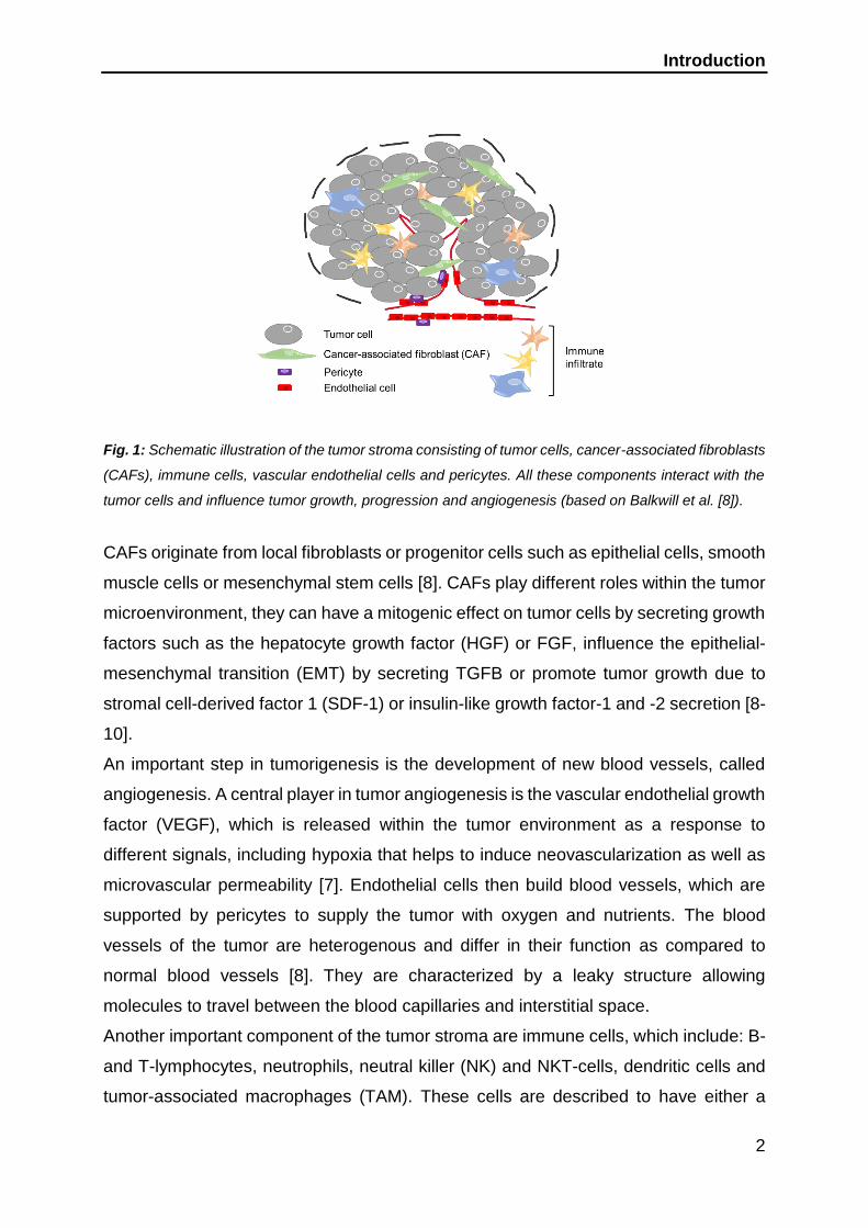
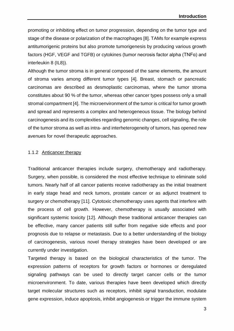
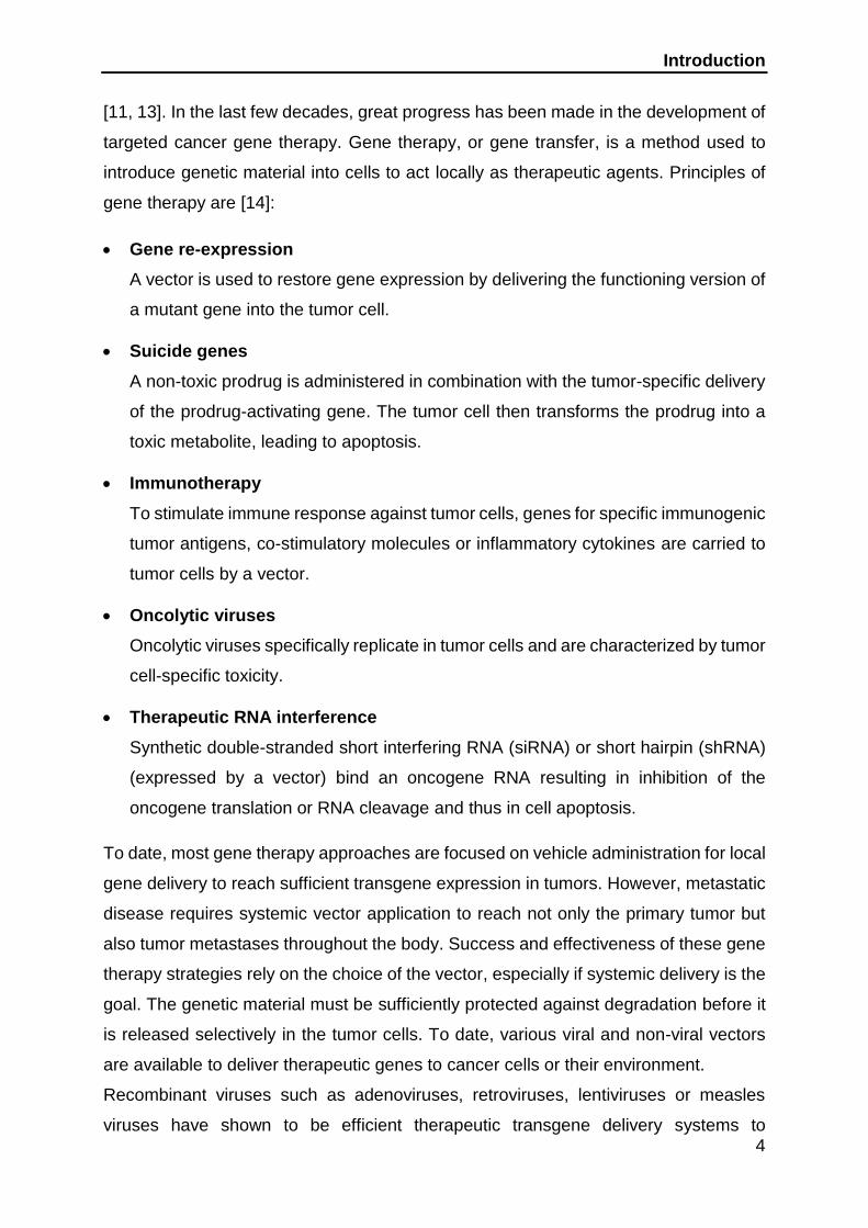
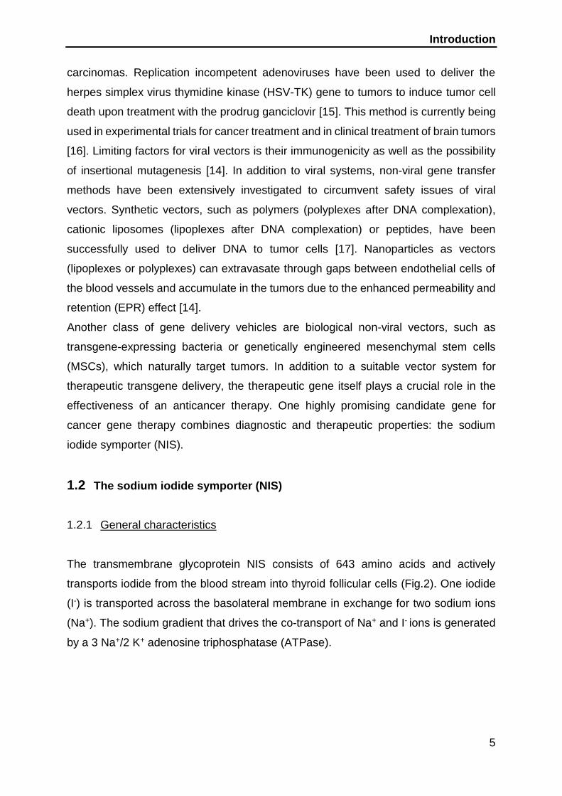
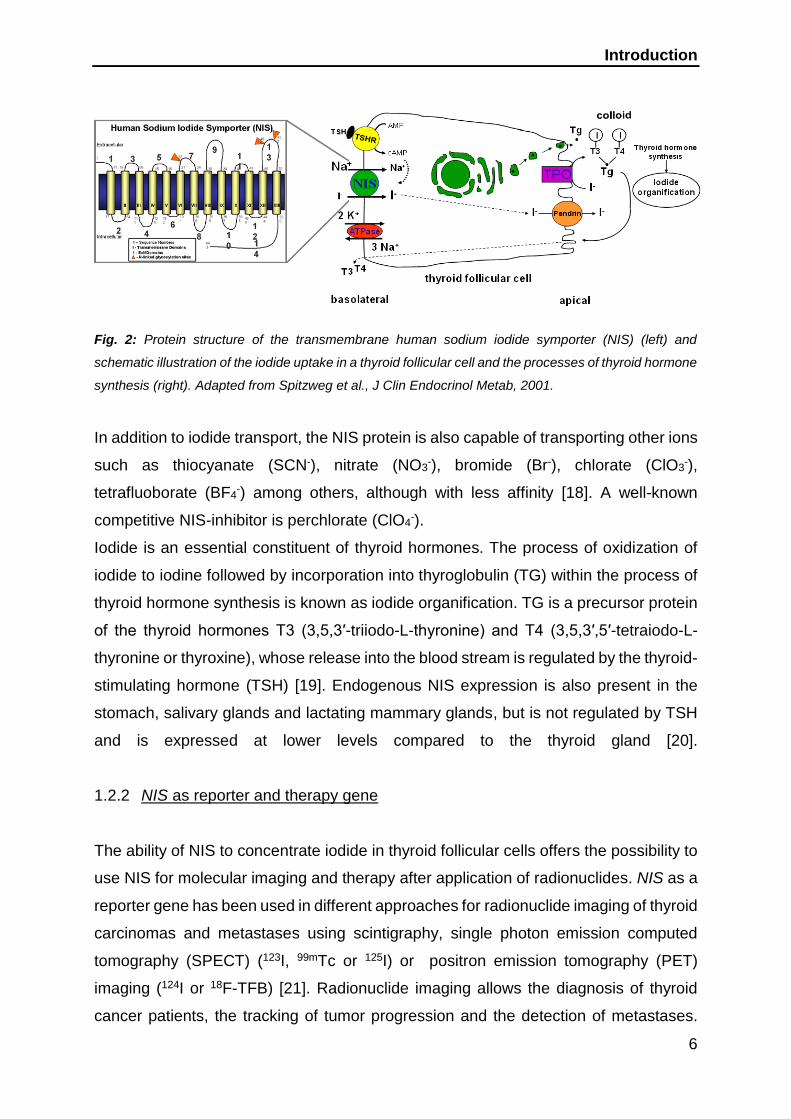

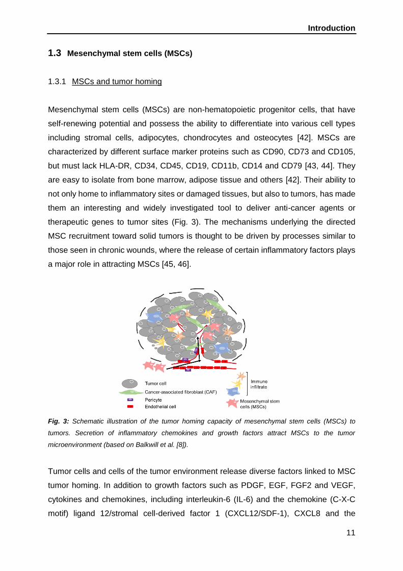
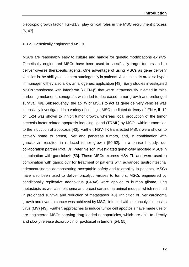
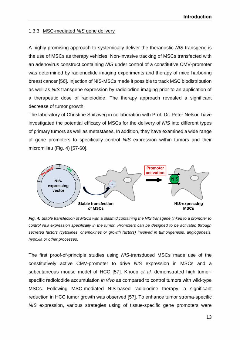

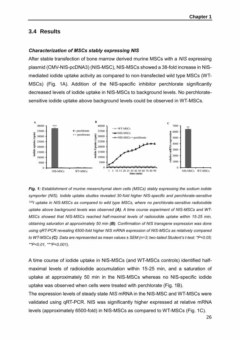

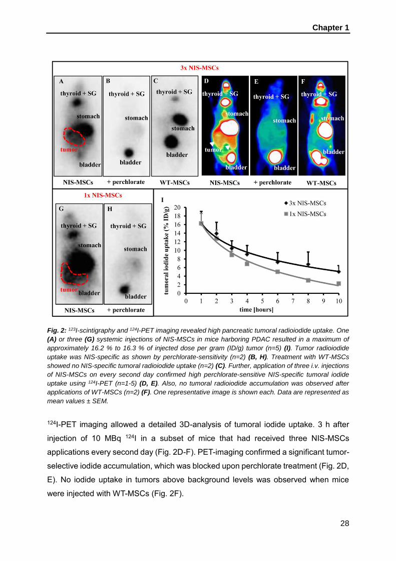
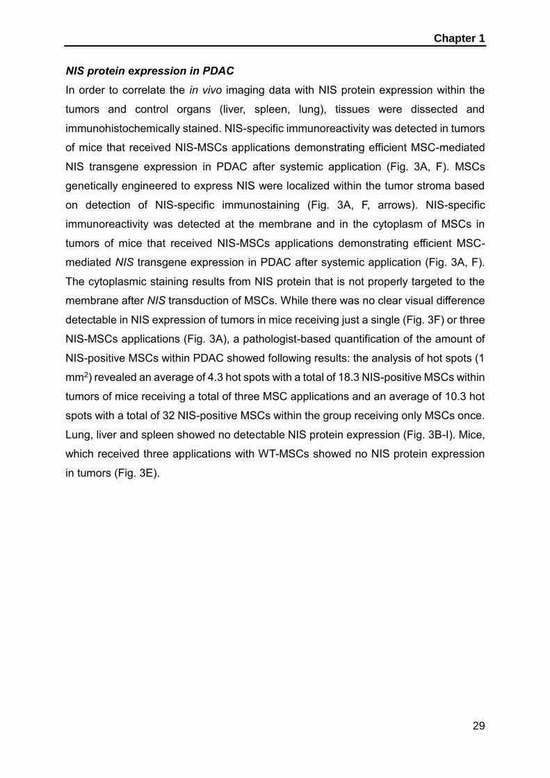
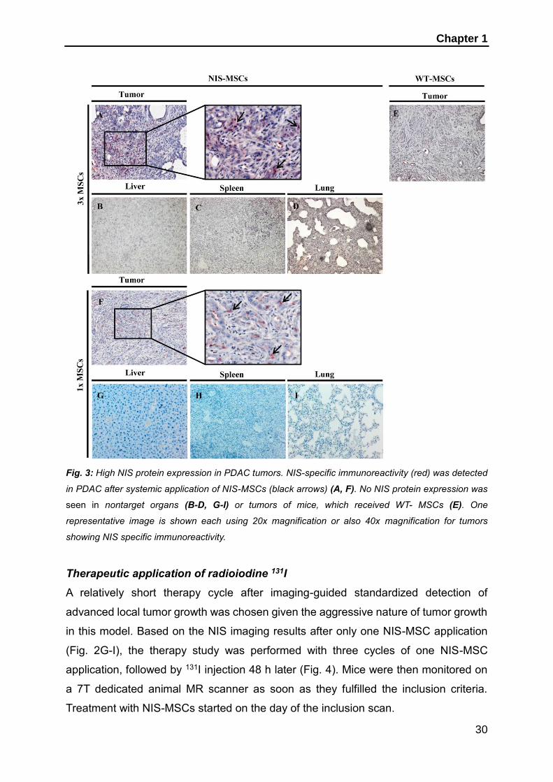
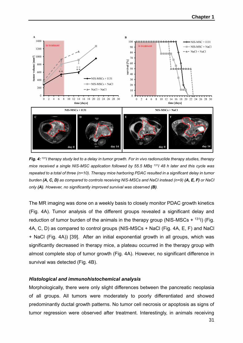
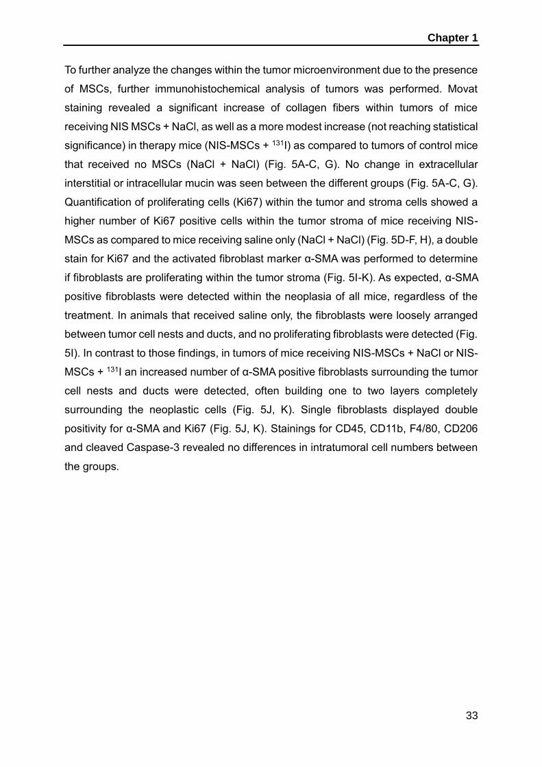


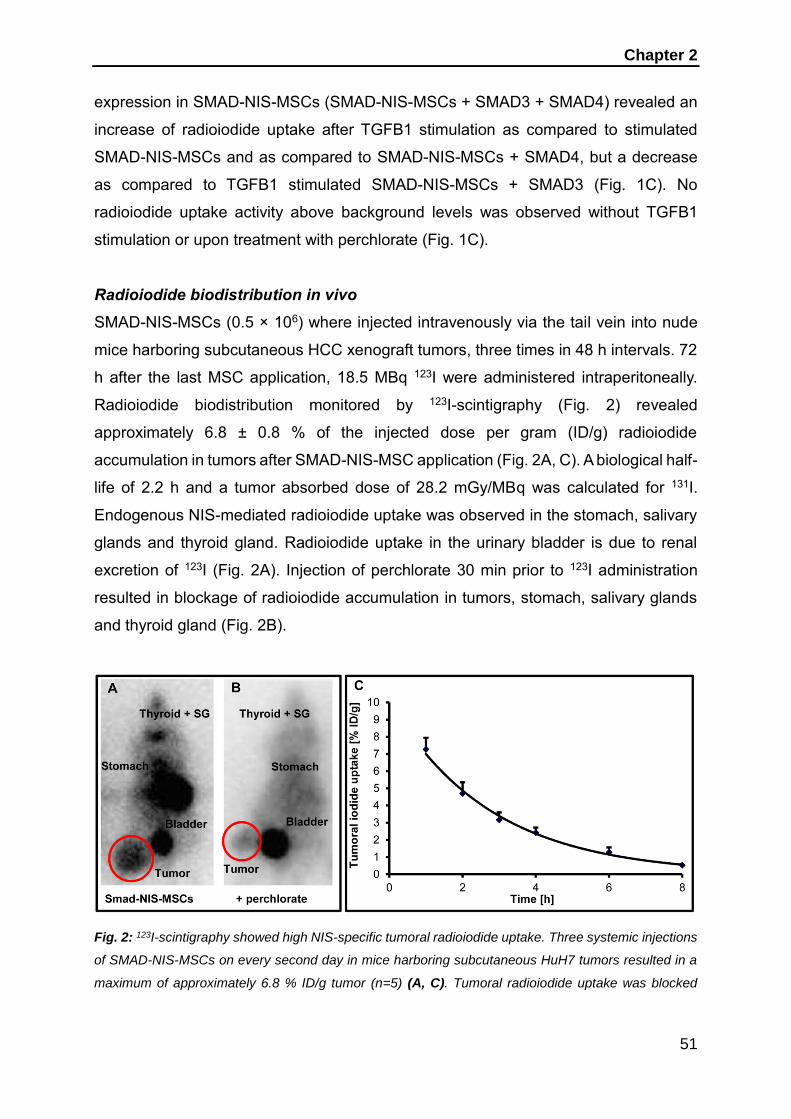
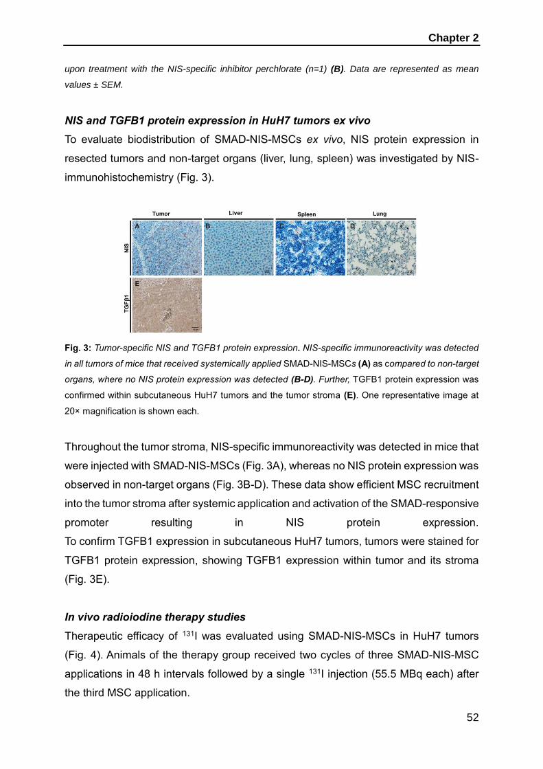
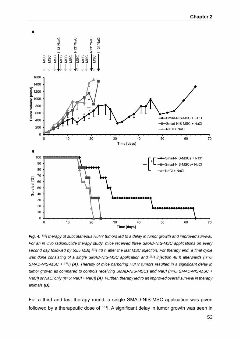

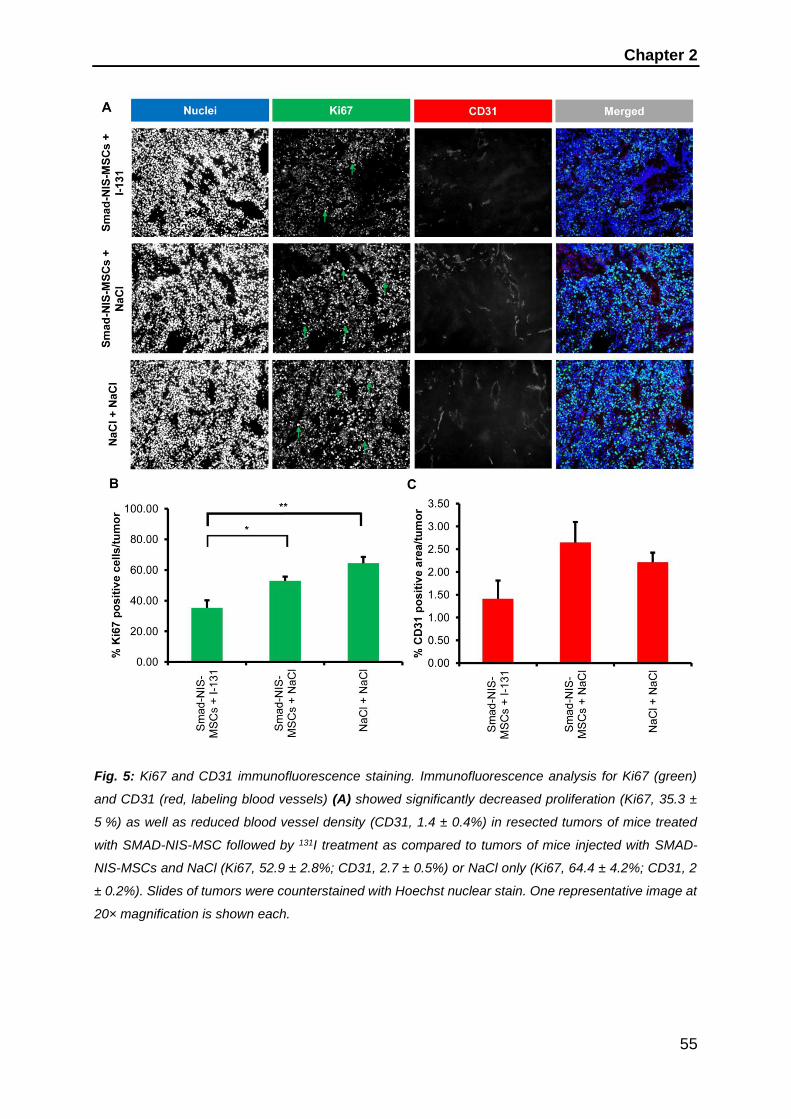


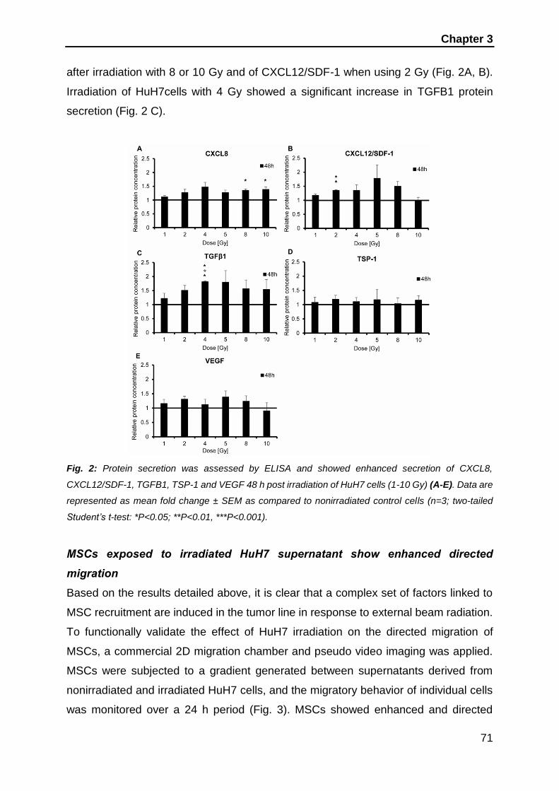
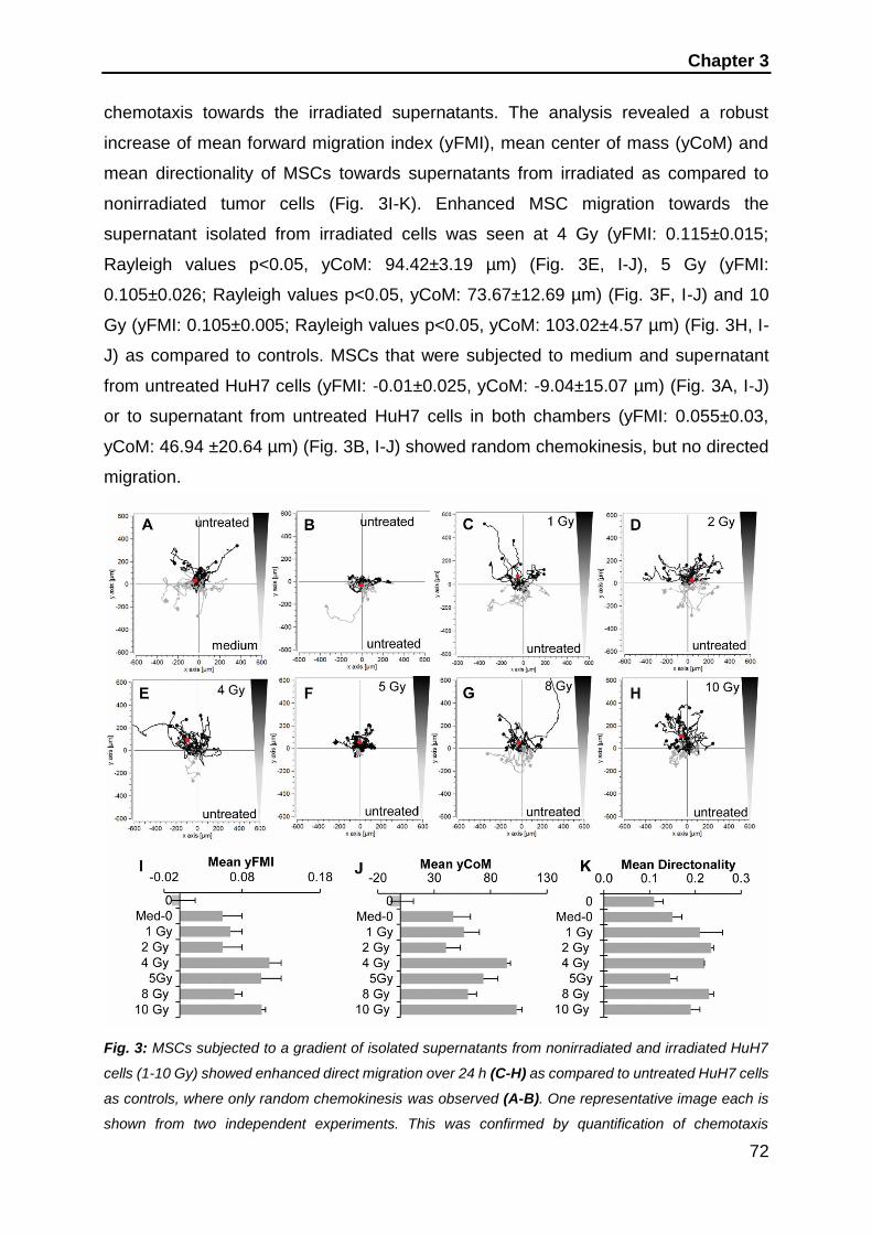
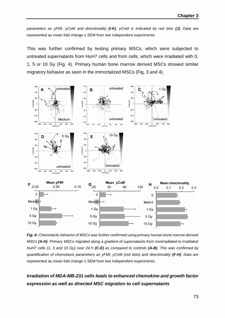
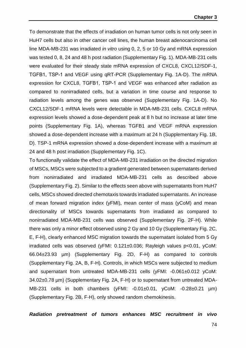
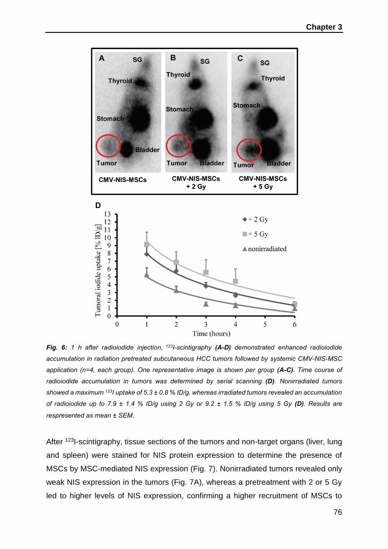
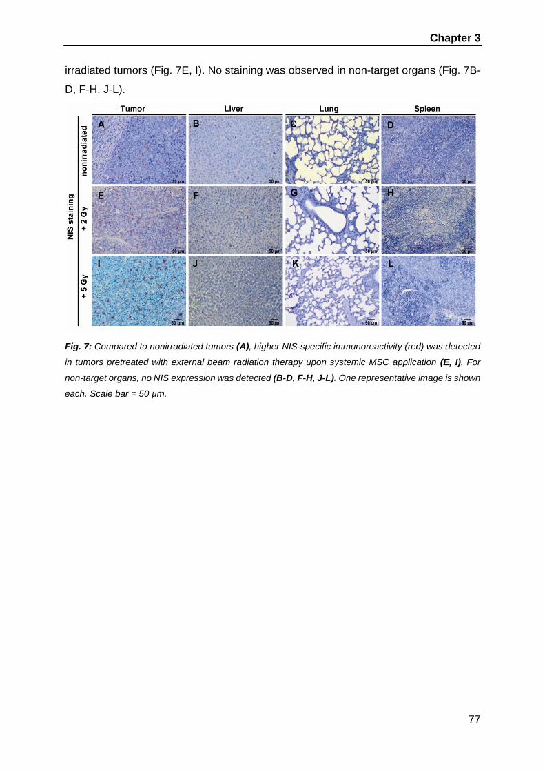

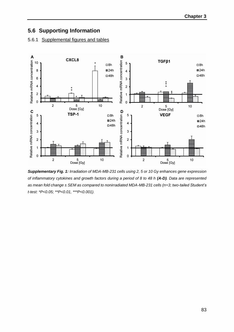
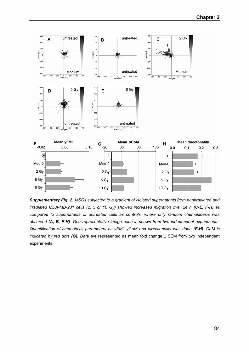


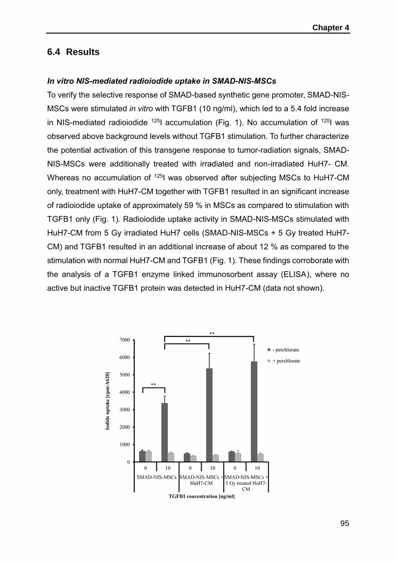
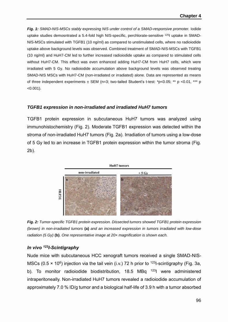
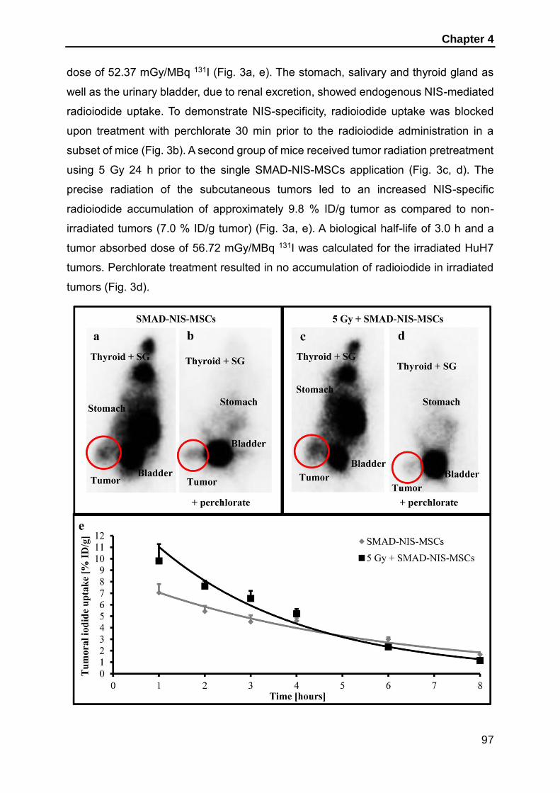
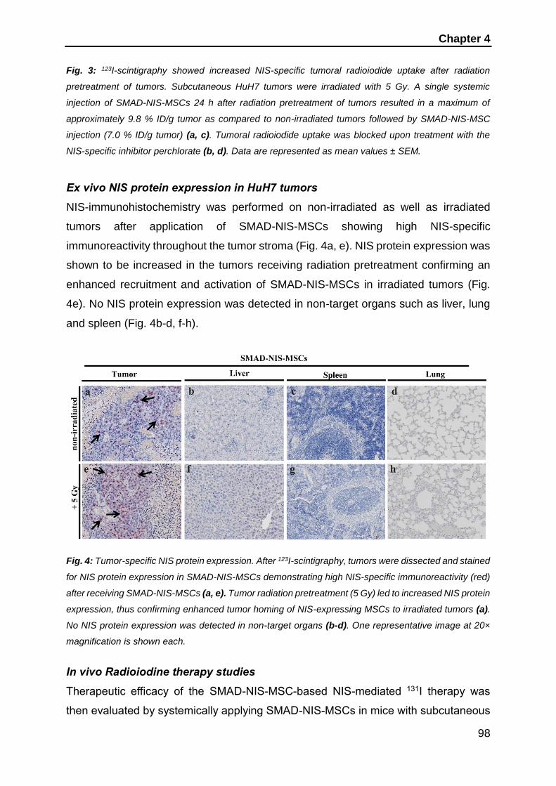
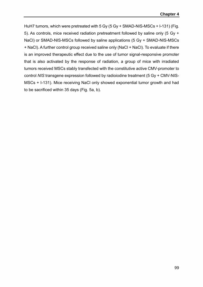
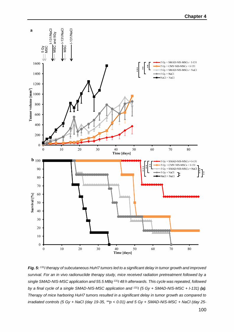
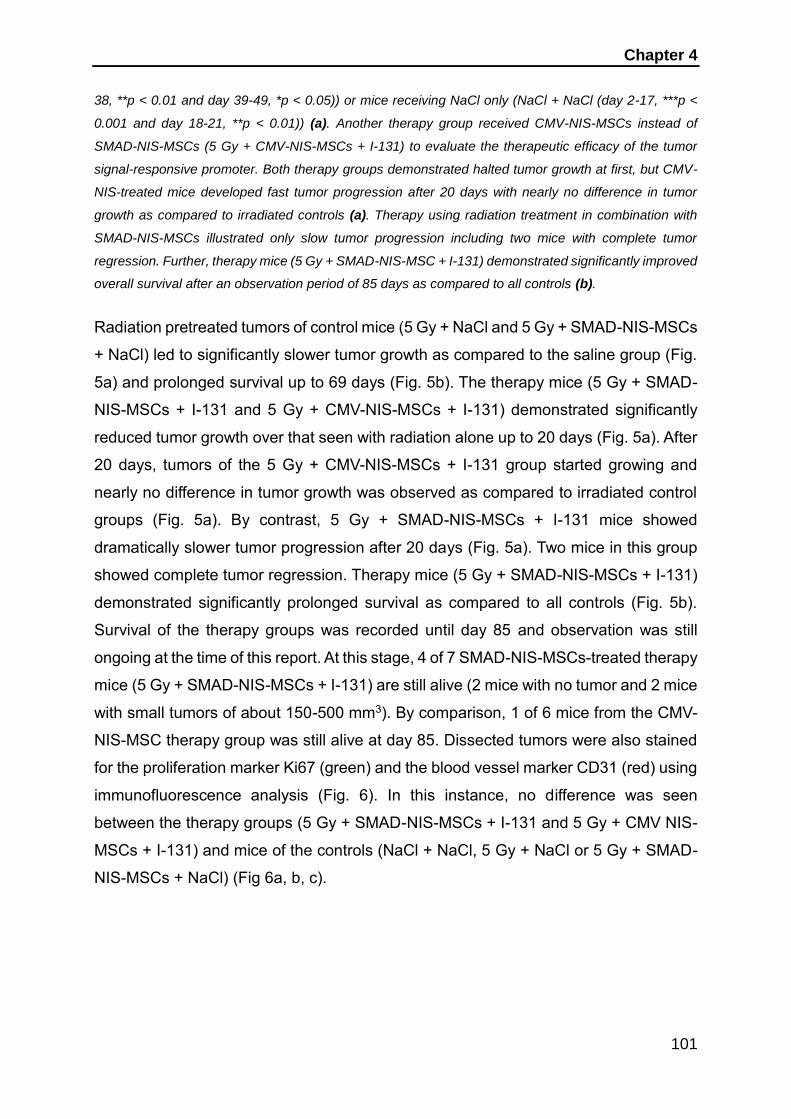

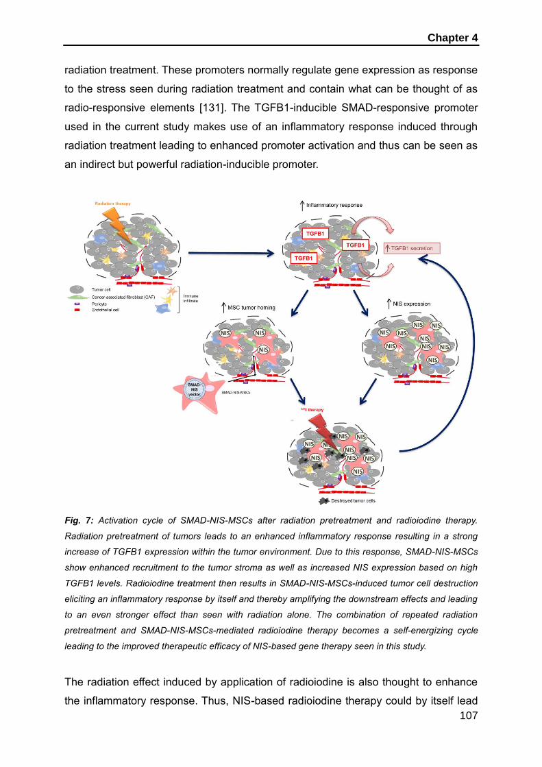


![Optical Surface Microtraps based on Evanescent Waves · Optical Surface Microtraps based on Evanescent Waves Dissertation zur Erlangung des Doktorgrades an der ... [Fol02]. While](https://static.documents.pub/doc/80x56/5bdee43809d3f2647f8b63e5/optical-surface-microtraps-based-on-evanescent-optical-surface-microtraps-based.jpg)


