Dissertati der Fa der Ludw Cyclin-depend Elucidating re and evaluating no Sabin ion zur Erlangung des Doktorgrade akultät für Chemie und Pharmazie wig-Maximilians-Universität München dent kinase 5 in endothelial cell migr egulatory mechanisms upstream of ovel Cdk inhibitors as anti-angiogen ne Bianca Monika Weitensteiner aus Tirschenreuth 2011 es n ration: Cdk5 nic drugs
Transcript
Dissertation zur Erlangung des Doktorgrades
der Fakultät für Chemie und Pharmazie
der Ludwig
Cyclin-dependent kinase
Elucidating regulatory mechanisms
and evaluating nov
Sabine Bianca Monika Weitensteiner
Dissertation zur Erlangung des Doktorgrades
der Fakultät für Chemie und Pharmazie
der Ludwig-Maximilians-Universität München
dependent kinase 5 in endothelial cell migration:
Elucidating regulatory mechanisms upstream of Cdk5
and evaluating novel Cdk inhibitors as anti-angiogenic drugs
Sabine Bianca Monika Weitensteiner
aus Tirschenreuth
2011
Dissertation zur Erlangung des Doktorgrades
Universität München
in endothelial cell migration:
upstream of Cdk5
angiogenic drugs
Erklärung
Diese Dissertation wurde im Sinne von § 13 Abs. 3 bzw. 4 der Promotionsordnung vom
29. Januar 1998 (in der Fassung der sechsten Änderungssatzung vom 16. August
2010) von Herrn Prof. Dr. Stefan Zahler am Lehrstuhl für Pharmazeutische Biologie
betreut.
Ehrenwörtliche Versicherung
Diese Dissertation wurde selbstständig und ohne unerlaubte Hilfe erarbeitet.
pazopanib (Votrient) and vandetanib (Caprelsa5, formerly Zactima) have been
approved for certain metastatic cancer types as monotherapy or in combination with
chemotherapy.6
However, in clinical use it has become apparent that anti-angiogenic tumor therapy is
more challenging than expected: Many tumors are refractory to VEGF-blockade, or
become resistant during treatment. This evasive resistance7 can be caused by a shift to
alternative angiogenic signaling pathways due to a pre-existing multiplicity of redundant
pro-angiogenic signals. Therefore novel targets in angiogenesis need to be identified
and characterized as a basis for future therapeutic concepts. If and how anti-
angiogenic therapy itself may contribute to increased metastasis is subject to
investigation.8, 9
1.1.1 The angiogenic cascade
The vessel wall of non-growing capillaries consists of a lining of endothelial cells tightly
interconnected by junction molecules like VE-cadherin and claudin, a basement
membrane and surrounding pericytes. When the quiescent vessels are activated by
angiogenic factors which are released from nearby hypoxic (tumor) tissue a series of
events occurs, as summarized in Figure 1:
1 INTRODUCTION 3
Figure 1 The angiogenic cascade. Pro-angiogenic factors like vascular endothelial growth factor (VEGF) or basic fibroblast growth factor (bFGF) bind to the corresponding receptors on endothelial cells and stimulate degradation of extracellular matrix (ECM) by secretion of digestive enzymes and initiate proliferation of endothelial cells. Newly formed endothelial cell sprouts further proliferate and migrate towards the tumor, navigating along the gradient of angiogenic cues. In order to coordinate endothelial cell movement, a tip cell is selected equipped with filopodia to sense guidance cues while the subsiding stalk cells proliferate and eventually form the lumen.10 Finally, the endothelial cells organize into hollow tubes and create a novel basement membrane. At the end, tight junctions and firm contacts to the ECM are formed and pericytes and smooth muscle cells are recruited to the mature vessel wall.3, 6, 11
1.2 Function and regulation of Cdks
Cyclin-dependent kinases (Cdks) are a family of small serine/threonine kinases which
are only active when they are bound to their regulatory subunits, the cyclins. The
presence or absence of the activating cyclin is therefore crucial for Cdk kinase activity.
At least 29 proteins have been designated as cyclins, sharing a conserved “cyclin box”.
For some classes of cyclins the Cdk binding partner has not yet been identified.12
The best-characterized members of the Cdk family are involved in cell cycle control:
the mitotic Cdk Cdk1, and the interphase Cdks Cdk2, Cdk4 and Cdk6. They pair with
A-, B-, D- and E-type cyclins, whose expression fluctuates during the cell cycle (which
explains the term “cyclins”) and this way regulates Cdk activity.13 Cdk3 is only little-
studied and is also implicated in cell cycle control. 14 Cdk7, Cdk8, Cdk9, Cdk10, Cdk11,
Cdk12 and Cdk13 regulate transcription and splicing. They are activated by Cyclin H
(Cdk7), C (Cdk8), T and K (Cdk9), and cyclins from the L-type (Cdk10-Cdk13). 12, 15-24
In a second function, Cdk7 regulates the activity of the cell cycle Cdks: It is part of the
Cdk-activating kinase (CAK) complex together with Cyclin H and the stabilizing protein
Mat125 (see Figure 2), and phosphorylates the cell-cycle Cdks in the T-loop for full
activation.26 Cdk kinase activity can further be modulated by inhibitory phosphorylations
or endogenous Cdk inhibitors (CKIs), as displayed in Figure 2.
CKIs regulate the activity of the cell cycle Cdks by direct interaction. The CKIs of the
INK4 family, p16INK4A, p15INK4B, p18INK4C and p19INK4D, specifically bind the monomeric
Cdk and thereby prevent activation via cyclins, whereas p21Cip1, p27Kip1 and p57Kip2
directly inhibit the Cdk-cyclin complex.12
Figure 2 Regulation of cell cycle Cdks . Cdks are only active when bound to their cyclin activators. The Cdk-cyclin complexes can be additionally modulated by phosphorylation: Phosphorylation by Cdk activating kinase (CAK) in the T-loop of the Cdk enhances kinase activity of the complex. By contrast, phosphorylation of a conserved threonine or tyrosine residue by Wee1 or Myt1 negatively regulates kinase activity. The Cdc25 phosphatases abrogate this inhibitory phosphorylation. Cdk inhibitors from the INK4 or Cip/Kip family block kinase activity by either stabilizing the monomeric Cdk or by binding to the cyclin-Cdk complex. (P: Phosphorylation, : Kinase activity; adapted from Malumbres & Barbacid12)
Cyclin
Cdk Cdk
Cyclin
Cdk
CyclinP
Cdk7
Cyclin H
Mat1
Cdk
Cyclin
PWee1Myt1
Cdc25
INK4
Cip/Kip
CAK
1 INTRODUCTION 5
1.3 Cdk5 as a unique Cdk in charge of cellular migration
1.3.1 Functions of Cdk5
Cdk5 has been discovered as a neuronal cdc2-like kinase (nclk) in 1992.27 Cdk5 is a
proline-directed serine/threonine kinase that phosphorylates serine or threonine
residues directly upstream of a proline, with a preference for a basic residue in the +3
position and the consensus sequence (S/T)PX(K/H/R).28 Despite its high sequence
homology with the mitotic Cdk1 (cdc2), Cdk5 is not involved in cell cycle control and
unique among the Cdks in its regulation and function. Cdk5 deficient mice die
perinatally due to severe defects in neuronal layering, as Cdk5 is crucial for the
cytoarchitecture of the CNS.29 On the cellular level, Cdk5 is well-described in neurons
as the key hub in the dynamic network of trafficking and transport, integrating signals in
cytoskeletal dynamics during neuronal migration, in synaptic plasticity and synaptic
vesicle endo- and exocytosis, cell adhesion and axon guidance, neuromuscular
development and pain signaling.30, 31 Deregulated Cdk5 activity in neurons is a major
feature of Alzheimer’s disease resulting in the aggregation of neurofibrillary tangles
comprised of Cdk5-hyperphosphorylated tau.32 Although Cdk5 expression and activity
is highest in the central nervous system27, Cdk5 is as well expressed in various tissues,
and an increasing body of research uncovers extraneuronal functions of Cdk5, where it
is involved in the regulation of migration, cell death and survival, glucose metabolism
and inflammation. 33, 34
1.3.2 Regulation of Cdk5
Monomeric Cdk5 requires the association with a regulatory subunit for activation; the
known Cdk5 activators p35 and p39 however are no cyclins. It has been shown that
Cyclin D, E and G bind to but do not activate Cdk5.35-38 The expression and cleavage of
p35 and p39 essentially determines Cdk5 activity and localization, as displayed in
Figure 3 for p35.39
Neuronal growth factor (NGF)40, tumor necrosis factor-alpha (TNF-α)41, 42 and
interferon-gamma (INF-γ)43 increase transcription and expression of p35 via
extracellular signal regulated kinase (ERK) / early growth response-1 (Egr-1) mediated
pathways.
6 1 INTRODUCTION
Figure 3 Regulation of Cdk5 . p35 activates Cdk5 and recruits the p35/Cdk5 complex to the membrane, as p35 carries a myristoylation.32, 44 The short-lived p35 is rapidly degraded by the ubiquitin-proteasome pathway after phosphorylation by Cdk5.45 The tyrosine kinases c-abl (via the adaptor protein cables)46 and fyn47 activate Cdk5 by phosphorylation of tyrosine 15. Neurotoxic events activate calpain which cleaves p35 into p2548, 49 and liberates the p25/Cdk5 complex from the membrane. p25 is more stable than p35 and dislocates Cdk5 activity to the cytoplasm, which leads to tau hyperphosphorylation32 (P: Phosphorylation, U: Ubiquitination,
: Kinase activity; adapted from Dhavan & Tsai39).
p39, which exhibits similar characteristics, is less explored: p39 carries a
myristoylation44 and is either degraded by the proteasome or cleaved by calpain into
p2950. p35/Cdk5 and p39/Cdk5 show similar substrate specificity51, although they seem
to have physiologically distinct functions. The expression patterns of p35 and p39 in the
developing brain differ52, they locate Cdk5 activity to different subcellular regions44 and
p39 can compensate only for some but not all functions of p35 in p35 knockout mice.53
Cdk5 activation by p35 and p39 has mainly been explored in neurons but p35-
dependent Cdk5 activity was detected in several non-neuronal tissues as well.54-58
Recently, Cyclin I was described as an alternative activator of Cdk5 in podocytes.59
In addition, Cdk5 regulation is unique in the family of cyclin-dependent kinases in two
further aspects: 1) The endogenous Cdk inhibitors (CKIs) are not involved in Cdk5
regulation and modulation. Yet, Cdk5 can phosphorylate and stabilize p27Kip1, which is
crucial for migration.60, 61 2) Modulation of Cdk5 activity by phosphorylation
fundamentally differs from the cell cycle Cdks. In contrast to the cell cycle Cdks,
p10fyn
Cdk5p35
Cdk5
Cdk5p25
p35UUU
cytopl. membrane
Cdk5
nucleus
proteasomep35
P
P
P
cablesc-abl
regulationof transcription
calpain
1 INTRODUCTION 7
phosphorylation by CAK in the T-loop seems dispensable for Cdk5 full activation. CAK
can phosphorylate Cdk5 on serine 159, but the function of this phosphorylation is
disputed.62-65 Activity of mitotic Cdks is inhibited by phosphorylation of conserved
threonine or tyrosine residues (T14 and Y15 in Cdk2) by Wee1 or Myt1 (see Figure 2).
Cdk5 is phosphorylated on T14 in vitro and this inhibits kinase activity.66 Cdk5 is not
inhibited by Wee164, but can be phosphorylated at tyrosine 15 by fyn and c-abl.47 46 In
contrast to the mitotic Cdks, this phosphorylation stimulates Cdk5 activity.
1.4 Cyclin-dependent kinase inhibitors
Tumor cells characteristically display limitless replicative potential which is caused by
alterations in cell-cycle control systems.2 Cdks as prominent regulators of the cell cycle
exhibit deregulated activity in tumors, resulting from overexpression and mutations in
cell cycle cyclins and Cdks, as well as from a loss of their endogenous inhibitors, the
CKIs.67, 68 To target Cdks is therefore a promising strategy in anticancer therapy and
several approaches are imaginable to alter Cdk activity – either by direct inhibition of
the catalytic Cdk subunit, or by indirectly modulating regulatory pathways that govern
Cdk activity, for example binding of cyclins, phosphorylation of the Cdk subunit or
interaction with the CKIs. Most small molecule Cdk inhibitors interact with the ATP-
binding site of the kinase subunit, which is fundamental for kinase activity. The ATP-
binding site is well conserved among the Cdks which is why adequate Cdk selectivity of
inhibitors remains a big challenge.69 If selective or broad-spectrum Cdk inhibitors are
more effective remains under discussion. Established Cdk modulators such as
flavopiridol (Alvocidib) and roscovitine (CYC202, seliciclib) inhibit a relatively wide
range of Cdks. Second generation Cdk inhibitors are under preclinical and clinical
investigation at present. They can be subdivided in three classes: 1) Broad spectrum
Cdk inhibitors that target both cell cycle and transcriptional Cdks, 2) Selective inhibitors
of Cdk2 or Cdk4/6 or 3) Compounds with combined activity against Cdks and additional
kinases with a benefit for anticancer therapy, for example receptor tryrosine kinases or
Aurora kinases. The most promising strategy for successful therapy with selective or
combined Cdk inhibitors is very likely depending on the genetic alterations present in
the tumor. 70
8 1 INTRODUCTION
1.4.1 Roscovitine
(R)-roscovitine or CYC-202/seliciclib – in the following referred to as roscovitine –
belongs to the class of 2,6,9-trisubstituted purines and is one of the best-studied Cdk
inhibitors.71
Figure 4 Structure of ( R)-roscovitine. The arrows indicate how the structure was modified for the tested Cdk inhibitors.
It is developed by Cyclacel Pharmaceuticals72 and currently tested in several Phase I
and Phase II clinical trials for tumor treatment.73 Roscovitine inhibits mainly Cdk1,
Cdk2, Cdk5, Cdk7 and Cdk9 and exerts anti-mitotic and pro-apoptotic effects in a wide
range of tumor cells.74 Cell-cycle independent actions of roscovitine mainly derive from
Cdk5 inhibition and include anti-angiogenic75 and anti-inflammatory76 effects, inhibition
of cell migration and motility77, 78 and modulation of glucose metabolism.79
The roscovitine derivatives evaluated for their anti-angiogenic potential were kindly
provided by V. Krystof and R. Jorda (Palacký University & Institute of Experimental
Botany, Olomouc, Czech Republic) and the structures are displayed in the Materials
and Methods section (Figure 5).
1.5 Aim of the study
As explained in the previous paragraphs, Cyclin-dependent kinases (Cdks) and their
activators, the cyclins, control the transitions of the cell cycle, which is deregulated in
many tumor types.67 Cdk inhibitors like roscovitine or flavopiridol are currently tested in
clinical trials as anti-proliferative anticancer drugs.
Anti-angiogenic actions of these Cdk inhibitors have been observed in vitro and in
vivo.80-82 Recently, it has been demonstrated that the anti-angiogenic effect of the Cdk
inhibitor roscovitine most likely results from impaired endothelial cell migration. The
effect on migration was traced down to Cdk5 inhibition which led to Rac1 inactivation
OH
NH
N
N N
N
NH
NH2
RN
1 INTRODUCTION 9
and lamellipodia disruption.75 A promising novel strategy in anti-angiogenic therapy
may therefore be Cdk5 inhibition. Up to date, improved Cdk inhibitors have mainly
been developed in order to block cancer cell proliferation but have not systematically
been optimized and evaluated for anti-angiogenic action by Cdk5 inhibition.
In contrast to the mitotic Cdks, Cdk5 is a cell-cycle independent Cdk that is known to
control migration of post-mitotic neurons during CNS development.
With regard to its regulation, Cdk5 is as well unique among the Cdks: First, Cdk5
activity and subcellular localization is directed by non-cyclin proteins (p35 and p39).
Second, phosphorylation of a conserved tyrosine 15 decreases activity of mitotic Cdks
but stimulates Cdk5 activity and third, endogenous Cdk inhibiting proteins (CKIs),
which control cell cycle Cdks, do not influence Cdk5 activity. Activation of Cdk5 in non-
neuronal cells, as investigated so far, parallels the neuronal Cdk5 activation pathways,
however distinct mechanisms have been reported as well.54 The mechanisms which
are responsible for the activation of Cdk5 during endothelial cell migration have not yet
been explored.
Aims of the study were therefore:
1. to elucidate the characteristics of Cdk5 regulation during endothelial
cell migration by investigating the role of the neuronal activators
p35/p25 and p39/p29 and the tyrosine 15 phosphorylation.
2. to evaluate the anti-angiogenic potency of novel roscovitine-derived
Cdk inhibitors in vitro and in vivo, thereby paying particular
consideration on their potency to inhibit Cdk5.
2 MATERIALS AND METHODS
12 2 MATERIALS AND METHODS
2.1 Materials
2.1.1 Biochemicals, inhibitors, dyes and cell cultu re reagents
Table 1 Biochemicals, inhibitors, dyes and cell culture reagents
Novel roscovitine derivatives were synthesized and kindly provided by V. Krystof and
R. Jorda (Palacký University & Institute of Experimental Botany, Olomouc, Czech
Republic).
Figure 5 Chemical Structures of the tested LGR comp ounds in comparison to ( R)-roscovitine. The LGR compounds have been modified in comparison to (R)-roscovitine in one or more of the following aspects: 1. The purine scaffold has been changed to pyrazolo[4,3-d]pyrimidine (LGR 1404, 1406, 1407, 1430, 1492, 1667, 1695), with LGR 1404 being a bioisoster of (R)-roscovitine. 2. In the aminobenzyl group, an additional ortho amino function is present (LGR 1430, 1492, 1695, 1730). 3. The residue at purine C2 or pyrazolo[4,3-d]pyrimidine C5 respectively, differs from (R)-roscovitine either in structure and/or stereochemistry. Defined configurations are shown in the chemical structures. LGR 1407 contains no stereocenter. LGR 1406 and LGR 1430 are an equal mixture of 4 stereoisomers: the trans enantiomers (R,S) and (S,R); and the cis enantiomers (R,R) and (S,S).
N
NH
N
NH
NHOH
NH2
N
LGR 1695 (S)
N
NH
N
N
NHOH
NH2
N
LGR 1730 (R)
N
NH
N
NH
NNHOH
LGR 1667 (R)
LGR 1406
** NN
H
NN
NH
NH
NH2
OH
NH
N
NN
NH
NH
LGR 1404 (R)
NH2
NH
NH
N
NN
NH
LGR 1407
NNH
NN
NHNH
NH2
NH2
**
LGR 1430
OHNH
N
NH
N
NH
N
NH2
LGR 1492 (R)
OH
NH
N
N N
N
NH
(R)-roscovitine
16 2 MATERIALS AND METHODS
2.2 Cell culture
2.2.1 Cell culture solutions and reagents
The following solutions were used for isolation and cultivation of endothelial cells:
Table 5 Cell culture solutions
Growth medium Stopping medium
ECGM 500 ml M 199 500 ml
Supplement Mix #C-39215 23.5 ml FCS 50 ml
FCS gold 50 ml
Penicillin/Streptomycin 5 ml
Amphothericin B 5 ml
Freezing medium Trypsin/EDTA (T/E)
FCS gold 10 % Trypsin 0.05 %
DMSO 10 % EDTA 0.20 %
Growth medium PBS
Collagen G
Collagen G 0.001 %
PBS
Before use, FCS gold was heat inactivated. First, FCS gold was partially thawed at
room temperature for 30 min, then completely thawed at 37°C. Heat inactivation was
carried out at 56°C for 30 min. FCS aliquots were s tored at -20°C
2.2.2 Endothelial cells
Endothelial cells (ECs) were cultured under constant humidity at 37°C with 5 % CO 2 in
an incubator (Heraeus, Hanau, Germany). Cells were routinely tested for mycoplasma
contamination with the Venor®GeM PCR detection kit (Minerva Biolabs, Berlin,
Germany). All cell culture flasks, Petri dishes and multiwell plates were coated with
Collagen G for 30 min in the incubator prior to plating the cells.
2 MATERIALS AND METHODS 17
2.2.2.1 HMEC-1 (Human microvascular endothelial cel ls)
The cell line CDC/EU.HMEC-1 was kindly provided by the Centers for Disease Control
and Prevention (Atlanta, GA, USA). The immortalized HMEC-1 cell line was created by
transfection of human dermal microvascular endothelial cells with a plasmid coding for
the transforming SV40 large T-antigen. HMEC-1 were shown to retain endothelial
morphologic, phenotypic, and functional characteristics.85, 86 HMEC-1 were used for
endothelial cell proliferation experiments, siRNA transfection and immunoprecipitations
for kinase assay and LC-ESI-MS/MS.
2.2.2.2 HUVECs (Human umbilical vein endothelial ce lls)
Human umbilical cords were kindly provided by Klinikum München Pasing,
Frauenklinik München West/Krüsmannklinik, Rotkreuzklinikum München, and
WolfartKlinik Gräfelfing. After childbirth, umbilical cords were placed in PBS+Ca2+/Mg2+
containing penicillin (100 U/ml) and streptomycin (100 µg/ml), and stored at 4°C. Cells
were freshly isolated every week. The umbilical vein was washed with PBS+Ca2+/Mg2+,
filled with 0.1 g/l collagenase A, and incubated for 45 min at 37°C. To isolate
endothelial cells, the vein was flushed with stopping medium and the cell suspension
was centrifuged (1,000 rpm, 5 min). Afterwards, cells were resuspended in growth
medium and plated in a 25 cm2 flask (passage #0). After reaching confluency, cells
were trypsinized and plated in a 75 cm2 flask. Unless otherwise indicated, experiments
were performed using cells at passage #3. HUVECs were used for all other
experiments except endothelial cell proliferation experiments, siRNA transfection and
immunoprecipitations for LC-ESI-MS/MS.
2.2.3 Passaging
After reaching confluency, cells were either sub-cultured 1:3 in 75 cm2 culture flasks or
seeded either in multiwell-plates or dishes for experiments. For passaging, medium
was removed and cells were washed twice with PBS before incubation with T/E for
1-2 min at 37°C. Thereafter, cells were gradually d etached and the digestion was
terminated using stopping medium. After centrifugation (1,000 rpm, 5 min), the pellet
was resuspended in growth medium and cells were plated.
18 2 MATERIALS AND METHODS
2.2.4 Freezing and thawing
For freezing, confluent HMEC-1 from a 150 cm2 flask were trypsinized, centrifuged in
stopping medium (1,000 rpm, 5 min) and resuspended to 2 x 10⁶ cells/ml in ice-cold
freezing medium. 1.5 ml aliquots were frozen in cryovials. After storage at -80°C for
24 h, aliquots were moved to liquid nitrogen for long term storage.
For thawing, a cryovial was warmed to 37°C and the content was immediately
dissolved in pre-warmed stopping medium. In order to remove DMSO, cells were
centrifuged (1,000 rpm, 5 min), resuspended in growth medium and transferred to a
75 cm2 culture flask.
2.3 Western blot analysis
2.3.1 Preparation of protein samples
Endothelial cells were treated as indicated, washed once with ice-cold PBS and
subsequently lysed in RIPA lysis buffer or in modified RIPA lysis buffer for phospho-
proteins. Immediately, cells were frozen at -80°C. Afterwards, cells were scraped off
and transferred to Eppendorf tubes (Peske, Aindling-Arnhofen, Germany) before
centrifugation (14,000 rpm, 10 min, 4°C). Tissue sa mples as positive controls were
homogenized in lysis buffer with a POLYTRON PT 1200 C homogenizer (Kinematica
AG, Lucerne, Switzerland), frozen at -80°C and cent rifuged twice (14,000 rpm, 10 min,
4°C). Protein concentration was determined in the s upernatant using either the BCA or
the Bradford assay. Afterwards, Laemmli sample buffer (3x) was added and samples
were heated at 95°C for 5 min. Samples were kept at -20°C until Western blot analysis.
2 MATERIALS AND METHODS 19
Table 6 Buffers for protein sample preparation
RIPA buffer Lysis buffer for phosphoproteins
Tris/HCl (pH 7.4) 50 mM Tris/HCl (pH 7.4) 50 mM
NaCl 150 mM NaCl 150 mM
Nonidet NP 40 1% Nonidet NP 40 1%
Deoxycholic acid 0.25% Deoxycholic acid 0.25%
SDS 0.10% SDS 0.10%
H2O Na3VO4 0.3 mM
Complete®mini EDTAfree 4.0 mM NaF 1.0 mM
PMSF 1.0 mM β-Glycerophosphate 3.0 mM
Na3VO4 1.0 mM Pyrophosphate 10 mM
NaF 1.0 mM H2O
Complete®mini EDTAfree 4.0 mM
PMSF 1.0 mM
H2O2 600 µM
5x SDS-sample buffer 3x Laemmli buffer
Tris/HCl (pH 6.8) 3.125 M Tris/HCl (pH 6.8) 187.5 mM
Glycerol 10 ml SDS 6%
SDS 5% Glycerol 30%
DTT 2% Bromphenol blue 0.025%
Pryonin Y 0.025% H2O
H2O β-Mercaptoethanol 12.5%
2.3.2 Membrane fractionation
HUVEC lysates were separated into a soluble (cytosolic) and a particulate
(membranous) fraction, as described previously by Li H et al.87 HUVECs were treated
as indicated, washed twice with ice-cold PBS+Ca2+/Mg2+, and homogenized in lysis
buffer. Protein quantification with Bradford Assay was used to obtain equal protein
amount in the samples. Lysates were centrifuged at 100,000 g and 4°C for 45 min. The
supernatant (cytosolic fraction) was collected and boiled with 5x SDS sample buffer
(5 min, 95°C). The pellet was washed in lysis buffe r containing 1.0 M NaCl and
centrifuged at 100,000 g and 4°C for 20 min. The su pernatant was discarded and the
pellet was solubilized with lysis buffer containing 20 mM CHAPS at 4°C for 30 min.
20 2 MATERIALS AND METHODS
After centrifugation at 100,000 g and 4°C for 45 mi n, the supernatant was kept as
membranous fraction and boiled with 5x SDS sample buffer (5 min, 95°C). The
cytosolic and membranous fractions were used for Western blotting.
Table 7 Buffer for membrane fractionation
Membrane fractionation lysis buffer
Tris/HCl (pH 7.5) 50 mM
EDTA 0.5 mM
EGTA 0.5 mM
Glycerol 10 %
Glutathion 7 mM
DTT 2 mM
PMSF 1 mM
Complete®mini EDTAfree 4 mM
H2O
2.3.3 Immunoprecipitation
Immunoprecipitation for Kinase Assay
Immunoprecipitations were used in order to obtain active Cdk5 complexes for Cdk5
kinase assay. Cells or tissue samples were lysed (see 2.3.1) in the indicated lysis
buffer (Table 6 and Table 8) and kept on ice for 30 min. Thereafter, the samples were
centrifuged and protein concentrations were determined in the supernatants. Cell
lysates were incubated with 2 µg of the indicated antibodies over night at 4°C under
gentle agitation. Thereafter, Protein A (for rabbit IgG) or Protein G (for mouse IgG)
Agarose beads (Sigma-Aldrich, Taufkirchen, Germany) were washed with lysis buffer
and 50 µl bead suspension (25 µl packed beads) was added to each sample. After 3 h
of incubation at 4°C, the beads were spun down and an aliquot of the supernatant was
collected as binding control and boiled with Laemmli sample buffer (3x) for 5 min at
95°C. Subsequently, two washing steps with lysis bu ffer and two washing steps with
kinase buffer were performed, and 50 µl kinase buffer (Table 9) were added to the
bead pellet. The beads were resuspended in the kinase buffer before taking a 10 µl
aliquot of the beads as binding control and boiling the aliquot with Laemmli sample
buffer (3x) for 5 min at 95°C. The rest of the bead s was used for Cdk5 kinase assay.
2 MATERIALS AND METHODS 21
For the HA-immunoprecipitation of the Cdk5-HA transfected cells, the Pierce
Mammalian HA-Tag IP/Co-IP Kit (Thermo Fisher Scientific, Rockford, IL, USA) was
used according to the manufacturer’s instructions. The kinase assay reaction was
carried out directly on the beads.
Immunoprecipitation for the detection of novel Cdk5-interacting proteins by LC-ESI-
MS/MS
HMEC-1 were treated as indicated, lysed in the Lysis buffer for phosphoproteins (Table
6) and immunoprecipitation with Cdk5 mouse monoclonal antibody was performed as
described in the paragraph above. After three washing steps with lysis buffer, the
beads were spun down, 25µl Laemmli sample buffer (3x) were added and the samples
were boiled for 5 min at 95°C and stored at -20°C u ntil gel electrophoresis and
subsequent analysis.
Table 8 Lysis buffers for Cdk5 immunoprecipitation/kinase assay
Kinase assay lysis buffer 1
(Miyamoto et al.47)
Homogeniz ation buffer
(Menn et al.88)
HEPES (pH 7.5) 50 mM MOPS (pH 7.2) 25 mM
NaCl 150 mM MgCl2 15 mM
EDTA 1 mM EGTA 15 mM
DTT 1 mM DTT 2 mM
MgCl2 20 mM Na3VO4 1 mM
Nonidet NP 40 0.5 % NaF 1 mM
PMSF 1 mM β-Glycerophosphate 60 mM
Na3VO4 1 mM p-Nitrophenylphosphate 10 mM
NaF 10 mM Disodiumphenylphosphate 1 mM
Leupeptin 1µg/ml Leupeptin 10 µg/ml
H2O Aprotinin 10 µg/ml
Soybean trypsin inhibitor 10 µg/ml
Benzamidine 100 µM
H2O
22 2 MATERIALS AND METHODS
MPER lysis buffer with inhibitors
mammalian protein extraction reagent (MPER), supplied in the HA Co-IP Kit
PMSF 1 mM
Na3VO4 1 mM
NaF 1 mM
Complete®mini EDTAfree
4 mM
2.3.4 Cdk5 kinase assay
Cdk5 for the kinase assay was immunoprecipitated from cell lysates as described in
Heidelberg, Germany) was filled into the lower compartment of µ-slide Angiogenesis
wells (ibidi GmbH, Martinsried, Germany) on ice. For polymerization of the MatrigelTM
Matrix, the slides were incubated at 37°C for 30 mi n. 12,000 HUVECs/well were
seeded onto the MatrigelTM and stimulated in quintuplets for 16h. The level of tube
formation was determined by light microscopy using the TILLvisON system.
Quantitative image analysis of tube length, number of branching points and tubes was
done by Wimasis GmbH, Munich.
2.9.5 Chemotaxis assay
The effect of the LGR compounds on endothelial cell chemotaxis was determined using
Collagen IV coated µ-slides Chemotaxis (ibidi, Martinsried, Germany).99 The slides and
the media were equilibrated overnight in the incubator before the experiment. A
HUVECs suspension of 5 x 10⁶ cells per ml was seeded into the observation channel of
the slides according to the protocol, and the cells were allowed to attach for 4 hours.
2 MATERIALS AND METHODS 39
Thereafter, the chambers of the chemotaxis slide were completely filled with serum-
free medium M 199; and growth medium containing 30 % FCS was added to one
chamber in order to generate an FCS-gradient from 0 % to 10 %. 10 µM of the
indicated compounds were added both to the M199 and to the 30 % FCS. Chemotaxis
was observed over 20 hours by live-cell imaging with a Zeiss LSM 510 META confocal
microscope equipped with a heating stage (EMBLem, Heidelberg, Germany). During
observation, cells were incubated with constant humidity at 37°C and with 5 % CO 2.
A time series was collected taking 1 picture every 10 minutes. For cell tracking and
data analysis, the manual tracking plug-in (Fabrice Cordelieres) and the Chemotaxis
and Migration Tool (Version 1.01, ibidi, Martinsried, Germany) for ImageJ were used.
2.9.6 Chorioallantoic membrane (CAM) assay
Preparation of cellulose discs
After preparing the cellulose solution, the mixture was autoclaved, resulting in a
homogenized, clear solution. For each disk, 200 µl of the warm solution were given into
the preformed circles of the lid of a 96 well plate and allowed to polymerize under a
laminar air flow for 48 h. Finally, the cellulose disks were removed using tweezers and
stored in a sterile Petri dish until use.
Table 25 Cellulose solution
Cellulose solution
Hydroxyethyl cellulose (HEC) 2.5 %
PVP 17 2 %
PEG 400 2 %
H2O
Preparation of the eggs and stimulation
Fertilized white leghorn eggs were incubated for 72 h at 37°C in humidified
atmosphere. After transferring the growing embryo into a Petri dish, a second
incubation period of 72 h followed. At day 6, two cellulose discs, one with
2.5 ng VEGF / 250 nmol compound and the other with 2.5 ng VEGF / DMSO as control
were placed on one CAM. After 24 h of stimulation, the vascular structure in the
stimulated areas of the CAM was visualized using a stereomicroscope and a CCD
camera (Olympus, Munich, Germany) and pictures were taken.
40 2 MATERIALS AND METHODS
2.10 In vivo tumor model
The HUH7 xenograft tumor model in SCID mice was performed in cooperation with M.
Günther (Pharmaceutical Biotechnology, Department of Pharmacy, Ludwig-
Maximilians-Universität, Munich, Germany).
2.10.1 Animals and cell line
Female SCID mice (8-10 weeks) were housed in individually ventilated cages under
specific pathogen free conditions with a 12 h day/night cycle and with free access to
food and water. All experiments were performed according to German legislation for
the protection of animals and approved by the local government authorities.
The HUH7 cell line was kindly provided by M. Günther (Pharmaceutical Biotechnology,
Department of Pharmacy, Ludwig-Maximilians-Universität, Munich, Germany) and
cultured in DMEM medium (PAA, Pasching, Austria) with 10% FCS gold.
2.10.2 Tumor cell implantation
HUH7 were harvested with T/E at approximately 70 % confluency. 5 x 10⁶ HUH7 cells
in 100 µl PBS were injected subcutaneously with a 25 G needle (Braun, Melsungen,
Germany) into the flank of SCID mice. Animals were checked regularly for tumor
progression. Tumor volume was determined in situ, using a digital measuring slide
(Digi-Met, Preisser, Gammertingen, Germany). Length (a), width (b) and height (c) of
the tumor were measured and tumor volume was calculated by the formula a·b·c·π/6;
with π/6 as the correction factor for tumor shape.
2.10.3 Intraperitoneal application of LGR 1407
The tumors were allowed to become established for 6 days before initiation of
treatment. On treatment day 1, mice were randomly assigned to the treatment (n=4) or
the control group (n=3). LGR 1407 was dissolved in DMSO (50 mg/ml) and freshly
diluted 1:10 with PBS / 40 mM HCl before injection. 30mg/kg/d LGR 1407 solution
(treatment) or the respective volume of vehicle only (control) was administered
intraperitoneally with a 25 G needle (Braun, Melsungen, Germany). The mice were
treated daily from day 1 to day 7.
2 MATERIALS AND METHODS 41
2.10.4 Isolation of tumors
For investigation of tumor size, mice were sacrificed by neck fracture. Tumors were
removed and weight and volume was determined. For immunohistochemistry and
determination of microvessel density according to 2.8.2.6, the tumors were fixed and
stained according to 2.8.2.
2.11 Statistical Analysis
The number of independently performed experiments is stated in the respective figure
legend. One representative image is shown. Bar graph data are mean values ± SEM.
Statistical analysis was performed with the GraphPad Prism software version 3.03
(GraphPad Software, San Diego, CA, USA). Unpaired t test was used to compare two
groups. To compare three or more groups, one-way ANOVA followed by Dunnett post
hoc test was used. Values of p < 0.05 were considered statistically significant.
3 RESULTS
44 3 RESULTS
3.1 Cdk5 regulation in endothelial cell migration
Our group has previously shown that Cdk5 is a central regulator of endothelial cell
migration and a potential target for anti-angiogenic compounds.75 Therefore, the first
part of the study aimed at the elucidation of the regulation of Cdk5 in endothelial cell
migration. To get a uniform activated migratory cell phenotype for Western blot or Real
Time RT-PCR, the cells were detached and freshly seeded for spreading, as indicated
in the graph labels.
3.1.1 Cdk5 expression level in endothelial cell mig ration
First, we tested the basal Cdk5 protein expression in comparison to brain tissue. In
endothelial cells Cdk5 protein expression was by trend lower than in human cortex
tissue, but the difference was not statistically significant (Figure 6)
A B
Figure 6 Cdk5 protein expression in endothelial cel ls in comparison to human cortex. Cdk5 protein amount was analyzed by Western blot in samples of human cortex (HC), confluent HUVECs (HU) and HMEC-1 (HM). β-actin served as a loading control and for normalization of protein amount. Relative quantification (A) and one representative image (B) of three individual blots are shown. Note the much lower protein loading in the HC sample in panel B (n=3, mean ± SEM, p>0.05, One Way ANOVA, Dunnett).
Cdk5 regulation mainly takes place by transcriptional control of its activators p35 and
p39, only to a minor degree by alteration of Cdk5 expression.39 In order to find out if
this also accounts for the regulation of Cdk5 in endothelial cell migration, we first
analyzed whether the protein levels of Cdk5 are changed during endothelial cell
spreading (Figure 7). The amount of Cdk5 protein was not significantly altered in the
migration-activated cells in comparison to confluent control.
1.0
0.8
0.6
0.4
0.2
0
Cdk
5 ba
nd in
tens
ity(x
-fol
d co
ntro
l)
1.2
Cdk5
β-actin
HC HU HM
3 RESULTS 45
A
B
Figure 7 Cdk5 expression in endothelial cells is no t changed during spreading. HUVECs were freshly seeded and allowed to spread for the indicated time periods or grown to confluency as control. Cdk5 protein amount was analyzed by Western blot. β-actin served as a loading control and for normalization of protein amount. Relative quantification (A) and one representative image (B) of three individual blots are shown (n=3, mean ± SEM, p>0.05, One Way ANOVA, Dunnett).
3.1.2 Cdk5 phosphorylation at tyrosine 15 is not cr ucial for endothelial cell migration
Cdk5 phosphorylation on tyrosine 15 by the non-receptor tyrosine kinases fyn or c-abl
enhances Cdk5 activity and stimulates migration in oligodendrocyte precursor cells47
and neurite outgrowth in cortical neurons46. So we assumed that Cdk5 phosphorylation
on tyrosine 15 might also be central for endothelial cell migration. First, we determined
if treatment with the fyn inhibitor SU6656 or with the c-abl inhibitor imatinib affected
endothelial cell migration. Neither inhibition of fyn nor inhibition of c-abl decreased
endothelial cell migration (Figure 8).
1.5
1.0
0.5
0
Cdk
5 ba
nd in
tens
ity(x
-fol
d co
ntro
l)
2.0
cell spreading
Cdk5
β-actin
Co 15’ 30’ 60’ 180’45’ 120’ cell spreading
46 3 RESULTS
A B
Figure 8 Neither fyn nor c-abl inhibition affect en dothelial cell migration. Confluent layers of HUVECs were scratched and allowed to migrate for 16 h in the presence or absence of the respective concentration of the inhibitors. The columns indicate the relative area re-covered by migrating cells. A: Effect of the fyn inhibitor SU6656 B: Effect of the c-abl inhibitor imatinib (A and B: n=3, mean ± SEM, p>0.05, One Way ANOVA, Dunnett).
Phosphorylation of Cdk5 on tyrosine 15 might still be crucial for endothelial cell
migration, because during endothelial cell migration other tyrosine kinases are
activated which could also phosphorylate Cdk5. Nevertheless, in Western blots we
found no increase of Cdk5 phosphorylation level during spreading in comparison to
confluent control (Figure 9).
1.2
1.0
0.8
0.6
0.4
0.2
0
Mig
ratio
n
(x-f
old
cont
rol)
SU6656
1.2
1.0
0.8
0.6
0.4
0.2
0
Mig
ratio
n
(x-f
old
cont
rol)
imatinib
3 RESULTS 47
A
B
Figure 9 Cdk5 phosphorylation at tyrosine 15 is not increased during spreading. HUVECs were freshly seeded and allowed to spread for the indicated time periods or grown to confluency as control. Phospho-Cdk5 Y15 and Cdk5 amount were determined by Western blot. The ratio pCdk5/Cdk5, normalized to control (A) and one representative image (B) of three individual blots are shown. (n=3, mean ± SEM, p>0.05, One Way ANOVA, Dunnett).
Overexpression of a non-phosphorylatable Y15F-Cdk5 in endothelial cells did not lead
to a decrease in migration as well (Figure 10). Therefore, Cdk5 tyrosine 15
phosphorylation does not seem to be crucial for endothelial cell migration.
Figure 10 Overexpression of a non-phosphorylatable Cdk5 mutant does not affect endothelial cell migration. HUVECs were transfected with the Y15F-Cdk5-EGFP plasmid or the empty EGFP vector as a control. 24h after transfection, the cells were scratched and allowed to migrate for 16h (n=3, mean ± SEM, p>0.05, t-test). Transfection efficiency of at least 45% was determined by FACS analysis of GFP positive cells (data not shown).
1.2
1.0
0.8
0.6
0.4
0.2
0
Ban
d in
tens
ity p
Cdk
5/C
dk5
(x
-fol
d co
ntro
l)
cell spreading
pCdk5 Y15
Cdk5β-actin
Co 15’ 30’ 60’ 180’45’ 120’ cell spreading
1.21.00.80.60.40.2
0
Mig
ratio
n
(x-f
old
cont
rol)
1.4
48 3 RESULTS
3.1.3 p35 and p39 are not the central Cdk5 activato rs in endothelial cell migration
3.1.3.1 Neuronal Cdk5 activator p35 is expressed at a low level in endothelial
cells
Cdk5, although in the beginning discovered in neurons, has now been described in
various non-neuronal cell types, for example epithelial cells100, endothelial cells101,
podocytes54, adipocytes55, pancreatic β-cells79, and several cancer cells56, 57, 102. For
p35, extraneuronal expression has also been reported.54-58
As p35 is described as the central regulator of Cdk5, we first determined whether p35
is present in endothelial cells and compared the amount of p35 mRNA in endothelial
cells to that in human cortex tissue. The detected amount of p35 mRNA in endothelial
cells was only about 0.3% of that in brain tissue. p35 protein is present in endothelial
cells, but to a substantially lesser degree than in brain tissue (Figure 11).
A B
Figure 11 In endothelial cells, p35 is expressed at a very low level in comparison to brain tissue. A: mRNA from human cortex (HC), confluent HUVECs (HU) and HMEC-1 (HM) was isolated and the amount of p35 mRNA was determined by Real Time RT-PCR (n=3, mean ± SEM, * p<0.05, One Way ANOVA, Dunnett). B: Western blot of p35 protein levels of human cortex and confluent HUVECs and HMEC-1. Note the much lower protein loading in the HC sample (n=1).
3.1.3.2 Cdk5 is translocated to the membrane during endothelial cell spreading
In neurons, Cdk5 is translocated to the membrane when coupled to its activator p35,
which carries a myristoylation motif that anchors it to the plasma membrane.32, 44
In endothelial cells, we found an increased amount of Cdk5 in the membrane fraction
after 45 min cell spreading (Figure 12).
1.2
0.8
0.40.01
0
p35
mR
NA
(x
-fol
d co
ntro
l)
*0.005 *
n.s.
p35β-actin
HC HU HM
3 RESULTS 49
Figure 12 Cdk5 is translocated to the membrane duri ng spreading. Confluent HUVECs or HUVECs which were allowed to spread for 45 min were lysed for membrane fractionation and the amount of Cdk5 in the cytosolic or membranous fraction was determined by Western blot. β-actin served as a loading control (confl. = confluent, spr. = 45 min spreading; n=3).
On the one hand p35 expression is very low in confluent endothelial cells; and on the
other hand a translocation of Cdk5 to the membrane takes place during spreading, in a
similar manner as described in neuronal cells, where this translocation is p35
dependent. This indicates that p35 expression might be increased during endothelial
cell migration.
We found that p35 mRNA levels are elevated in spreading cells in comparison to
confluent cells, with peaks at 45 and 240 minutes after plating. The Real time RT-PCR
experiments shown in Figure 13 have been performed under my supervision by Suvi
Heiskanen for her Master Thesis.
Figure 13 p35 mRNA expression is regulated in sprea ding cells, with peaks at 45 and 240 min. HUVECs were freshly seeded for the indicated time periods or as control grown to confluency. p35 mRNA amount was analyzed by Real-time RT-PCR (n=3, mean ± SEM, * p<0.05, One Way ANOVA, Dunnett).
3.1.3.3 The pro-angiogenic factor VEGF does not aff ect p35 protein levels in
endothelial cells
To further investigate the potential role of p35 in endothelial cell migration, we tested
the influence of growth factor signaling on the level of p35. p35 expression has been
Cdk5
β-actin
confl. spr. confl. spr.
cytosol membrane
2.0
1.5
1.0
0.5
0
p35
mR
NA
(x-f
old
cont
rol)
cell spreading
**
*
50 3 RESULTS
shown to be under the control of NGF signaling in neurons via an ERK/Egr1 pathway.40
Treatment with VEGF did not lead to increased levels of p35 (Figure 14). Treatment
with PDGF-B and bFGF did not augment p35 protein levels as well (data not shown).
A
B
Figure 14 p35 protein level is not increased upon V EGF treatment. HUVECs were serum-starved for 4 h and subsequently treated with 50 ng/ml VEGF for the indicated time periods or left untreated as control. p35 protein amount was determined by Western blot. β-actin served as a loading control and for normalization of protein amount. Relative quantification and one representative image of three individual blots is shown (n=3, mean ± SEM, p>0.05, One Way ANOVA, Dunnett).
3.1.3.4 p35 levels in endothelial cells are regulat ed by proteasomal degradation
but not by calpain.
p35 protein level is known to be controlled by two degradation pathways: On the one
hand, it can be degraded via the ubiquitin-proteasome pathway after
autophosphorylation by p35/Cdk5 in a negative feedback loop.45, 103 On the other hand,
p35 can be cleaved by calpain into p10 and the more stable Cdk5 activator p25, whose
binding leads to a translocation of Cdk5 from the membrane to the cytosol.48 We
investigated therefore whether p35 levels in endothelial cells are influenced by
proteasomal degradation or by calpain activity. Our hypothesis was that the
degradation of p35 by the proteasome or calpain might be involved in Cdk5 activation
and redistribution within the cell. Further, inhibiting the degradation of the unstable p35
protein might be useful for detection.
1.21.00.80.60.40.2
0
p35
band
inte
nsity
(x
-fol
d co
ntro
l)1.4
VEGF
p35β-actin
VEGF15‘ 30‘ 60‘ 120‘ Co
3 RESULTS 51
Treatment with the proteasome inhibitor MG132 led to an increase of p35 protein levels
in HUVECs. Both antibodies used for Western blot are able to detect also the p35
cleavage product p25 besides p35. The two proteins can be distinguished by their
molecular weight of 25 kDa or 35 kDa respectively. No 25 kDa band could be detected
in control cells or after MG132 treatment, which should result in an increased cleavage
of p35 via the calpain pathway, suggesting that p25 is not present in endothelial cells
(Figure 15).
Figure 15 p35 protein level increases upon proteaso me inhibition. HUVECs were treated with 10 µM MG132 for the indicated time periods or left untreated as control. p35 protein amount was determined by Western blot. β-actin served as a loading control. p25 is not detectable (n=3).
In contrast to the effect of proteasome inhibition, treatment of HUVECs with the calpain
inhibitor A-705253.13 did not lead to increased p35 protein levels and had no effect on
endothelial cell migration (Figure 16).
p35/p25
β-actin
MG13215‘ 30‘ 60‘ 120‘ Co
35kDa
25 kDa
52 3 RESULTS
A
B
C
Figure 16 p35 protein levels and migration are not affected upon calpain inhibition in endothelial cells. A , B: HUVECs were treated with 10 µM of the calpain inhibitor A-705253.13 for the indicated time periods or left untreated as control. p35 protein amount was determined by Western blot. β-actin served as a loading control and for normalization of protein amount. Relative quantification (A) and one representative image (B) of three individual blots are shown. C: Confluent layers of HUVECs were scratched and allowed to migrate for 16 h in the presence or absence of the respective concentration of the calpain inhibitor A-705253.13. The columns indicate the relative area re-covered by migrating cells (A-C: n=3, mean ± SEM, p>0.05, One Way ANOVA, Dunnett).
1.0
0.5
0
p35
band
inte
nsity
(x
-fol
d co
ntro
l)
1.5
A-705253.13
p35β-actin
A-705253.131 h 2 h 4 h 8 h 24 hCo
1.0
0.8
0.6
0.4
0.2
0
Mig
ratio
n(x
-fol
d co
ntro
l)
1.2
A-705253.13
3 RESULTS 53
3.1.3.5 siRNA-mediated downregulation of p35 does n ot influence endothelial
cell migration
Although p35 does not seem to be involved in growth-factor induced signaling in the
endothelium, the fact that it is regulated in spreading cells implicates a role in
cytoskeletal rearrangement during endothelial cell migration. To finally elucidate the
role of p35 in endothelial cell migration, p35 siRNA experiments were performed in
HMEC-1. A concentration-dependent decrease in migration after Cdk5 inhibition with
roscovitine was confirmed in the microvascular HMEC-1, comparable to the effect in
HUVECs (Figure 17).
Figure 17 Cdk5 inhibition leads to decreased migrat ion not only in HUVECs but also in HMEC-1. Confluent layers of HMEC-1 were scratched and allowed to migrate for 8-12 h in the presence or absence of the respective concentrations of (R)-roscovitine. The columns indicate the relative area re-covered by migrating cells (n=3, mean ± SEM, * p<0.05, One Way ANOVA, Dunnett).
For the RNAi experiments, p35 downregulation was assured by Real Time RT-PCR.
p35 downregulation with siRNA did not lead to a decrease of endothelial cell migration
(Figure 18).
1.2
1.0
0.8
0.6
0.4
0.2
0
Mig
ratio
n
(x-f
old
cont
rol)
*
roscovitine
54 3 RESULTS
A B
Figure 18 p35 RNAi knockdown does not affect migrat ion of endothelial cells. HMEC-1 were transfected with p35 siRNA or non-targeting (nt) siRNA. 40h after transfection, the cells were scratched and allowed to migrate for 8-12h. Downregulation of p35 was assured by Real Time RT-PCR. A: The columns indicate the relative area re-covered by migrating cells. (n=3, mean ± SEM, p>0.05, t-test). B: Representative images of one scratch assay are shown with the corresponding downregulation control. p35 mRNA was reduced by 50-60% in the three experiments (single experiment data not shown).
On protein level, the silencing was visible only after treatment with the proteasome
inhibitor MG132 (Figure 19), due to the short half life of p35.
Figure 19 p35 downregulation can be detected on pro tein level after proteasome inhibition. HMEC-1 were transfected with p35 siRNA or non-targeting (nt) siRNA as a control and seeded. After 38 h or 46 h respectively, the cells were treated with 10 µM of the proteasome inhibitor MG132 for two hours and then harvested (40 h or 48 h after transfection) for Western blot. β-actin served as a loading control (n=1).
As we could detect p39 in endothelial cells at a very low level (Figure 20), we
hypothesized that p35 downregulation might be compensated by p39 upregulation
which would mask an effect on migration.53, 98 Yet, no significant increase of p39 mRNA
levels was found after downregulation of p35 (Figure 21). This demonstrates that p39
does not compensate p35 downregulation and is probably not crucial for endothelial
cell migration.
1.21.00.80.60.40.2
0
Mig
ratio
n
(x-f
old
cont
rol)
1.4
nt
p35
1.0
0.8
0.6
0.4
0.2
0p3
5 m
RN
A(x
-fol
d co
ntro
l)
1.2
start end
p35
β-actin
siRNAMG132
nt p35+ +
nt p35+ +
40 h after transfection48 h
3 RESULTS 55
Figure 20 In endothelial cells, p39 mRNA is detecta ble but at an insignificant level in comparison to brain tissue. mRNA from human cortex (HC) and confluent HUVECs (HU) was isolated and the amount of p39 mRNA was determined by Real Time RT-PCR. Lower panel: Agarose gel bands of p39 RT-PCR product (n=1).
Figure 21 Downregulation of p35 does not lead to up regulation of p39. HMEC-1 were transfected with p35 siRNA or non-targeting (nt) siRNA as a control and harvested 40 h after transfection .The relative expression of p39 mRNA was determined by Real Time RT-PCR. p35 downregulation of 50-60% was assured in the p35 siRNA samples by Real Time RT-PCR (n=3, mean ± SEM, p>0.05, t-test).
3.1.3.6 p35 knockout mice do not show visible defec ts in their vascular
phenotype
In order to finally clarify the role of p35 in endothelial cell migration and angiogenesis in
vivo, we investigated the vascular phenotype of p35 deficient mice. p35 knockout mice
are viable and show defects in the cortical layering similar to Cdk5 deficient mice.98 To
detect severe defects in vascular patterning in p35 k.o. mice, organ sections of brain,
heart, lung, liver and kidney were stained for CD31 and analyzed. No obvious
differences in vessel patterning were found between the wild type and p35 mice. Figure
22A displays representative images of liver and kidney sections. For quantitative
1.2
0.8
0.41 x 10⁻⁴
0
p39
mR
NA
(x
-fol
d co
ntro
l)
0.5 x 10⁻⁴
1.0
0.8
0.6
0.4
0.2
0
p39
mR
NA
(x
-fol
d co
ntro
l)
1.2
56 3 RESULTS
evaluation, the vessel density was determined for brain and liver sections. No
significant difference in vessel density between wild type and p35 k.o. mice could be
detected (Figure 22B and C). The experiments for Figure 22 were performed under my
supervision by Tanja Tran for her Bachelor Thesis.
A
B C
Figure 22 p35-deficient mice display a regular vasc ular phenotype. A: Representative images of liver and kidney of wild type and p35 -/- mice (40x magnification; red: CD31 staining; blue: nuclei). B: Representative images of the cortex and liver of wild type and p35 -/- mice (cortex: 10x magnification; liver: 40x magnification; HE staining). C: Vessel density of cortex and liver, determined in the HE-stained sections and expressed as vessels / mm2 (wt: n=3, p35 -/-: n=4, mean ± SEM, p>0.05, t-test).
wt p35 -/-
liver
kidney
2,000
1,000
0
vess
els
/ mm
2
3,000
cortex liver
wt p35 -/-
cortex
liver
3 RESULTS 57
3.1.3.7 TNF does not affect p35 levels in endotheli al cells
The previous results excluded p35 as the crucial regulator of Cdk5 in endothelial cell
migration and angiogenesis. Recently, our group was able to show that Cdk5 inhibition
targets the endothelium in a second central process: Cdk5 is responsible for the
regulation of inflammation, where it is in control of TNF-α-induced expression of
adhesion molecules in endothelial cells.76 Therefore we hypothesized that p35 might
not be central for Cdk5 regulation in endothelial cell migration but in inflammatory
activated endothelial cells. For this reason we tested whether TNF-α treatment of
endothelial cells leads to an increase of p35 expression, as it has been shown for
PC12 cells and adipocytes.41, 42
In HUVECs, TNF-α treatment for short and long periods did not cause a significant
elevation of p35 protein levels (Figure 23).
58 3 RESULTS
A
B
Figure 23 p35 protein levels are not significantly increased upon TNF- α treatment. HUVECs were treated with 10 ng/ml TNF-α for the indicated time periods or left untreated as control. p35 protein amount was determined by Western blot. β-actin served as a loading control and for normalization of protein amount. Relative quantification and representative image of at least three individual blots is shown (A and B: n≥3, mean ± SEM, p>0.05, One Way ANOVA, Dunnett).
3.1.4 Cdk5 kinase activity in endothelial cells
Inhibition of Cdk5 kinase activity both by roscovitine and overexpression of a dominant
negative kinase dead D144N-Cdk5 mutant decreases endothelial cell migration, as well
as Cdk5 downregulation by siRNA.75 These findings point to a crucial role of Cdk5
activity during endothelial cell migration. In order to measure Cdk5 activity, we set up a
1.5
1.0
0.5
0
band
inte
nsity
p35
(x-f
old
cont
rol)
2.0
TNF-α
p35β-actin
TNF-α5‘ 15‘ 30‘ 60‘ 120‘Co
p35β-actin
TNF-α4 h 6 h 8 h 12 h 24 hCo
1.5
1.0
0.5
0
band
inte
nsity
p35
(x-f
old
cont
rol)
2.0
TNF-α
3 RESULTS 59
kinase assay according to the following procedural method: Immunoprecipitation of
Cdk5 (or p35) – kinase reaction with [γ-32P]-ATP and Histone H1 as a substrate – SDS-
PAGE and autoradiography, as previously described in different variations.104
3.1.4.1 Cdk5 immunoprecipitation
Cdk5 was precipitated as described in the Materials and Methods section, using a
buffer 1 and Kinase buffer 1. To assure Cdk5-specific activity, 10 µM roscovitine were
added to one probe as negative control. Successful immunoprecipitation was verified
by Western blot.
In this setting, only a weak signal was obtained, and the negative control did not
consistently differ from the samples (Figure 24).
Figure 24 Kinase Assay after Cdk5 immunoprecipitati on. HUVECs were freshly seeded, allowed to spread for the indicated time periods, and lysed in Kinase assay lysis buffer 1. Cdk5 activity was assayed after Cdk5 immunoprecipitation in Kinase buffer 1. 10 µM roscovitine were added to one assay reaction as negative control. Western blot for Cdk5 served as immunoprecipitation control (n=3).
3.1.4.2 p35 immunoprecipitation
In order to obtain a higher amount of active kinase, we changed the protocol to
immunoprecipitation of the putative activator p35, as we had not yet fully excluded p35
for Cdk5 activation in endothelial cell migration.
The lysis conditions were altered to avoid disruption of the Cdk5/p35 complex:
detergent-free Homogenization buffer and sonication were used for the lysis, and the
kinase reaction was carried out in Kinase buffer 2 according to the protocol of S. Bach
(Station Biologique, Roscoff, Bretagne, France, personal communication). The amount
of roscovitine was increased in order to assure complete inhibition (Figure 25).
Cdk5, supernatant Cdk5, beads
kinase activity
rosc 15’ 30’ 45’ 60’
cell spreading
60 3 RESULTS
Figure 25 Kinase Assay after p35 immunoprecipitatio n. HUVECs were freshly seeded, allowed to spread for the indicated time periods, and lysed in Homogenization buffer. Cdk5 activity was assayed after p35 immunoprecipitation in Kinase buffer 2. p35 immunoprecipitation from mouse brain tissue lysate served as positive control. 100 µM roscovitine were added to one assay reaction as negative control. (n=4).
The positive control from brain consistently displayed a very strong signal. This
confirms the functioning of the assay. For the endothelial cell samples, the signal was
consistently similar to the negative control. Densitometric quantification of the signals
with the roscovitine control as zero point showed no significant differences and resulted
partly in negative values (data not shown).
In order to rule out that the weak signal in endothelial cells was due to unspecific
binding, we compared immunoprecipitations with p35, Cdk5, irrelevant rabbit polyclonal
IgG and beads only from endothelial cells. The autoradiography (Figure 26) showed no
difference in the signal.
Figure 26 Kinase activity after IP. HUVECs were freshly seeded, allowed to spread for 15 min, and then lysed in Homogenization buffer. Cdk5 activity was assayed after p35 (p35) or Cdk5 (Cdk5) immunoprecipitation or mock IP with irrelevant IgG (IgG) or no IgG (-) in Kinase buffer 2. (n=1).
3.1.4.3 Cdk5 overexpression
According to our previous findings, we concluded that Cdk5 activity in endothelial cells
is present, but probably very weak. In order to increase the Cdk5 amount and activity,
we overexpressed Cdk5 in endothelial cells. To reduce unspecific binding, we used a
HA-tagged Cdk5 together with the Pierce ProFoundTM Mammalian HA-tag IP/Co-IP kit,
where the anti-HA antibody is directly coupled to the beads without Protein A or G.
The kinase assay was carried out in Kinase Buffer 3 directly on the beads. A
comparable approach is used in Brinkkoetter et al.59 Overexpression of Cdk5-HA and
equal loading was assured by Western blot of the supernatants for Cdk5 and β-actin,
respectively. In order to check if a “dilution effect” in the activator/Cdk5 ratio caused by
MB rosc 15’ 30’ 45’ 60’
cell spreading
p35 - Cdk5 IgG
3 RESULTS 61
Cdk5 overexpression would decrease the signal, we also co-transfected Cdk5-HA with
p35-myc. Cells transfected with the empty vector were used as negative control.
Overexpression of Cdk5-HA did not lead to a stronger signal in comparison to negative
control (empty vector). p35-myc co-expression with Cdk5-HA did also not result in an
increased signal (Figure 27).
Figure 27 Kinase activity after Cdk5-HA overexpress ion and HA immunoprecipitation. HUVECs were transfected with Cdk5-HA only, Cdk5-HA and p35-myc or empty vector as control. Kinase activity was assayed after immunoprecipitation with the ProFoundTM Mammalian HA-tag IP/Co-IP kit in Kinase Buffer 3. Overexpression of plasmids and equal loading was confirmed in the supernatant by Western blot for HA-tag, p35 and β-actin. (Cdk5-HA overexpression: n≥3, Cdk5-HA/p35-myc co-transfection: n=1)
3.1.4.4 Evaluation of different Cdk5 antibodies wit h recombinant Cdk5/p35
To finally exclude that the IP procedure and the buffers would interfere with Cdk5
activity, we first tested the used buffers with an active recombinant Cdk5/p35 complex
(Millipore, Billerica, MA, USA). An additional set of a lysis and a kinase buffer was also
tested, which had been used for Cdk5 kinase assay in endothelial cells by Cho et al.105
(data not shown). Kinase assay lysis buffer 1 and Kinase buffer 3 were chosen for the
evaluation of antibodies.
We compared the previously used mouse monoclonal Cdk5 antibody with a rabbit
polyclonal Cdk5 (C-8) and a mouse monoclonal Cdk5 (J-3) antibody (both from Santa
Cruz Biotechnology, Santa Cruz, CA, USA). IP with beads only served as a negative
control. As a positive control, 20 ng of active recombinant Cdk5/p35 were added to one
lysate before the IP. All antibodies were able to immunoprecipitate the recombinant
Cdk5 activity. However no difference between Cdk5 immunoprecipitation (IP) and
unspecific IP was detected in the endothelial cell lysates (Figure 28).
Cdk5HA
Cdk5HA+
p35 myc
emptyvector
HA p35β-actin
kinase activity
62 3 RESULTS
Figure 28 Evaluation of Cdk5 antibodies for Cdk5 ki nase assay. For Cdk5 (Inv.) and Cdk5 (J-3) HMEC-1 lysates, and for Cdk5 (C-8), HUVEC lysates obtained with Kinase assay lysis buffer 1 were used for immunoprecipitations with or without active recombinant Cdk5/p35 added as indicated in the figure legend. Immunoprecipitation without IgG served as negative control. Kinase activity was assayed in Kinase buffer 3. (n=1).
3.1.5 A proteomics approach to reveal novel Cdk5 in teracting proteins
According to the experimental data up to now, Cdk5 regulation in endothelial cells
seems to differ from the “canonical” pathway with p35 and p39 as central activators.
For detection of significant levels of Cdk5 kinase activity, it is important to identify the
endothelial specific activator(s), as immunoprecipitation of activators leads to increased
Cdk5 kinase activity in the assay.
In order to find novel Cdk5 interacting proteins, we used immunoprecipitation of Cdk5
and a proteomics approach after separation of the samples in SDS-PAGE gels. For this
experiment, we compared confluent and 45 min spreading HMEC-1 as well as starved
and FCS treated HMEC-1. Figure 29 shows the Coomassie stained SDS-PAGE gel
after Cdk5 immunoprecipitation. Cdk5 is clearly precipitated as a band at 33 kDa is
visible in the precipitation samples in comparison to IgG IP. The bands marked in
Figure 29B and C appear to be regulated upon spreading or stimulation with FCS.
Cdk5 (J-3)
+-
++
--
Cdk5 abCdk5/p35 rek.
Cdk5 (C-8)
+-
++
--
Cdk5 abCdk5/p35 rek.
Cdk5 (Inv.)
+-
++
--
Cdk5 abCdk5/p35 rek.
3 RESULTS 63
Figure 29 Coomassie stained SDS-PAGE gel after CDK5 immunoprecipitation. A : In comparison to negative control IP (IgG/confl., lane 6), precipitated Cdk5 is present as a 33kDa band. B: At 40-50 kDa, a weak negatively regulated band can be seen. C: At about 75 kDa a pair of bands is visible, positively regulated during spreading or FCS stimulation.
Bands A, B and C were kindly analyzed by G. Maccarrone in the laboratory of C. Turck
(Max Planck Institute of Psychiatry, Munich). The results are displayed on the next
page in Table 26.
- confl. spr. starv. -confl. treatmentFCS
Cdk5 Ab
IgG control
kDa
- Cdk5 Cdk5 Cdk5 -IgG IPCdk5
34
27
43557295
C
B
A
64 3 RESULTS
Table 26 Proteins co-immunoprecipitated with Cdk5 and identified by LC-ESI-MS/MS.
Ban
dU
niP
rot
Pro
tein
nam
eM
asco
tS
core
Mr(D
a)N
o.of
pept
ide
mat
ches
Seq
uenc
eco
vera
ge(%
)
A
CD
K1_
HU
MA
NC
elld
ivis
ion
prot
ein
kina
se 1
1175
3413
142
61.6
CD
K5_
HU
MA
NC
elld
ivis
ion
prot
ein
kina
se 5
586
3373
925
38.4
CD
K2_
HU
MA
NC
elld
ivis
ion
prot
ein
kina
se 2
245
3407
914
30.2
GB
LP_H
UM
AN
Gua
nine
nucl
eotid
e-bi
ndin
gpr
otei
nsu
buni
tbet
a-2-
like
117
735
511
611
BA
CT
B_H
UM
AN
Act
in, c
ytop
lasm
ic1
1057
4205
256
38.4
PO
TE
E_H
UM
AN
PO
TE
ank
yrin
dom
ain
fam
ilym
embe
rE26
612
2882
176.
7
C,
uppe
r
GR
P78
_HU
MA
N78
kD
agl
ucos
e-re
gula
ted
prot
ein
1371
7240
261
44.8
DLG
4_H
UM
AN
Dis
ks la
rge
hom
olog
470
880
788
3624
.6
LMN
A_H
UM
AN
Lam
in-A
/C O
S=
Hom
o sa
pien
s40
474
380
1217
.8
C,
low
er
HS
P7C
_HU
MA
NH
eats
hock
cogn
ate
71 k
Da
prot
ein
1448
7108
257
43.8
HS
P71
_HU
MA
NH
eats
hock
70 k
Da
prot
ein
178
970
294
3030
.1
GR
P75
_HU
MA
NS
tres
s-70
pro
tein
, m
itoch
ondr
ial
608
7392
025
32.7
HS
P72
_HU
MA
NH
eats
hock
-rel
ated
70 k
Da
prot
ein
258
470
263
2220
HS
71L_
HU
MA
NH
eats
hock
70 k
Da
prot
ein
1L48
070
730
1718
.4
DLG
4_H
UM
AN
Dis
ks la
rge
hom
olog
441
880
788
1117
.7
DE
SP
_HU
MA
ND
esm
opla
kin
395
3340
2122
6.2
HS
P76
_HU
MA
NH
eats
hock
70 k
Da
prot
ein
631
471
440
1415
.1
3 RESULTS 65
3.2 Novel Cdk inhibitors with increased Cdk5 selectivity show anti-angiogenic effects in vitro and in vivo
We identified Cdk5 as a novel target in endothelial cell migration and angiogenesis
using roscovitine as a molecular tool. Roscovitine does however not only inhibit Cdk5
but also Cdk2, further Cdk1, Cdk7 and Cdk9.106, 107 Therefore, we tested novel
roscovitine derivatives in order to identify highly potent anti-angiogenic Cdk inhibitors
with an increased selectivity for Cdk5. The compounds LGR 1404, LGR 1406,
LGR 1407, LGR 1492, LGR 1667, LGR 1695 and LGR 1730 were kindly provided by V.
Krystof and R. Jorda (Palacký University & Institute of Experimental Botany, Olomouc,
Czech Republic). For the chemical structures see the Materials and Methods section
(Figure 5).
3.2.1 The LGR compounds do not show acute toxicity on endothelial cells
To rule out potential toxic effects, the impact of the novel Cdk inhibitors on cell viability
was tested. No lower cell viability was found for 10 µM of each of the inhibitors in
comparison to control. By contrast, 30 µM of LGR 1404, 1406, 1407, 1492, 1695 and
1730 displayed a weak but significant decrease of viability. Therefore, in the functional
assays, the effects at 10 µM were used as selection criterion (Figure 30).
Figure 30 The tested LGR compounds do not affect ce ll viability of endothelial cells in the concentration range used in the functional a ssays. Confluent HUVECs were treated for 16 h with the indicated compounds or left untreated as control. Cell viability was determined in CellTiter-BlueTM assays (n=3, mean ± SEM, * p<0.05, One Way ANOVA, Dunnett).
1.2
1.0
0.8
0.6
0.4
0.2
0
10 µM 30 µM
Cel
l via
bilit
y
(x-f
old
cont
rol) *
* * * * *
66 3 RESULTS
3.2.2 The LGR compounds inhibit endothelial cell pr oliferation
Figure 31 The LGR compounds concentration-dependent ly inhibit endothelial cell proliferation. HMEC-1 were left untreated as control or treated with 0.3-1-3-10-30 µM of the indicated LGR Cdk inhibitors. After 72 h, cell proliferation was determined by crystal violet staining (n=3, mean ± SEM, IC50 values calculated with Graph Pad Prism).
LGR 1404 IC50 = 9.94 µM
1.5
1.0
0.5
0
-0.5 0 0.5 1 1.5 log10c
Pro
lifer
atio
n (x
-fol
d co
ntro
l)LGR 1406 IC50 = 0.93 µM
1.5
1.0
0.5
0
-0.5 0 0.5 1 1.5 log10c
Pro
lifer
atio
n (x
-fol
d co
ntro
l)
LGR 1407 IC50 = 3.66 µM
1.5
1.0
0.5
0
-0.5 0 0.5 1 1.5 log10c
Pro
lifer
atio
n (x
-fol
d co
ntro
l)
LGR 1430 IC50 = 6.16 µM
1.5
1.0
0.5
0
-0.5 0 0.5 1 1.5 log10c
Pro
lifer
atio
n (x
-fol
d co
ntro
l)
LGR 1492 IC50 = 5.36 µM
1.5
1.0
0.5
0
-0.5 0 0.5 1 1.5 log10c
Pro
lifer
atio
n (x
-fol
d co
ntro
l)
LGR 1667 IC50 = 9.54 µM
1.5
1.0
0.5
0
-0.5 0 0.5 1 1.5 log10c
Pro
lifer
atio
n (x
-fol
d co
ntro
l)
LGR 1695 IC50 = 10.17 µM
1.5
1.0
0.5
0
-0.5 0 0.5 1 1.5 log10c
Pro
lifer
atio
n (x
-fol
d co
ntro
l)
LGR 1730 IC50 = 18.19 µM
1.5
1.0
0.5
0
-0.5 0 0.5 1 1.5 log10c
Pro
lifer
atio
n (x
-fol
d co
ntro
l)
3 RESULTS 67
As a first screening step, the novel inhibitors were tested in crystal violet proliferation
assays with HMEC-1 (Figure 31). All eight compounds concentration-dependently
showed an impact on endothelial cell proliferation, with an IC50 between approximately
1 µM (LGR 1406) and 20 µM (LGR 1730).
3.2.3 LGR 1404, 1406, 1407 and 1492 significantly r educe endothelial cell migration at a concentration of 10 µM
Endothelial cell migration is the subsequent crucial step in angiogenesis after the
activation of the quiescent endothelial cells to proliferate. All eight LGR compounds
were tested for their effect on migration at 10 µM and 30 µM. LGR 1404, 1406, 1407
and 1492 were able to significantly decrease endothelial cell migration at 10 µM.
Treatment with 10 µM of the most potent substances, LGR 1406 and 1407, reduced
migration by 51 % and 31 %, respectively (Figure 32).
68 3 RESULTS
A
B
Figure 32 The four compounds LGR 1404, 1406, 1407 a nd 1492 significantly reduce endothelial cell migration at 10 µM. Confluent layers of HUVECs were scratched and the cells were allowed to migrate for 16 h in the presence or absence of the respective concentration of the compounds. A: The columns indicate the area re-covered by migrating cells. (n=3, mean ± SEM, * p<0.05, One Way ANOVA, Dunnett). B: Scratches at endpoint, representative images taken out of three experiments.
The most powerful compounds from the migration experiments, LGR 1404, 1406, 1407
and 1492 were chosen for tube formation assays. 10 µM of LGR 1404, 1406 and 1407
showed a significant reduction of tube and branching point numbers as well as of total
tube length. LGR 1406 and 1407 again showed the strongest effects. 10 µM of
LGR 1406 decreased tube length and number of branching points by 56 %, and the
tube number by 42 %. Treatment with 10 µM of LGR 1407 resulted in an about 30 %
reduction of tube number and total tube length; and to a 35 % reduction in the number
of branching points (Figure 33).
A
B
1.2
1.0
0.8
0.6
0.4
0.2
0
10 µM
No.
of t
ubes
(x-f
old
cont
rol)
30 µM
*
** *
***
1.2
1.0
0.8
0.6
0.4
0.2
0
10 µM
No.
of b
ranc
hing
poi
nts
(x-f
old
cont
rol)
30 µM
*
*
*
*
**
**
70 3 RESULTS
C
Figure 33 LGR 1404, 1406, 1407 and 1492 concentrati on-dependently inhibit tube formation. HUVECs were seeded onto a matrix of growth-factor reduced MatrigelTM in the presence or absence of the compounds in the respective concentration. After 16h, images were taken and tube characteristics were quantified. A: Number of tubes B: Number of branching points C: Tube total length (A, B, C: n=3, mean ± SEM, * p<0.05, One Way ANOVA, Dunnett).
3.2.5 Overall motility of HUVECs in a chemotactic g radient is affected by LGR 1404, 1406 and 1407
LGR 1404, 1406 and 1407 inhibit endothelial cell migration and also tube formation,
where cytoskeletal rearrangement and migration-like processes take place as well.
In order to study the impact of LGR 1404, 1406 and 1407 on migration in more detail,
chemotaxis experiments in an FCS gradient were performed with HUVECs in the
presence of 10 µM of the respective compound. Cell paths were analyzed for
accumulative and Euclidean distance, velocity and y-forward migration index.
Accumulative distance maps the complete distance a cell migrates, whereas the
Euclidean distance represents the linear distance between starting and end point of
migration. The y-forward migration index describes the orientation in the chemotactic
gradient. All three compounds led to a decrease in accumulative distance and velocity,
which are indicators for general cell motility. Orientation did not seem to be clearly
affected, as the y-forward migration index was not significantly reduced. However, the
Euclidean distance, as a second indicator for orientation, was decreased (Figure 34).
1.2
1.0
0.8
0.6
0.4
0.2
0
10 µM
Tot
al tu
be le
ngth
(x-f
old
cont
rol)
30 µM
*
*
*
* **
*
3 RESULTS 71
A
B
Figure 34 LGR 1404, 1406 and 1407 decrease chemotax is of endothelial cells. Chemotaxis of HUVECs in the presence or absence of 10 µM of the indicated compounds was determined in µ-slides Chemotaxis. A: Quantitative evaluation of accumulated and Euclidean distance, velocity and y-forward migration index (n=3, mean ± SEM, * p<0.05, One Way ANOVA, Dunnett). B: Representative cell tracking plots of untreated and LGR treated cells.
*
**
**
* ** *
1.2
1.0
0.8
0.6
0.4
0.2
0
x-fo
ld c
ontr
ol
y-forward migration index
VelocityEuclidean distance
Accumulated distance
x-axis [units]
800
400
0
-400
-800
y-ax
is [u
nits
]
LGR 1404Co
FC
S
10%
0%
x-axis [units]
800
400
0
-400
-800
y-ax
is [u
nits
]
LGR 1407LGR 1406
FC
S
10%
0%
72 3 RESULTS
3.2.6 LGR 1404, 1406 and 1407 completely inhibit ve ssel formation in the CAM assay
The anti-angiogenic potency of the three most effective compounds has been
evaluated in vitro so far. In order to substantiate these findings, chorioallantoic
membrane (CAM) assays were performed with LGR 1404, 1406 and 1407, where they
Figure 35 LGR 1404, 1406 and 1407 completely inhibi t VEGF-induced vessel formation in the CAM assay . Cellulose discs with 2.5 ng VEGF / 250 nmol compound or 2.5 ng VEGF / DMSO as control were placed on the membrane for 24 h. Representative images of the stimulated areas of at least three independent experiments are shown.
3.2.7 Kinase profile of LGR 1406 and 1407 shows pre ferential inhibition of Cdk5 and Cdk2
We found that LGR 1406 and 1407 were the most potent compounds in the in vitro
assays. Therefore it was of interest to see which kinases, especially Cdks, are inhibited
by those compounds. The kinase profiling was performed by ProQinase (Freiburg,
Germany). For LGR 1404, synthesis, kinase profiling and biological effects on cancer
cells have recently been published.108
LGR 1406 and 1407 were tested for their IC50 in a panel of 24 kinases, including the
Cdk/Cyclin complexes Cdk1/Cyclin B, Cdk2/Cyclin A, Cdk2/Cyclin E, Cdk4/Cyclin D1,
Cdk5/p25NCK, CDK6/Cyclin D1, CDK7/Cyclin H/MAT1 and Cdk9/Cyclin T. The other
kinases tested were
− PTK6 (Protein tyrosine kinase 6, also known as breast tumor kinase, BRK)
− EGFR (Epidermal growth factor receptor)
VEGF/DMSO
LGR 1406LGR 1404 LGR 1407
VEGF/compound
3 RESULTS 73
− FAK (Focal adhesion kinase)
− FGFR1 and FGFR2 (Fibroblast growth factor receptor 1 and 2)
− NLK (Nemo-like kinase)
− PAK4 (p21 protein (Cdc42/Rac)-activated kinase 4)
− VEGFR1 and VEGFR2 (Vascular endothelial growth factor receptor 1 and 2)
− MEK1 (Mitogen-activated protein kinase kinase 1, MAP2K1)
− ROCK1 (Rho-associated, coiled-coil containing protein kinase 1)
The IC50 [M] of LGR 1406 and LGR 1407 for the Cdk/Cyclin complexes are shown in
Table 27.
Table 27 Cdk profile of the two most potent substan ces LGR 1406 and 1407. Both compounds show increased selectivity for Cdk2 and Cdk5.
Both compounds inhibit mainly Cdk2 and Cdk5, and to some extent Cdk9 and Cdk1.
Concerning the other tested kinases, FAK, PAK4, RSK3 and Aurora kinase A are
inhibited by LGR 1406 with an IC50 below 1 x 10⁻⁵ M. LGR 1407 only inhibits Aurora
kinase A (IC50 ≤ 1 x 10⁻⁵ M) in addition to the Cdks displayed in Table 27.
> 10-4 [M]< 10-7
Cdk1/CycB
Cdk2/CycA
Cdk2/CycE
Cdk4/CycD1
Cdk5/p25
Cdk6/CycD1
Cdk7/CycH/MAT1
Cdk9/CycT
LGR1406
3,2·10-6
9,9 ·10-7
5,9·10-7
1,5·10-5
4,4·10-7
>10-4 > 10-4 1,0·10-6
LGR1407
5,8 ·10-6
1,5·10-6
9,9·10-7
6,6·10-5
1,6·10-6
9,1·10-5
> 10-4 1,9·10-6
74 3 RESULTS
3.2.8 LGR 1404, 1406 and 1407 reduce lamellipodia f ormation and Rac1 localization to lamellipodia, indicating a Cdk5-dependent mode of action
In order to gain insight into the anti-angiogenic action of the three most potent LGR, we
analyzed their effect on the lamellipodia formation in migrating endothelial cells. It is
known that Cdk5 inhibition leads to impaired endothelial cell migration via a
downstream inactivation of Rac1 and resulting in decreased Rac1 localization to the
leading edge and breakdown of lamellipodia.75
LGR 1404, 1406 and 1407 significantly diminished the formation of lamellipodia by
54 % (LGR 1404) to 67 % (LGR 1406) at 10 µM. This can be seen in the corresponding
images stained for f-actin. The strong decrease in lamellipodia formation was a first
indication for a Cdk5-dependent mode of action (Figure 36).
A B
Figure 36 LGR 1404, 1406 and 1407 significantly dec reased lamellipodia formation in migrating endothelial cells. Confluent HUVECs were scratched and allowed to migrate in the presence of 10 µM or 30 µM of the respective compounds for 8 h. The cells were fixed, stained for f-actin and the cells with prominent lamellipodia were counted at the scratch front from 10x magnification fluorescence images. A: The graph shows the number of cells with prominent lamellipodia per 100 cells at scratch front (n=3, mean ± SEM, * p<0.05, One Way ANOVA, Dunnett). B: Representative images of the scratch front (f-actin, 10x magnification).
To substantiate this finding, we examined the localization of Rac1 to the cell front of
migrating cells. In immunofluorescence stainings we found a decreased Rac1
80
60
40
20
0cells
with
lam
ellip
odia
(per
100
cel
ls a
t scr
atch
)
10 µM 30 µM
*
* * * **
Co
LGR 1406 10µM
LGR 1404 10µM
LGR 1407 10µM
3 RESULTS 75
localisation to lamellipodia as displayed in Figure 37. Cortactin served as a marker
protein for lamellipodia. Again, LGR 1406 and LGR 1407 showed the strongest effects.
Figure 37 The LGR compounds 1404, 1406 and 1407 dec reased colocalization of Rac1 and cortactin at lamellipodia. Confluent HUVECs were scratched and allowed to migrate in the presence of 10 µM of the respective compounds for 8 h. The cells were fixed and stained for f-actin (red), cortactin (green) and Rac1 (white) and pictures were taken. Representative images out of three experiments are shown (CLSM, 40x magnification).
3.2.9 LGR 1407 significantly inhibits tumor growth and vascularization in a hepatoma xenograft mouse model
In the in vitro and ex vivo angiogenesis assays, LGR 1406 and LGR 1407 have
consistently shown the strongest effects, with LGR 1406 being slightly more potent. In
order to prove the anti-angiogenic and anti-tumor potential of the inhibitors, we
evaluated the effects of LGR 1407 on tumor growth and vascularization in an in vivo
tumor model of xenograft hepatocellular carcinoma in SCID mice. LGR 1407 was
chosen because of its potency and higher selectivity for Cdks in the kinase panel.
f-actin cortactin Rac1 merge
Co
LGR 1404
LGR 1406
LGR 1407
76 3 RESULTS
LGR 1407 was able to clearly reduce tumor growth after seven days of treatment of the
xenograft tumors (Figure 38).
A
B C
Figure 38 LGR 1407 significantly impairs tumor grow th in a hepatoma xenograft mouse model. SCID mice with established subcutaneous HUH7 xenograft tumors were treated with 30 mg/kg/day LGR 1407 or vehicle for 7 days. A: Images of the tumors after extraction (scale: mm) B: Tumor volume during treatment (l x w x d x π/6; Co: n=3, LGR 1407: n=4, mean ± SEM) C: Tumor weight at end point (Co: n=3, LGR 1407: n=4, mean ± SEM, * p<0.05, t-test).
Figure 38A displays that the treated tumors are not only smaller, but also seem to lack
sufficient vascularization and blood supply as the tissue appears in part colorless. This
was further analyzed in CD31 stained tumor sections. In the treated tumors, the
microvessel density was decreased by 70 % in comparison to the controls (Figure 39).
Co
LGR 1407
150
100
50
0T
umor
wei
ght [
mg]
*
100
80
60
40
20
0
Tum
orvo
lum
e [m
m3 ] Co
LGR 1407
0 2 6 8
Time [d]
4
3 RESULTS 77
A B
Figure 39 LGR 1407 significantly decreases microves sel density in the hepatoma xenograft tumors. Microvessel density per mm2 tumor tissue was determined after CD31 staining of tumor sections. A: Microvessel density of Control and LGR 1407 treated tumors (Co: n=3, LGR 1407: n=4, mean ± SEM, * p<0.05, t-test). B: Representative images of Control and LGR 1407 treated tumor sections (40x magnification, CD31 (red) and hematoxylin counterstaining).
200
100
0
CD
31 p
os. m
icro
vess
els
per
mm
2
*
Co
LGR 1407
4 DISCUSSION
80 4 DISCUSSION
4.1 Elucidation of novel endothelial signaling path ways as the basis for future anti-angiogenic therapy
For the first time, we have demonstrated that regulation of Cdk5 in endothelial cell
migration differs from the “canonical” pathway reported in neurons and cancer cells.
This finding is of importance with regard to established anti-angiogenic tumor therapy.
Therapeutic agents targeting VEGF or its signaling pathways in clinical use show only
transient benefits. The tumor adapts to anti-angiogenic therapy by switching to
alternative pro-angiogenic pathways.7 Therefore it is of central interest to elucidate
novel signaling pathways in tumor angiogenesis to provide the knowledge for future
therapeutic exploitation. Our group has recently identified Cdk5 as a potential new
target in endothelial cell migration and angiogenesis. In order to increase our
understanding of the mechanisms of Cdk5 regulation and to find possible links to
established angiogenesis signaling pathways, it is necessary to reveal the
characteristics of Cdk5 activation in endothelial cell migration. New insight into Cdk5
regulation in endothelial cells might as well lead the way to Cdk5 specific and at the
same time endothelium-specific inhibitors that target activator-kinase binding. This has
been established for neurons: the p35-derived CIP109 and p5110 are specific p25/Cdk5
interaction inhibitors without disturbing p35/Cdk5 activity.
4.2 Cdk5 regulation in endothelial cell migration e xerts non-canonical characteristics
In neurons, p35 and p39, and their truncated forms p25 and p29, respectively, activate
Cdk5. Additionally, Cdk5 can be regulated by phosphorylation on tyrosine 15 and
serine 159 (reviewed in Dhavan & Tsai39). Aim of the work was to investigate if Cdk5
regulation in endothelial cell migration follows the same principles as in neuronal
migration.
4.2.1 Cdk5 phosphorylation on tyrosine 15 is dispen sable in endothelial cell migration
In our work, we concentrated on tyrosine 15 phosphorylation, which is well described
as activating in neuronal and in non-neuronal cells, and has also been implicated in
migration and actin dynamics.42, 47, 96 Further phosphorylation sites of Cdk5 on serine
4 DISCUSSION 81
159 and threonine 14 have been reported, but their significance in cell signaling
mechanisms is not clearly defined.62-66 Cdk5 inhibition in migrating endothelial cells
leads to defects in the actin cytoskeleton and lamellipodia formation, so our hypothesis
was that Cdk5 may be activated during endothelial cell migration via phosphorylation of
tyrosine 15.
Fyn47 and c-abl 46 reportedly phosphorylate Cdk5 at tyrosine 15. Yet, treatment of
endothelial cells with fyn inhibitor SU6656 or the c-abl inhibitor imatinib did not affect
migration. Moreover, phosphorylation of Cdk5 during endothelial cell spreading by so
far unidentified tyrosine kinases is obviously not the case, since Cdk5 tyrosine 15
phosphorylation was not increased. Finally, a crucial function of phospho-Cdk5Y15 in
endothelial cell migration was ruled out by overexpression of a non-phosphorylatable
Cdk5 mutant.
For Cdk5, it has not conclusively been shown that phosphorylation is essential for full
activation after binding to its activator. Regarding the structure of activator/Cdk5
complex, phosphorylation of serine 159 in Cdk5 is not required for reaching the fully
active conformation.65 If Cdk5 needs to be phosphorylated at tyrosine 15 for full
activation has not been proven. According to our data, phosphorylation on tyrosine 15
is not essential for Cdk5 regulation in endothelial cell migration. Recently, a novel
mode of Cdk5 regulation has been shown: Cdk5 can be activated by S-nitrosylation.111,
112 To investigate a regulation of Cdk5 by nitrosylation during endothelial cell migration
may be of interest, as it has been reported that Cdk5 can inactivate eNOS by
phosphorylation.105 Moreover, eNOS co-localizes with actin at the cell front during
HUVEC migration113, and is also connected to Rac1 signaling.114, 115
4.2.2 p35 and p39 are not central for Cdk5 regulati on in endothelial cell migration
Expression of p35 and activation of Cdk5 by p35 has been described in non-neuronal
cells.54-58 We have shown that p35 and p39 are expressed in endothelial cells as well,
but reach only a fraction of 3 x 10⁻³ and 1 x 10⁻⁴ of the mRNA expression in brain
tissue, respectively. In comparison, the protein levels of Cdk5 in endothelial cells are by
trend lower than in human brain, but not significantly. The extremely low expression
level of the classical Cdk5 regulators in endothelial cells is in line with the investigations
of Cho et al.105 In contrast to their findings, we were able to identify p35 on protein level
in HUVECs and also in HMEC-1.
82 4 DISCUSSION
As the expression of p39 was hardly detectable on mRNA level, we focused on the role
of p35 in endothelial cells, in order to identify its function in migration and angiogenesis.
To examine the role of p35 during endothelial cell migration, we used spreading cells
as a model for cytoskeletal activation in comparison to confluent cells. During
endothelial cell spreading, Cdk5 is targeted to the membrane and p35 expression is
weakly increased after 45 min and 240 min. As p35 carries a myristoylation which
targets Cdk5 to the membrane in neuronal cells44, these findings suggested that 1) p35
might be involved in Cdk5 regulation during migration and 2) p35 may be responsible
for the membrane localization of Cdk5 during spreading. However, we could not
consistently detect p35 protein in membrane fractions due to its low amount.
As Cdk5 inhibition affects growth factor-induced endothelial cell migration75, the
influence of VEGF, a fundamental stimulus in angiogenesis and endothelial cell
migration, was investigated. For p35, growth factor (NGF) induced increase in protein
level has already been shown in neurons.40 p35 protein expression in endothelial cells,
though, is not induced upon treatment with VEGF, neither with PDGF-B and bFGF.
This further indicates that p35 is not involved in Cdk5 regulation in endothelial cell
migration.
However, we supposed that the degradation of p35 by the proteasome or calpain might
be involved in Cdk5 activation and redistribution within the cell. Further, stabilization of
unstable p35 protein by inhibiting degradation could facilitate detection. Indeed, we
found that p35 protein level is controlled by the proteasome, since treatment with a
proteasome inhibitor leads to increased protein levels of p35. This effect was utilized to
detect the p35 siRNA downregulation on protein level. The impact of MG132 on p35
protein amount indicates a rapid cycle of p35 proteasomal degradation, as has been
reported for neurons.45 The significance of p35 stabilization for endothelial cell
migration cannot be determined by MG132 treatment, as this substance strongly
inhibits NF-κB and this in itself affects endothelial cell migration.116
The alternative degradation pathway of p35 via the protease calpain leads to the
cleavage product p25. In neurons, p25 deregulates Cdk5 activity with regard to
cytosolic localization and prolonged activation. Calpain and the endogenous calpain
inhibitor calpastatin are active in endothelial cells117 and calpain can be activated
during VEGF-induced endothelial cell migration.118 However, p25 was not detected in
endothelial cells, neither after stabilization of the p35 protein by proteasome inhibition,
which should favor the second degradation pathway. Furthermore, calpain inhibition
had no effect on endothelial cell migration and did not lead to increased p35 levels.
4 DISCUSSION 83
These findings demonstrate that calpain cleavage of p35 to p25 is not involved in the
regulation of Cdk5 during endothelial migration.
In p35 siRNA experiments, we could finally rule out a key role of p35 in endothelial cell
migration. Since it has been shown that p39 is able to counteract the loss of p3553,
compensation of p35 downregulation by rise of p39 was also excluded.
The conclusion drawn from the in vitro experiments – p35 is not crucial for endothelial
cell migration – was substantiated in vivo by our investigations in p35 deficient mice.
The vascular pattern and the vessel density in p35-/- animals did not differ from those of
wild type mice. If p35 knockout had affected endothelial cell migration during
developmental and physiological angiogenesis, obvious defects should have been
visible in organ vascularization and vessel density, and this is not the case.
Altogether, the results presented in this study exclude p35 and p39 as key regulators in
endothelial cell migration. Therefore we, for the first time, have demonstrated that Cdk5
in endothelial cell migration is not dependent on p35/p25 and p39/p29 as activators.
4.2.3 p35 is not increased in Cdk5-dependent endoth elial inflammatory reaction
Our group has previously proven that additional anti-inflammatory properties of the Cdk
inhibitor roscovitine result from Cdk5 inhibition in the endothelium.76 This means two
central functions of the endothelium, migration/angiogenesis and inflammation, are
regulated by Cdk5. Therefore, the question was, whether p35 might be crucial for Cdk5
in the endothelial inflammatory response although it has been proven to be extraneous
in endothelial cell migration. It has already been shown that Cdk5 can be differentially
modulated by disparate activators in the same cell type.59, 119
Thus, we investigated the influence of TNF-α treatment on p35 expression in
endothelial cells, as TNF-α stimulation can increase p35 levels in neuronal and non-
neuronal cells.42, 120 TNF-α had no impact on the amount of p35 protein in endothelial
cells, which may indicate that p35 has no key role for Cdk5 regulation in inflammatory
activated endothelium as well. This supports the findings from our investigation of p35
and p39 in endothelial cell migration, and points out to a specific regulatory mechanism
for Cdk5 in the endothelium.
84 4 DISCUSSION
4.2.4 Evaluation of Cdk5 activity in endothelial ce lls
The effect of Cdk5 inhibition on endothelial cell migration is derived from inhibition of
the kinase activity, since the inhibitor roscovitine blocks the ATP binding-pocket of the
kinase.74 Furthermore, a dominant negative kinase dead mutant of Cdk5 exerts the
same effects as Cdk5 loss after siRNA downregulation.75 Thus, it is the activity, and not
only the presence of Cdk5 that is crucial for endothelial cell migration.
Measuring the influence of stimuli on Cdk5 kinase activity is of central importance in
order to explore activators and activating processes of Cdk5. To assess Cdk5 activity,
we first immunoprecipitated Cdk5, p35, or overexpressed Cdk5-HA. Cdk5-specific
kinase activity was evaluated by comparison to a negative control, which was obtained
by addition of Cdk5 inhibitor roscovitine to the kinase reaction, by immunoprecipitation
with irrelevant or no IgG, or by using HA-untransfected cell lysate, respectively. In all
settings, the kinase activity in endothelial cells did not significantly differ from the
negative control. This indicates that endogenous Cdk5 activity is very low in endothelial
cells and difficult to distinguish from background signal. Methodical issues were ruled
out by using recombinant Cdk5/p35 kinase to check buffers and antibodies for their
compatibility with the assay. With p35 immunoprecipitation, it was possible to obtain a
strong kinase activity from neuronal cell lysate – demonstrating that the assay worked
– however the signal from the endothelial cells was not distinguishable from
background. This could now be explained by the finding that p35 does not seem to be
involved in Cdk5 regulation in endothelial cell migration. After Cdk5-HA overexpression
and p35-myc co-transfection in order to increase Cdk5 activity and to improve the
signal/background ratio the signal was still not increased over background. Cdk5
overexpression alone may lead to an inappropriate activator/kinase ratio. As this could
not be overcome by p35 co-transfection, our previous findings that p35 is no central
Cdk5 activator in endothelial cells are further substantiated.
As Cdk5 activity is reliant on the pairing with its activating subunit, the crucial step in
the assay is the co-immunoprecipitation of the activator. According to our findings so
far, Cdk5 activity in endothelial cells is presumably much lower than in neuronal cells.
In order to get sufficient kinase activity, it is therefore essential to identify the
endothelial activator of Cdk5 and precipitate the activator with Cdk5, as it is described
for Cdk5/p35 by Nicolic & Tsai.104
The challenges that remain for the evaluation of Cdk5 activity in migrating endothelial
cells are 1) to identify the so far unknown endothelial Cdk5 activator and 2) to optimize
the kinase assay with the precipitation of activator, in order to obtain sufficient kinase
4 DISCUSSION 85
activity, as Cdk5 activity in endothelial cells is supposedly much lower than in neuronal
cells but central for migration.
4.2.5 A proteomics approach provides novel insights into endothelial Cdk5 signaling
In literature, few works have investigated the role of Cdk5 in the endothelium; and the
part of the canonical activators p35/p25 and p39/p29 has not been thoroughly
examined yet, except by this work. The fact that the central endothelial-related
processes inflammation and angiogenesis which are regulated by Cdk5 do not depend
on p35 and p39 raises the question, which up to date unknown activator is responsible
for Cdk5 activity in endothelial cells.
We chose a proteomics approach with LC-ESI-MS/MS identification of protein bands
after Cdk5 immunoprecipitation in order to find novel Cdk5 interacting proteins. This
approach aims first to identify the activator of Cdk5 in endothelial cell migration.
Second, up to now unknown Cdk5 substrates upstream from Rac1 could be
determined in order to reveal the mechanism of Rac1 inactivation by Cdk5 inhibition
during endothelial cell migration.75 We used two migration-related stimulations for the
investigations – 45 min spreading and starved/FCS treated cells – in comparison to
confluent or starved control cells. Three bands were analyzed, as described in the
results part (list of identified proteins: see Results part, Table 26). The LC-ESI-MS/MS
identified also Cdk2 and Cdk1 in the band containing Cdk5. This could be either due to
the about 60% sequence homology of Cdk5 with Cdk1 and Cdk2 respectively27, which
would lead to overlapping peptides; or owing to partly unspecific antibody binding, as
the Cdk5 antibody is raised against the whole Cdk5 protein. GBLP (guanine nucleotide-
binding protein subunit beta-2-like 1), also known as RACK1 (Receptor for activated
protein C kinase 1) belongs to the RACK family of proteins identified as adaptor
proteins in PKC signaling.121 RACK1 has been shown to regulate VEGFR1-mediated
endothelial cell migration: RACK1 silencing led to impaired migration, suppressed
PI3K/AKT cascade activation and inhibited Rac1 activation and membrane ruffling.122
However, inactivation of Rac1 by Cdk5 inhibition with roscovitine did not affect AKT
phosphorylation, which suggests an AKT independent pathway. If RACK1 might be the
link between Cdk5 and Rac1 in endothelial cell migration, still needs to be elucidated.
RACK1 could be a novel Cdk5 substrate as it carries potential phosphorylation sites for
Cdk5 (determined with GPS Version 2.1123, 124).
86 4 DISCUSSION
Cdk5 plays an important role in the regulation of the actin cytoskeleton125, and it is
localized to the dense actin network of lamellipodia in endothelial cell migration75. This
could explain the presence of actin in the immunoprecipitation, although no direct
interaction of Cdk5 and actin has been reported, only via its neuronal activators p35
and p39.126, 127
Various HSP 70 Heat shock proteins co-immunoprecipitated with Cdk5. Heat shock
proteins are molecular chaperons which guide protein folding and are involved in
cellular transport. They are upregulated in response to cellular stress but some are also
constitutively expressed.128 In neurons, Heat shock cognate 71 kDa protein HSP7C can
bind to, and regulate degradation of Cdk5-hyperphosphorylated tau in neurons; a direct
Cdk5/HSP7C interaction is not reported.129 The role of HSP 70 in angiogenesis has
been investigated by Shiota et al.: They showed that global HSP 70 inhibition
decreased endothelial cell migration, VEGF-induced eNOS phosphorylation and
angiogenesis in vivo.130 This again might allude to a connection of Cdk5 and eNOS
during endothelial cell migration.
The co-IP of lamin A and disks large homolog 4 (or PSD-95), both substrates of Cdk5
in neurons, proves that our approach of Cdk5-IP and LC-ESI-MS/MS is suitable for
identifying Cdk5 interacting proteins in endothelial cell migration. Lamin A is a nuclear
envelope protein, and its phosphorylation by Cdk5 in neuronal cells results in nuclear
envelope dispersion, which precedes cell death.131 Lamin A can be cleaved during
endothelial cell apoptosis.132 Further, a lamin A mutant (progerin) is responsible for the
vascular symptoms in Hutchinson–Gilford progeria syndrome.133 Disks large homolog 4
(PSD-95) is a post-synaptic density (PSD) protein in glutamatergic synapses, and is
known as a substrate of Cdk5 in hippocampal neurons.134 Its presence as a synaptic
protein in endothelial cells is striking and a function in endothelial cells is so far
unknown. With neurexin and neurogilin two other synaptic proteins have been shown to
play a role in the vasculature; and the parallels between signaling in nerves and
vessels very likely exceed the field of guiding cues.135
Desmoplakin is an intracellular part of desmosomes, tight intercellular junctions, and
anchors intermediate filaments to these structures. Desmoplakin localizes with VE-
cadherin in HUVECs at tight junctions136 and also is involved in tube formation of
endothelial cells in vitro.137 In epithelial cells, Cdk5 is involved in the maintenance of
cell-cell-adhesion.138, 139 The presence of desmoplakin may therefore indicate a role for
Cdk5 in endothelial barrier function.
4 DISCUSSION 87
POTE ankyrin domain family member E (POTE-2) belongs to the primate specific
POTE family140, which share an ankyrin repeat domain and sequence homology with
actin. POTE proteins were discovered in prostate, ovary, testis and placenta (POTE=
prostate ovary testis expressed protein) but have been reported to be overexpressed
in certain tumors141. POTE proteins are palmitoylated and located at the membrane.142
Nothing so far is known about their role in the endothelium or a link to Cdk5.
In order to find the endothelial specific activator a comparison between IPs with
different antibodies needs to be carried out. It cannot be excluded that the antibody
interfered with the binding of the activator to Cdk5, as it was not yet possible to
precipitate a potential candidate for Cdk5 activation. The antibody used was tested in
the kinase assay with recombinant Cdk5/p35 and does not disturb p35 binding, but the
binding and amount of the endothelial specific activator might be weaker or lower,
respectively. Besides p35/p25 and p39/p29, there are reports that Cdk5 can bind to
several cyclins59,35-38, of which only Cyclin I is able to activate Cdk5.59 Cyclin I is
expressed in endothelial cells (data not shown), but if it is involved in Cdk5 activation
needs to be elucidated. Nevertheless, our approach identified promising novel Cdk5
interacting proteins in endothelial cells. For example RACK1, desmoplakin, lamin A and
PSD-95 could be worth closer investigations, possibly revealing novel roles of Cdk5 in
PKC signaling, barrier function, (endothelial cell) apoptosis, and so far unknown
functions of neuronal synaptic proteins in endothelial cells.
4.3 Novel LGR inhibitors are potent anti-angiogenic compounds and validate Cdk5 as a target
4.3.1 In vitro potency of LGR compounds is confirmed in the CAM assay in vivo
Our approach to use the effect on endothelial migration as the crucial selection criterion
resulted in the identification of compounds that were tested further in functional assays
for their tube formation and directed migration in a chemotactic gradient. Finally, the
most potent compounds LGR 1404, 1406 and 1407 proved their anti-angiogenic
potency in CAM-assays, where they completely inhibited VEGF-induced vessel
formation. We have identified three potent novel roscovitine derivatives that display
increased anti-angiogenic activity in comparison to their mother substance.
88 4 DISCUSSION
4.3.2 Kinase data and Cdk5-dependent mode of action of LGR 1404, 1406 and 1407
Roscovitine inhibits several Cdks, mainly Cdk1, Cdk2, Cdk5, Cdk7 and Cdk9. The
selectivity data depend on the kinase panel referred to.74, 108, 143, 144 LGR 1407 is equally
potent in inhibition of Cdk2 and Cdk5, and inhibits Cdk1 and Cdk9 to some extent.
LGR 1406 is by one order of magnitude more selective towards Cdk5 and Cdk2 in
comparison to Cdk1 and Cdk9. Both compounds inhibited preferably Cdks in our
kinase panel, with LGR 1407 showing a higher Cdk selectivity. Comparing the two
most powerful compounds LGR 1406 and 1407, the lower IC50 for Cdk5 and the higher
selectivity for Cdk5 (and Cdk2) of LGR 1406 mirror the effect in the angiogenesis
assays. LGR 1407 is more selective towards Cdk5 in comparison to LGR 1404, which
mainly inhibits Cdk2 (IC50 for Cdk2 0.22 µM, for Cdk5 0.94 µM according to Jorda et
al.108). This is probably the reason why LGR 1404 is the least potent anti-angiogenic
compound of the three with regard to the in vitro data. Determining the effect of
LGR 1404, 1406 and 1407 on lamellipodia formation and Rac1 localization, we suggest
that their mode of action is the potent inhibition of Cdk5 and not Cdk2. The lower
selectivity of LGR 1404 for Cdk5 becomes also apparent in the lamellipodia
quantification and the Rac1/lamellipodia immunofluorescence images: the disruption of
lamellipodia and the effect on Rac1 is not that prominent as with LGR 1406 and
LGR 1407.
4.3.3 Structure and anti-angiogenic potency of the LGR
In order to optimize the structure of the Cdk inhibitors for optimal anti-angiogenic
potential, the relation of structure changes and anti-angiogenic effect is of interest. For
the LGR compounds as roscovitine derivatives, the structure was modified in three
points:
1 - Changing the purine scaffold to a pyrazolo[4,3-d]pyrimidine
In general, the change of the scaffold led to a higher anti-angiogenic potency of the
substances. All substances chosen for further evaluation after the migration assay
share the pyrazolo[4,3-d]pyrimidine scaffold. Direct comparison of the potency of
roscovitine and its pyrazolo[4,3-d]pyrimidine bioisoster, LGR 1404, substantiates this
observation. The only compound tested with a purine scaffold, LGR 1730, showed the
weakest effect on proliferation and only a minor impact on migration.
4 DISCUSSION 89
2 - ortho-amino function in the aminobenzyl group at C6 (purine) or C7 (pyrazolo[4,3-
d]pyrimidine)
The presence of an amino group rather seems to decrease the anti-angiogenic
potential of the compounds. The compounds LGR 1430 and LGR 1492 differ from
LGR 1406 and LGR 1404, respectively, only in the presence of the amino function, and
show both weaker effects. This is especially obvious in the comparison of LGR 1406
and LGR 1430 as LGR 1406 was the most potent compound in the assays, whereas
LGR 1430 showed no detectable effect on migration at 30 µM (data not shown).
3 - Variation of the side chain at C2 (purine) or C5 (pyrazolo[4,3-d]pyrimidine)
Evaluating the impact of different side chains on the anti-angiogenic effect is difficult as
the compounds differ from roscovitine in more than one structural property and no
direct comparison is possible. By trend, a bulky side chain like the substituted sec-
butyl- (e.g. LGR 1404) or cyclohexyl- (e.g. LGR 1406) groups seem to increase anti-
angiogenic potency.
4.3.4 Novel Cdk5 inhibitors validate Cdk5 as an ant i-angiogenic target in vitro and in the LGR 1407 tumor xenograft
For further evaluation of Cdk5 as a target in angiogenesis, novel inhibitors with
increased Cdk5 selectivity need to be synthesized and examined for their anti-
angiogenic potential. We have demonstrated that LGR 1404, 1406 and 1407 are able
to potently inhibit angiogenesis in vitro via a Cdk5-dependent mechanism and show a
higher potency and selectivity for Cdk5 in comparison to the established Cdk5 inhibitor
roscovitine. Additionally, LGR 1407 was well tolerated in the in vivo mouse model and
led to a clear decrease in tumor vascularization and tumor growth. The conclusions we
can draw from this experiment are clear: On the one hand, with LGR 1407 we have a
potent anti-tumor agent displaying low toxicity in vivo. On the other hand, for the first
time we have shown the efficacy of a Cdk5 inhibitor on tumor angiogenesis in vivo.
This further validates Cdk5 as a promising target in angiogenesis and the therapeutic
potential of Cdk5 inhibitors.
90 4 DISCUSSION
4.4 Conclusion and future aspects
4.4.1 Cdk5 upstream regulation in endothelial cells
This work provides first insight into Cdk5 upstream regulation in endothelial cells, which
we have for the first time proven to substantially differ from the “canonical” pathway via
p35/p25 and p39/p29 and tyrosine 15 phosphorylation. A goal for future research is the
identification of the so far unknown endothelial specific Cdk5 activator(s). A possible
candidate for further investigations is Cyclin I, which is expressed in HUVECs and
HMEC-1, and has been shown to activate Cdk5. A useful approach for further research
is provided by the Cdk5 immunoprecipitation and LC-ESI-MS/MS: on the one hand it
showed that neuronal substrates of Cdk5 are probably also involved in endothelial cell
signaling, and on the other hand revealed potential novel Cdk5 interacting partners,
with RACK1 as a possible link to Rac1 activation.
4.4.2 Cdk inhibitors with higher potency and select ivity to Cdk5
The concept of Cdk5 inhibition to target angiogenesis has recently been introduced by
our group. In this study, novel roscovitine derivatives were tested in order to find
effective and non-toxic small molecule inhibitors for a possible therapeutic application.
Three potent compounds were identified: LGR 1404, 1406 and 1407 strongly inhibit
angiogenesis in vitro and in vivo. Their impact on Cdk5 parallels the efficacy in the
angiogenesis assays which supports the strategy of Cdk5 inhibition as a powerful new
approach in anti-angiogenic therapy. Moreover, for the first time the efficacy of a Cdk5
inhibitor on tumor angiogenesis in a HCC xenograft mouse model has been
demonstrated with the compound LGR 1407. For the further development of anti-
angiogenic roscovitine derivatives, comparison of the structures of the tested LGR
inhibitors shows a positive correlation to anti-angiogenic potency for the pyrazolo[4,3-
d]pyrimidine scaffold and a negative correlation for an additional amino function in the
benzyl group.
5 SUMMARY
92 5 SUMMARY
The discovery, characterization and validation of novel targets in angiogenesis is of
central interest, as clinical use of anti-angiogenic drugs in anticancer therapy has
shown that in many cases resistance and sustained tumor growth occur in response to
blockade of VEGF-induced angiogenesis by a shift towards alternative angiogenic
pathways.
Cyclin-dependent kinase inhibitors, which were actually designed as anti-proliferative
drugs affecting the cell cycle of the limitless proliferating tumor cells, exert anti-
angiogenic properties as well. The atypical cyclin-dependent kinase Cdk5 has recently
been identified as a potential target protein for the anti-angiogenic effects. Cdk5
inhibition leads – via Rac1 inactivation – to actin disorganization, lamellipodia
breakdown and consequently to a decrease of endothelial cell migration.75
In the first part of the presented study, the question was addressed if Cdk5 is regulated
during endothelial cell migration by the known mechanisms: via its neuronal non-cyclin
activators p35 or p39, or by tyrosine 15 phosphorylation.
The activating tyrosine 15 phosphorylation of Cdk5 was found to be dispensable for
Cdk5 regulation in endothelial cell migration.
A low amount of p35 is expressed in endothelial cells on mRNA and protein level,
whereas p39 mRNA is at the limit of detection. p25, the calpain cleavage product of
p35 and alternative Cdk5 activator, though, is not present in endothelial cells.
Downregulation of p35 does not affect endothelial cell migration and does not lead to a
compensatory upregulation of p39. In vivo, p35 deficient mice do not display vascular
aberrations. These findings exclude p35 as a central regulator of endothelial cell
migration during angiogenesis. Cdk5 kinase activity in endothelial cells is very likely
independent of p35 as well. There was also no strong hint for a prominent role of p35 in
TNF-α induced inflammatory reaction of endothelial cells, a second major process in
the endothelium under the control of Cdk5. Mass spectrometric identification of proteins
co-immunoprecipitated with Cdk5 revealed novel potential Cdk5 interactants in
endothelial cells. If Cyclin I, an alternative Cdk5 activator which is also expressed in
endothelial cells, is involved in Cdk5 regulation in endothelial cells still needs to be
elucidated.
The second part of the study dealt with the evaluation of prospective anti-angiogenic
Cdk inhibitors, paying particular consideration on their potency to inhibit Cdk5. Eight
novel roscovitine derivatives were assessed in vitro for their impact on endothelial
proliferation, migration, chemotaxis and tube formation. The three most potent
compounds LGR 1404, 1406 and 1407 were selected and proved their anti-angiogenic
5 SUMMARY 93
potency in chorioallantoic membrane assays in vivo. LGR 1406 and LGR 1407 showed
preferred inhibition of Cdk5 (and Cdk2), whereas the slightly less effective LGR 1404
inhibited Cdk2 rather than Cdk5. Immunostainings demonstrated that the three LGR
compounds affect endothelial cell migration by lamellipodia breakdown and cortactin
and Rac1 mislocalization, which suggests a Cdk5-dependent mode of action. LGR
1407 decreased tumor growth and angiogenesis in a hepatocellular carcinoma
xenograft in SCID mice. For the future development of anti-angiogenic compounds,
comparison of the structures of the tested LGR inhibitors shows a positive correlation
to anti-angiogenic potency for the pyrazolo[4,3-d]pyrimidine scaffold and a negative
correlation for an additional amino function in the benzyl group.
From these findings, we conclude that regulation of Cdk5 in endothelial cells follows an
alternative mechanism different from the canonical pathway via p35 and p39 described
in neurons. Moreover, we present a promising approach to identify novel Cdk5
interacting proteins and suggest Cyclin I as a candidate for upcoming investigations.
Using novel potent Cdk inhibitors, our results further support the idea of Cdk5 inhibition
as a future approach for anti-angiogenic therapy, highlighting the compound LGR 1407
as a potent inhibitor of tumor growth and vascularization.
6 REFERENCES
96 6 REFERENCES
1. Lamalice L, Le Boeuf F, Huot J. Endothelial cell migration during angiogenesis. Circ Res. Mar 30 2007;100(6):782-794.
2. Hanahan D, Weinberg RA. The hallmarks of cancer. Cell. Jan 7 2000;100(1):57-70.
3. Tonini T, Rossi F, Claudio PP. Molecular basis of angiogenesis and cancer. Oncogene. Sep 29 2003;22(42):6549-6556.
4. Hanahan D, Folkman J. Patterns and emerging mechanisms of the angiogenic switch during tumorigenesis. Cell. Aug 9 1996;86(3):353-364.
5. Zeneca A. AstraZeneca announces trade name CAPRELSA® for vandetanib. 2011; http://www.astrazeneca-us.com/about-astrazeneca-us/newsroom/all/12379191?itemId=12379191. Accessed September 20, 2011, 2011.
6. Carmeliet P, Jain RK. Molecular mechanisms and clinical applications of angiogenesis. Nature. May 19 2011;473(7347):298-307.
7. Bergers G, Hanahan D. Modes of resistance to anti-angiogenic therapy. Nat Rev Cancer. Aug 2008;8(8):592-603.
8. Loges S, Mazzone M, Hohensinner P, Carmeliet P. Silencing or fueling metastasis with VEGF inhibitors: antiangiogenesis revisited. Cancer Cell. Mar 3 2009;15(3):167-170.
9. Paez-Ribes M, Allen E, Hudock J, et al. Antiangiogenic therapy elicits malignant progression of tumors to increased local invasion and distant metastasis. Cancer Cell. Mar 3 2009;15(3):220-231.
10. Herbert SP, Stainier DY. Molecular control of endothelial cell behaviour during blood vessel morphogenesis. Nat Rev Mol Cell Biol. 2011;12(9):551-564.
11. Fischer C SM, Carmeliet P. Principles and Therapeutic Implications of Angiogenesis, Vasculogenesis and Arteriogenesis. In: Moncada S HA, ed. The Vascular Endothelium II. Berlin Heidelberg: Springer-Verlag; 2006.
12. Malumbres M, Barbacid M. Mammalian cyclin-dependent kinases. Trends Biochem Sci. Nov 2005;30(11):630-641.
13. Malumbres M, Barbacid M. Cell cycle, CDKs and cancer: a changing paradigm. Nat Rev Cancer. Mar 2009;9(3):153-166.
15. Fisher RP. Secrets of a double agent: CDK7 in cell-cycle control and transcription. J Cell Sci. Nov 15 2005;118(Pt 22):5171-5180.
16. Pinhero R, Liaw P, Bertens K, Yankulov K. Three cyclin-dependent kinases preferentially phosphorylate different parts of the C-terminal domain of the large subunit of RNA polymerase II. Eur J Biochem. Mar 2004;271(5):1004-1014.
6 REFERENCES 97
17. Akoulitchev S, Chuikov S, Reinberg D. TFIIH is negatively regulated by cdk8-containing mediator complexes. Nature. Sep 7 2000;407(6800):102-106.
18. Fu TJ, Peng J, Lee G, Price DH, Flores O. Cyclin K functions as a CDK9 regulatory subunit and participates in RNA polymerase II transcription. J Biol Chem. Dec 3 1999;274(49):34527-34530.
19. Garriga J, Grana X. Cellular control of gene expression by T-type cyclin/CDK9 complexes. Gene. Aug 4 2004;337:15-23.
20. Yokoyama H, Gruss OJ, Rybina S, et al. Cdk11 is a RanGTP-dependent microtubule stabilization factor that regulates spindle assembly rate. J Cell Biol. Mar 10 2008;180(5):867-875.
21. Wilker EW, van Vugt MA, Artim SA, et al. 14-3-3sigma controls mitotic translation to facilitate cytokinesis. Nature. Mar 15 2007;446(7133):329-332.
22. Petretti C, Savoian M, Montembault E, Glover DM, Prigent C, Giet R. The PITSLRE/CDK11p58 protein kinase promotes centrosome maturation and bipolar spindle formation. EMBO Rep. Apr 2006;7(4):418-424.
23. Chen HH, Wong YH, Geneviere AM, Fann MJ. CDK13/CDC2L5 interacts with L-type cyclins and regulates alternative splicing. Biochem Biophys Res Commun. Mar 16 2007;354(3):735-740.
24. Chen HH, Wang YC, Fann MJ. Identification and characterization of the CDK12/cyclin L1 complex involved in alternative splicing regulation. Mol Cell Biol. Apr 2006;26(7):2736-2745.
25. Tassan JP, Jaquenoud M, Fry AM, Frutiger S, Hughes GJ, Nigg EA. In vitro assembly of a functional human CDK7-cyclin H complex requires MAT1, a novel 36 kDa RING finger protein. EMBO J. Nov 15 1995;14(22):5608-5617.
26. Larochelle S, Merrick KA, Terret ME, et al. Requirements for Cdk7 in the assembly of Cdk1/cyclin B and activation of Cdk2 revealed by chemical genetics in human cells. Mol Cell. Mar 23 2007;25(6):839-850.
27. Hellmich MR, Pant HC, Wada E, Battey JF. Neuronal cdc2-like kinase: a cdc2-related protein kinase with predominantly neuronal expression. Proc Natl Acad Sci U S A. Nov 15 1992;89(22):10867-10871.
28. Songyang Z, Lu KP, Kwon YT, et al. A structural basis for substrate specificities of protein Ser/Thr kinases: primary sequence preference of casein kinases I and II, NIMA, phosphorylase kinase, calmodulin-dependent kinase II, CDK5, and Erk1. Mol Cell Biol. Nov 1996;16(11):6486-6493.
29. Ohshima T, Ward JM, Huh CG, et al. Targeted disruption of the cyclin-dependent kinase 5 gene results in abnormal corticogenesis, neuronal pathology and perinatal death. Proc Natl Acad Sci U S A. Oct 1 1996;93(20):11173-11178.
30. Smith DS, Tsai LH. Cdk5 behind the wheel: a role in trafficking and transport? Trends Cell Biol. Jan 2002;12(1):28-36.
98 6 REFERENCES
31. Su SC, Tsai LH. Cyclin-Dependent Kinases in Brain Development and Disease. Annu Rev Cell Dev Biol. Oct 29 2010.
32. Patrick GN, Zukerberg L, Nikolic M, de la Monte S, Dikkes P, Tsai LH. Conversion of p35 to p25 deregulates Cdk5 activity and promotes neurodegeneration. Nature. Dec 9 1999;402(6762):615-622.
33. Liebl J, Furst R, Vollmar AM, Zahler S. Twice switched at birth: Cell cycle-independent roles of the "neuron-specific" cyclin-dependent kinase 5 (Cdk5) in non-neuronal cells. Cell Signal. Nov 2011;23(11):1698-1707.
34. Rosales JL, Lee KY. Extraneuronal roles of cyclin-dependent kinase 5. Bioessays. Oct 2006;28(10):1023-1034.
35. Xiong Y, Zhang H, Beach D. D type cyclins associate with multiple protein kinases and the DNA replication and repair factor PCNA. Cell. Oct 30 1992;71(3):505-514.
36. Guidato S, McLoughlin DM, Grierson AJ, Miller CC. Cyclin D2 interacts with cdk-5 and modulates cellular cdk-5/p35 activity. J Neurochem. Jan 1998;70(1):335-340.
37. Miyajima M, Nornes HO, Neuman T. Cyclin E is expressed in neurons and forms complexes with cdk5. Neuroreport. May 30 1995;6(8):1130-1132.
38. Kanaoka Y, Kimura SH, Okazaki I, Ikeda M, Nojima H. GAK: a cyclin G associated kinase contains a tensin/auxilin-like domain. FEBS Lett. Jan 27 1997;402(1):73-80.
39. Dhavan R, Tsai LH. A decade of CDK5. Nat Rev Mol Cell Biol. Oct 2001;2(10):749-759.
40. Harada T, Morooka T, Ogawa S, Nishida E. ERK induces p35, a neuron-specific activator of Cdk5, through induction of Egr1. Nat Cell Biol. May 2001;3(5):453-459.
41. Utreras E, Futatsugi A, Rudrabhatla P, et al. Tumor necrosis factor-alpha regulates cyclin-dependent kinase 5 activity during pain signaling through transcriptional activation of p35. J Biol Chem. Jan 23 2009;284(4):2275-2284.
42. Nohara A, Okada S, Ohshima K, Pessin JE, Mori M. CDK5 is a key molecule in TNF-{alpha} induced insulin resistance. J Biol Chem. Aug 3 2011.
43. Song JH, Wang CX, Song DK, Wang P, Shuaib A, Hao C. Interferon gamma induces neurite outgrowth by up-regulation of p35 neuron-specific cyclin-dependent kinase 5 activator via activation of ERK1/2 pathway. J Biol Chem. Apr 1 2005;280(13):12896-12901.
44. Asada A, Yamamoto N, Gohda M, Saito T, Hayashi N, Hisanaga S. Myristoylation of p39 and p35 is a determinant of cytoplasmic or nuclear localization of active cyclin-dependent kinase 5 complexes. J Neurochem. Aug 2008;106(3):1325-1336.
6 REFERENCES 99
45. Patrick GN, Zhou P, Kwon YT, Howley PM, Tsai LH. p35, the neuronal-specific activator of cyclin-dependent kinase 5 (Cdk5) is degraded by the ubiquitin-proteasome pathway. J Biol Chem. Sep 11 1998;273(37):24057-24064.
46. Zukerberg LR, Patrick GN, Nikolic M, et al. Cables links Cdk5 and c-Abl and facilitates Cdk5 tyrosine phosphorylation, kinase upregulation, and neurite outgrowth. Neuron. Jun 2000;26(3):633-646.
47. Miyamoto Y, Yamauchi J, Tanoue A. Cdk5 phosphorylation of WAVE2 regulates oligodendrocyte precursor cell migration through nonreceptor tyrosine kinase Fyn. J Neurosci. Aug 13 2008;28(33):8326-8337.
48. Kusakawa G, Saito T, Onuki R, Ishiguro K, Kishimoto T, Hisanaga S. Calpain-dependent proteolytic cleavage of the p35 cyclin-dependent kinase 5 activator to p25. J Biol Chem. Jun 2 2000;275(22):17166-17172.
49. Lee MS, Kwon YT, Li M, Peng J, Friedlander RM, Tsai LH. Neurotoxicity induces cleavage of p35 to p25 by calpain. Nature. May 18 2000;405(6784):360-364.
50. Patzke H, Tsai LH. Calpain-mediated cleavage of the cyclin-dependent kinase-5 activator p39 to p29. J Biol Chem. Mar 8 2002;277(10):8054-8060.
51. Yamada M, Saito T, Sato Y, et al. Cdk5--p39 is a labile complex with the similar substrate specificity to Cdk5--p35. J Neurochem. Sep 2007;102(5):1477-1487.
52. Hisanaga S, Saito T. The regulation of cyclin-dependent kinase 5 activity through the metabolism of p35 or p39 Cdk5 activator. Neurosignals. Sep-Oct 2003;12(4-5):221-229.
53. Ko J, Humbert S, Bronson RT, et al. p35 and p39 are essential for cyclin-dependent kinase 5 function during neurodevelopment. J Neurosci. Sep 1 2001;21(17):6758-6771.
54. Griffin SV, Hiromura K, Pippin J, et al. Cyclin-dependent kinase 5 is a regulator of podocyte differentiation, proliferation, and morphology. Am J Pathol. Oct 2004;165(4):1175-1185.
55. Okada S, Yamada E, Saito T, et al. CDK5-dependent phosphorylation of the Rho family GTPase TC10(alpha) regulates insulin-stimulated GLUT4 translocation. J Biol Chem. Dec 19 2008;283(51):35455-35463.
56. Lin H, Juang JL, Wang PS. Involvement of Cdk5/p25 in digoxin-triggered prostate cancer cell apoptosis. J Biol Chem. Jul 9 2004;279(28):29302-29307.
57. Feldmann G, Mishra A, Hong SM, et al. Inhibiting the cyclin-dependent kinase CDK5 blocks pancreatic cancer formation and progression through the suppression of Ras-Ral signaling. Cancer Res. Jun 1 2010;70(11):4460-4469.
58. Ubeda M, Kemp DM, Habener JF. Glucose-induced expression of the cyclin-dependent protein kinase 5 activator p35 involved in Alzheimer's disease regulates insulin gene transcription in pancreatic beta-cells. Endocrinology. Jun 2004;145(6):3023-3031.
100 6 REFERENCES
59. Brinkkoetter PT, Olivier P, Wu JS, et al. Cyclin I activates Cdk5 and regulates expression of Bcl-2 and Bcl-XL in postmitotic mouse cells. J Clin Invest. Sep 1 2009.
60. Kawauchi T, Chihama K, Nabeshima Y, Hoshino M. Cdk5 phosphorylates and stabilizes p27kip1 contributing to actin organization and cortical neuronal migration. Nat Cell Biol. Jan 2006;8(1):17-26.
61. Besson A, Gurian-West M, Schmidt A, Hall A, Roberts JM. p27Kip1 modulates cell migration through the regulation of RhoA activation. Genes Dev. Apr 15 2004;18(8):862-876.
62. Sharma P, Sharma M, Amin ND, Albers RW, Pant HC. Regulation of cyclin-dependent kinase 5 catalytic activity by phosphorylation. Proc Natl Acad Sci U S A. Sep 28 1999;96(20):11156-11160.
63. Rosales J, Han B, Lee KY. Cdk7 functions as a cdk5 activating kinase in brain. Cell Physiol Biochem. 2003;13(5):285-296.
64. Poon RY, Lew J, Hunter T. Identification of functional domains in the neuronal Cdk5 activator protein. J Biol Chem. Feb 28 1997;272(9):5703-5708.
65. Tarricone C, Dhavan R, Peng J, Areces LB, Tsai LH, Musacchio A. Structure and regulation of the CDK5-p25(nck5a) complex. Mol Cell. Sep 2001;8(3):657-669.
66. Matsuura I, Wang JH. Demonstration of cyclin-dependent kinase inhibitory serine/threonine kinase in bovine thymus. J Biol Chem. Mar 8 1996;271(10):5443-5450.
67. Hall M, Peters G. Genetic alterations of cyclins, cyclin-dependent kinases, and Cdk inhibitors in human cancer. Adv Cancer Res. 1996;68:67-108.
68. Malumbres M, Barbacid M. To cycle or not to cycle: a critical decision in cancer. Nat Rev Cancer. Dec 2001;1(3):222-231.
70. Malumbres M, Pevarello P, Barbacid M, Bischoff JR. CDK inhibitors in cancer therapy: what is next? Trends Pharmacol Sci. Jan 2008;29(1):16-21.
71. Meijer L, Raymond E. Roscovitine and other purines as kinase inhibitors. From starfish oocytes to clinical trials. Acc Chem Res. Jun 2003;36(6):417-425.
72. Guzi T. CYC-202 Cyclacel. Curr Opin Investig Drugs. Dec 2004;5(12):1311-1318.
73. Aldoss IT, Tashi T, Ganti AK. Seliciclib in malignancies. Expert Opin Investig Drugs. Dec 2009;18(12):1957-1965.
74. Meijer L, Bettayeb K, Galons H. (R)-Roscovitine (CYC202, Seliciclib). In: Smith PJ, Yue EW, eds. Inhibitors of cyclin dependent kinases as anti-tumor agents: CRC Press Taylor & Francis Group; 2006.
6 REFERENCES 101
75. Liebl J, Weitensteiner SB, Vereb G, et al. Cyclin-dependent kinase 5 regulates endothelial cell migration and angiogenesis. J Biol Chem. Nov 12 2010;285(46):35932-35943.
76. Berberich N, Uhl B, Joore J, et al. Roscovitine blocks leukocyte extravasation by inhibition of cyclin-dependent kinases 5 and 9. Br J Pharmacol. Jul 2011;163(5):1086-1098.
77. Strock CJ, Park JI, Nakakura EK, et al. Cyclin-dependent kinase 5 activity controls cell motility and metastatic potential of prostate cancer cells. Cancer Res. Aug 1 2006;66(15):7509-7515.
78. Tanaka T, Serneo FF, Tseng HC, Kulkarni AB, Tsai LH, Gleeson JG. Cdk5 phosphorylation of doublecortin ser297 regulates its effect on neuronal migration. Neuron. Jan 22 2004;41(2):215-227.
80. Kerr JS, Wexler RS, Mousa SA, et al. Novel small molecule alpha v integrin antagonists: comparative anti-cancer efficacy with known angiogenesis inhibitors. Anticancer Res. Mar-Apr 1999;19(2A):959-968.
81. Zahler S, Liebl J, Furst R, Vollmar AM. Anti-angiogenic potential of small molecular inhibitors of cyclin dependent kinases in vitro. Angiogenesis. Sep 2010;13(3):239-249.
82. Krystof V, Rarova L, Liebl J, et al. The selective P-TEFb inhibitor CAN508 targets angiogenesis. Eur J Med Chem. Sep 2011;46(9):4289-4294.
83. Neuhof C, Fabiunke V, Deibele K, et al. Reduction of myocardial infarction by calpain inhibitors A-705239 and A-705253 in isolated perfused rabbit hearts. Biol Chem. Nov 2004;385(11):1077-1082.
84. Blake RA, Broome MA, Liu X, et al. SU6656, a selective src family kinase inhibitor, used to probe growth factor signaling. Mol Cell Biol. Dec 2000;20(23):9018-9027.
85. Ades EW, Candal FJ, Swerlick RA, et al. HMEC-1: establishment of an immortalized human microvascular endothelial cell line. J Invest Dermatol. Dec 1992;99(6):683-690.
86. Bouis D, Hospers GA, Meijer C, Molema G, Mulder NH. Endothelium in vitro: a review of human vascular endothelial cell lines for blood vessel-related research. Angiogenesis. 2001;4(2):91-102.
87. Li H, Oehrlein SA, Wallerath T, et al. Activation of protein kinase C alpha and/or epsilon enhances transcription of the human endothelial nitric oxide synthase gene. Mol Pharmacol. Apr 1998;53(4):630-637.
102 6 REFERENCES
88. Menn B, Bach S, Blevins TL, Campbell M, Meijer L, Timsit S. Delayed treatment with systemic (S)-roscovitine provides neuroprotection and inhibits in vivo CDK5 activity increase in animal stroke models. PLoS One. 2010;5(8):e12117.
89. Tsai LH, Takahashi T, Caviness VS, Jr., Harlow E. Activity and expression pattern of cyclin-dependent kinase 5 in the embryonic mouse nervous system. Development. Dec 1993;119(4):1029-1040.
90. Smith PK, Krohn RI, Hermanson GT, et al. Measurement of protein using bicinchoninic acid. Anal Biochem. Oct 1985;150(1):76-85.
91. Bradford MM. A rapid and sensitive method for the quantitation of microgram quantities of protein utilizing the principle of protein-dye binding. Anal Biochem. May 7 1976;72:248-254.
92. Laemmli UK. Cleavage of structural proteins during the assembly of the head of bacteriophage T4. Nature. Aug 15 1970;227(5259):680-685.
93. Towbin H, Staehelin T, Gordon J. Electrophoretic transfer of proteins from polyacrylamide gels to nitrocellulose sheets: procedure and some applications. Proc Natl Acad Sci U S A. Sep 1979;76(9):4350-4354.
94. Kurien BT, Scofield RH. Protein blotting: a review. J Immunol Methods. Mar 1 2003;274(1-2):1-15.
95. Pfaffl MW. A new mathematical model for relative quantification in real-time RT-PCR. Nucleic Acids Res. May 1 2001;29(9):e45.
96. Qiao F, Gao CY, Tripathi BK, Zelenka PS. Distinct functions of Cdk5(Y15) phosphorylation and Cdk5 activity in stress fiber formation and organization. Exp Cell Res. Nov 15 2008;314(19):3542-3550.
97. van den Heuvel S, Harlow E. Distinct roles for cyclin-dependent kinases in cell cycle control. Science. Dec 24 1993;262(5142):2050-2054.
98. Chae T, Kwon YT, Bronson R, Dikkes P, Li E, Tsai LH. Mice lacking p35, a neuronal specific activator of Cdk5, display cortical lamination defects, seizures, and adult lethality. Neuron. Jan 1997;18(1):29-42.
99. Zengel P, Nguyen-Hoang A, Schildhammer C, Zantl R, Kahl V, Horn E. mu-Slide Chemotaxis: A new chamber for long-term chemotaxis studies. BMC Cell Biol. 2011;12(1):21.
100. Gao CY, Zakeri Z, Zhu Y, He H, Zelenka PS. Expression of Cdk5, p35, and Cdk5-associated kinase activity in the developing rat lens. Dev Genet. 1997;20(3):267-275.
101. Sharma MR, Tuszynski GP, Sharma MC. Angiostatin-induced inhibition of endothelial cell proliferation/apoptosis is associated with the down-regulation of cell cycle regulatory protein cdk5. J Cell Biochem. Feb 1 2004;91(2):398-409.
during mitogenic stimulation of G(1)-arrested human breast cancer cells. Oncogene. Jun 6 1996;12(11):2315-2324.
103. Saito T, Onuki R, Fujita Y, et al. Developmental regulation of the proteolysis of the p35 cyclin-dependent kinase 5 activator by phosphorylation. J Neurosci. Feb 15 2003;23(4):1189-1197.
104. Nikolic M, Tsai LH. Activity and regulation of p35/Cdk5 kinase complex. Methods Enzymol. 2000;325:200-213.
105. Cho DH, Seo J, Park JH, et al. Cyclin-dependent kinase 5 phosphorylates endothelial nitric oxide synthase at serine 116. Hypertension. Feb 2010;55(2):345-352.
106. Meijer L, Borgne A, Mulner O, et al. Biochemical and cellular effects of roscovitine, a potent and selective inhibitor of the cyclin-dependent kinases cdc2, cdk2 and cdk5. Eur J Biochem. Jan 15 1997;243(1-2):527-536.
107. Wang D, de la Fuente C, Deng L, et al. Inhibition of human immunodeficiency virus type 1 transcription by chemical cyclin-dependent kinase inhibitors. J Virol. Aug 2001;75(16):7266-7279.
108. Jorda R, Havlicek L, McNae IW, et al. Pyrazolo[4,3-d]pyrimidine bioisostere of roscovitine: evaluation of a novel selective inhibitor of cyclin-dependent kinases with antiproliferative activity. J Med Chem. Apr 28 2011;54(8):2980-2993.
109. Zheng YL, Li BS, Amin ND, Albers W, Pant HC. A peptide derived from cyclin-dependent kinase activator (p35) specifically inhibits Cdk5 activity and phosphorylation of tau protein in transfected cells. Eur J Biochem. Sep 2002;269(18):4427-4434.
110. Zheng YL, Amin ND, Hu YF, et al. A 24-residue peptide (p5), derived from p35, the Cdk5 neuronal activator, specifically inhibits Cdk5-p25 hyperactivity and tau hyperphosphorylation. J Biol Chem. Oct 29 2010;285(44):34202-34212.
111. Zhang P, Yu PC, Tsang AH, et al. S-nitrosylation of cyclin-dependent kinase 5 (cdk5) regulates its kinase activity and dendrite growth during neuronal development. J Neurosci. Oct 27 2010;30(43):14366-14370.
112. Qu J, Nakamura T, Cao G, Holland EA, McKercher SR, Lipton SA. S-Nitrosylation activates Cdk5 and contributes to synaptic spine loss induced by {beta}-amyloid peptide. Proc Natl Acad Sci U S A. Aug 15 2011.
113. Bulotta S, Cerullo A, Barsacchi R, et al. Endothelial nitric oxide synthase is segregated from caveolin-1 and localizes to the leading edge of migrating cells. Exp Cell Res. Apr 1 2006;312(6):877-889.
114. Rao GK, Bender JR. Rac, PAK, and eNOS ACTion. Circ Res. Aug 15 2008;103(4):328-330.
115. Sawada N, Salomone S, Kim HH, Kwiatkowski DJ, Liao JK. Regulation of endothelial nitric oxide synthase and postnatal angiogenesis by Rac1. Circ Res. Aug 15 2008;103(4):360-368.
104 6 REFERENCES
116. Park SY, Yi EY, Jung M, Lee YM, Kim YJ. Ethyl pyruvate, an anti-inflammatory agent, inhibits tumor angiogenesis through inhibition of the NF-kappaB signaling pathway. Cancer Lett. Apr 28 2011;303(2):150-154.
117. Fujitani K, Kambayashi J, Sakon M, et al. Identification of mu-, m-calpains and calpastatin and capture of mu-calpain activation in endothelial cells. J Cell Biochem. Aug 1 1997;66(2):197-209.
118. Su Y, Cui Z, Li Z, Block ER. Calpain-2 regulation of VEGF-mediated angiogenesis. FASEB J. Jul 2006;20(9):1443-1451.
119. Brinkkoetter PT, Wu JS, Ohse T, et al. p35, the non-cyclin activator of Cdk5, protects podocytes against apoptosis in vitro and in vivo. Kidney Int. Apr 2010;77(8):690-699.
120. Utreras E, Futatsugi A, Pareek TK, Kulkarni AB. Molecular Roles of Cdk5 in Pain Signaling. Drug Discov Today Ther Strateg. Sep 2009;6(3):105-111.
121. Schechtman D, Mochly-Rosen D. Adaptor proteins in protein kinase C-mediated signal transduction. Oncogene. Oct 1 2001;20(44):6339-6347.
122. Wang F, Yamauchi M, Muramatsu M, Osawa T, Tsuchida R, Shibuya M. RACK1 regulates VEGF/Flt1-mediated cell migration via activation of a PI3K/Akt pathway. J Biol Chem. Mar 18 2011;286(11):9097-9106.
123. GPS 2.1 [computer program]: CUCKOO Workgroup, Hefei National Laboratory for Physical Sciences at Microscale and School of Life Sciences, University of Science & Technology of China, Hefei, Anhui 230027, China; 2011.
124. Xue Y, Ren J, Gao X, Jin C, Wen L, Yao X. GPS 2.0, a tool to predict kinase-specific phosphorylation sites in hierarchy. Mol Cell Proteomics. Sep 2008;7(9):1598-1608.
125. Smith D. Cdk5 in neuroskeletal dynamics. Neurosignals. Sep-Oct 2003;12(4-5):239-251.
126. He L, Zhang Z, Yu Y, Ahmed S, Cheung NS, Qi RZ. The neuronal p35 activator of Cdk5 is a novel F-actin binding and bundling protein. Cell Mol Life Sci. May 2011;68(9):1633-1643.
127. Humbert S, Dhavan R, Tsai L. p39 activates cdk5 in neurons, and is associated with the actin cytoskeleton. J Cell Sci. Mar 2000;113 ( Pt 6):975-983.
128. Meimaridou E, Gooljar SB, Chapple JP. From hatching to dispatching: the multiple cellular roles of the Hsp70 molecular chaperone machinery. J Mol Endocrinol. Jan 2009;42(1):1-9.
129. Shimura H, Schwartz D, Gygi SP, Kosik KS. CHIP-Hsc70 complex ubiquitinates phosphorylated tau and enhances cell survival. J Biol Chem. Feb 6 2004;279(6):4869-4876.
6 REFERENCES 105
130. Shiota M, Kusakabe H, Izumi Y, et al. Heat shock cognate protein 70 is essential for Akt signaling in endothelial function. Arterioscler Thromb Vasc Biol. Mar 2010;30(3):491-497.
131. Chang KH, Multani PS, Sun KH, et al. Nuclear envelope dispersion triggered by deregulated Cdk5 precedes neuronal death. Mol Biol Cell. May 2011;22(9):1452-1462.
132. Ohta T, Eguchi R, Suzuki A, Miyakaze S, Ayuzawa R, Kaji K. Hypoxia-induced apoptosis and tube breakdown are regulated by p38 MAPK but not by caspase cascade in an in vitro capillary model composed of human endothelial cells. J Cell Physiol. Jun 2007;211(3):673-681.
133. McClintock D, Gordon LB, Djabali K. Hutchinson-Gilford progeria mutant lamin A primarily targets human vascular cells as detected by an anti-Lamin A G608G antibody. Proc Natl Acad Sci U S A. Feb 14 2006;103(7):2154-2159.
134. Morabito MA, Sheng M, Tsai LH. Cyclin-dependent kinase 5 phosphorylates the N-terminal domain of the postsynaptic density protein PSD-95 in neurons. J Neurosci. Jan 28 2004;24(4):865-876.
135. Bottos A, Rissone A, Bussolino F, Arese M. Neurexins and neuroligins: synapses look out of the nervous system. Cell Mol Life Sci. Aug 2011;68(16):2655-2666.
136. Valiron O, Chevrier V, Usson Y, Breviario F, Job D, Dejana E. Desmoplakin expression and organization at human umbilical vein endothelial cell-to-cell junctions. J Cell Sci. Aug 1996;109 ( Pt 8):2141-2149.
137. Zhou X, Stuart A, Dettin LE, Rodriguez G, Hoel B, Gallicano GI. Desmoplakin is required for microvascular tube formation in culture. J Cell Sci. Jul 1 2004;117(Pt 15):3129-3140.
138. Negash S, Wang HS, Gao C, Ledee D, Zelenka P. Cdk5 regulates cell-matrix and cell-cell adhesion in lens epithelial cells. J Cell Sci. May 15 2002;115(Pt 10):2109-2117.
139. Nakano N, Nakao A, Ishidoh K, et al. CDK5 regulates cell-cell and cell-matrix adhesion in human keratinocytes. Br J Dermatol. Jul 2005;153(1):37-45.
140. Bera TK, Zimonjic DB, Popescu NC, et al. POTE, a highly homologous gene family located on numerous chromosomes and expressed in prostate, ovary, testis, placenta, and prostate cancer. Proc Natl Acad Sci U S A. Dec 24 2002;99(26):16975-16980.
141. Bera TK, Saint Fleur A, Lee Y, et al. POTE paralogs are induced and differentially expressed in many cancers. Cancer Res. Jan 1 2006;66(1):52-56.
142. Das S, Ise T, Nagata S, Maeda H, Bera TK, Pastan I. Palmitoylation of POTE family proteins for plasma membrane targeting. Biochem Biophys Res Commun. Nov 23 2007;363(3):751-756.
106 6 REFERENCES
143. De Azevedo WF, Leclerc S, Meijer L, Havlicek L, Strnad M, Kim SH. Inhibition of cyclin-dependent kinases by purine analogues: crystal structure of human cdk2 complexed with roscovitine. Eur J Biochem. Jan 15 1997;243(1-2):518-526.
144. McClue SJ, Blake D, Clarke R, et al. In vitro and in vivo antitumor properties of the cyclin dependent kinase inhibitor CYC202 (R-roscovitine). Int J Cancer. Dec 10 2002;102(5):463-468.
7 APPENDIX
108 7 APPENDIX
7.1 Publications
7.1.1 Original publications
Weitensteiner SB, Liebl J, Krystof V, Günther, M, Pechan P, Strnad M, Fürst R,
Vollmar AM, Zahler S
Novel Cdk inhibitors as potent anti-angiogenic comp ounds in vitro and in vivo.


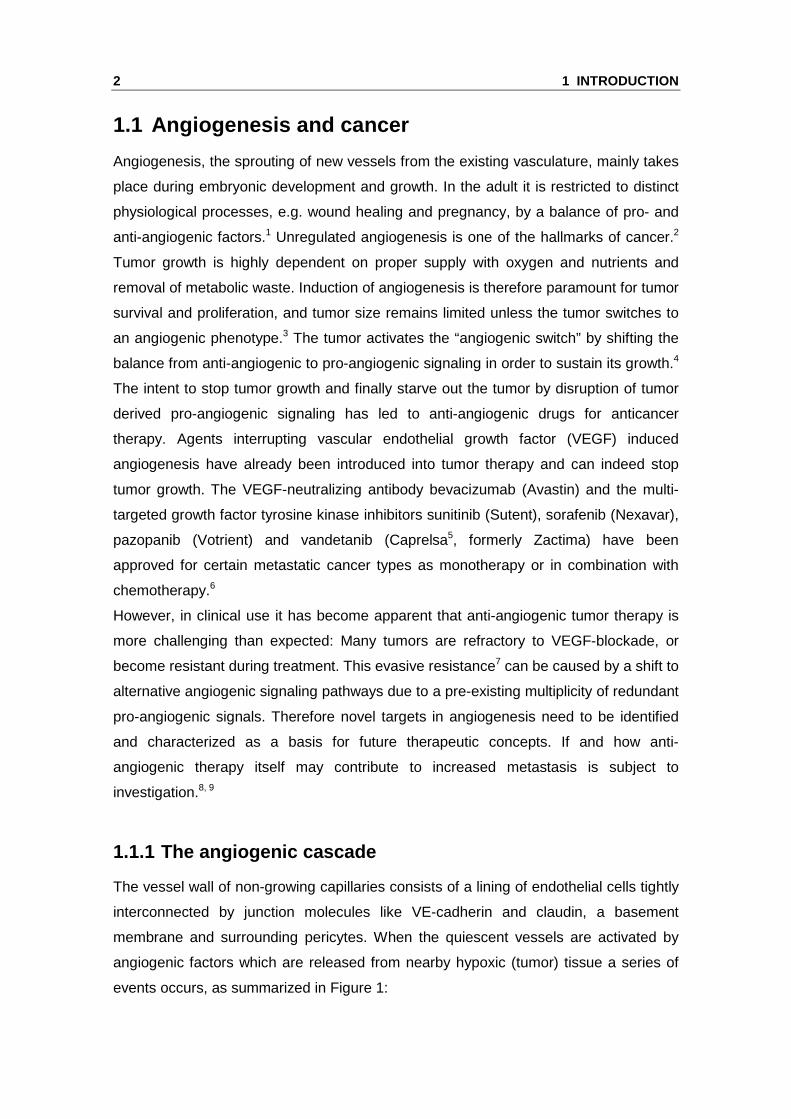
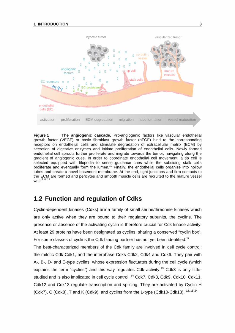
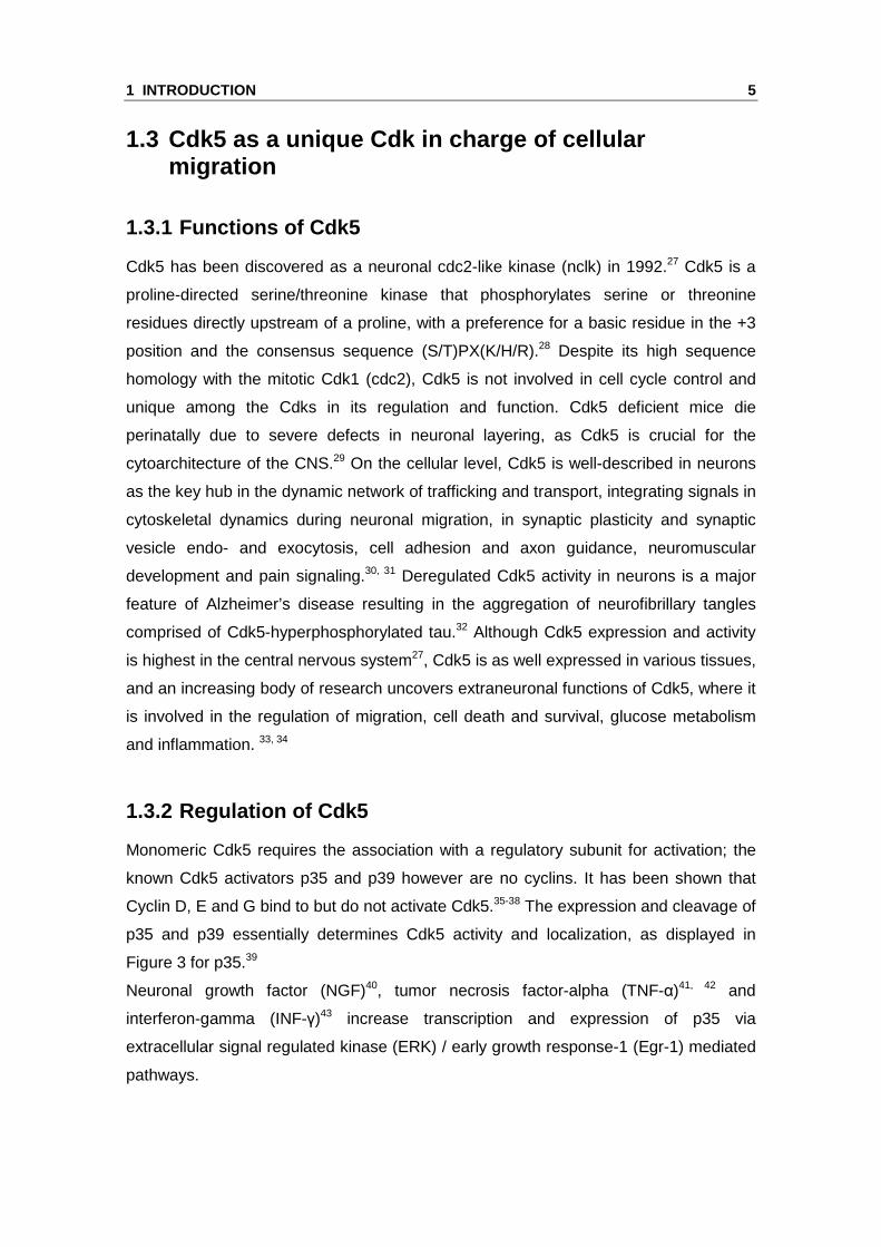
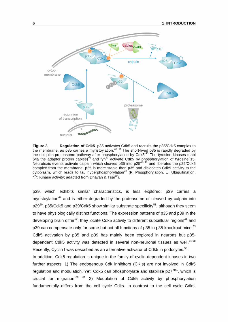
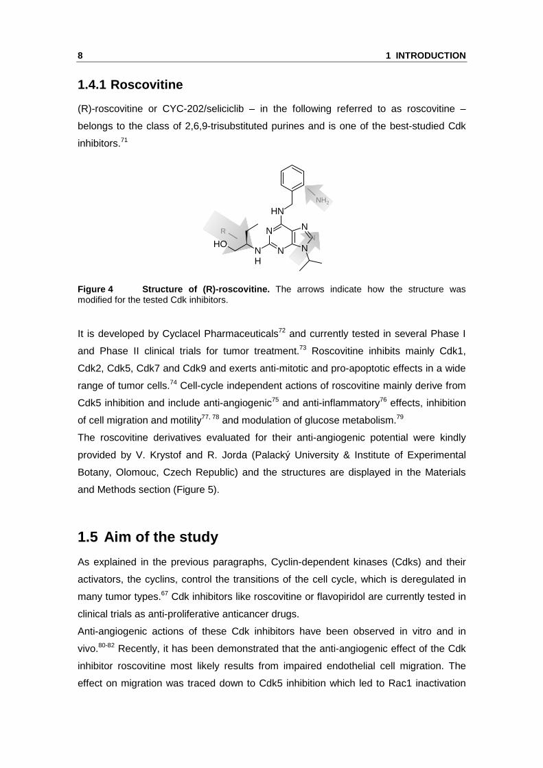


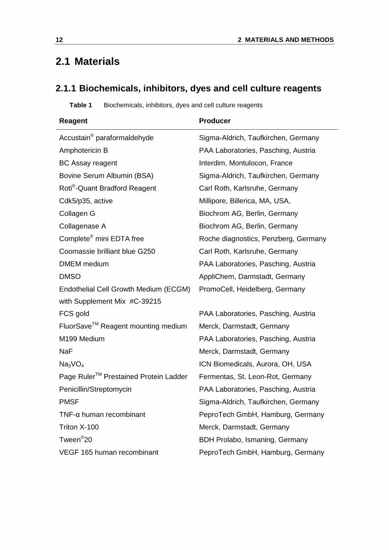
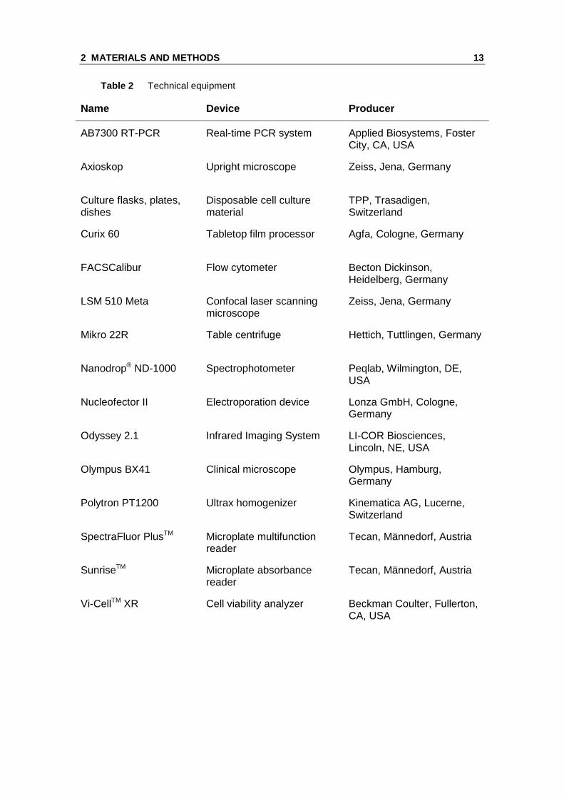
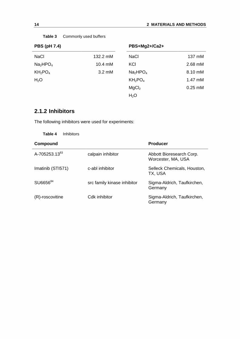
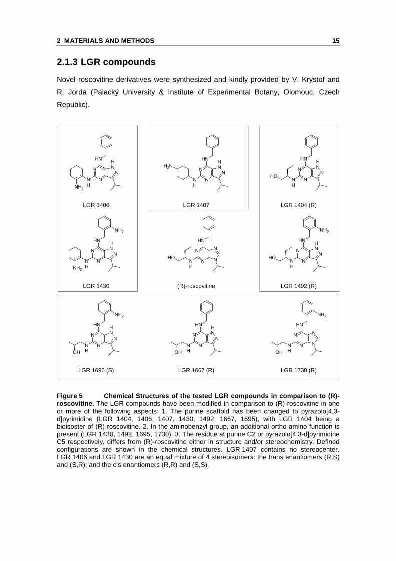
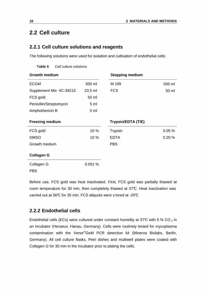
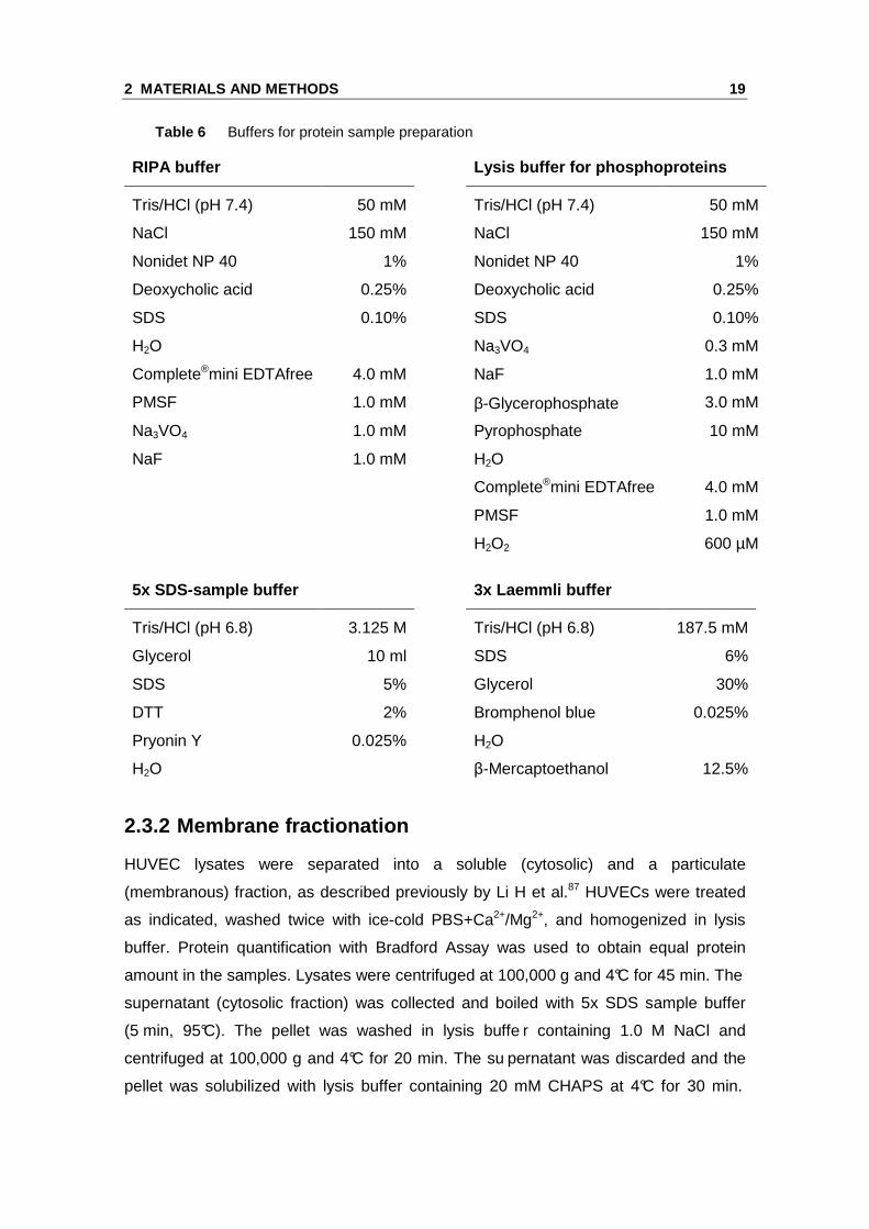
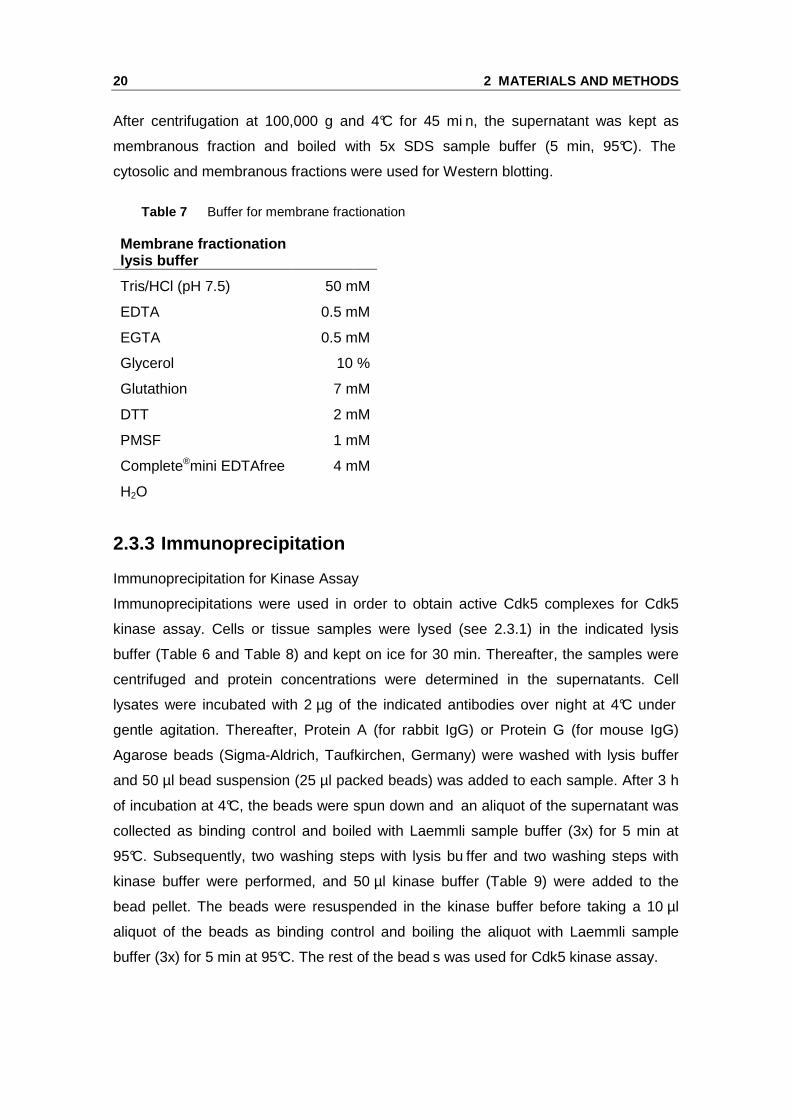
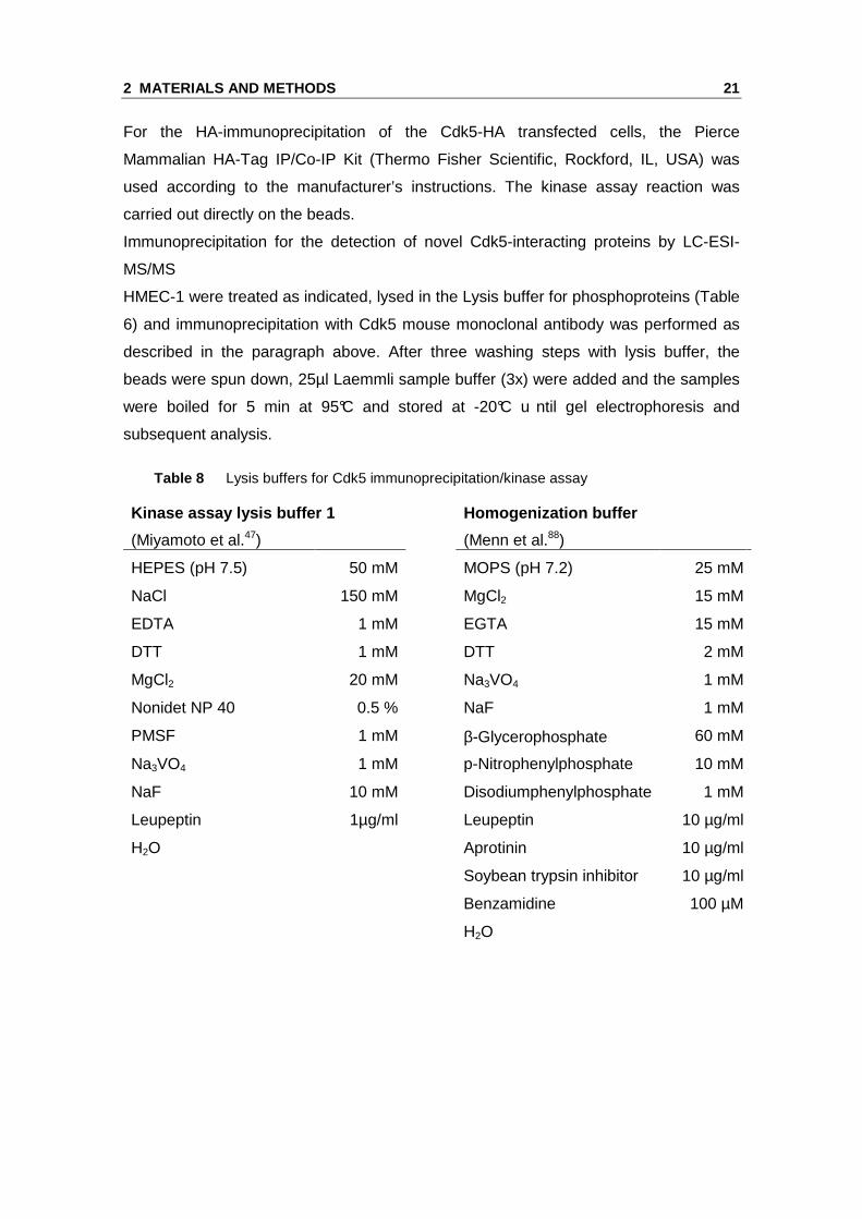
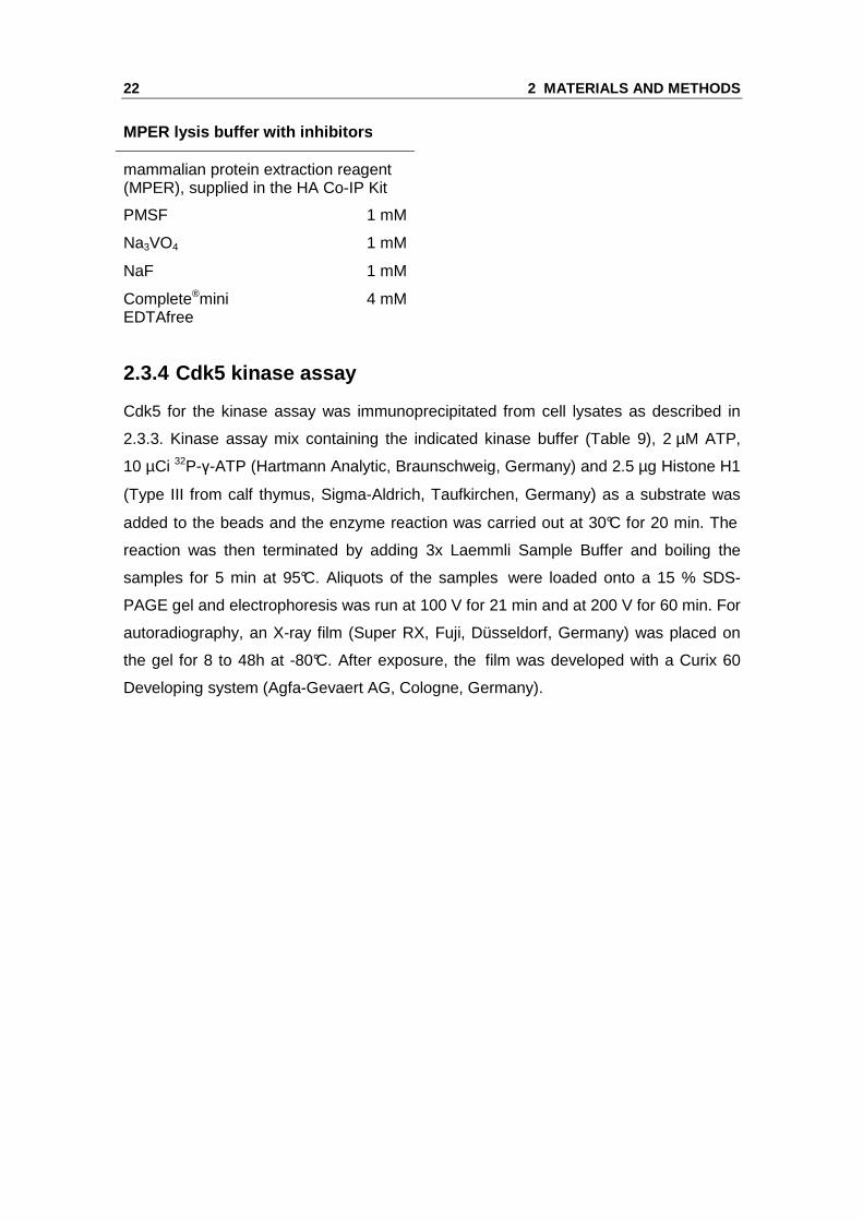
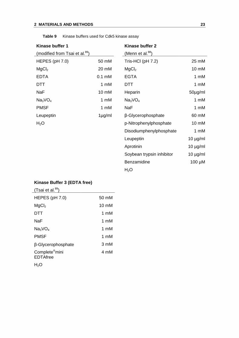
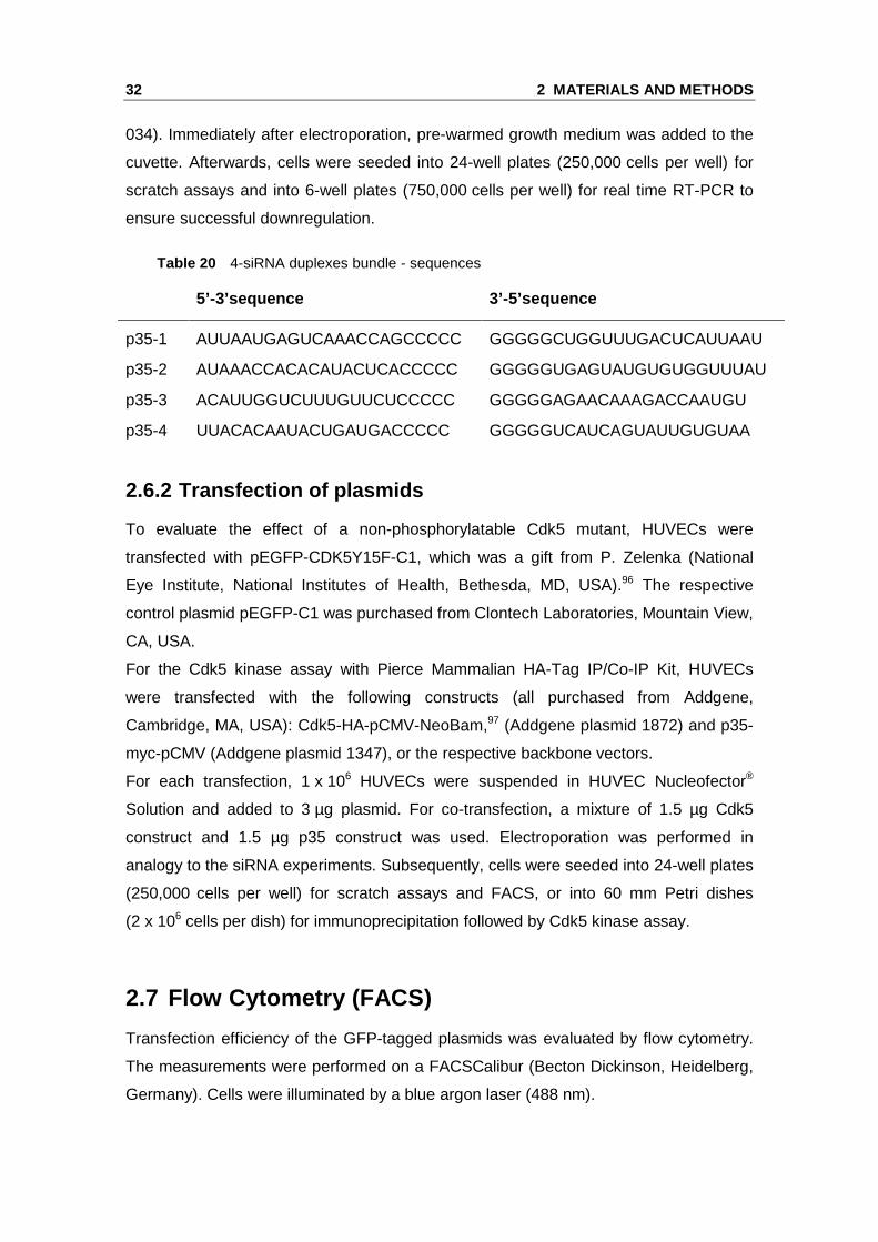
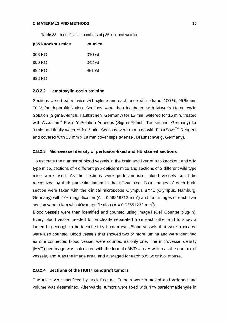
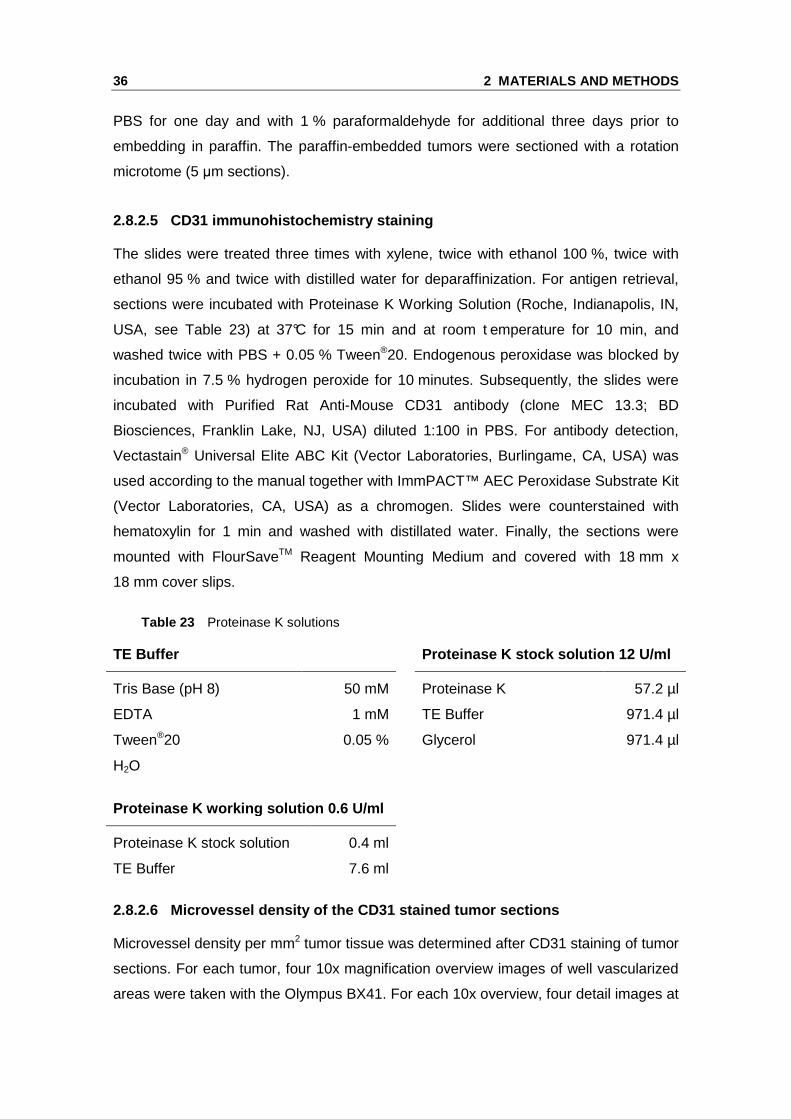
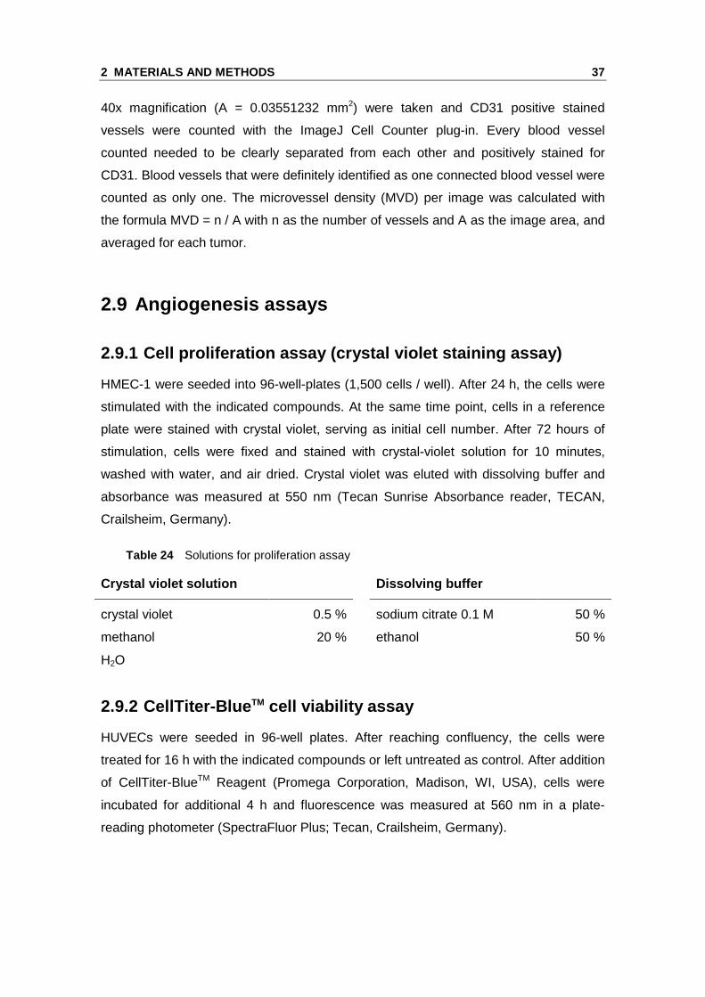


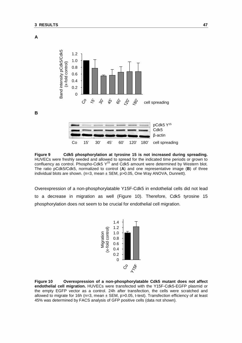
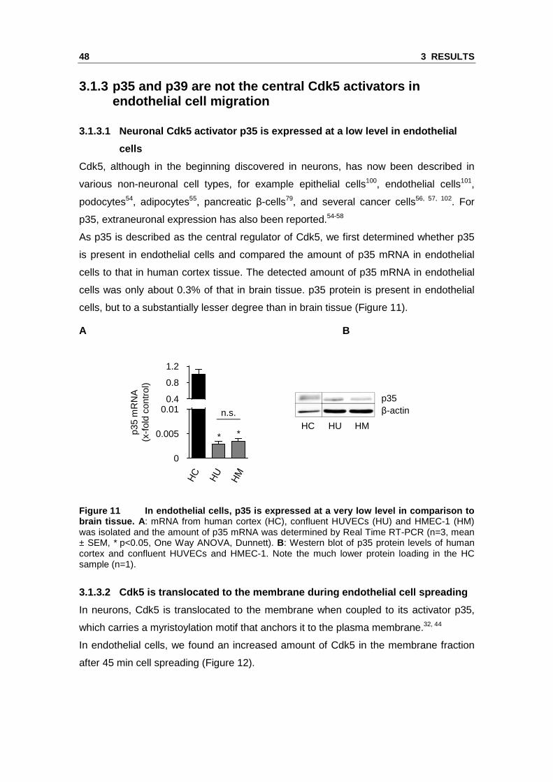
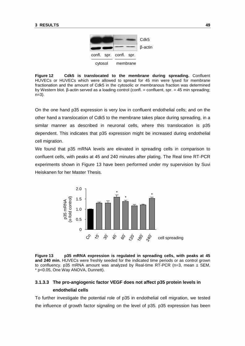
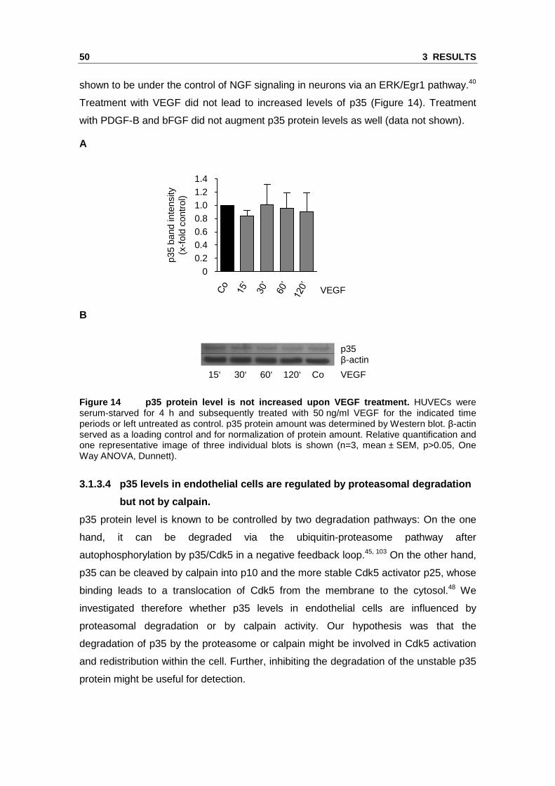
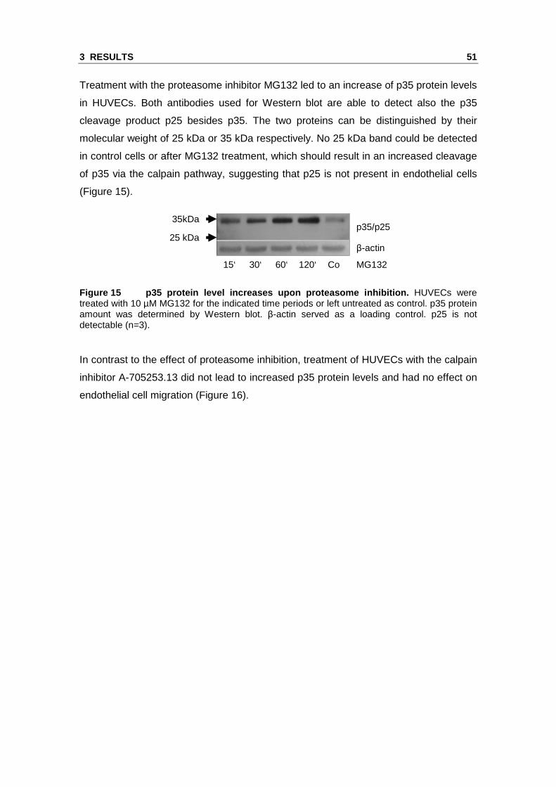
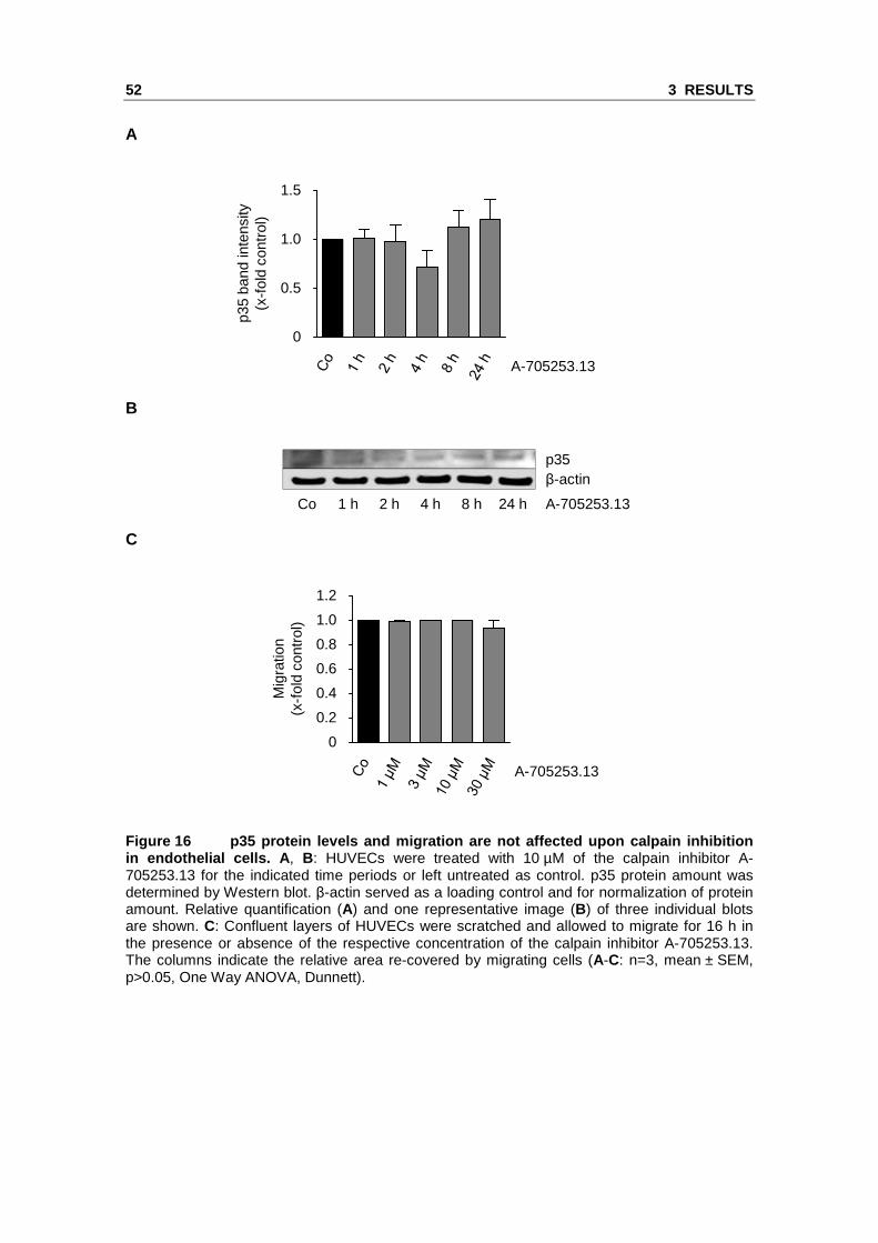
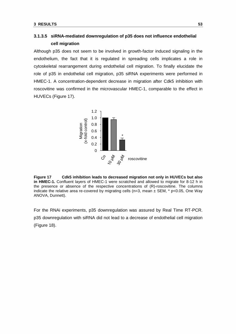
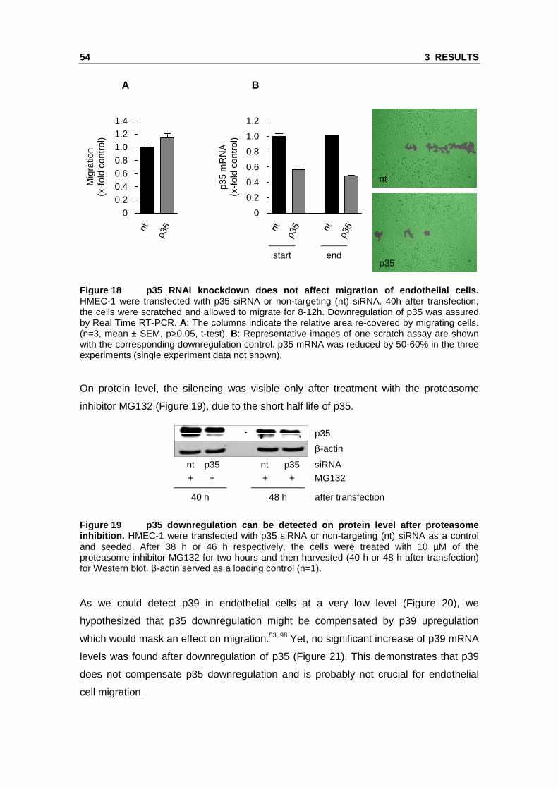
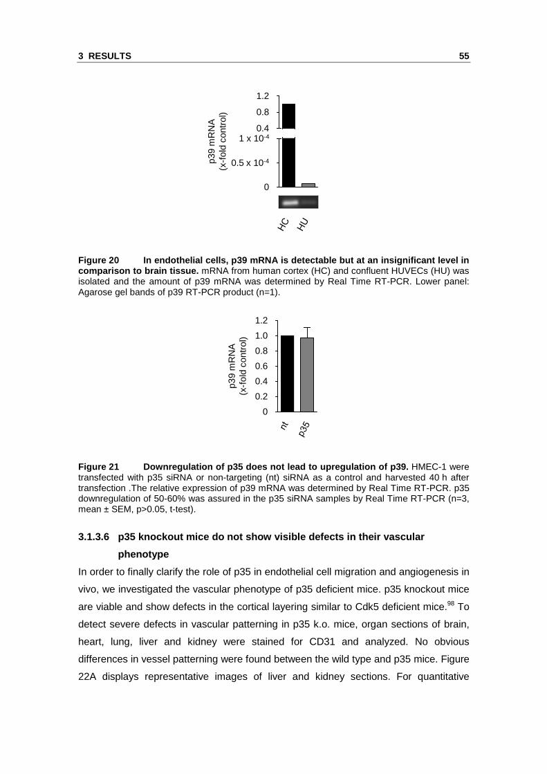
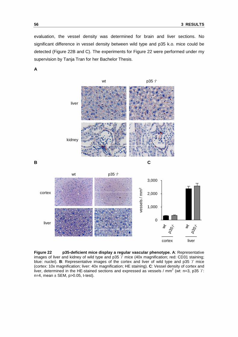
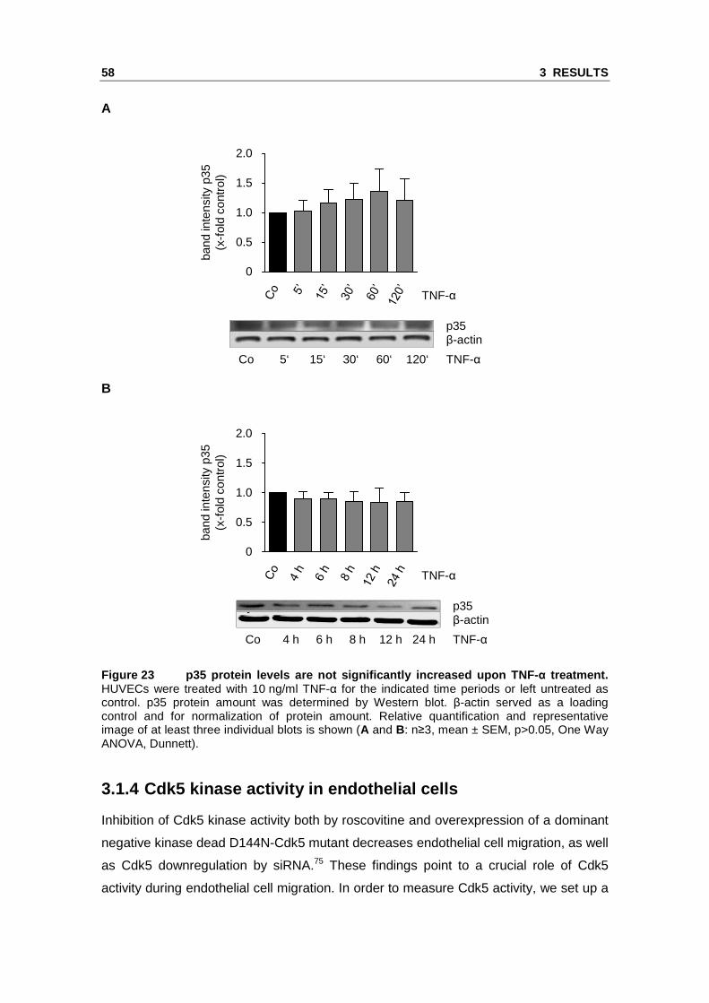
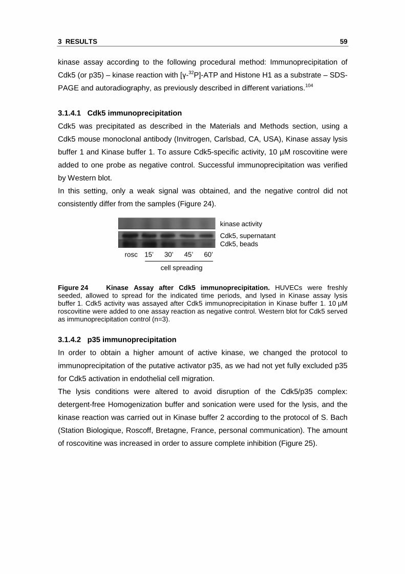
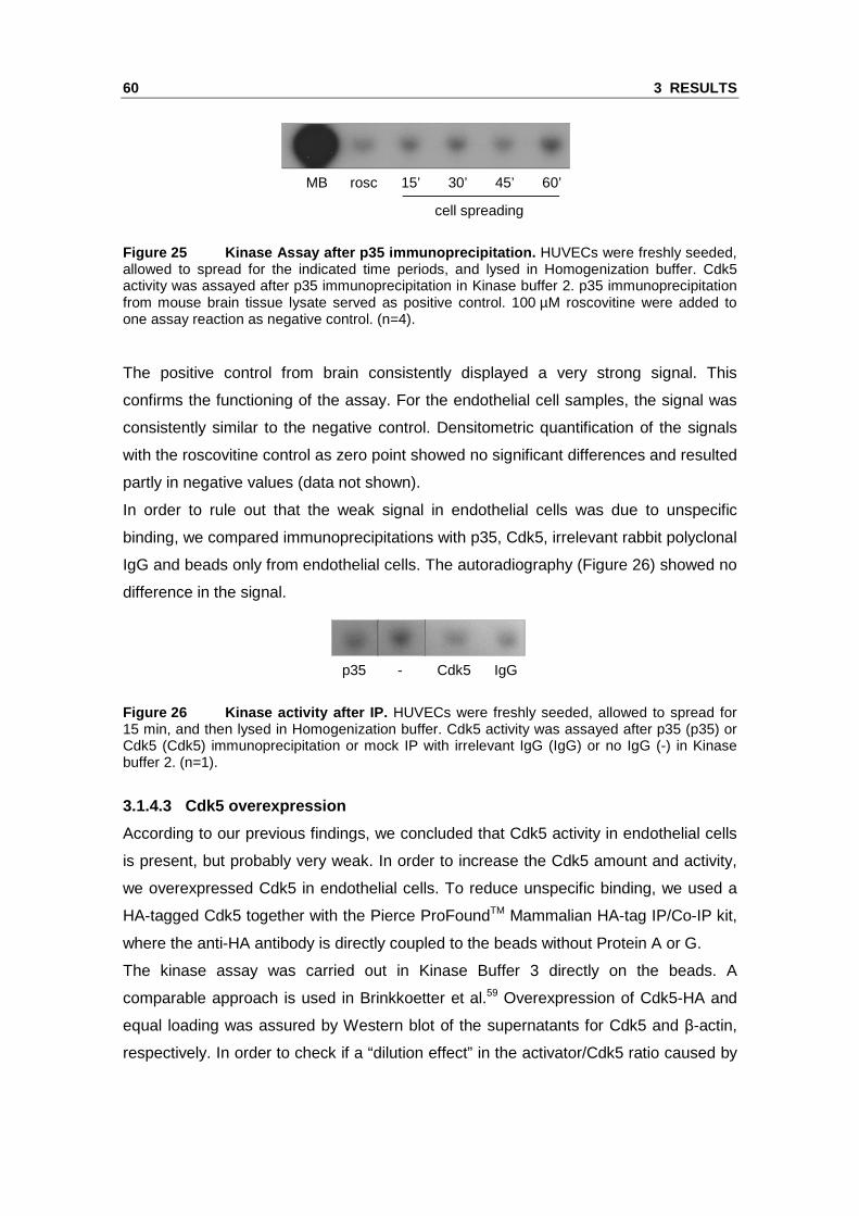
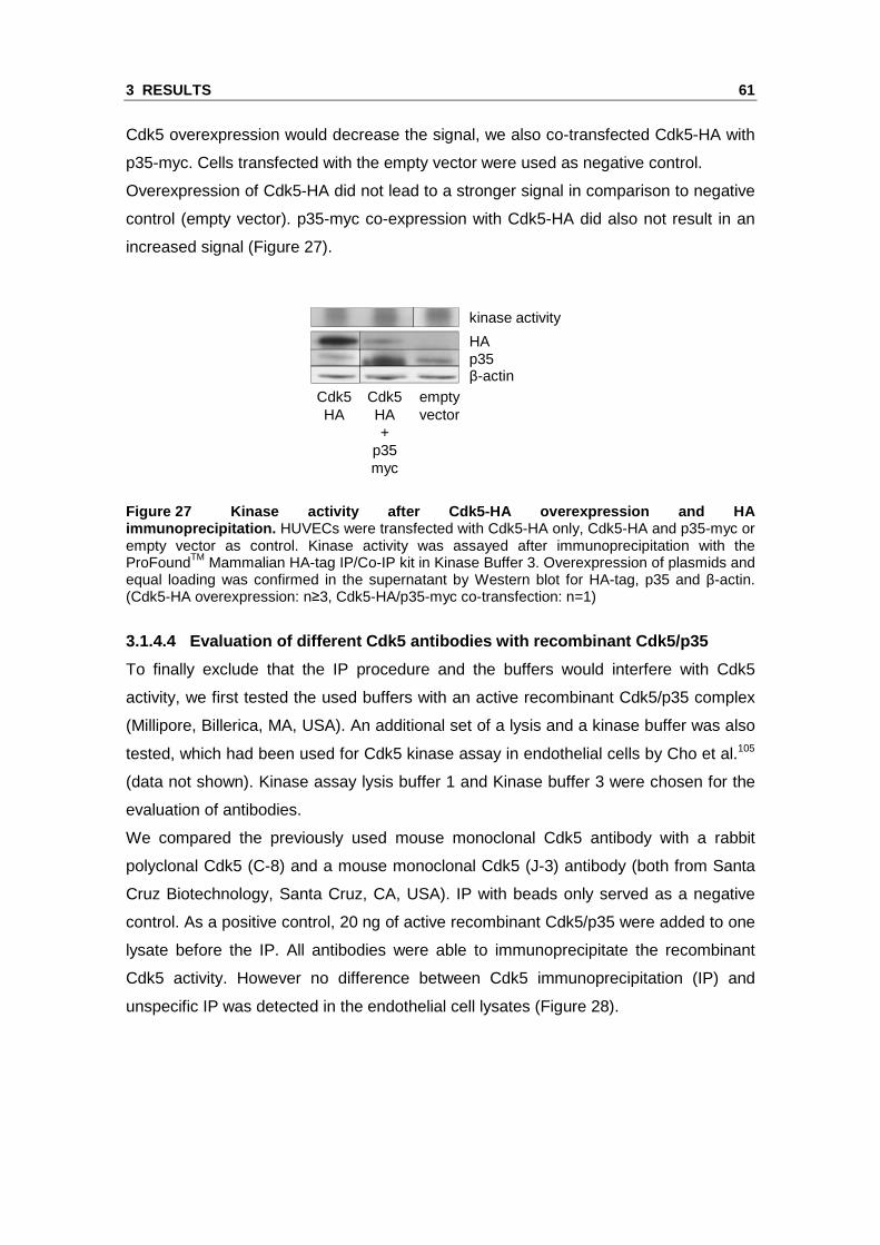
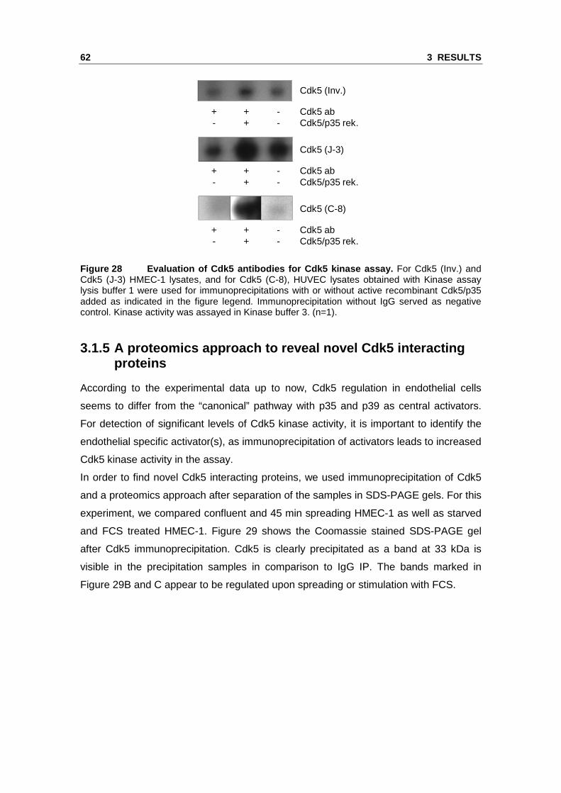
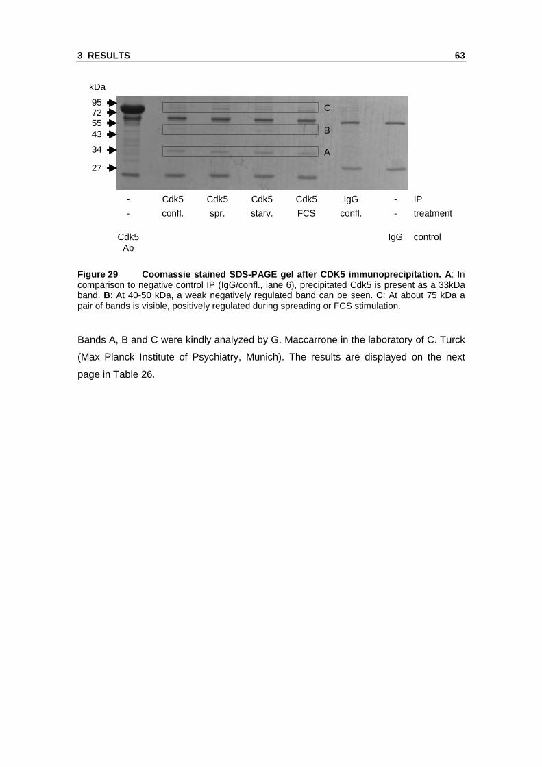
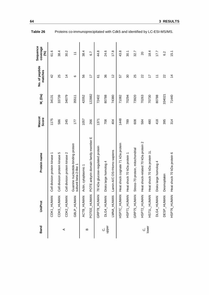
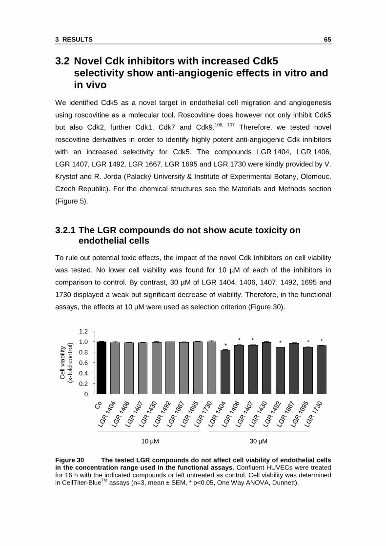
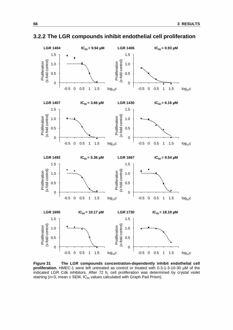
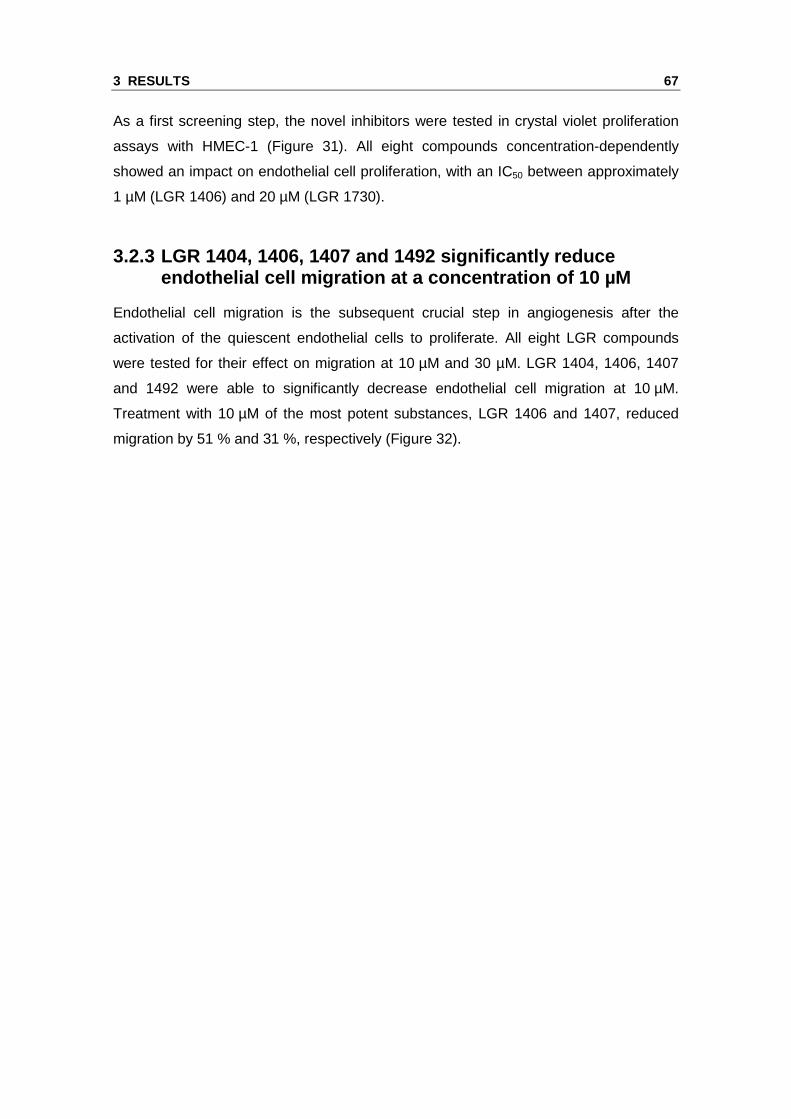
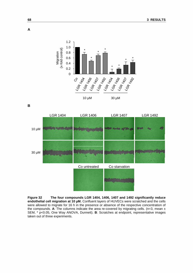
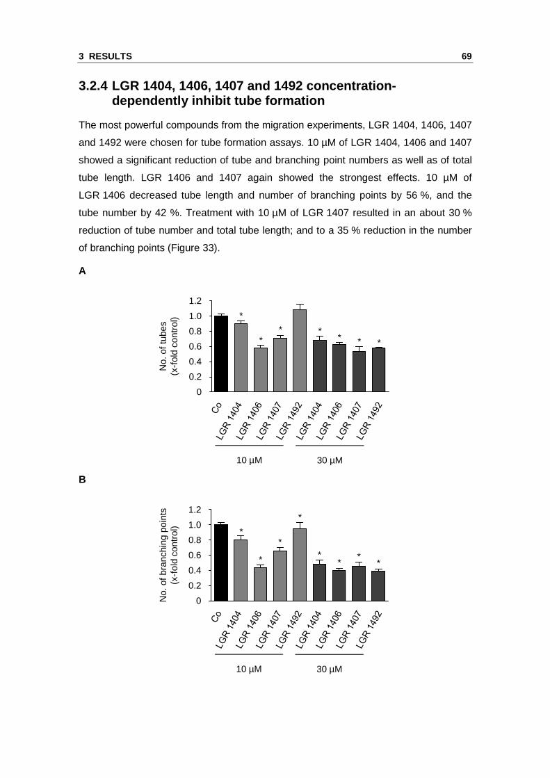
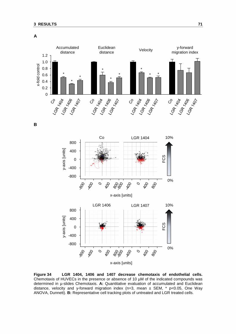
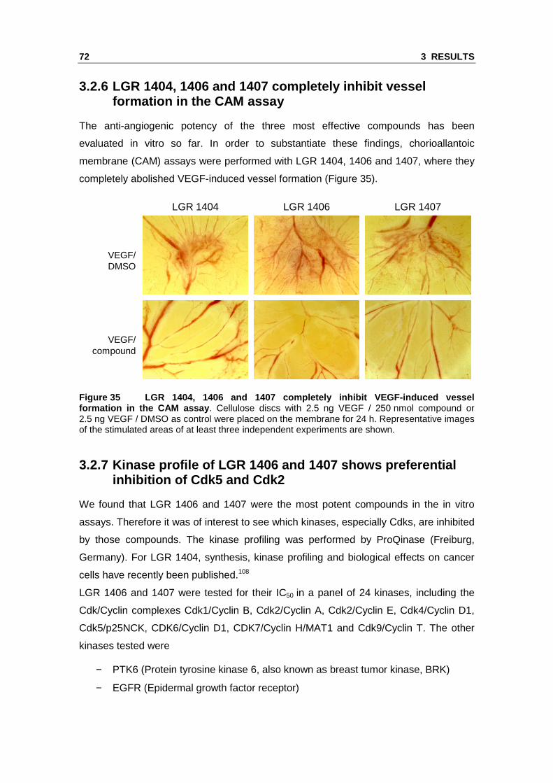
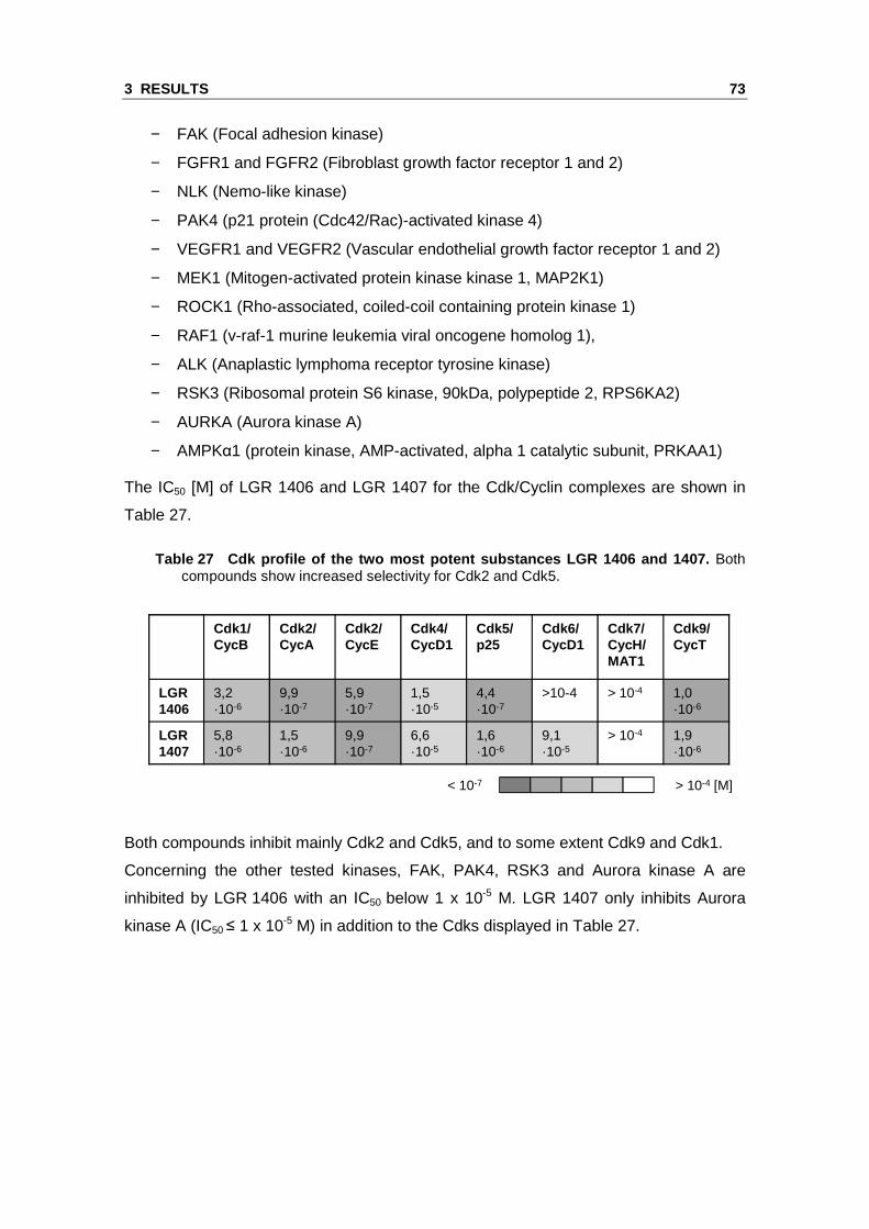
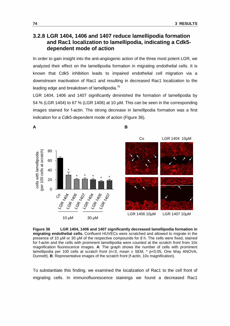
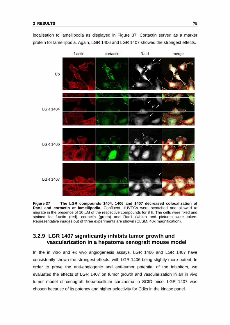
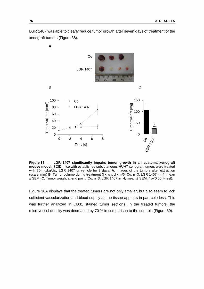
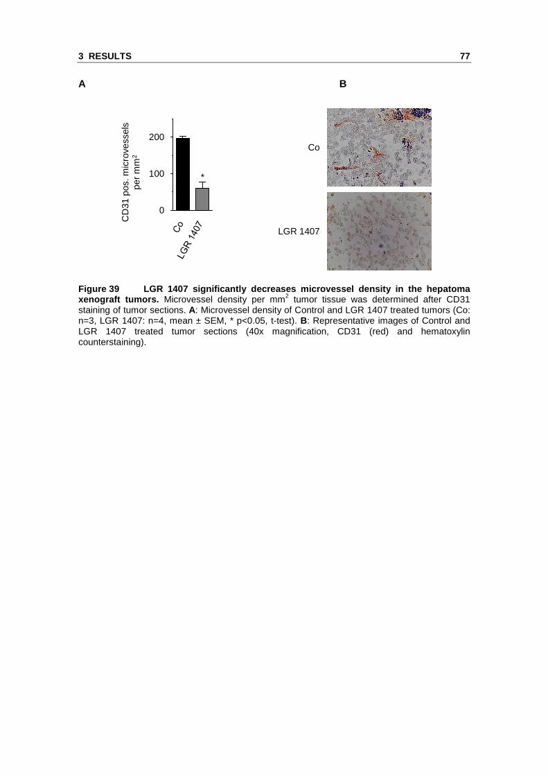


![Optical Surface Microtraps based on Evanescent Waves · Optical Surface Microtraps based on Evanescent Waves Dissertation zur Erlangung des Doktorgrades an der ... [Fol02]. While](https://static.documents.pub/doc/80x56/5bdee43809d3f2647f8b63e5/optical-surface-microtraps-based-on-evanescent-optical-surface-microtraps-based.jpg)


