237 The comparison of the marginal gaps of zirconia framework luted with different types of phosphate based-resin cements Darunee Owitayakul, Wipada Lertrid, Chuchai Anatamana, Piyapanna Pittayachawan Darunee Owitayakul 1 , Wipada Lertrid 2 , Chuchai Anatamana 3 , Piyapanna Pittayachawan 2 1 Department of Family and Community Dentistry, Chiang Mai University. 2 Department of Advanced General Dentistry, Mahidol University. 3 Department of Prosthodontic, Mahidol University. The comparison of the marginal gaps of zirconia framework luted with different types of phosphate based-resin cements Corresponding author: Piyapanna Pittayachawan Department of Advanced General Dentistry, Mahidol University, 6 Yothi Street, Phayathai, Bangkok, 10400 Thailand Email: [email protected]Received: 11 August 2015 Accepted: 12 October 2015 Abstract Objective: Phosphase based-resin cements are known to chemically bond to zirconia ceramics. Additionally, they are the cement of choice for these materials. Therefore, the aim of this study was to evaluate the marginal gaps of zirconia frameworks luted with two types of phosphate based resin cements. Materials and methods: Thirty maxillary premolar teeth were randomly divided into two groups (n=15); a self-etch (Panavia F2.0; Kuraray Medical, Japan) and self-adhesive (Rely X U100; 3MESPE, USA) resin cements. The teeth were prepared with round shoulder and the 6° convergence angle. The zirconia frameworks were fabricated using TDS® CAD/CAM system (Total Dental Solution, Changhua, Taiwan). The marginal fits of zirconia frameworks were evaluated under a microscope (MM-11C, Nikon, Tokyo, Japan) (30x magnification) at the following sequences: before cementation, after cementation and after 20,000 thermocycles. The differences in the marginal gaps (after and before cementation: delta diff1, after thermocycling and after cementation: delta diff2) between two resin cements were analyzed with Student’s t-test and Mann Whitney U test. The differences between two processes in each group were tested with paired t-test and Wilcoxon signed-rank test (p<0.05). Results: After cementation, the marginal gaps of two resin cements were significantly higher than before cementation (p<0.001). The slight differences were found in mean of delta diff2 (self-etch: 0.88 µm, self-adhesive: 0.26 µm). The marginal gaps of zirconia frameworks cemented with self-adhesive resin cement were significantly smaller than self-etch resin cement both after cementation and after thermocycling (p<0.05). Conclusions: Long-term thermocycling had no clinical effect on the marginal fit. The type of resin cements influenced the marginal fit. The marginal gaps achieved by both resin cements were within clinically accepted standards (<120 µm). Keywords: marginal gaps, zirconia ceramic, cement thickness, phosphate based cement, thermocycling, resin cement How to cite: Owitayakul D, Lertrid W, Anatamana C, Pittayachawan P. The comparison of the marginal gaps of zirconia framework luted with different types of phosphate based-resin cements. M Dent J 2015; 35: 237-51. Dental Journal Original Article Mahidol Dental Journal
Transcript
237The comparison of the marginal gaps of zirconia framework luted with different types of phosphate based-resin cementsDarunee Owitayakul, Wipada Lertrid, Chuchai Anatamana, Piyapanna Pittayachawan
1 Department of Family and Community Dentistry, Chiang Mai University. 2 Department of Advanced General Dentistry, Mahidol University. 3 Department of Prosthodontic, Mahidol University.
The comparison of the marginal gaps of zirconia framework luted with different types of phosphate based-resin cements
Corresponding author: Piyapanna Pittayachawan Department of Advanced General Dentistry, Mahidol University, 6 Yothi Street, Phayathai, Bangkok, 10400 Thailand Email: [email protected] Received: 11 August 2015 Accepted: 12 October 2015
AbstractObjective: Phosphase based-resin cements are known to chemically bond to zirconia ceramics. Additionally, they are the cement of choice for these materials. Therefore, the aim of this study was to evaluate the marginal gaps of zirconia frameworks luted with two types of phosphate based resin cements.
Materials and methods: Thirty maxillary premolar teeth were randomly divided into two groups (n=15); a self-etch (Panavia F2.0; Kuraray Medical, Japan) and self-adhesive (Rely X U100; 3MESPE, USA) resin cements. The teeth were prepared with round shoulder and the 6° convergence angle. The zirconia frameworks were fabricated using TDS® CAD/CAM system (Total Dental Solution, Changhua, Taiwan). The marginal fits of zirconia frameworks were evaluated under a microscope (MM-11C, Nikon, Tokyo, Japan) (30x magnification) at the following sequences: before cementation, after cementation and after 20,000 thermocycles. The differences in the marginal gaps (after and before cementation: delta diff1, after thermocycling and after cementation: delta diff2) between two resin cements were analyzed with Student’s t-test and Mann Whitney U test. The differences between two processes in each group were tested with paired t-test and Wilcoxon signed-rank test (p<0.05).
Results: After cementation, the marginal gaps of two resin cements were significantly higher than before cementation (p<0.001). The slight differences were found in mean of delta diff2 (self-etch: 0.88 µm, self-adhesive: 0.26 µm). The marginal gaps of zirconia frameworks cemented with self-adhesive resin cement were significantly smaller than self-etch resin cement both after cementation and after thermocycling (p<0.05).
Conclusions: Long-term thermocycling had no clinical effect on the marginal fit. The type of resin cements influenced the marginal fit. The marginal gaps achieved by both resin cements were within clinically accepted standards (<120 µm).
How to cite: Owitayakul D, Lertrid W, Anatamana C, Pittayachawan P. The comparison of the marginal gaps of zirconia framework luted with different types of phosphate based-resin cements. M Dent J 2015; 35: 237-51.
Dental Journal Original ArticleMahidol Dental Journal
238 The comparison of the marginal gaps of zirconia framework luted with different types of phosphate based-resin cements The comparison of the marginal gaps of zirconia framework luted with different types of phosphate based-resin cementsDarunee Owitayakul, Wipada Lertrid, Chuchai Anatamana, Piyapanna Pittayachawan
M Dent J Volume 35 Number 3 September-December 2015
The comparison of the marginal gaps of zirconia framework luted with different types of phosphate based-resin cements
Introduction The demand for esthetic restorations has increased during the past few years. All-ceramic restorations offer a highly esthetic appearance, chemical stability and biocompatibility.1,2 However, the major disadvantage of using conventional ceramics is that they involve risk of fracture because of their low mechanical properties.1,2
Zirconia has recently been developed and introduced for application in dentistry due to the superior mechanical properties, which is attributed to a process known as transformation toughening. Zirconia is attractive for restorative dentistry and produced by computer-aided design/computer-aided manufacturing (CAD/CAM). It can be used for anterior and posterior crowns, long span bridges and also implant abutments.3 Apart from the mechanical properties and esthetics, the long-term clinical success of all ceramic restorations can be influenced by marginal adaptation, which is a very important factor for the longevity of the restored teeth. Poor marginal adaptation results in secondary caries, periodontal disease, pulp sensitivity, pulp necrosis and esthetic problems such as staining or marginal discoloration.4,5 It also influences the wear of the luting cement 6,7 and its ability to withstand loading.7 Sailer et al.8 studied the success rate of 3-5 units zirconia frameworks for posterior fixed partial dentures (FPDs). The result showed that there was no fracture occurred after three years. Therefore, the success rate of zirconia framework was 100%. The survival rate was 84.8% because of secondary caries (10.9%) and chipping of veneering ceramic (13.0%). For 5 years of clinical evaluation, the study found that the success rate was decreased to 97.8% resulting from one 5-unit framework fractured through the connector area from trauma. The survival rate was 73.9% due to secondary caries (21.7%) and chipping of veneering ceramic
(15.2%). There was no significant difference regarding the periodontal parameters between the test and control teeth. Furthermore, all patients were satisfied with the esthetics of the all ceramic restorations, and 91.7% were satisfied with functional aspects.9 As a result of various biologic and technical problems, the overall survival rate of zirconia FPDs in this study was 73.9%. More than 20% were found secondary caries resulting from marginal discrepancies.9 Therefore, the marginal fit is one of the important factors that determine the clinical performance. It is also closely related to the longevity of the restoration. The definition of the internal gap is the perpendicular measurement from the axial wall of the preparation to the internal surface of restoration.10 The marginal gap is the perpendicular measurement from the margin of the restored tooth to the margin of the restoration.11 Gap measurements at margin are frequently used to quantify fit. There are many methods for measuring marginal gap. The standardized methods are classified in four basic categories, which are 1.) direct view, 2.) cross-sectional, 3.) impression technique and 4.) explorer and visual examination.12 The direct view method is easy, convenient and rapid because the crowns are retrievable. Moreover, it does not require additional steps. This technique allows measurement at various stages of crown fabrication. However, the repeated seating on the master dies can damage the margin by abrasion. In addition, the inaccuracies in repositioning crowns on the master dies will cause increased standard deviation and decreases statistical significance. The other disadvantage is that it is difficult to determine with a rounded margin because there is no reference points for repeating the measurement at the curved surface. It is hard to assess the marginal discrepancy of a overcontouring crown margin by direct view method. For the cross-sectional
The comparison of the marginal gaps of zirconia framework luted with different types of phosphate based-resin cements 239The comparison of the marginal gaps of zirconia framework luted with different types of phosphate based-resin cementsDarunee Owitayakul, Wipada Lertrid, Chuchai Anatamana, Piyapanna Pittayachawan
M Dent J Volume 35 Number 3 September-December 2015
method, the processes of this method are that the crowns are placed on dies or teeth and then specimens are embedded in the clear acrylic resin. After that, they are sectioned by diamond disc. This technique has been used to measure marginal discrepancies on both non-cemented and cemented crowns. This method requires additional steps by embedding and sectioning; therefore, it is more time-consuming. However, the cross-sectional method allows greater precision in measurement and the reference points can be made, which lead to evaluate the gaps more accurate and repeatable.12 The degree of horizontal discrepancies can also be determined. Furthermore, crowns and dies are in the same units, which avoids a potential damage of master dies from repeated measurements.12
The final method is the impression technique or the replica technique, which is popular and non-invasive method13 to assess the marginal and internal gaps that used the sectioned elastomeric materials without destroying the specimens. This method is quick to detect an obviously inaccurate restoration.14
McLean and von Fraunhoter15 suggested that the marginal gaps for restorations below 120 µm were clinically acceptable. Poor marginal adaptation results in cement dissolution, microleakage, plaque accumulation, and secondary caries.16,17 One study suggested that a minimum of 50 measurements should be done along margins on both non-cemented and cemented crowns, whether the measurement sites are selected in a systematic or random manner. The measurement above 50 points per crown provides acceptable results.18 Bindl and Mörmann16 studied the marginal and internal fits of all ceramic CAD/CAM crown copings. The results showed that the mean of marginal gaps of the DCS® crown copings (densely sintered zirconium oxide ceramic) was 33±20 µm., which did not differ from any other CAD/CAM copings or conventional crowns such as slip-cast and
heat-pressing. Beuer and colleagues19 found that the type of the restorations influenced on the marginal gaps. The mean of marginal gaps for 14-unit zirconia fixed bridges and single-crown copings were 25±29 and 13±12 µm, respectively. The location of the abutment tooth influenced on the marginal gaps of fixed bridges; however, it did not affect the gaps of single-crown copings. Different luting cements may provide different effects on marginal adaptation of restorations.20
Various luting cements, which are available on the market, have different properties. Resin cements were recommended as the materials of choice for ceramic restorations because of several advantages such as the retention and marginal adaptation of the restorations are improved and they have low solubility in the oral environment and less microleakage compared with conventional cements.21,22 Additionally, the marginal discrepancy of the restoration is compensated by the use of a resin composite luting agent.23 Phosphate based resin cement is recommended to use with zirconia restoration. However, very few studies investigated the marginal adaptation of the zirconia frameworks luted with this resin cement.16 In addition, there have been limited studies about the effect of the long-term thermocycling to the marginal adaptation of the zirconia frameworks. The objective of this study was to compare the marginal and internal gaps of zirconia frameworks luted with two types of phosphate based resin cements before cementation, after cementation and after thermocycling. The null hypothesis is that there were no significant differences of marginal and internal gaps between two phosphate based resin cements.
Materials and methods Thirty permanent upper premolars without any lesions were extracted for orthodontic reasons. All teeth were stored in 10% formalin
240 The comparison of the marginal gaps of zirconia framework luted with different types of phosphate based-resin cements The comparison of the marginal gaps of zirconia framework luted with different types of phosphate based-resin cementsDarunee Owitayakul, Wipada Lertrid, Chuchai Anatamana, Piyapanna Pittayachawan
M Dent J Volume 35 Number 3 September-December 2015
The comparison of the marginal gaps of zirconia framework luted with different types of phosphate based-resin cements
solution for 2 weeks24 and then they were thoroughly cleaned, and periodontium were removed with surgical blades. They were embedded into clear epoxy resin and positioned with the long axis of the teeth perpendicular to the floor of the mould. The clear epoxy resin blocks were left for 24 hours to assure complete curing. The teeth were prepared using an Operat ing Instruct ion SCHICK-ceramic-milling set; Master S3-Nr. 2650 (Schick-Dentalgeräter, Schemmerhofen, Germany) and diamond rotary cutting instruments under water cooling. Rounded-shoulder finishing margins (1.0-mm circumference) at the cementoenamel junction and six degree convergence angle were produced using high-speed, round-end, tapered diamonds (No. 837314016; Jota AG, Switzerland) with diameters of approximately 1.0 mm and six degrees of taper. Preparation heights of 4.0 mm were achieved using high-speed, cylindrical diamonds (No. 837314016; Jota AG, Switzerland). The transition from the axial to the occlusal surfaces was rounded with round white stones (No.3-10-02-03; Shofu Inc., Kyoto, Japan) (Figure 1). During the study, all specimens were stored in distilled water and kept in an incubator at 37°C. All prepared specimens were sent to the dental laboratory to fabricate the zirconia
frameworks via the TDS® CAD/CAM (Total Dental Solution, Changhua, Taiwan). A wall thickness of 0.5 mm and a cement space of 30 µm were set for the frameworks. The flat occlusal surfaces of the frameworks were prepared parallel to the horizontal plane. The marginal fits of all zirconia frameworks were evaluated at the following sequence points: before cementation, after cementation and after thermocycling. The framework margin and the tooth margin were marked with different colored markers. The frameworks were then seated on the abutment teeth and the framework-abutment specimens were held in the position using a screw device to seat the frameworks during measurement (Figure 2). The marginal gaps of the zirconia frameworks were measured under a microscope (MM-11C, Nikon, Tokyo, Japan) at 30x magnification around the circumference of the tooth before cementation for 60 measurements.18
The values of the marginal gaps (µm) could be recorded from the digital counter monitor. All the measurements were performed two times per specimen to calibrate and find the measurement error of a recorder. Additionally, the same person performed all measurements to minimize variation in the measured values.
Figure 1 The abutment was prepared with a 1.0 mm rounded shoulder margin, an occlusocervical dimension of 4.0 mm and the 6 degrees convergence angle.
Figure 2 The framework was placed on the abutment and held in the position using a screw device to seat the frameworks completely during measurement.
The comparison of the marginal gaps of zirconia framework luted with different types of phosphate based-resin cements 241The comparison of the marginal gaps of zirconia framework luted with different types of phosphate based-resin cementsDarunee Owitayakul, Wipada Lertrid, Chuchai Anatamana, Piyapanna Pittayachawan
M Dent J Volume 35 Number 3 September-December 2015
The specimens (n=30) were divided into two groups: self-etching (SE group) and self-adhesive resin cements (SA group) (Table 1). The zirconia frameworks were cemented according to the cement manufacturers’ instructions. Panavia™ F2.0 (Kuraray Medical, Tokyo, Japan) was used in the SE group. Liquid A and liquid B (ED primer 2.0) were mixed in 1:1 ratio and applied to the tooth surfaces. Paste A and paste B were then mixed in a 1:1 ratio (within 20 seconds) and applied on the inner surfaces of the framework. The zirconia framework was then seated on the abutment teeth, using a loading device, for seven minutes with a load of 50 N.16,19 The cement was partially light-cured for three seconds and the excess cement removed with the aid of an explorer. The cement was then light-cured for 40 seconds on all four sides of the specimen to accelerate polymerization. Finally, a protective gel, Oxyguard II® (Kuraray Medical, Tokyo, Japan) was applied on the margin for three minutes to prevent the formation of an oxygen-inhibited layer and then rinsed it off with distilled water. RelyX™ U100 (3M ESPE; St. Paul, MN, USA) was employed in the SA group. This cement does not require any prior tooth surface treatment before bonding. Base
and catalyst pastes were mixed together on a mixing pad for 20 seconds. Resin cement was applied on the inner surface of the framework and the process utilized was the same as with the SE group. After 24 hours storage in distilled water at 37°C, all specimens were thermocycled for 20,000 times between 5°C and 55°C with a dwell time of 20 seconds in each bath. The interval time between baths was 10 seconds.25
After thermocycling, the marginal gaps of zirconia frameworks were recorded around the circumference of the tooth under an optical microscope (x 30 magnification) according to the same method described above. All specimens were embedded in clear epoxy resin to prevent fracture and debonded frameworks during sectioning. They were sectioned bucco-palatally and mesio-distally by an Accutom-50 precision cut-off machine (Struers, Compenhagen, Denmark) and grinding machine using water-cooled diamond saw (Struers, Compenhagen, Denmark) to produce four pieces (eight surfaces) of specimens. The marginal gaps, marginal discrepancy and cement thickness of each surface were measured by taking photographs using a camera (Coolpix E
242 The comparison of the marginal gaps of zirconia framework luted with different types of phosphate based-resin cements The comparison of the marginal gaps of zirconia framework luted with different types of phosphate based-resin cementsDarunee Owitayakul, Wipada Lertrid, Chuchai Anatamana, Piyapanna Pittayachawan
M Dent J Volume 35 Number 3 September-December 2015
The comparison of the marginal gaps of zirconia framework luted with different types of phosphate based-resin cements
990, Nikon, Tokyo, Japan) that connected to the Eclipse E400 POL microscope (Nikon, Tokyo, Japan) and the specimens were evaluated at 250x magnification using a digital image analysis system (Image Pro Plus, version 3.0, Media Cyberntics Incs, Bethesda, MD, USA). The measurements were performed to evaluate the marginal discrepancies and cement thickness at the margins, the corners, the middle of the axial walls, the tip of the cusps and the occlusal floor in the mesio-distal and bucco-palatal directions (Figure 3); therefore, a total of twenty-two points per specimens were done. For measuring the marginal discrepancies, the distance of the finishing line of the tooth margin to the framework margin were recorded. Measurements of the gaps at the margin were recorded as the 90 degree distance between tooth margin and framework margin. Measurement at the corner was performed at the center point of the shoulder finishing line (Figure 4). Measurements along the axial wall were employed approximately half way between the corner and the tip of cusp. The measurements at the tip of cusp were performed approximately at the tips. The cement thickness
at the occlusal surface was evaluated at the occlusal floor only once in every section.
Statistic analysis The differences in the cement thickness at margins and inner surfaces of frameworks (µm) between the two resin cements were analyzed using independent t-test and the Mann-Whitney U test (marginal discrepancy, axial wall, cusp tips) (p<0.05).
Figure 4 Measurement points for evaluation of cement thickness. AB: Marginal discrepancies (the distance of finishing line of shoulder to zirconia coping). AC: The margin.
The comparison of the marginal gaps of zirconia framework luted with different types of phosphate based-resin cements 243The comparison of the marginal gaps of zirconia framework luted with different types of phosphate based-resin cementsDarunee Owitayakul, Wipada Lertrid, Chuchai Anatamana, Piyapanna Pittayachawan
M Dent J Volume 35 Number 3 September-December 2015
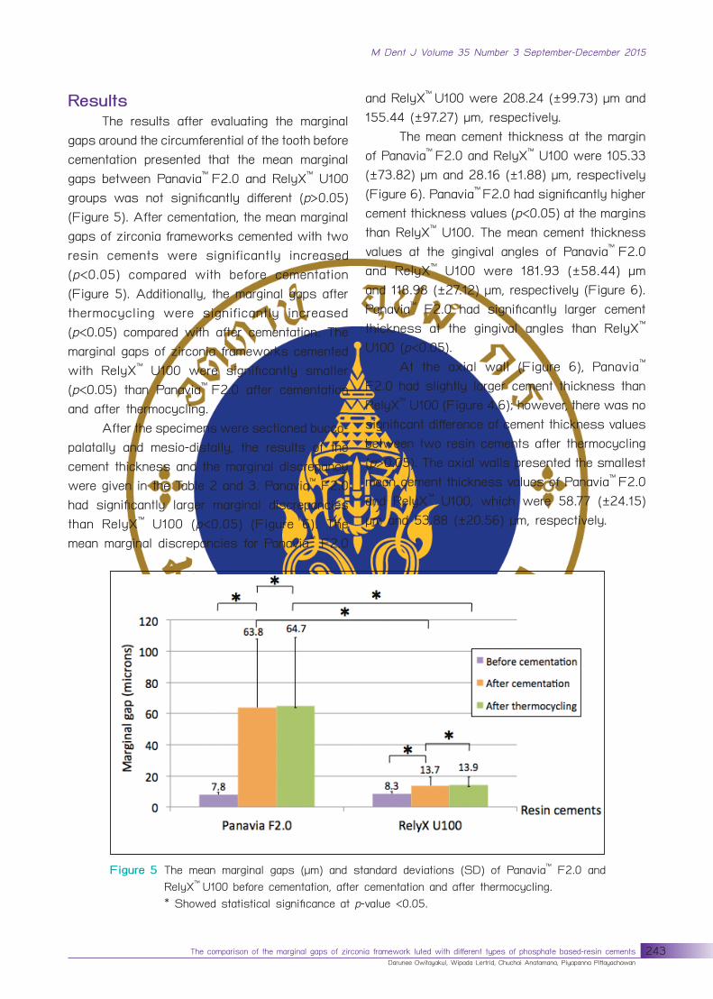
Results The results after evaluating the marginal gaps around the circumferential of the tooth before cementation presented that the mean marginal gaps between Panavia™ F2.0 and RelyX™ U100 groups was not significantly different (p>0.05) (Figure 5). After cementation, the mean marginal gaps of zirconia frameworks cemented with two resin cements were significantly increased (p<0.05) compared with before cementation (Figure 5). Additionally, the marginal gaps after thermocycling were significantly increased (p<0.05) compared with after cementation. The marginal gaps of zirconia frameworks cemented with RelyX™ U100 were significantly smaller (p<0.05) than Panavia™ F2.0 after cementation and after thermocycling. After the specimens were sectioned bucco-palatally and mesio-distally, the results of the cement thickness and the marginal discrepancy were given in the Table 2 and 3. Panavia™ F2.0 had significantly larger marginal discrepancies than RelyX™ U100 (p<0.05) (Figure 6). The mean marginal discrepancies for Panavia™ F2.0
and RelyX™ U100 were 208.24 (±99.73) µm and 155.44 (±97.27) µm, respectively. The mean cement thickness at the margin of Panavia™ F2.0 and RelyX™ U100 were 105.33 (±73.82) µm and 28.16 (±1.88) µm, respectively (Figure 6). Panavia™ F2.0 had significantly higher cement thickness values (p<0.05) at the margins than RelyX™ U100. The mean cement thickness values at the gingival angles of Panavia™ F2.0 and RelyX™ U100 were 181.93 (±58.44) µm and 118.98 (±27.12) µm, respectively (Figure 6). Panavia™ F2.0 had significantly larger cement thickness at the gingival angles than RelyX™ U100 (p<0.05). At the axial wall (Figure 6), Panavia™ F2.0 had slightly larger cement thickness than RelyX™ U100 (Figure 4.6); however, there was no significant difference of cement thickness values between two resin cements after thermocycling (p>0.05). The axial walls presented the smallest mean cement thickness values of Panavia™ F2.0 and RelyX™ U100, which were 58.77 (±24.15) µm and 53.88 (±20.56) µm, respectively.
Figure 5 The mean marginal gaps (µm) and standard deviations (SD) of Panavia™ F2.0 and RelyX™ U100 before cementation, after cementation and after thermocycling.
* Showed statistical significance at p-value <0.05.
244 The comparison of the marginal gaps of zirconia framework luted with different types of phosphate based-resin cements The comparison of the marginal gaps of zirconia framework luted with different types of phosphate based-resin cementsDarunee Owitayakul, Wipada Lertrid, Chuchai Anatamana, Piyapanna Pittayachawan
M Dent J Volume 35 Number 3 September-December 2015
The comparison of the marginal gaps of zirconia framework luted with different types of phosphate based-resin cements
Figure 6 The mean of cement thickness values (µm) and standard deviations at the marginal discrepancies, margins, gingival angles, middle of axial walls, cusp tips and occlusal floors in zirconia frameworks cemented with Panavia™ F2.0 and RelyX™ U100.
Table 2 The mean cement thickness after thermocycling at margins and inner surfaces of frameworks (µm) including standard deviations in the different areas of zirconia frameworks luted with RelyX™ U100.
AreaRelyX™ U100 (mean ± SD)
Buccal Palatal Mesial Distal AverageMarginal
discrepancies149.46±16.78
154.14±18.74
158.97±17.63
159.18±18.66
155.44±97.27
Margin21.43±3.34
22.34±2.90
35.90±4.03
32.96±4.09
28.16±20.62
Angle123.95±5.13
134.82±4.46
109.57±4.20
107.56±4.44
118.98±27.12
Axial57.40±3.71
62.74±4.70
46.39±2.36
48.97±3.23
53.88±20.56
Cusp tip129.54±6.10
134.07±8.68
101.99±4.90
117.98±4.30
120.90±35.88
Bucco-palatal Mesio-distal
Occlusal105.29±7.70
98.82±7.26
101.70±40.32
Panavia™ F2.0. had significantly higher cement thickness at the cusp tips than RelyX™ U100 (Figure 6) (p<0.05). The mean cement thickness and standard deviations of Panavia™
F2.0 and RelyX™ U100 were 186.07±15.47 µm and 120.90±35.88 µm, respectively. Furthermore,
Panavia™ F2.0 had significantly higher internal fit at the occlusal floors than RelyX™ U100 (p<0.05). The mean cement thickness values of Panavia™
F2.0 and RelyX™ U100 were 186.07 (±79.19) µm and 101.70 (±40.32) µm, respectively.
The comparison of the marginal gaps of zirconia framework luted with different types of phosphate based-resin cements 245The comparison of the marginal gaps of zirconia framework luted with different types of phosphate based-resin cementsDarunee Owitayakul, Wipada Lertrid, Chuchai Anatamana, Piyapanna Pittayachawan
M Dent J Volume 35 Number 3 September-December 2015
In summary, the means of cement thickness of the two resin cements were significantly different thermocycling (p<0.05). RelyX™ U100 had significantly better marginal and internal fits than Panavia™ F2.0 (Table 4) at all measurement points except at the axial wall that there was no difference between those two resin cements. The highest cement thickness values at inner surfaces of zirconia frameworks were found at the cusp tips and the occlusal floors. Whereas, the lowest cement thickness values at inner surfaces of zirconia frameworks were observed at the axial walls in both resin cements.
Discussion In this study, the marginal gaps before cementation were measured because the precision of TDS® system can be investigated without effects of luting cements. The mean marginal gaps of zirconia frameworks before cementation were 7.76 (± 1.51) µm for Panavia™ F2.0 group and 8.33 (±1.65) µm for RelyX™ U100 group, which were not significantly different. This is identified that TDS® system had a good precision. However, after cementation, the mean marginal gaps of Panavia™ F2.0 (63.8±43.9 µm) and RelyX™ U100 (13.7±5.3
Table 3 The mean cement thickness after thermocycling at margins and inner surfaces of frameworks (µm) including standard deviations in the different areas of zirconia frameworks luted with Panavia™ F2.0.
AreaPanavia™F2.0 (mean ± SD)
Buccal Palatal Mesial Distal AverageMarginal
discrepancies223.63±16.86
226.62±20.48
193.01±17.88
189.69±17.23
208.24±99.73
Margin 102.02±11.22
109.32±17.49
105.58±11.94
104.41±13.04
105.33±73.82
Angle 194.54±9.09
190.42±13.50
166.68±9.78
176.08±9.46
181.93±58.44
Axial 67.80±6.18
65.83±4.24
50.10±2.44
51.32±2.94
58.77±24.15
Cusp tip 202.42±13.15
177.63±11.92
169.04±10.74
195.20±11.47
186.07±15.47
Bucco-palatal Mesio-distal
Occlusal 191.35±14.58
181.43±14.52
186.39±79.19
Table 4 Conclusion of the mean overall of cement thickness values (µm) including standard deviations of zirconia frameworks luted with two resin cements.
246 The comparison of the marginal gaps of zirconia framework luted with different types of phosphate based-resin cements The comparison of the marginal gaps of zirconia framework luted with different types of phosphate based-resin cementsDarunee Owitayakul, Wipada Lertrid, Chuchai Anatamana, Piyapanna Pittayachawan
M Dent J Volume 35 Number 3 September-December 2015
The comparison of the marginal gaps of zirconia framework luted with different types of phosphate based-resin cements
µm) were significantly larger compared with before cementation, which were consistent with the previous studies.26-28 Additionally, this study found that the marginal and internal gaps of zirconia frameworks cemented with RelyX™ U100 were significantly smaller than Panavia™ F2.0 after cementation. Therefore, the null hypothesis of this study was rejected as there was the difference in the marginal and internal gaps between two types of phosphate based resin cements. The difference of the internal and marginal gaps between two types of resin cements in this study may be due to several factors such as preparation design, cement space, seating force, and types of resin cements. The preparation design such as convergence angle and marginal configuration can influence the marginal gaps of restorations.20,29 A rounded shoulder margins were prepared in this study as several studies reported that there was no significant difference in marginal gaps of ceramic crowns between shoulder and chamfer margins.30-32 For the convergence angle, the clinical convergence angle was approximately 10-20 degrees33, which was larger than the theoretical guidelines that reccomerded to use 6-10 degrees convergence angles.34 Therefore, the convergence angle of the abutment set at 6 degrees in this study may be too small and not represented the clinical convergence angle of abutment. In addition, it was difficult to place the framework using Panavia™ F2.0 resin cement compared to RelyX™ U100. Therefore, it is suggested that the convergence angle in the experimental study should be prepared more than 6 degree when high viscosity resin cement was used to minimize the error during placing the framework. Cement space is one of the factors influences the marginal gap. It is performed similar function as die spacer of a conventional technique fabricated crown. Cement space
creates a space for the cement film on the occlusal and axial surfaces of a prepared tooth, relieves the hydraulic pressure during the initial stage of cementation, and facilitates distribution of cement with minimal friction resistance or filtration along the axial walls. When internal relief is not provided, the cement film between the restoration and the prepared tooth hinders seating of restorations. According to Pilo et al.35, the more precisely the restoration fits the prepared tooth, the more difficult it is for cement to escape from the inner surface of the restoration and the surface of prepared tooth. Nakamura et al.36 suggested that the luting space should set at 30 µm because they found that this luting space could create the good marginal fits that were not affected by the occlusal convergence angle of the abutment. Thus a cement space of 30 µm was chosen for the zirconia frameworks in this study. In addition, the seating force was standardized by using a load of 50 N following the methodology described by previous studies.16,19,37 It has already been demonstrated that marginal adaptation is not improved with a seating force excesses 5 kg (49 N).38 According to Jorgensen’s study39, the optimum cementation force required to reduce the film thickness of cement was 5 kg. However, both studies carried out with zinc phosphate cement. Many authors found that the higher seating forces significantly improved the crown seating.20,40 White et al..41 reported that the film thickness of resin cement was decreased when heavy forces were applied. In addition, it is suggested that the heavy force should be maintained on the restoration until the cement setting is completed.41
Apart from the seating forces, another factor that affects the marginal and internal gaps is the type of resin luting cements. Different types of resin cements have different compositions, which affect film thickness after loading restorations and result in the different marginal fit of restorations. High viscosity
The comparison of the marginal gaps of zirconia framework luted with different types of phosphate based-resin cements 247The comparison of the marginal gaps of zirconia framework luted with different types of phosphate based-resin cementsDarunee Owitayakul, Wipada Lertrid, Chuchai Anatamana, Piyapanna Pittayachawan
M Dent J Volume 35 Number 3 September-December 2015
luting agents provide larger marginal interfacial widths than low viscosity materials.42 As low viscosity property, microfilled resin composite provides more wear resistance and better flow than hybrid resin composite.10,43 However, other investigators supported the use of high viscosity resin composite because highly filled resin cement may improve abrasion resistance at the margin area, facilitate removal of the excess cement and reduce polymerization shrinkage.22
According to the manufacturers’ information based on ISO 4049, the film thickness of Panavia™ F2.0 and RelyX™ U100 are 24 µm and 17 µm, respectively, that meet the ISO standard of 50 µm maximum film thickness for resin-based cements. However, Panavia™ F2.0 has higher film thickness and viscosity than RelyX™ U100 that results in a low of flow rate. This made the Panavia™ F2.0 was difficult to be placed on the abutment and created the higher gaps. Additionally, one factor that affects the viscosity and film thickness of luting cements is the mixing technique. Panavia™ F2.0 consists of two syringes of pastes, which have to be twisted for dispensing two pastes. Consequently, the mixing ratio might not be equal in each mixing process. Whereas, RelyX™ U100 has a clicker dispenser, which achieves the exact dosage of mixing ratio for dispensed pastes. Moreover, some authors suggested that dentists should manipulate resin cements carefully and quickly and apply heavy seating force because of their viscosity.27,41,44 As stated, it is suggested that the high placing pressure should be used if the Panavia F resin cement is selected for treatment. In addition, it is not suitable for a parallel and long abutment as its high viscosity make the crown is difficult to place. The mean marginal gaps of zirconia frameworks after thermocycling in the present study were 64.7 (± 44.2) µm for Panavia™ F2.0 and 13.9 (± 5.5) µm for RelyX™ U100.
Although the result from the analysis showed the significant difference between before and after thermocycling, the data showed the marginal gap values were slightly increased after long-term thermocycling. The previous studies found that the marginal gaps of three-unit fixed partial dentures constructed from ceramic systems was not significantly different after artificial aging.45-47 In contrast, some authors demonstrated that the marginal gaps was significantly increased after thermocycling 1,500 cycles.48 In this study, the marginal gaps measured by direct view method were significantly smaller than cross-sectional method. There were many possible reasons that might be causes the error in the marginal gap investigation by direct view with external measurements. First, the use of resin cements, which are tooth-colored materials, is hard to precisely distinguish the resin cements from the tooth margins. Second, the excess resin cements are hard to remove because of their high mechanical properties. Consequently, the excess resin cements may cover measurement points that results in the exceeding marginal gap values. Another reason was that if the zirconia framework has marginal overhang, the real marginal gap could not be precisely investigated. According to Ferrari et al.49 concluded that the absolute marginal discrepancy appears to be better defined on a section when compared to direct viewing of the margins, and thus easier to determine. That finding was similar to this project that some specimens especially zirconia frameworks cemented with Panavia™ F2.0 showed the excess cement remaining on the marginal areas after sectioning. Moreover, some specimens had overhanging margins of restorations. Therefore, the advantage of cross-sectioning method is not only more precise than non-invasive technique, but also results in statistically significant higher values.48 According to McLean and von Fraunhofer15,
248 The comparison of the marginal gaps of zirconia framework luted with different types of phosphate based-resin cements The comparison of the marginal gaps of zirconia framework luted with different types of phosphate based-resin cementsDarunee Owitayakul, Wipada Lertrid, Chuchai Anatamana, Piyapanna Pittayachawan
M Dent J Volume 35 Number 3 September-December 2015
The comparison of the marginal gaps of zirconia framework luted with different types of phosphate based-resin cements
the clinically acceptable marginal gap should be within 120 µm for restorations. In this study, the mean marginal gaps luted with Panavia™ F2.0 and RelyX™ U100 were within the range of clinical acceptance in all sequence points; before cementaion, after cementation and after thermocycling. Another point should be mentioned is the marginal discrepancy. The larger marginal discrepancy of restorations increases plaque retention and changes distribution of microflora, which can induce the onset of periodontal disease.50 The mean marginal discrepancies of zirconia frameworks in this study were 208.24 (± 99.73) µm for Panavia™ F2.0 and 155.44 (± 97.27) µm for RelyX™ U100. There were significant differences of the marginal discrepancies between two resin cements. The marginal discrepancies of zirconia frameworks cemented with Panavia™ F2.0 and RelyX™ U100 in this study were not within clinical acceptable limit (120 µm)15,45 investigated the marginal fit of alumina and zirconia-based fixed partial dentures. The study showed that the mean marginal discrepancies ranged from 60.5-74.0 µm. However, the values were evaluated without cementation. The large of marginal discrepancies in this study may cause by the skill of the technician, the accuracy of the scanning process51,52 and effects of luting cements.48 Therefore, clinicians should examine the framework’s margin and correct it to achieve the proper margin before cementation. Moreover, the mixing technique of luting cements and heavy forces applied are important to complete seating of restorations. The results of the mean marginal and internal gap values using cross-sectional method for RelyX™ U100 were 28.16 (±20.62) µm and 98.44 (±41.93) µm. For Panavia™ F2.0. The mean marginal and internal gaps values were 105.33 (±73.82) µm and 148.56 (±80.45) µm, respectively. The marginal gaps of zirconia
frameworks luted with two resin cements using the cross-sectional method were within clinically accepted standards (≤120 µm).15
This study revealed that RelyX™ U100 had better both marginal and internal gap values than Panavia™ F2.0. The reasons may be the different characteristic of two resin cements that are discussed above. In addition, the mean internal gap dimensions at angle, cusp tip and occlusal surface were higher than other areas; 118.98 (±27.12) µm (angle), 120.90 (±35.88) µm (cusp tip) and 101.70 (±40.32) µm (occlusal surface) for RelyX™ U100, and 181.93 (±58.44) µm (angle), 186.07 (±15.47) µm (cusp tip) and 186.39 (±79.19) µm (occlusal surface) for Panavia™ F2.0. The highest mean internal gaps were found at the cusp tips and the occlusal floors, which were similar to the previous studies.53 Nevertheless, Kokubo et al.17
demonstrated that the round slope of the chamfer presented the largest gap values. This finding may result from the limitation of the probe tip of the scanner that could not reach the deepest point of the chamfer. Moreover, the former study concluded that thick adhesive cement under a ceramic restoration may reduce the support from the tooth, thus increasing the risk of ceramic fracture.54 Another point to be mentioned was that the internal gaps at the axial walls showed the lowest values. According to Tuntiprawon and Wilson report55, all ceramic crowns with smaller gap dimensions at the axial walls and the margins demonstrated the best compressive strength. In summary, there were several factors influence on marginal and internal fits in this study such as convergence angle, margin of zirconia framework and viscosity of resin cements. Therefore, zirconia framework margin should be checked and adjusted properly before cementation. Moreover, high viscosity resin cements should be more precisely selected to use with taper abutment.
The comparison of the marginal gaps of zirconia framework luted with different types of phosphate based-resin cements 249The comparison of the marginal gaps of zirconia framework luted with different types of phosphate based-resin cementsDarunee Owitayakul, Wipada Lertrid, Chuchai Anatamana, Piyapanna Pittayachawan
M Dent J Volume 35 Number 3 September-December 2015
In conclusion: under the conditions of testing of this study, the following conclusions may be drawn: the marginal gap was significantly increased after cementation compared with before cementaion. Long-term thermocycling may have no clinical effect on marginal gap. In addition, the marginal and internal gaps achieved by Panavia™ F2.0 and RelyX™ U100 were within clinically accepted standards (≤120 µm) except at cusp tips and occlusal floor that exceeded 120 µm. Panavia™ F2.0 showed significantly larger marginal gap than RelyX™ U100.
Acknowledgement The authors would like to thank the staff of the research unit and Prosthodontic Department, Faculty of Dentistry, Mahidol University for the laboratory support. In addition, the authors thank 3M ESPE (Thailand) and Kuraray (Thailand) Co., Ltd. for supplying resin cements for this project The Author declares that there are no conflicts of interest.
Funding: Faculty of Dentistry, Mahidol University (Grant 14/2552)Competing interests: noneEthical Approval: COE-COE 2009/015.0411
References 1. Craig RG, Powers JM. Restorative dental
materials. 11 th ed. Mosby Inc: St. Louis; 2002.2. van Noort R. Introduction to dental materials. 2
nd ed. London: Mosby; 2002.3. Parker R. The use of zirconia in restorative
dentistry. Dent today 2007:114-9.4. Yeo IS, Yang JH, Lee JB. In vitro marginal fit of
three all-ceramic crown systems. J Prosthet Dent 2003; 90: 459-64.
5. Kumbuloglu O, Lassila LV, User A, Vallittu PK. A study of the physical and chemical properties of four resin composite luting cements. Int J Prosthodont 2004; 17: 357-63.
6. Rosenstiel SF, Land MF, Crispin BJ. Dental luting agents: A review of the current literature. J
Prosthet Dent 1998; 80: 280-301.7. Addi S, Hedayati-Khams A, Poya A, Sjogren G.
Interface gap size of manually and CAD/CAM-manufactured ceramic inlays/onlays in vitro. J Dent 2002; 30: 53-8.
8. Sailer I, Feher A, Filser F, Luthy H, Gauckler LJ, Scharer P, et al. Prospective clinical study of zirconia posterior fixed partial dentures: 3-year follow-up. Quintessence Int 2006; 37: 685-93.
9. Sailer I, Feher A, Filser F, Gauckler LJ, Luthy H, Hammerle CH. Five-year clinical results of zirconia frameworks for posterior fixed partial dentures. Int J Prosthodont 2007; 20: 383-8.
10. Kawai K, Isenberg BP, Leinfelder KF. Effect of gap dimension on composite resin cement wear. Quintessence Int 1994; 25: 53-8.
11. Holmes JR, Bayne SC, Holland GA, Sulik WD. Considerations in measurement of marginal fit. J Prosthet Dent 1989; 62: 405-8.
12. Sorensen JA. A standardized method for determination of crown margin fidelity. J Prosthet Dent 1990; 64: 18-24.
13. Thordrup M, Isidor F, Horsted-Bindslev P. Comparison of marginal fit and microleakage of ceramic and composite inlays: an in vitro study. J Dent 1994; 22: 147-53.
14. Rosenstiel S, Land MF and Fuj imoto J. Contemporary Fixed Prosthodontics. 2nd ed. St Louis: Mosby; 1995: 605-6.
15. McLean J, von Fraunhofer JA. The estimation of cement film thickness by an in vivo technique. Br Dent J 1971; 131: 107-11.
16. Bindl A, MÖrmann WH. Marginal and internal fit of all-ceramic CAD/CAM crown-copings on chamfer preparations. J Oral Rehabil 2005; 32: 441-7.
17. Kokubo Y, Ohkubo C, Tsumita M, Miyashita A, Vult von Steyern P, Fukushima S. Clinical marginal and internal gaps of Procera AllCeram crowns. J Oral Rehabil 2005; 32: 526-30.
18. Groten M, Axmann D, Probster L, Weber H. Determination of the minimum number of marginal gap measurements required for practical in-vitro testing. J Prosthet Dent 2000; 83: 40-9.
19. Beuer F, Neumeier P, Naumann M. Marginal fit of 14-unit zirconia fixed dental prosthesis retainers. J Oral Rehabil 2009; 36: 142-9.
20. Wang CJ, Millstein PL, Nathanson D. Effects of cement, cement space, marginal design, seating aid materials, and seating force on crown cementation.
250 The comparison of the marginal gaps of zirconia framework luted with different types of phosphate based-resin cements The comparison of the marginal gaps of zirconia framework luted with different types of phosphate based-resin cementsDarunee Owitayakul, Wipada Lertrid, Chuchai Anatamana, Piyapanna Pittayachawan
M Dent J Volume 35 Number 3 September-December 2015
The comparison of the marginal gaps of zirconia framework luted with different types of phosphate based-resin cements
J Prosthet Dent 1992; 67: 786-90.21. White SN, Sorensen JA, Kang SK, Caputo AA.
Microleakage of new crown and fixed partial denture luting agents. J Prosthet Dent 1992; 67: 156-61.
22. Krämer N, Lohbauer U, Frankenberger R. Adhesive luting of indirect restorations. Am J Dent 2000; Nov; 13: 60D-76D.
23. Sertgoz A, Gemalmaz D, Alkumru H, Yoruc B. Luting composite thickness of two ceramic inlay systems. Eur J Prosthodont Restor Dent 1995; 3: 151-4.
24. Lee JJ, Nettey-Marbell A., Cook A Jr., Pimenta LA., Leonard R., Ritter AV. Using extracted teeth for research: the effect of storage medium and sterilization on dentin bond strengths. J Am Dent Assoc 2007; 138: 1599-603.
25. Internation Organization for standardization. ISO No. 4049: 2009. Dentistry--polymer-based restorative materials.
26. Kern M, Schaller HG, Strub JR. Marginal fit of restorations before and after cementation in vivo. Int J Prosthodont 1993; 6: 585-91.
27. White SN, Kipnis V. Effect of adhesive luting agents on the marginal seating of cast restorations. J Prosthet Dent 1993; 69: 28-31.
28. Blatz MB, Sadan A, Kern M. Resin-ceramic bonding: a review of the literature. The J Prosthet Dent 2003; 89: 268-74.
29. Beuer F, Aggstaller H, Richter J, Edelhoff D, Gernet W. Influence of preparation angle on marginal and internal fit of CAD/CAM-fabricated zirconia crown copings. Quintessence Int 2009; 40: 243-50.
30. Shearer B, Gough MB, Setchell DJ. Influence of marginal configuration and porcelain addition on the fit of In-Ceram crowns. Biomaterials 1996; 17: 1891-5.
31. Tsitrou EA, Northeast SE, van Noort R. Evaluation of the marginal fit of three margin designs of resin composite crowns using CAD/CAM. J Dent 2007; 35: 68-73. Epub 2006 Jun 15.
32. Baig MR, Tan KB, Nicholls JI. Evaluation of the marginal fit of a zirconia ceramic computer-aided machined (CAM) crown system. J Prosthet Dent 2010; 104: 216-27.
33. Goodacre CJ, Campagni WV, Aquilino SA. Tooth preparations for complete crowns: an art form based on scientific principles. J Prosthet Dent 2001; 85: 363-76.
34. Shillingburg HT, Hobo S, Whitsett LD. Fundamental of fixed prosthodontics. Chicago, IL: Quintessence Publishing Co; 1981: 79.
35. Pilo R, Cardash HS, Baharav H, Helft M. Incomplete seating of cemented crowns: a literature review. J Prosthet Dent 1988; 59: 429-33.
36. Nakamura T, Dei N, Kojima T, Wakabayashi K. Marginal and internal fit of Cerec 3 CAD/CAM all-ceramic crowns. Int J Prosthodont 2003; 16: 244-8.
37. Beuer F, Aggstaller H, Edelhoff D, Gernet W, Sorensen J. Marginal and internal fits of fixed dental prostheses zirconia retainers. Dent Mater 2009; 25: 94-102.
38. Grajower R, Lewinstein I, Zeltser C. The effective minimum cement thickness of zinc phosphate cement for luted non-precious crowns. J Oral Rehabil 1985; 12: 235-45.
39. Jorgensen KD. Factors affecting the film thickness of zinc phosphate cements. Acta Odontol Scand 1960; 18: 479-90.
40. Piemjai M, Nakabayashi N. Effect of dentin conditioners on wet bonding of 4-META/MMA-TBB resin. J Adhes Dent 2001; 3: 325-31.
41. White SN, Yu Z. Film thickness of new adhesive luting agents. J Prosthet Dent 1992; 67: 782-5.
42. Bindl A, Mormann WH. Clinical and SEM evaluation of all-ceramic chair-side CAD/CAM-generated partial crowns. Eur J Oral Sci 2003; 111: 163-9.
43. Zuellig-Singer R, Bryant RW. Three-year evaluation of computer-machined ceramic inlays: influence of luting agent. Quintessence Int 1998; 29: 573-82.
44. White SN, Yu Z, Kipnis V. Effect of seating force on film thickness of new adhesive luting agents. J Prosthet Dent 1992; 68: 476-81.
45. Tinschert J, Natt G, Mautsch W, Augthun M, Spiekermann H. Fracture resistance of lithium disilicate-, alumina-, and zirconia-based three-unit fixed partial dentures: a laboratory study. Int J Prosthodont 2001; 14: 231-8.
46. Stappert CF, Dai M, Chitmongkolsuk S, Gerds T, Strub JR. Marginal adaptation of three-unit fixed partial dentures constructed from pressed ceramic systems. Br Dent J 2004; 196: 766-70; discussion 0, quiz 80.
47. Att W, Komine F, Gerds T, Strub JR. Marginal adaptation of three different zirconium dioxide three-unit fixed dental prostheses. J Prosthet Dent 2009; 101: 239-47.
The comparison of the marginal gaps of zirconia framework luted with different types of phosphate based-resin cements 251The comparison of the marginal gaps of zirconia framework luted with different types of phosphate based-resin cementsDarunee Owitayakul, Wipada Lertrid, Chuchai Anatamana, Piyapanna Pittayachawan
M Dent J Volume 35 Number 3 September-December 2015
48. Hung SH, Hung KS, Eick JD, Chappell RP. Marginal fit of porcelain-fused-to-metal and two types of ceramic crown. J Prosthet Dent 1990; 63: 26-31.
49. Ferrari M, Dagostin A, Fabianelli A. Marginal integrity of ceramic inlays luted with a self-curing resin system. Dent Mater 2003; 19: 270-6.
50. Lang NP, Kiel RA, Anderhalden K. Clinical and microbiological effects of subgingival restorations with overhanging or clinically perfect margins. J Clin Periodontol 1983; 10: 563-78.
51. Martinez-Rus F, Suarez MJ, Rivera B, Pradies G. Evaluation of the absolute marginal discrepancy of zirconia-based ceramic copings. J Prosthet Dent 2011; 105: 108-14.
52. May KB, Russell MM, Razzoog ME, Lang BR. Precision of fit: the Procera AllCeram crown. J Prosthet Dent 1998; 80: 394-404.
53. Davis DR. Comparison of fit of two types of all-ceramic crowns. J Prosthet Dent 1988; 59: 12-6.
54. Molin MK, Karlsson SL, Kristiansen MS. Influence of film thickness on joint bend strength of a ceramic/resin composite joint. Dent Mater 1996; 12: 245-9.
55. Tuntiprawon M, Wilson PR. The effect of cement thickness on the fracture strength of all-ceramic crowns. Aust Dent J 1995; 40: 17-21.