The Exonuclease Trex1 Restrains MacrophageProinflammatory Activation
Selma Pereira-Lopes,* Teja Celhar,† Gloria Sans-Fons,* Maria Serra,*
Anna-Marie Fairhurst,† Jorge Lloberas,* and Antonio Celada*
The three-prime repair exonuclease 1 (TREX1) is the most abundant exonuclease in mammalian cells. Mutations in Trex1 gene are
being linked to the development of Aicardi–Goutieres syndrome, an inflammatory disease of the brain, and systemic lupus
erythematosus. In clinical cases and in a Trex1-deficient murine model, chronic production of type I IFN plays a pathogenic role.
In this study, we demonstrate that Trex12/2 mice present inflammatory signatures in many different organs, including the brain.
Trex1 is highly induced in macrophages in response to proinflammatory stimuli, including TLR7 and TLR9 ligands. Our findings
show that, in the absence of Trex1, macrophages displayed an exacerbate proinflammatory response. More specifically, following
proinflammatory stimulation, Trex12/2 macrophages exhibited an increased TNF-a and IFN-a production, higher levels of CD86,
and increased Ag presentation to CD4+ T cells, as well as an impaired apoptotic T cell clearance. These results evidence an
unrevealed function of the Trex1 as a negative regulator of macrophage inflammatory activation and demonstrate that macro-
phages play a major role in diseases associated with Trex1 mutations, which contributes to the understanding of inflammatory
signature in these diseases. The Journal of Immunology, 2013, 191: 6128–6135.
Three-prime repair exonuclease 1 (TREX1) is a homodi-meric exonuclease (1–3) that degrades DNA in a 39→59manner (4). TREX1 activity has a propensity toward
certain DNA sequences and requires magnesium and manganesein the active site for proper functioning (5). In humans, mutationsin TREX1 are associated with the Aicardi–Goutieres syndrome(AGS), a neurologic disease characterized by chronic productionof IFN in the CNS (6). Such mutations are also associated withfamilial chilblain lupus (7) and with systemic lupus erythematosus(SLE), both of which have partial symptom overlap with AGSphenotype (8). About 0.5% of SLE patients have mutations inTREX1, which indicates that mutations in this gene are the mostcommonly known monogenic cause for SLE (9).The mechanism by which TREX1 deficiency leads to a disease
is still not entirely clear. The mouse model, Trex12/2, has a dra-matically reduced life expectancy due to the development of in-
flammatory myocarditis (10). An increase of intracellular DNAtriggers IFN-ab production that causes a so-called antiviral state(11), which, in turn, leads to autoimmunity. There are currentlytwo predominant explanations for DNA origin. First, it has beenshown that TREX1 deficiency impairs G1/S cell cycle transitions.This, in turn, leads to the accumulation of ssDNA produced in Sphase (12). Second, alternative evidence shows that ssDNA orig-inates from the replication of endogenous retroelements beingaccumulated in Trex1-deficient mice (13). In fact, it has beenfound that Trex1 also neutralizes part of the function of endoge-nous retroelements in synovial fibroblasts, which in consequenceleads to rheumatoid arthritis when Trex1 is not present (14).The IFN-ab production, which contributes to the above-men-
tioned, has been shown to be dependent on stimulator of IFN genes(STING) pathway (15). In addition, according to further studies,TBK1, IFN regulatory factor (IRF)3, and IRF7 are also required forthe expression of the antiviral genes when TREX1 is absent or non-functional (16).Pattern-recognition receptors when triggered lead to an innate
immune activation by inducing cytokine and IFN expression (17).More precisely, in the case of nucleotides being the trigger, TLR7recognizes ssRNA, whereas TLR9 recognizes hypomethylatedCpG DNA. Both TLR7 and TLR9 trigger IRF7 and NF-kB andinduce the expression of type I IFN and cytokines, respectively. InSLE and other autoimmune diseases, a plethora of immune cellsare dysfunctional, including T and B cells, which together resultin high levels of circulating autoreactive Abs (18). Macrophagesfrom SLE patients are more active, with increased cytokine pro-duction and an increase in the ability to present self-Ags to T cells,together with a decreased ability to perform apoptotic clearance(19).Our work shows that, in mice, Trex1 has a different tissue
distribution with higher expression in macrophages after proin-flammatory activation when comparing with other cell types as Band T cells. In the current investigation, we examined the role ofTrex1 in activated macrophages. In brief, our findings show thatTrex1 is required for the correct function of macrophages. Defi-
*Grupo Biologıa del Macrofago, Departamento de Fisiologıa e Inmunologıa, Uni-versitat de Barcelona, 08028 Barcelona, Spain; and †Singapore Immunology Net-work, Immunos, Singapore 138648, Singapore
Received for publication June 17, 2013. Accepted for publication October 2, 2013.
This work was supported by Ministerio de Educacion, Cultura y Deporte Formaciondel Profesorado Universitario Proyecto AP2010-5396 (to S.P.-L.), European MolecularBiology Organization Short Term Fellowship ASTF 206-2012 (to S.P.-L.), Ministeriode Economıa y Competividad Grants BFU2007-63712/BMC and BFU2011-23662 (toA.C.), and European Union’s Seventh Framework Programme (FP7/2007-2013) Grant241779 (Nuclease Immune Mediated Brain and Lupus-Like Conditions: natural his-tory, pathophysiology, diagnostic, and therapeutic modalities with application to otherdisorders of autoimmunity). This work was also supported by core funding from theSingapore Immunology Network at A*STAR, Singapore (to A.-M.F.).
Address correspondence and reprint requests to Prof. Antonio Celada, University ofBarcelona, Baldiri Reixac 10, 08028 Barcelona, Spain. E-mail address: [email protected]
The online version of this article contains supplemental material.
Abbreviations used in this article: AGS, Aicardi–Goutieres syndrome; BMDDC,bone marrow–derived dendritic cell; BMDM, bone marrow–derived macrophage;IRF, IFN regulatory factor; qPCR, quantitative PCR; SLE, systemic lupus erythema-tosus; STING, stimulator of IFN gene; TREX1, three-prime repair exonuclease 1;WT, wild type.
Copyright� 2013 by TheAmericanAssociation of Immunologists, Inc. 0022-1767/13/$16.00
ciency results in a more proinflammatory phenotype characterizedby increased cytokine production and increased capacity to activateT cells. Furthermore, the loss of Trex1 weakens the macrophageability to perform an apoptotic clearance. This lack of auto-regulation may explain the generalized inflammation associatedwith deficiency.
Materials and MethodsMice
C57BL6J mice were purchased at Charles River Laboratories (Wilmington,MA). Trex12/2 mice (10) were provided by D. Bonthron (University ofLeeds). OTII mice expressing transgenic TCRs specific for I-Ab plusOVA323–339 were provided by J. L. Rodriguez (Centro de InvestigacionesBiologicas, Consejo Superior de Investigaciones Cientificas, Madrid,Spain). The generation of Tlr9-deficient mice has been described earlier(20). Mice were at least eight generations C57BL6J; therefore, C57BL6Jwere used as wild type (WT). All mice were maintained and used in aspecific pathogen-free facility at the Parc Cientific de Barcelona or theBiological Resource Centre in Singapore. The care and use of laboratoryanimals conformed to the National Institutes of Health guidelines, and allexperimental procedures conformed to an Institutional Animal Care andUse Committee–approved animal protocol (Animal Research Committeeof the Government of Catelonia, number 2523; A*STAR BRC IACUC100539).
Reagents
Murine rIFN-g, rIFN-a, rTNF-a, rIL-4, and rIL-10 were purchased fromR&D Systems (Minneapolis, MN). CpGB and R848 were obtained fromInvivoGen (San Diego, CA). OVA was purchased from Hyglos. All otherchemicals used were of the highest available purity grade and were pur-chased from Sigma-Aldrich (St. Louis, MO).
Histochemistry
Animals were euthanized, and tissues were fixed in 4% paraformaldehydeand paraffin embedded. H&E sections were measured, and the grade ofmononuclear inflammatory infiltration was semiquantified by using an or-dinal scale as follows: 2, minimal or no evidence of mononuclear infiltra-tion; +, mild mononuclear infiltration; ++, moderate to severe mononuclearinfiltration; and +++, severe mononuclear infiltration.
Cell culture and purification
Mouse fibroblasts (L929 cell line from American Type Culture Collection)were maintained in DMEM 10% heat-inactivated FCS supplementedwith 2-ME and flutamax from Invitrogen (Carlsbad, CA), supplementedwith 100 U/ml penicillin and 100 mg/ml streptomycin. Bone marrow–derivedmacrophages (BMDM) were generated from 6- to 8-wk-old mice. Bonemarrow cells from femora and tibia were flushed and cultured in plastictissue culture dishes (150 mm) in DMEM containing 20% FCS (PAALaboratories, Pasching, Austria) and 30% of L-cell conditioned media asa source of M-CSF (21). Media was supplemented with 100 U/ml peni-cillin and 100 mg/ml streptomycin. Cells were incubated at 37˚C in ahumidified 5% CO2 atmosphere. After 7 d of culture, a homogeneous pop-ulation of adherent macrophages was obtained (.99% CD11b and F4/80).To obtain bone marrow–derived dendritic cells (BMDDC), bone marrowcells were incubated in DMEM containing 10% FCS with GM-CSF (20 ng/ml). On days 3, 6, and 8, fresh media was added to cells. On day 10, cellswere recovered (22).
Peritoneal macrophages were obtained by lavage following euthanasia.Cells were cultured in 10% FCS DMEM for 3 h at 37˚C to enable adherenceof macrophages. All nonattached cells were washed using warm DMEM.Purity of the macrophage population was determined by flow cytometry(.90% CD11b and F4/80).
T and B cells were sorted from fresh spleen, using CD3-allophycocyaninCy7 and B220-AF700, respectively, from eBioscience. Sorting was per-formed using a BD FACSAria SORP.
RNA extraction and real-time RT-PCR
Total RNAwas extracted with Tri Reagent, purified, and DNase treated withPureLink RNA Mini Kit, as described by the manufacturer (Ambion, LifeTechnologies). For cDNA synthesis, 400 ng total RNA andMoloneymurineleukemia virus reverse-transcriptase RNase H Minus, Point Mutant, oligo(dT)15 primer, and PCR nucleotide mix were used, as described by themanufacturer (Promega). Quantitative PCR (qPCR) was performed in
triplicate using the SYBR Green Master Mix (Applied Biosystems) ina final volume of 10 ml using a 7900 HT Fast Real Time PCR System(Applied Biosystems). Data were normalized to the housekeeping gene,hprt1 and/or l14. Data are expressed as relative mRNA levels comparedwith the untreated control. Sequences of primers used are detailed inSupplemental Table I.
Western blot protein analysis
Cells were lysed, as described (23), in lysis buffer (1% Triton X-100, 10%glycerol, 50 mM HEPES [pH 7.5], 150 mM NaCl, protease inhibitors, and1 mM sodium ortovanadate). After 20 min of rotation at 4˚C, cell extractswere centrifuged at 12,000 3 g and supernatants were kept at 280˚C.Protein concentration was measured by the Bio-Rad protein analysis kit. Atotal of 20 mg protein extracts was heated at 95˚C in Laemmli SDSloading buffer, resolved by SDS-PAGE, and transferred to polyvinylidenedifluoride membranes (Amersham). Membranes were blocked with PBSwith 5% of dry milk for 1 h and then incubated with Abs against Trex1and b-actin. Secondary Abs were peroxidase-labeled anti-mouse. ECL(Amersham) was used for detection.
TNF-a and IL-6 were detected in supernatant using Mouse ELISAReady-SET-Go (eBioscience).
Flow cytometry
On day 7, BMDM were collected and 5 3 105 cells were used to assess thephenotypewith flow cytometry using anti–CD115-PE, anti–F4/80-PECy5, anti–CD45-PECy7, anti–CD11b-allophycocyanin, and anti–GR1-allophycocyaninCy7 Abs from eBioscience plus DAPI to gate out dead cells. To deter-mine changes in expression of surface markers after activation, cellswere plated in 12-well plates, left to adhere, and treated with differentstimuli for 21 h. Anti–CD86-PECy7 and anti–CD40-allophycocyaninwere used to detect activation levels after stimulation. Samples wereacquired in BD LSRFortessa, BD FACSCanto II, or Gallios flowcytometer from Beckman Coulter depending on experimental designand availability.
Phagocytosis
Jurkat cells were washed three times in plain RPMI 1640 media and stainedwith CFSE (Molecular Probes, Life Technologies) at 1.25 mM for 10 min.For inducing apoptosis, labeled cells were incubated with 30 mM etoposidefor 18 h. Apoptosis level was assessed with the Annexin V ApoptosisDetection Kit APC (eBioscience) (Supplemental Fig. 1). Apoptotic cellswere incubated with stimulated macrophages at a ratio 4:1 (apoptotic cells:macrophage) after 1 h, media was discarded, and macrophages were col-lected by scraping on ice. Macrophages were stained on ice with specificAbs anti–CD45-PECY7, anti–CD11b-allophycocyanin Cy7, or anti–Ly108-PE plus DAPI to distinguish live and dead cells. Macrophages andT cells were acquired and analyzed using FlowJo. The percentage ofCD11b- and CFSE-positive macrophages was used as a measure of phago-cytosis.
T cell proliferation
Macrophages were plated in 96-well plate at a concentration of 5 3 104
cells/well. Macrophages were left to attach for 4 h and stimulated for 18 hwith LPS, CpGB, or R848. Media was removed, and cells were washedonce and then pulsed with 100 mg/ml OVA for 3 h; then 5 3 105 of CFSE-stained splenocytes from OTII mice were added to each well. After 5-dincubation period, the proliferation of T cells was assessed by flowcytometry. Cells were stained with anti–CD4-PerCpCy5.5, anti–Va2-allophycocyanin, and anti–CD45-PECy7, as well as with DAPI for dis-crimination between live and dead cells.
Statistical analysis
Data were analyzed by using a two-tailed Student t test for comparing twogroups or ANOVA for multiple groups. Bonferroni post hoc correction wasused to compare pairs. Statistical analysis was performed with GraphPadSoftware Prism 4.
ResultsTrex12/2 mice have systemic inflammation
Trex12/2 mice were originally reported to have a mortality rate of50% at 20 wk of age, due to myocarditis (10). More recently, thet1/2 has been reported to be just 10 wk, with multiple organsexhibiting extensive inflammation (15). This may suggest that
housing conditions may alter the pathological impact of Trex1deficiency.Given the difference in pathology between the studies, we
undertook an anatomopathological study of Trex12/2 mice tobetter understand the phenotypes present when these knockoutmice were kept in our specific pathogen-free animal facility. Tothat end, Trex12/2 and WT littermates (controls) were eutha-nized at 6–8 wk old, and the extent of mononuclear infiltrationwas measured in different tissues using H&E staining (Fig. 1A,Table I). Examination of the heart demonstrated severe diffuselymphocytic infiltration (Fig. 1A). This was more prominent inthe myocardium of the left ventricle and atrium and less so in theright ventricle (data not shown). We also observed a moderate tosevere mononuclear inflammatory infiltration in multiple organs,including the lung, the liver, the smooth muscle of the uterus, andthe salivary gland with periductal infiltration. Inflammatory in-filtration was minimal to mild in other tissues and organs, in-cluding skeletal muscle, lamina propia of glandular stomach, andlamina propia of urinary bladder, kidney, pancreas, and brain ofTrex12/2 mice.Given the fact that humans with AGS present severe encepha-
lopathy, we decided to examine the brain further, hypothesizingthat the effects of Trex1 deletion may be evident prior to pathologicalpresentation. We determined that brain tissue from Trex12/2 micehad increased expression of Il1b, Tnfa, Nos2, and cxcl10 comparedwith controls (Fig. 1B).
Trex1 is highly expressed in macrophages afterproinflammatory activation
Due to the varying levels of cellular infiltration and inflammationin different organs, we decided to define in detail the anatomicaldistribution of Trex1. RNA expression of different C57BL6Jmouse tissues was determined by qPCR. Our results show thatTrex1 is expressed in all tissues tested to some degree, and that thespleen, thymus, and uterus express the higher levels when com-pared with the remaining tissues examined (Fig. 2A).Given the differing levels of Trex1 within tissues, we decided to
examine the expression in purified cell populations using qPCR.
Furthermore, because Trex1 deletion is associated with systemicinflammation, we determined expression following stimulation. Trex1was absent in a murine fibroblast cell line, L929, but was detectedin splenic B and T cells, peritoneal macrophages, BMDDCs, andBMDMs (Fig. 2B). As a contrast, Trex1 levels were significantlyhigher in all immune cells. This suggests the importance of Trex1expression in the immune cells analyzed.We have shown previously that Trex1 is upregulated in mac-
rophages following stimulation with IFN-g (24). We thereforesought to determine whether other inflammatory stimuli couldmodulate Trex1 expression across multiple cell types. Interest-ingly, expression in T cells and fibroblasts was unaffected by allstimuli used (Fig. 2B). However, incubation with type I IFN (IFN-a) resulted in a significant upregulation of Trex1 mRNA in pu-rified B cells, peritoneal macrophages, BMDMs, and BMDDCs
FIGURE 1. Trex12/2 mice develop multiple organ inflammation. (A) Representative H&E tissue sections are shown for both WT and Trex12/2 mice.
Mononuclear infiltration (indicated with the white arrows) can be observed in heart, liver, lung, and pancreas sections from Trex2/2 mice compared with
WT, but not brain. Scale bars, 50 mm. (B) RNA was extracted from brains of Trex12/2 mice and controls. The level of RNA expression of the indicated
genes was determined using qPCR. Trex12/2 mice brains present higher expression of proinflammatory genes in comparison with controls. All assays are
representative of at least three independent experiments showing similar results. The results shown are mean 6 SD. *p , 0.05, ***p , 0.001 in relation
to the controls when all the independent experiments have been compared.
Table I. Histological mononuclear infiltration observed in the differenttissues and organs of Trex12/2 animals
Four intensity levels have been defined, as follows: 2, absence of mononuclearinfiltration; +, mild mononuclear infiltration; ++, moderate to severe mononuclearinfiltration; and +++, severe mononuclear infiltration.
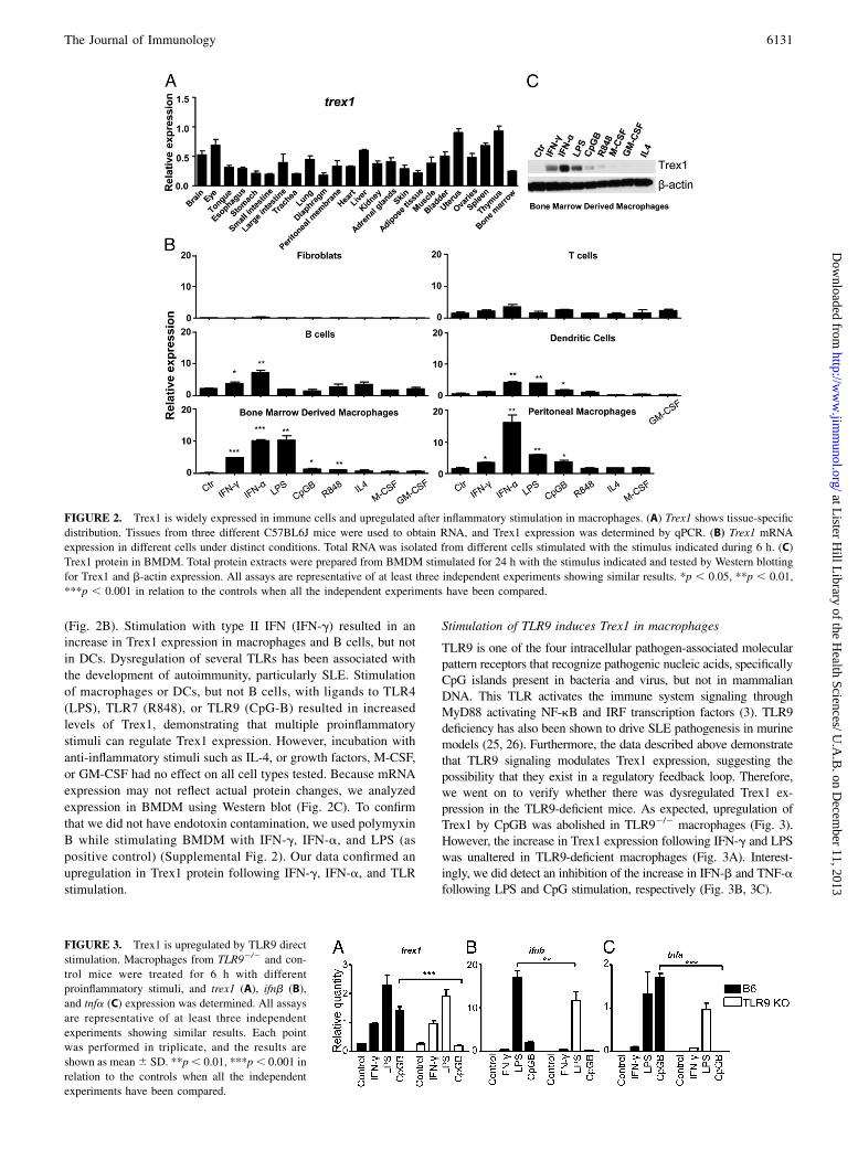
(Fig. 2B). Stimulation with type II IFN (IFN-g) resulted in anincrease in Trex1 expression in macrophages and B cells, but not
in DCs. Dysregulation of several TLRs has been associated with
the development of autoimmunity, particularly SLE. Stimulation
of macrophages or DCs, but not B cells, with ligands to TLR4
(LPS), TLR7 (R848), or TLR9 (CpG-B) resulted in increased
levels of Trex1, demonstrating that multiple proinflammatory
stimuli can regulate Trex1 expression. However, incubation with
anti-inflammatory stimuli such as IL-4, or growth factors, M-CSF,
or GM-CSF had no effect on all cell types tested. Because mRNA
expression may not reflect actual protein changes, we analyzed
expression in BMDM using Western blot (Fig. 2C). To confirm
that we did not have endotoxin contamination, we used polymyxin
B while stimulating BMDM with IFN-g, IFN-a, and LPS (as
positive control) (Supplemental Fig. 2). Our data confirmed an
upregulation in Trex1 protein following IFN-g, IFN-a, and TLR
stimulation.
Stimulation of TLR9 induces Trex1 in macrophages
TLR9 is one of the four intracellular pathogen-associated molecularpattern receptors that recognize pathogenic nucleic acids, specificallyCpG islands present in bacteria and virus, but not in mammalianDNA. This TLR activates the immune system signaling throughMyD88 activating NF-kB and IRF transcription factors (3). TLR9deficiency has also been shown to drive SLE pathogenesis in murinemodels (25, 26). Furthermore, the data described above demonstratethat TLR9 signaling modulates Trex1 expression, suggesting thepossibility that they exist in a regulatory feedback loop. Therefore,we went on to verify whether there was dysregulated Trex1 ex-pression in the TLR9-deficient mice. As expected, upregulation ofTrex1 by CpGB was abolished in TLR92/2 macrophages (Fig. 3).However, the increase in Trex1 expression following IFN-g and LPSwas unaltered in TLR9-deficient macrophages (Fig. 3A). Interest-ingly, we did detect an inhibition of the increase in IFN-b and TNF-afollowing LPS and CpG stimulation, respectively (Fig. 3B, 3C).
FIGURE 2. Trex1 is widely expressed in immune cells and upregulated after inflammatory stimulation in macrophages. (A) Trex1 shows tissue-specific
distribution. Tissues from three different C57BL6J mice were used to obtain RNA, and Trex1 expression was determined by qPCR. (B) Trex1 mRNA
expression in different cells under distinct conditions. Total RNA was isolated from different cells stimulated with the stimulus indicated during 6 h. (C)
Trex1 protein in BMDM. Total protein extracts were prepared from BMDM stimulated for 24 h with the stimulus indicated and tested by Western blotting
for Trex1 and b-actin expression. All assays are representative of at least three independent experiments showing similar results. *p , 0.05, **p , 0.01,
***p , 0.001 in relation to the controls when all the independent experiments have been compared.
FIGURE 3. Trex1 is upregulated by TLR9 direct
stimulation. Macrophages from TLR92/2 and con-
trol mice were treated for 6 h with different
proinflammatory stimuli, and trex1 (A), ifnb (B),
and tnfa (C) expression was determined. All assays
Trex12/2 macrophages exhibit an exacerbatedproinflammatory response
In view of the significantly higher induction of Trex1 in macro-phages compared with other immune cell types, we decided toexamine whether the lack of Trex1 in this population could affectphenotype and function. We analyzed the surface expression ofmaturation markers in Trex12/2 and control BMDMs, which area homogeneous population of primary quiescent cells. There wereno significant differences in the surface expression of CD11b,F4/80, CD115 (also known as M-CSFR), and GR-1 (also Ly6G) ifTrex1 was present or not (Fig. 4A).We then examined the functional cytokine response of Trex1-
deficient BMDMs to TLR4, TLR7, and TLR9 stimulation. LPSstimulation revealed an increase in IL-1b mRNA with a concom-itant decrease in IFN-b (Fig. 4B). There were no measurabledifferences in the release of TNF-a or IL-6 following LPS stim-ulation (Fig. 4C). Further examination revealed that the increasedproduction of TNF-a following TLR9 stimulation was augmentedin BMDMs lacking Trex1 (Fig. 4C). Incubation with the TLR7ligand (R848) resulted in increased IFN-b expression in Trex12/2
BMDMs compared with controls (Fig. 4B). The other cytokinestested did not show any significant differences. Analysis of cellsurface costimulation molecules revealed a higher upregulation ofCD86 following LPS stimulation in Trex12/2 BMDMs comparedwith controls. There were no detectable differences followingTLR7 or TLR9 incubation (Fig. 4D). The presence of Trex1 didnot modify the levels of CD40 following any TLR stimulation(Fig. 4D). These results suggest that Trex1 has an important rolein macrophage response to proinflammatory activation.
Trex1 represses Ag presentation by activated macrophages
As absence of Trex1 in macrophages induced higher expressionof CD86 (a costimulator protein in APCs), we decided to determinethe ability of Trex1-deficient macrophages to induce T cell pro-liferation following Ag processing and presentation. To that end,we pulsed previously stimulated macrophages with OVA for 3 h.
Thereafter, we added splenocytes from OTII mice (mice witha transgenic TCR that specifically recognizes the OVA 323–339peptide) that were previously stained with CFSE. T cell prolifer-ation was assessed by flow cytometry on day 5. We determinedthat, whereas there were no differences in naive macrophageAg presentation between control and Trex1-deficient mice, mac-rophages from Trex12/2 mice induce significantly more T cellproliferation after stimulation with LPS and R848 (Fig. 5).These findings demonstrate the importance of Trex1 in mac-rophage Ag presentation that is exacerbated after specific TLRstimulation.
Trex1 is necessary for efficient phagocytosis by activatedmacrophages
A defect in macrophage clearance of apoptotic cells has beendescribed in SLE patients and murine models (27). Therefore, wedecided to analyze whether and to what extent the lack of Trex1would affect this function. Macrophages from Trex12/2 andcontrol mice were stimulated with different proinflammatorystimuli and thereafter further incubated with apoptotic CFSE-labeled T cells for 1 h, as described in Materials and Methods.Our results show that macrophages lacking Trex1 have an im-paired phagocytic ability following overnight incubation withTLR4, TLR7, and TLR9 ligands (Fig. 6). These treatments did notaffect significantly the expression of macrophage markers CD11band Ly108. To determine whether the change in apoptotic clear-ance could impact the levels of released nucleic acids, we mea-sured the DNA present in the supernatant following incubationwith apoptotic cells (Supplemental Fig. 3). No significant differ-ence was found. These data, together with our results describedabove, suggest that Trex1 controls the response of macrophages toTLR stimulation and has an important role in the resolution ofinflammation.
DiscussionDeficiency of Trex1 in human patients and murine models ischaracterized by systemic inflammation, which includes autoim-
FIGURE 4. Macrophages from Trex12/2 mice produce higher levels of inflammatory cytokines in response to TLR ligands. (A) Macrophages obtained
from Trex12/2 and control mice present similar phenotype. CD11b, F4/80, CD115, and GR1 expression were measured by flow cytometry. (B) Macro-
phages from Trex12/2 mice upregulate il1b to a greater extent, but regulate ifnb to a lesser extent following TLR4 simulation. Incubation with R848 (TLR7
ligation) resulted in an increase of ifnb by Trex12/2 macrophages. Levels of expression were determined by qPCR at 9 and 3 h for il1b and ifnb, re-
spectively. (C) Macrophages from Trex12/2 produce more TNF-a after 24-h stimulation. Supernatants were collected, and the levels of TNF-a and IL-6
were measured. (D) Increased expression of CD86 in macrophages of Trex12/2 mice after LPS treatment. The expression of CD86 and CD40 was de-
termined with flow cytometry after 24-h stimulation. All assays are representative of at least three independent experiments showing similar results. Results
are shown as mean 6 SD. *p , 0.05, **p , 0.01 in relation to the controls when all the independent experiments have been compared.
munity (11). In the studies presented in this work, we demonstratethat Trex1 plays an important regulatory role in activated macro-phages. Deficiency results in an increased activated proinflammatoryphenotype associated with an enhanced Ag presentation. Further-more, Trex12/2 mature macrophages exhibit a defect in the phago-cytosis of apoptotic cells, which is an important mechanism for theresolution of inflammation.Our findings also confirm and extend previous observations that
Trex12/2 mice present severe inflammation at multiple sites (15),including the brain, despite measureable monocyte infiltration inthis organ. In fact, recently, it has been demonstrated that in vitroIFN-a treatment promotes astrocyte activation, supporting theidea of inflammation as a key feature in the pathogenesis of AGS(28). We also showed that Trex1 has a tissue-specific distribution
with the highest levels in spleen, thymus, and uterus. This mayexplain why the level of tissue inflammation in the mice variesacross various tissues and why cardiac disease is the most fre-quently described trait of Trex1-deficient mice.Examination of cellular expression revealed high levels of Trex1
in immune cells, which were highly induced by proinflammatorystimuli and not by anti-inflammatory cytokines or growth factorsin macrophages. This is concurrent with the observation made inhuman monocyte-derived macrophages in which Trex1 was alsoupregulated after proinflammatory stimuli (29). Additionally, it hasalso been reported that Trex1 is upregulated by genotoxic drugs(30, 31), indicating that Trex1 is needed for both pathogen DNAdegradation and also small strands of nuclear DNA originatedfrom genotoxicity. This suggests that, under the normal physio-
FIGURE 5. Activatedmacrophages from Trex12/2
mice induce increased T cell proliferation. Macro-
phages were stimulated for 24 h, incubated with OVA
for 3 h, and then cocultured with splenocytes from
OT-II mice for 5 d. Floating cells were collected,
stained, and analyzed by FACS. In the top panels, an
example is shown of the gating strategy. First and
second panel gates for single cells are followed by
a third panel gate where only CD45 live cells are se-
lected. From these, the positive CD4 cells with TCR
Va2 chain were gated, and the percentage of T cell
proliferating cells was determined by the loss of CFSE
fluorescence, as this staining is diluted after cell pro-
liferation. In thebottompanels, themean6SDof three
independent experiments is shown. **p , 0.01,
***p , 0.001 in relation to the controls.
FIGURE 6. Apoptotic clearance is impaired in macrophages from Trex12/2 mice. (A) Apoptotic cells were labeled with CFSE (cytoplasmatic dye) and
incubated with stimulated or control BMDM at a ratio of 4:1 for 1 h. Supernatant was removed and macrophages were collected, stained, and analyzed by
flow cytometry. Macrophages that phagocyted apoptotic cells were detected as CFSE positive, when compared with the control (macrophages incubated
with no CFSE-labeled cells). Phagocytosis values are presented as fold change of percentage of positive CFSE live macrophages. (B) The cumulative graphs
of phagocytosis show a reduction in Trex12/2 macrophages following TLR stimulation. (C) The expression levels of macrophage surface markers are
shown; no significant differences were seen here. All assays are representative of at least three independent experiments showing similar results. Results are
shown as mean 6 SD. **p , 0.01, ***p , 0.001 in relation to the controls when all the independent experiments have been compared.
logical environment, Trex1 is upregulated following immune orgenotoxicity challenge to degrade an excess of cytoplasmaticDNA and also to regulate the host immune response. This hy-pothesis is based upon findings demonstrating that deficiency ofTrex1 increases the production of proinflammatory cytokines suchas TNF-a, IFN type 1, and IL-1b following TLR ligand exposure.These Trex12/2 TLR-activated macrophages have higher levelsof surface CD86 and an increased capability in Ag presentation,which leads to activation of adaptive immune system with primingautoreactive T cells. The accumulation of DNA further triggersintracellular DNA receptors as STING (32) and in consequenceoveractivates macrophages. Furthermore, Trex12/2 macrophagespresent a reduced ability to phagocyte apoptotic material. Allthese facts may explain the exacerbated inflammatory phenotypein Trex1-associated diseases.Excessive macrophage activation has numerous damaging effects,
exemplified in septic shock, which can lead to multiple organdysfunction syndrome and death through a surplus of TNF-a pro-duction among other cytokines (33). In other situations, persistenceof proinflammatory activity results in the development of chronicinflammation and autoimmunity. This defect is arguably related tothe excessive production of type I of IFN, signature of the AGS (6)that is also associated with autoimmune diseases.A strong activation of macrophages could result in self-damage.
LPS activation of macrophages can induce apoptosis throughthe autocrine production of TNF-a (34). To prevent undesirableeffects, macrophages have several mechanisms to protect againstexcessive activation that prevents their self-destruction (35). Forexample, CDK inhibitor p21Waf1 is induced by IFN-g and protectsfrom apoptosis (23). In the absence of this CDK inhibitor, micedevelop a lupus-like phenotype characterized by anti-DNA Absand glomerulonephritis (36, 37). p21Waf1 is a negative regulatorthat curbs excessive macrophage activation by providing a nega-tive feedback system of TNF-a and IL-1b production (38, 39). Wepropose a similar function for Trex1 as macrophage controller ofactivation.Impaired macrophage function, specifically with regard to the
clearance of dead cells, has been widely described in both patientsand murine models of SLE (40). This is an important mechanismfor the resolution of inflammation. Also, this process generatescirculating DNA, which is often found increased in patients withSLE (41, 42). In our system, we did not observe any differences inthe release of DNA by macrophages, despite the remarkable re-duction in phagocytosis by Trex12/2 macrophages, indicating thatthese are two independent mechanisms. Trex1 is a highly pro-cessive exonuclease potentially affecting the digestion of apoptoticDNA engulfed by macrophages. Therefore, it is possible that theuptake of external apoptotic bodies may also accumulate outsidethe cell, affecting the net results. Furthermore, a recent publicationhas described dysregulation of lysosomes associated with Trex1deficiency that may affect apoptotic cell processing (16). Thiswould top with further activation of the macrophage, as it isknown that macrophages switch to an anti-inflammatory profileafter phagocytosis of apoptotic cells (43), and the lower phagocyticcapacity could contribute to a delayed switch.A percentage of SLE patients exhibits a loss in Trex1 function
(8). In addition, we have shown that both TLR7 and TLR9 (19),which have been associated with the development of SLE, up-regulate Trex1, and that, in its absence, responses to these TLRligands result in an augmented inflammatory response.Our data demonstrate that Trex1 plays a key role in the in-
flammatory processes by macrophages. These cells are scavengersfor apoptotic cells and, in the absence of Trex1, unprocessed DNAcould induce in a STING-dependent or independent fashion the
production of cytokines and the exacerbation of the immunesystem explaining the inflammatory process. Our data suggestthat this pathwaymay play a role in the development of autoimmunediseases.
AcknowledgmentsWewholeheartedly thank Gemma Lopez, Natalia Plana, and the staff of the
Laboratory Animal Applied Research Platform of the Barcelona Science
Park for excellent technical assistance. We remain indebted to all Nuclease
Immune Mediated Brain and Lupus-Like Conditions Consortium members
for insightful and useful comments. The Nuclease Immune Mediated Brain
and Lupus-Like Conditions Consortium is composed of David Bonthron,
Genetics Section, Leeds Institute of Molecular Medicine, St. James’s
University Hospital (Leeds, U.K.); Yanick Crow, Genetic Medicine,
Manchester Academic Health Science Centre (Manchester, U.K.); Taco
Kuijpers, Academic Medical Center, University of Amsterdam (Amster-
dam, The Netherlands); Arn van den Maagdenberg, Departments of Hu-
man Genetics and Neurology, Leiden University Medical Centre (Leiden,
The Netherlands); Simona Orcesi, Department of Child Neurology and
Psychiatry, Instituto di Ricovero e Cura a Carattere Scientifico, Mondino
Institute of Neurology Foundation (Pavia, Italy); Dan Stetson, Department
of Immunology, University of Washington (Seattle, WA); and Adeline
Vanderver, Children Research Institute (Washington, D.C.).
DisclosuresThe authors have no financial conflicts of interest.
References1. Mazur, D. J., and F. W. Perrino. 2001. Structure and expression of the TREX1
and TREX2 39—.59 exonuclease genes. J. Biol. Chem. 276: 14718–14727.2. Brucet, M., J. Querol-Audı, M. Serra, X. Ramirez-Espain, K. Bertlik, L. Ruiz,
J. Lloberas, M. J. Macias, I. Fita, and A. Celada. 2007. Structure of the dimericexonuclease TREX1 in complex with DNA displays a proline-rich binding sitefor WW domains. J. Biol. Chem. 282: 14547–14557.
3. Kawai, T., and S. Akira. 2010. The role of pattern-recognition receptors in innateimmunity: update on Toll-like receptors. Nat. Immunol. 11: 373–384.
4. Mazur, D. J., and F. W. Perrino. 1999. Identification and expression of theTREX1 and TREX2 cDNA sequences encoding mammalian 39—.59 exonu-cleases. J. Biol. Chem. 274: 19655–19660.
5. Brucet, M., J. Querol-Audı, K. Bertlik, J. Lloberas, I. Fita, and A. Celada. 2008.Structural and biochemical studies of TREX1 inhibition by metals: identificationof a new active histidine conserved in DEDDh exonucleases. Protein Sci. 17:2059–2069.
6. Crow, Y. J., B. E. Hayward, R. Parmar, P. Robins, A. Leitch, M. Ali, D. N. Black,H. van Bokhoven, H. G. Brunner, B. C. Hamel, et al. 2006. Mutations in the geneencoding the 39-59 DNA exonuclease TREX1 cause Aicardi-Goutieres syndromeat the AGS1 locus. Nat. Genet. 38: 917–920.
7. Rice, G., W. G. Newman, J. Dean, T. Patrick, R. Parmar, K. Flintoff, P. Robins,S. Harvey, T. Hollis, A. O’Hara, et al. 2007. Heterozygous mutations in TREX1cause familial chilblain lupus and dominant Aicardi-Goutieres syndrome. Am. J.Hum. Genet. 80: 811–815.
8. Lee-Kirsch, M. A., M. Gong, D. Chowdhury, L. Senenko, K. Engel, Y. A. Lee,U. de Silva, S. L. Bailey, T. Witte, T. J. Vyse, et al. 2007. Mutations in the geneencoding the 39-59 DNA exonuclease TREX1 are associated with systemic lupuserythematosus. Nat. Genet. 39: 1065–1067.
9. Stewart, A. E., S. Dowd, S. M. Keyse, and N. Q. McDonald. 1999. Crystalstructure of the MAPK phosphatase Pyst1 catalytic domain and implications forregulated activation. Nat. Struct. Biol. 6: 174–181.
10. Morita, M., G. Stamp, P. Robins, A. Dulic, I. Rosewell, G. Hrivnak, G. Daly,T. Lindahl, and D. E. Barnes. 2004. Gene-targeted mice lacking the Trex1(DNase III) 39—.59 DNA exonuclease develop inflammatory myocarditis. Mol.Cell. Biol. 24: 6719–6727.
11. Tanoue, T., T. Yamamoto, and E. Nishida. 2002. Modular structure of a dockingsurface on MAPK phosphatases. J. Biol. Chem. 277: 22942–22949.
12. Yang, Y. G., T. Lindahl, and D. E. Barnes. 2007. Trex1 exonuclease degradesssDNA to prevent chronic checkpoint activation and autoimmune disease. Cell131: 873–886.
13. Stetson, D. B., J. S. Ko, T. Heidmann, and R. Medzhitov. 2008. Trex1 preventscell-intrinsic initiation of autoimmunity. Cell 134: 587–598.
14. Neidhart, M., E. Karouzakis, G. G. Schumann, R. E. Gay, and S. Gay. 2010.Trex-1 deficiency in rheumatoid arthritis synovial fibroblasts. Arthritis Rheum.62: 2673–2679.
15. Owens, D. M., and S. M. Keyse. 2007. Differential regulation of MAP kinasesignalling by dual-specificity protein phosphatases. Oncogene 26: 3203–3213.
16. Wang, Z., C. Zang, K. Cui, D. E. Schones, A. Barski, W. Peng, and K. Zhao.2009. Genome-wide mapping of HATs and HDACs reveals distinct functions inactive and inactive genes. Cell 138: 1019–1031.
17. Cao, W., C. Bao, E. Padalko, and C. J. Lowenstein. 2008. Acetylation ofmitogen-activated protein kinase phosphatase-1 inhibits Toll-like receptor sig-naling. J. Exp. Med. 205: 1491–1503.
18. Celhar, T., R. Magalhaes, and A. M. Fairhurst. 2012. TLR7 and TLR9 in SLE:when sensing self goes wrong. Immunol. Res. 53: 58–77.
19. Byrne, J. C., J. Nı Gabhann, E. Lazzari, R. Mahony, S. Smith, K. Stacey,C. Wynne, and C. A. Jefferies. 2012. Genetics of SLE: functional relevance formonocytes/macrophages in disease. Clin. Dev. Immunol. 2012: 582352.
20. Burkatovskaya, M., G. P. Tegos, E. Swietlik, T. N. Demidova, A. P Castano, andM. R. Hamblin. 2006. Use of chitosan bandage to prevent fatal infections de-veloping from highly contaminated wounds in mice. Biomaterials 27: 4157–4164.
21. Celada, A., P. W. Gray, E. Rinderknecht, and R. D. Schreiber. 1984. Evidence fora gamma-interferon receptor that regulates macrophage tumoricidal activity. J.Exp. Med. 160: 55–74.
22. Lutz, M. B., N. Kukutsch, A. L. Ogilvie, S. Rossner, F. Koch, N. Romani, andG. Schuler. 1999. An advanced culture method for generating large quantities ofhighly pure dendritic cells from mouse bone marrow. J. Immunol. Methods 223:77–92.
23. Xaus, J., M. Cardo, A. F. Valledor, C. Soler, J. Lloberas, and A. Celada. 1999.Interferon gamma induces the expression of p21waf-1 and arrests macrophagecell cycle, preventing induction of apoptosis. Immunity 11: 103–113.
24. Lu, T. C., Z. Wang, X. Feng, P. Chuang, W. Fang, Y. Chen, S. Neves, A. Maayan,H. Xiong, Y. Liu, et al. 2008. Retinoic acid utilizes CREB and USF1 in a tran-scriptional feed-forward loop in order to stimulate MKP1 expression in humanimmunodeficiency virus-infected podocytes. Mol. Cell. Biol. 28: 5785–5794.
25. Santiago-Raber, M. L., I. Dunand-Sauthier, T. Wu, Q. Z. Li, S. Uematsu,S. Akira, W. Reith, C. Mohan, B. L. Kotzin, and S. Izui. 2010. Critical role ofTLR7 in the acceleration of systemic lupus erythematosus in TLR9-deficientmice. J. Autoimmun. 34: 339–348.
26. Hasselgren, P. O. 2007. Ubiquitination, phosphorylation, and acetylation: triplethreat in muscle wasting. J. Cell. Physiol. 213: 679–689.
27. Xu, G., Y. Zhang, L. Zhang, A. I. Roberts, and Y. Shi. 2009. C/EBPbeta mediatessynergistic upregulation of gene expression by interferon-gamma and tumornecrosis factor-alpha in bone marrow-derived mesenchymal stem cells. StemCells 27: 942–948.
28. Cuadrado, E., M. H. Jansen, J. Anink, L. De Filippis, A. L. Vescovi, C. Watts,E. Aronica, E. M. Hol, and T. W. Kuijpers. 2013. Chronic exposure of astrocytesto interferon-a reveals molecular changes related to Aicardi-Goutieres syn-drome. Brain 136: 245–258.
29. Cobos Jimenez, V., T. Booiman, S. W. de Taeye, K. A. van Dort, M. A. Rits,J. Hamann, and N. A. Kootstra. 2012. Differential expression of HIV-1 inter-fering factors in monocyte-derived macrophages stimulated with polarizingcytokines or interferons. Sci. Rep. 2: 763.
30. Christmann, M., M. T. Tomicic, D. Aasland, N. Berdelle, and B. Kaina. 2010.Three prime exonuclease I (TREX1) is Fos/AP-1 regulated by genotoxic stressand protects against ultraviolet light and benzo(a)pyrene-induced DNA damage.Nucleic Acids Res. 38: 6418–6432.
31. Tomicic, M. T., D. Aasland, T. Nikolova, B. Kaina, and M. Christmann. 2013.Human three prime exonuclease TREX1 is induced by genotoxic stress andinvolved in protection of glioma and melanoma cells to anticancer drugs. Bio-chim. Biophys. Acta 1833: 1832–1843.
32. Gall, A., P. Treuting, K. B. Elkon, Y. M. Loo, M. Gale, Jr., G. N. Barber, andD. B. Stetson. 2012. Autoimmunity initiates in nonhematopoietic cells andprogresses via lymphocytes in an interferon-dependent autoimmune disease.Immunity 36: 120–131.
33. Casals-Casas, C., E. Alvarez, M. Serra, C. de la Torre, C. Farrera, E. Sanchez-Tillo, C. Caelles, J. Lloberas, and A. Celada. 2009. CREB and AP-1 activationregulates MKP-1 induction by LPS or M-CSF and their kinetics correlate withmacrophage activation versus proliferation. Eur. J. Immunol. 39: 1902–1913.
34. Xaus, J., M. Comalada, A. F. Valledor, J. Lloberas, F. Lopez-Soriano,J. M. Argiles, C. Bogdan, and A. Celada. 2000. LPS induces apoptosis inmacrophages mostly through the autocrine production of TNF-alpha. Blood 95:3823–3831.
35. Valledor, A. F., M. Comalada, L. F. Santamarıa-Babi, J. Lloberas, and A. Celada.2010. Macrophage proinflammatory activation and deactivation: a question ofbalance. Adv. Immunol. 108: 1–20.
36. Balomenos, D., J. Martın-Caballero, M. I. Garcıa, I. Prieto, J. M. Flores,M. Serrano, and C. Martınez-A. 2000. The cell cycle inhibitor p21 controlsT-cell proliferation and sex-linked lupus development. Nat. Med. 6: 171–176.
37. Salvador, J. M., M. C. Hollander, A. T. Nguyen, J. B. Kopp, L. Barisoni,J. K. Moore, J. D. Ashwell, and A. J. Fornace, Jr. 2002. Mice lacking the p53-effector gene Gadd45a develop a lupus-like syndrome. Immunity 16: 499–508.
38. Scatizzi, J. C., M. Mavers, J. Hutcheson, B. Young, B. Shi, R. M. Pope, E. M.Ruderman, D. S. Samways, J. A. Corbett, T. M. Egan, and H. Perlman. 2009.The cyclin dependent kinase domain of p21 is a suppressor of IL-1b-mediatedinflammation in activated macrophages. Eur. J. Immunol. 39: 820-825.
39. Trakala, M., C. F. Arias, M. I. Garcıa, M. C. Moreno-Ortiz, K. Tsilingiri,P. J. Fernandez, M. Mellado, M. T. Dıaz-Meco, J. Moscat, M. Serrano, et al.2009. Regulation of macrophage activation and septic shock susceptibilityvia p21(WAF1/CIP1). Eur. J. Immunol. 39: 810-819.
40. Szwench, E., M. P. Czkowska, K. Marczewski, A. Klisiewicz, I. Micha Owska,I. Ciuba, M. Januszewicz, A. Prejbisz, P. Hoffman, and A. Januszewicz. 2011.Phaeochromocytoma in a 86-year-old patient presenting with reversible myo-cardial dysfunction. Blood Press 20: 383-386.
41. De Vlas, S. J., D. Engels, A. L. Rabello, B. F. Oostburg, L. Van Lieshout,A. M. Polderman, G. J. Van Oortmarssen, J. D. Habbema, and B. Gryseels. 1997.Validation of a chart to estimate true Schistosoma mansoni prevalences fromsimple egg counts. Parasitology 114: 113–121.
42. Gaipl, U. S., L. E. Munoz, G. Grossmayer, K. Lauber, S. Franz, K. Sarter,R. E. Voll, T. Winkler, A. Kuhn, J. Kalden, et al. 2007. Clearance deficiency andsystemic lupus erythematosus (SLE). J. Autoimmun. 28: 114–121.
43. Cvetanovic, M., J. E. Mitchell, V. Patel, B. S. Avner, Y. Su, P. T. van der Saag,P. L. Witte, S. Fiore, J. S. Levine, and D. S. Ucker. 2006. Specific recognition ofapoptotic cells reveals a ubiquitous and unconventional innate immunity. J. Biol.Chem. 281: 20055–20067.