31
The Glymphatic System, Sleep & other states of unconsciousness Helene Benveniste, MD, PhD Department of Anesthesiology & Radiology, Stony Brook University, NY
The Glymphatic System, Sleep & other states of
unconsciousness
Helene Benveniste, MD, PhD
Department of Anesthesiology & Radiology,
Stony Brook University, NY
Disclosures
• Current funding: NIH, SUNY-BNE, Anonymous
• Other financial relationships: None
• Conflicts of interest: None
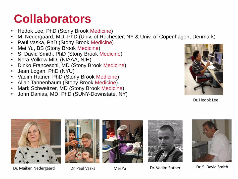
Collaborators• Hedok Lee, PhD (Stony Brook Medicine)• M. Nedergaard, MD, PhD (Univ. of Rochester, NY & Univ. of Copenhagen, Denmark)• Paul Vaska, PhD (Stony Brook Medicine)• Mei Yu, BS (Stony Brook Medicine)• S. David Smith, PhD (Stony Brook Medicine)• Nora Volkow MD, (NIAAA, NIH)• Dinko Franceschi, MD (Stony Brook Medicine)• Jean Logan, PhD (NYU)• Vadim Ratner, PhD (Stony Brook Medicine)• Allan Tannenbaum (Stony Brook Medicine)• Mark Schweitzer, MD (Stony Brook Medicine)• John Danias, MD, PhD (SUNY-Downstate, NY)
Dr. Maiken Nedergaard
Dr. Hedok Lee
Dr. Paul Vaska Dr. S. David SmithMei Yu
Collaborators
Dr. Vadim Ratner

Significance
• The brain is one of the most metabolically active organs in the body
• Removal of excess fluids and waste products is critical for normal brain function
• The brain parenchyma has no authentic lymphatic vessels for detoxification

The brain & metabolic waste: Example - Ammonia (NH3)
The glutamate-glutamine
cycle leads to production of
ammonia (NH3)….at the
same time ammonia is
needed in astrocytes for the
synthesis of glutamine from
glutamate.
Ammonia is rapidly
eliminated by the brain
From: Schousboe et al. Frontiers in Endocrinology, 2013

The brain & metabolic waste: Proteins
• Cytosolic proteins are constantly renewed
• Proteins incorporated in the cytoskeleton and membranes are constantly renewed
• Lack of specialized BBB transporters for most peptides and protein
• Recycling/degradation of protein: ubiquitination and autophagy
Schematic overview of autophagyRubinzstein, NATURE, Vol 443; 206

Significance
• Age-related decline in the efficiency of protein degradation has been implicated in pathological protein aggregation
• Neurodegenerative diseases, are characterized by accumulation of aggregation-prone mutated, misfolded or hyperphosphorylated proteins
• These proteins are present intracellular and extracellular
http://neuropathology-web.org/
Neurofibrillary tangle and amyloid plaque
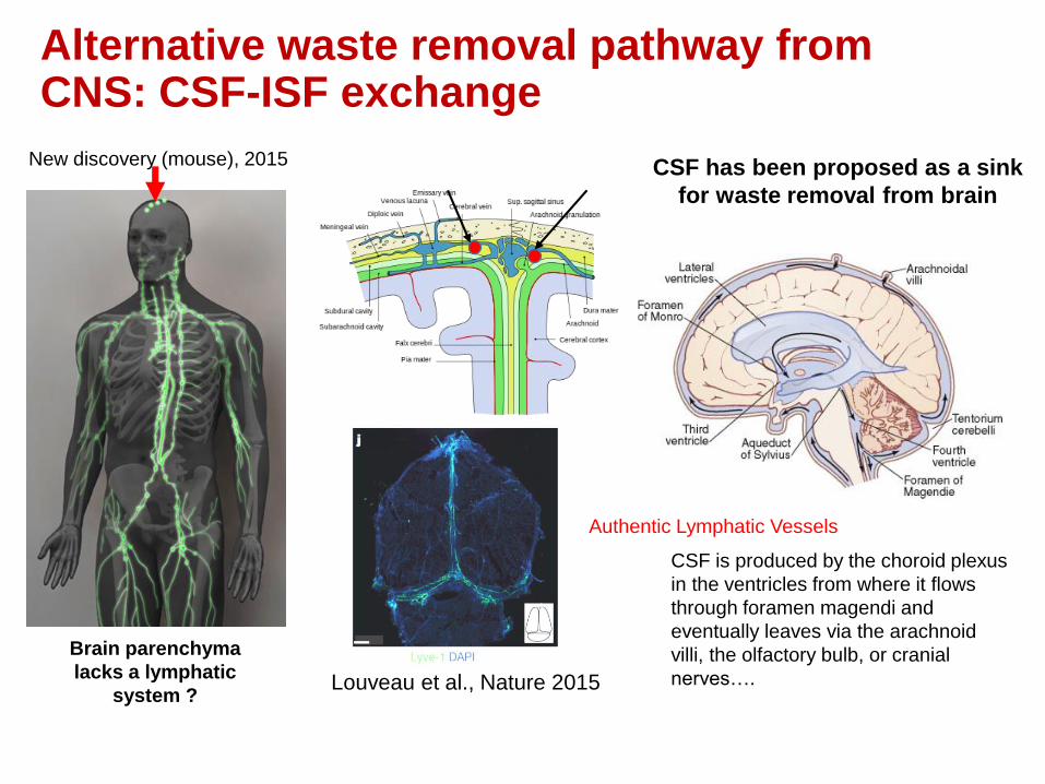
Alternative waste removal pathway from CNS: CSF-ISF exchange
Brain parenchyma
lacks a lymphatic
system ?
CSF has been proposed as a sink
for waste removal from brain
CSF is produced by the choroid plexus
in the ventricles from where it flows
through foramen magendi and
eventually leaves via the arachnoid
villi, the olfactory bulb, or cranial
nerves….
New discovery (mouse), 2015
Louveau et al., Nature 2015
Authentic Lymphatic Vessels

Dr. Quincke
Quincke injected cinnabar
granules into CSF of animals
(dogs, cats, rabbits)
1 mm
Cinnabar is a large molecule
and while it is transported in
CSF it does not go into
parenchyma…. From: “Modern CSF Research and Heinrich
Quincke’s Seminal Paper on the Distribution of
Cinnabar in Freely Moving Animals. Benveniste
et al., JCN 2015
Waste removal via CSF characterized by Heinrich Quincke in 1872

State-of-the-art knowledge of the glymphatic pathway
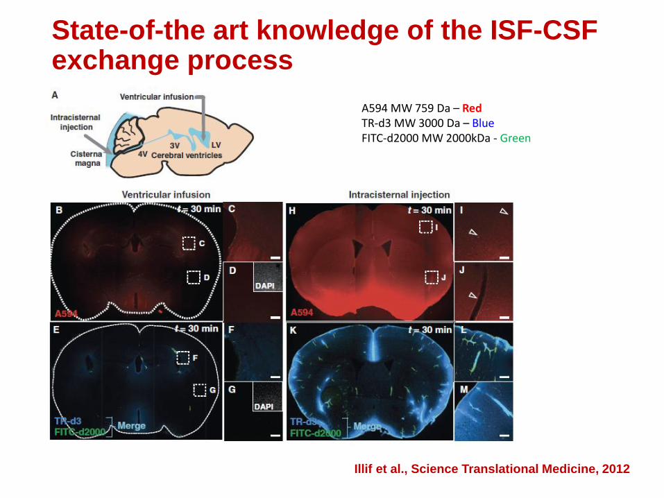
State-of-the art knowledge of the ISF-CSF exchange process
A594 MW 759 Da – RedTR-d3 MW 3000 Da – BlueFITC-d2000 MW 2000kDa - Green
Illif et al., Science Translational Medicine, 2012
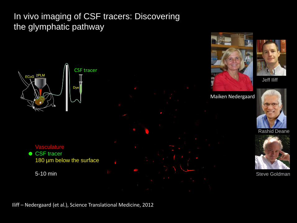
In vivo imaging of CSF tracers: Discovering
the glymphatic pathway
Vasculature
CSF tracer
180 µm below the surface
5-10 min
CSF tracer
Jeff Iliff
Rashid Deane
Steve Goldman
Iliff – Nedergaard (et al.), Science Translational Medicine, 2012
Maiken Nedergaard
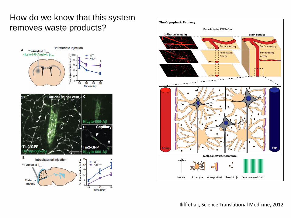
Iliff et al., Science Translational Medicine, 2012
How do we know that this system
removes waste products?

State-of-the-art-knowledge:
Visualizing glymphatic transport in real time using MRI
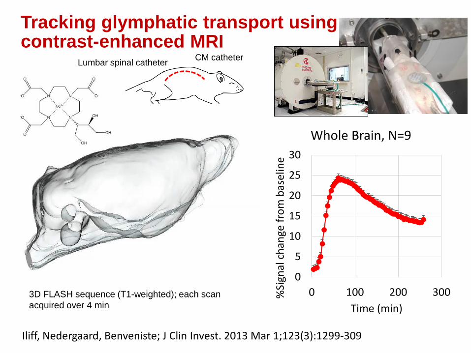
Tracking glymphatic transport using contrast-enhanced MRI
Gd-DTPA (MW: 938 Da)
Iliff, Nedergaard, Benveniste; J Clin Invest. 2013 Mar 1;123(3):1299-309
Lumbar spinal catheterCM catheter
Pineal Recess
3D FLASH sequence (T1-weighted); each scan
acquired over 4 min
0
5
10
15
20
25
30
0 100 200 300%Si
gnal
ch
ange
fro
m b
asel
ine
Time (min)
Whole Brain, N=9

Dr. Jean Logan, NYU
k2
k3 k4
K1CisternaMagna C1
C2
2-Compartment Model
“Retention” = k3/k4
“loss” = k2/(1+k3/k4)
Lee et al., J Neuroscience. 2015
0
50
100
150
200
250
300
350
400
450
0
5
10
15
20
25
30
0 50 100 150 200
CIS
TE
RN
A M
AG
NA
%S
ignal C
hange f
rom
baselin
e
Whole
bra
in
Time (min)
Gd-DTPA CSF RAT BRAIN
Whole Brain Cisterna Magna
Quantification of Gd-DTPA
brain-wide transport

Brain loss (clearance) and ‘retention’ of Gd-DTPA after CSF administration in rodent whole brain
0
2
4
6
8
10
12
14
0 100 200
% S
ign
al c
han
ge f
rom
bas
elin
e
Time (min)
2-Compartment model
Rat_072414_WB_TAC
Rat_072414_2C_model
0
5
10
15
k3/k
4=
rete
nti
on
Retention
0
0.1
0.2
0.3
0.4
0.5
k2/(
1+k
3/k
4)=
loss
‘Loss’ - Clearance
Lee et al., J Neuroscience 2015

Factors that influence glymphatic pathway function:
• AQP4
• Adrenergic tone
• Pulsatility
• Sleep / hypnotics
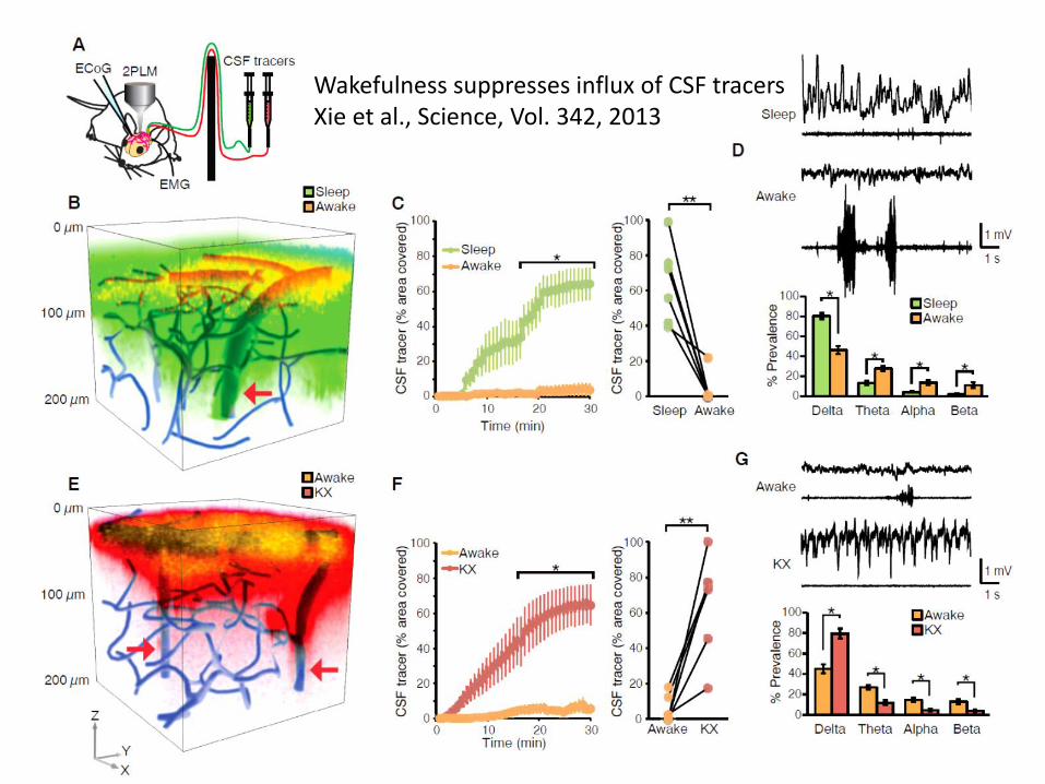
Wakefulness suppresses influx of CSF tracersXie et al., Science, Vol. 342, 2013
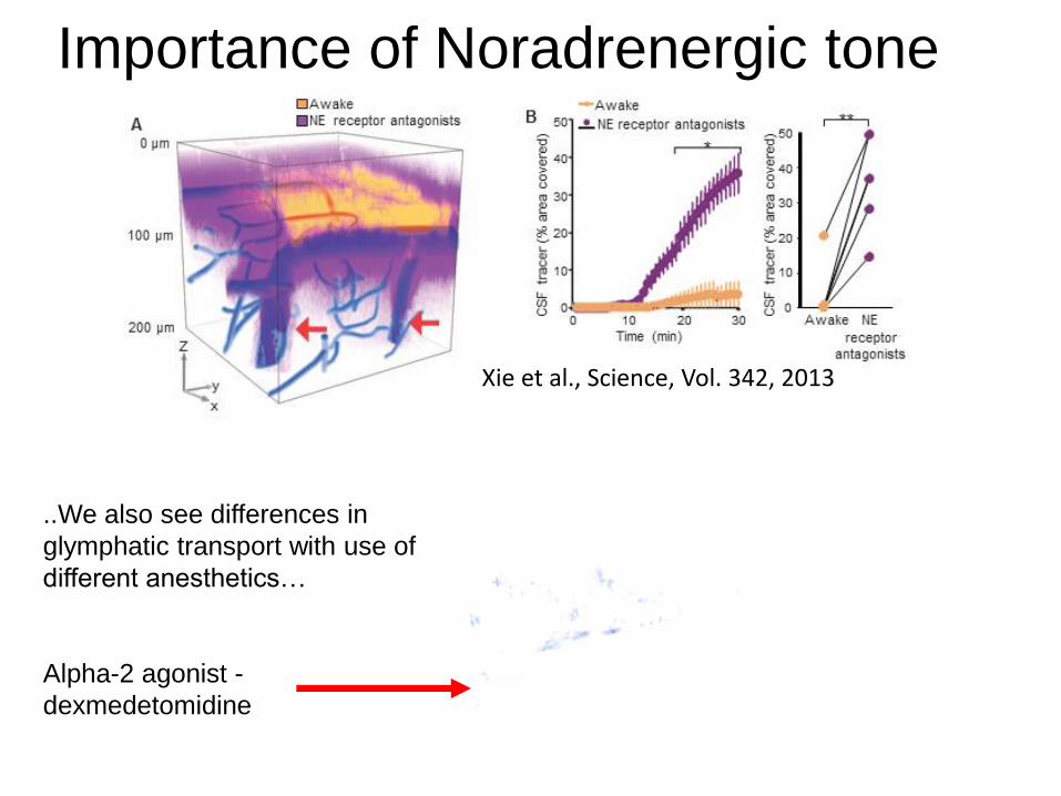
Importance of Noradrenergic tone
Xie et al., Science, Vol. 342, 2013
..We also see differences in
glymphatic transport with use of
different anesthetics…
Alpha-2 agonist -
dexmedetomidine

Knowledge gap:
If unconsciousness enhance brain waste removal – what about body position during the unconscious state?

How does body position during sleep/anesthesia influence glymphatictransport?
http://bestadjustablemattress.com/how-to-find-the-best-sleeping-positions/
“Consistently, poor sleepers spent more time on their backs with their heads straight” De Konincket al., Sleep 1983;6 (1):52-9
The most favored position is right lateral decubitus (Sleep 1983;6 (1):52-9)
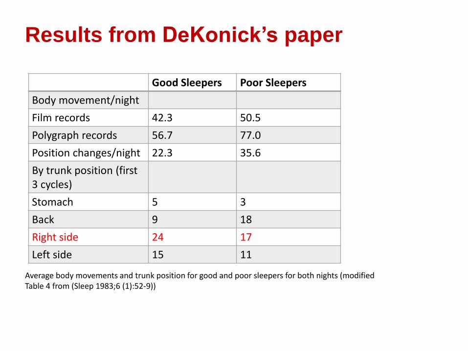
Results from DeKonick’s paper
Average body movements and trunk position for good and poor sleepers for both nights (modified Table 4 from (Sleep 1983;6 (1):52-9))
Good Sleepers Poor Sleepers
Body movement/night
Film records 42.3 50.5
Polygraph records 56.7 77.0
Position changes/night 22.3 35.6
By trunk position (first 3 cycles)
Stomach 5 3
Back 9 18
Right side 24 17
Left side 15 11

Sleeping gorillas
http://www.freepik.com/free-photo/gorilla-sleeping_352304.htm

Sleeping rats
http://blogs.discovermagazine.com/sciencenotfiction/2010/08/10/inception-and-the-neuroscience-of-sleep/#.VLZl0CvF8j4
“Slow wave sleep (SWS). During this stage, subjects assumed a reclining posture, sometimes on their abdomen….sometimes curled in a fetal position with eyes closed.” (Hobson; Behavioral Neruoscience; 2000, Vol 114; No. 6, 1239-1244).
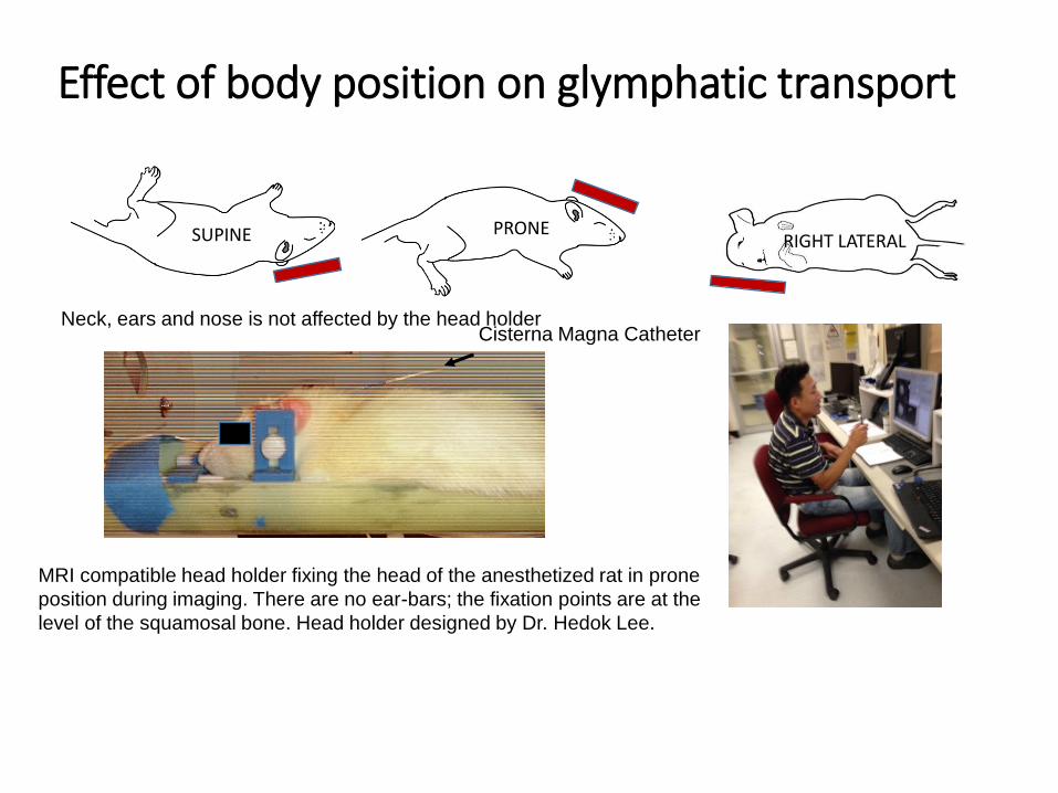
Effect of body position on glymphatic transport
SUPINE PRONE
MRI compatible head holder fixing the head of the anesthetized rat in prone
position during imaging. There are no ear-bars; the fixation points are at the
level of the squamosal bone. Head holder designed by Dr. Hedok Lee.
Cisterna Magna CatheterNeck, ears and nose is not affected by the head holder
RIGHT LATERAL
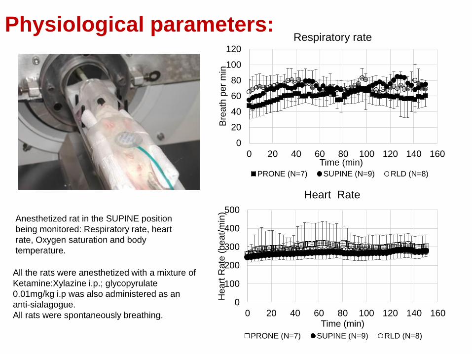
Physiological parameters:
Anesthetized rat in the SUPINE position
being monitored: Respiratory rate, heart
rate, Oxygen saturation and body
temperature.
All the rats were anesthetized with a mixture of
Ketamine:Xylazine i.p.; glycopyrulate
0.01mg/kg i.p was also administered as an
anti-sialagogue.
All rats were spontaneously breathing.
0
100
200
300
400
500
0 20 40 60 80 100 120 140 160
He
art
Ra
te (
be
at/
min
)
Time (min)
Heart Rate
PRONE (N=7) SUPINE (N=9) RLD (N=8)
0
20
40
60
80
100
120
0 20 40 60 80 100 120 140 160
Bre
ath
pe
r m
in
Time (min)
Respiratory rate
PRONE (N=7) SUPINE (N=9) RLD (N=8)
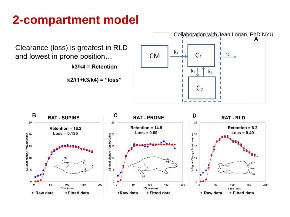
Collaboration with Jean Logan, PhD NYU
2-compartment model
Clearance (loss) is greatest in RLD
and lowest in prone position…
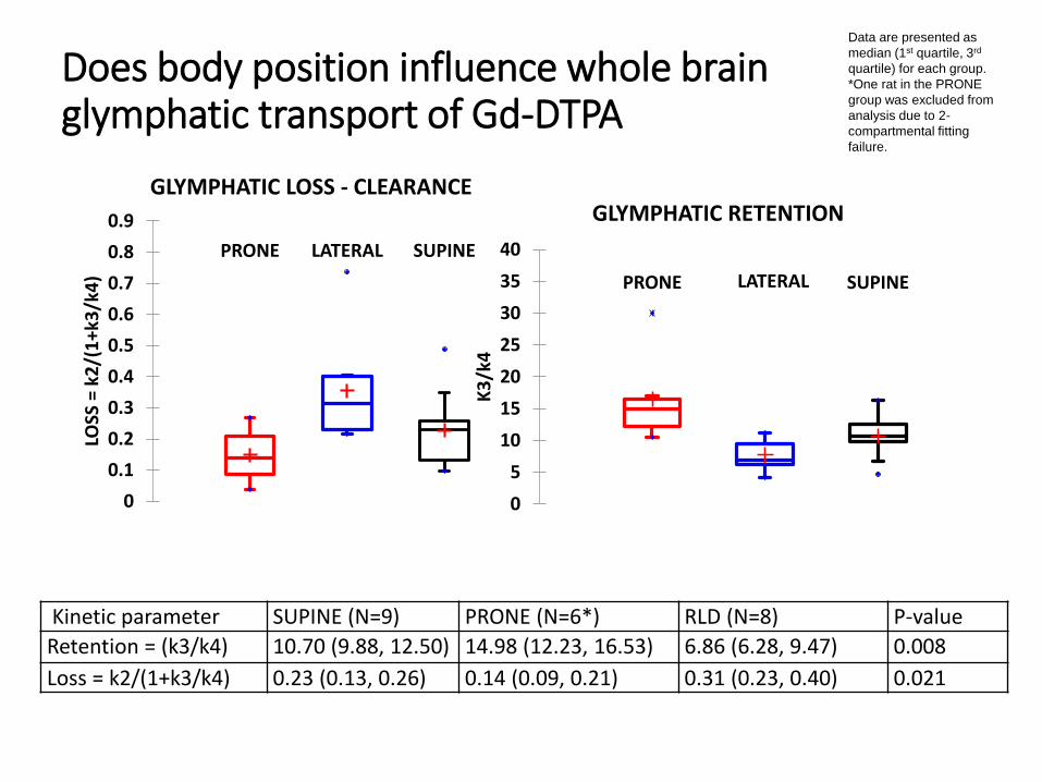
Does body position influence whole brain glymphatic transport of Gd-DTPA
Kinetic parameter SUPINE (N=9) PRONE (N=6*) RLD (N=8) P-value
Retention = (k3/k4) 10.70 (9.88, 12.50) 14.98 (12.23, 16.53) 6.86 (6.28, 9.47) 0.008
Loss = k2/(1+k3/k4) 0.23 (0.13, 0.26) 0.14 (0.09, 0.21) 0.31 (0.23, 0.40) 0.021
Data are presented as
median (1st quartile, 3rd
quartile) for each group.
*One rat in the PRONE
group was excluded from
analysis due to 2-
compartmental fitting
failure.
PRONE LATERAL SUPINE
0
0.1
0.2
0.3
0.4
0.5
0.6
0.7
0.8
0.9
LOSS
= k
2/(
1+k
3/k
4)
GLYMPHATIC LOSS - CLEARANCE
PRONE LATERAL SUPINE
0
5
10
15
20
25
30
35
40
K3
/k4
GLYMPHATIC RETENTION
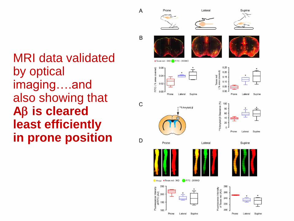
MRI data validated by optical imaging….and also showing that Ab is cleared least efficiently in prone position
Research opportunities
• Understand how the cardiac sympatho-vagal balance influence central nervous system arousal and glymphatic pathway function in the context of sleep and aging
• Understand how perivascular neurons, gliovascularinteractions and intramural vascular signaling change and interferes with glymphatic pathway functioning in normal aging.
• Mapping of the glymphatic, perivascular ‘connectome’; need to understand CSF-ISF streaming pattern in health and disease


