THE JOURNAL OF BIOLOGICAL CHEMISTRY 0 1993 by The American Society for Biochemistry and Molecular Biology, Inc. Vol. 268, No. 23, Issue of August 15, pp. 17240-17246,1993 Printed in U. S.A. Activation of Dynamin GTPase by Acidic Phospholipids and Endogenous Rat Brain Vesicles* (Received for publication, February 17, 1993, and in revised form, April 22, 1993) Pamela L. Tuma, Mary C. Stachniak, and Christine A. Collins$ From the Department of Cell. Molecular, and Structural Biology, Northwestern University Medical School, . . Chicago, Illinois 6061 1 Dynamin is a GTPase thought to play a role in en- docytosis based on genetic analysis of its homolog in Drosophila melanogaster shibire. Previous studies have stressed an in vitro association with microtu- bules, though additional evidence suggests that dyna- min associates with membranous organelles. In an analysis of the enzymatic and membrane binding prop- erties of dynamin, we have found that the acidic phos- pholipids, phosphatidylserine, phosphatidylglycerol, and phosphatidylinositol, are able to stimulate GTP hydrolysis in a manner similar to activation previously shown with microtubules. A neutral phospholipid, phosphatidylcholine, had no effect on dynamin GTPase. Activation of dynamin was biphasic, with a decrease in activity back to basal levels with increased concentrations of either microtubules or liposomes. A comparison between GTPase stimulation induced by microtubules and that by phospholipids suggests that ionic interactions between the basic C-terminal domain of dynamin and the negatively charged microtubule or phospholipid head group are important. In support of this, GTPase stimulation by these agents in combina- tion was not additive. A salt-extracted membrane frac- tion from brain tissue also activated dynamin GTPase, though to a lower extent than pure phospholipids. These results suggest that membrane componentscould be responsible for some aspects of the regulation of dynamin function in vivo. Dynamin is a GTP-binding protein that was first identified in microtubule preparations from calf brain as a 100-kDa nucleotide-sensitive microtubule-associated protein (1). It has been analyzed at themolecular level by cloning and sequenc- ing a rat cDNA (2) and a homolog in Drosophila named shibire (3,4). Based on the analysis of temperature-sensitive mutants of shibire, this protein is thought to be required for endocy- tosis, in particular for pinching-off of coated vesicles from the plasma membrane (5-7). The predicted amino acid sequence of dynamin (shibire) encodes the tripartite GTP binding consensus sequence (8), and dynamin has been shown to specifically hydrolyze GTP (9). As dynamin was first described in association with micro- tubules, the effect of this polymer on GTPase activity has been investigated. GTPase activity was found to be potently * The costs of publication of this article were defrayed in part by the payment of page charges. This article must therefore be hereby marked “advertisement” in accordance with 18 U.S.C. Section 1734 solely to indicate this fact. $To whom correspondence should be addressed Dept. of Cell, Molecular, and Structural Biology, Northwestern, University Medical School, 303 E. Chicago Ave., Chicago, IL 60611. Tel.: 312-503-4269; Fax: 312-503-7912. stimulated by relatively low concentrations of microtubules (9). Soluble tubulin and other cytoskeletal polymers were found to be unable to stimulate GTP hydrolysis, suggesting a specific regulatory role for microbutules in the physiological function of dynamin. However, the data obtained from the Drosophila studies suggest a role for dynamin in the early stages of endocytosis, a process that is thought to occur independently of microtubules (10). In addition, data from Scaife and Margolis (11) indicate that dynamin cosediments with membrane fractions from rat brain and is not found associated with microtubules by immunofluorescence analysis of PC12 cells. The apparent discrepancy between the enzy- mological characteristics of dynamin and cellular function as suggested by the genetic data (reviewed in Ref. 12) prompted us to explore the possibility that membrane components are involved in the regulation of dynamin GTPase. We show here that acidic phospholipids lead to activation of dynamin GTPase to levels comparable with those obtained with micro- tubules. Endogenous brain vesicles also activate dynamin. The properties of these activation processes indicate that phospholipids and/or other membrane components could be regulators of dynamin function in uiuo. EXPERIMENTAL PROCEDURES Materials-Male Sprague-Dawley rats between 150and 250 g were used as the source of brain tissue. Reagents for gel electrophoresis were from ICN Biomedicals and Bio-Rad. Phospholipids were ob- tained from both Sigma and Avanti Polar Lipids. Spermine, polyglu- tamate, and alkaline phosphatase-conjugated secondary antibodies were purchased from Sigma. Antibody to clathrin heavy chain was from ICN Biomedicals. Taxol was kindly provided by Nancita Lomax at the Drug Synthesis and Chemistry Branch, Division of Cancer Research, National Cancer Institute, and stored as a stock solution of 10 mM in dimethyl sulfoxide at -80 “C. Dynamin Purification-Dynamin was prepared from 4-5 g of rat brain tissue by coassembly with microtubules in extraction buffer (50 mM Pipes,’ 50 mM Hepes, 2 mM MgC12, 1 mM EDTA, pH 7.0) according to the procedure of Shpetner and Vallee (1) with minor modifications. Prior to the microtubule assembly step, brain cytosolic extract was fractionated over a 10-ml Bio-Gel P-6 column (10DG columns, Bio-Rad) equilibrated in extraction buffer. This procedure removes nucleotides and other low molecular weight compounds and in our bands is an essential step when using freshly isolated tissues as a source of protein. Following taxol assembly and a wash step in buffer lacking nucleotide, dynamin was extracted from the microtu- bules with a 7.5 mM GTP and 2.5 mM AMP-PNP and further purified by sucrose density gradient centrifugation (1). Sucrose gradient- purified dynamin was used for the GTPase studies reported here. Dynamin was further purified by ion exchange chromatography (1) for use as an immunogen for the production of a rabbit polyclonal antibody. Tissue Fractionation-A crude coated vesicle preparation was iso- lated by homogenization of 4-5 g of rat brain in extraction buffer and ’ The abbreviations used are: Pipes, 1,4-piperazinediethanesulfonic acid; AMP-PNP, adenyl-5”yl imidodiphosphate. 17240
Transcript
THE JOURNAL OF BIOLOGICAL CHEMISTRY 0 1993 by The American Society for Biochemistry and Molecular Biology, Inc.
Vol. 268, No. 23, Issue of August 15, pp. 17240-17246,1993 Printed in U. S.A.
Activation of Dynamin GTPase by Acidic Phospholipids and Endogenous Rat Brain Vesicles*
(Received for publication, February 17, 1993, and in revised form, April 22, 1993)
Pamela L. Tuma, Mary C. Stachniak, and Christine A. Collins$ From the Department of Cell. Molecular, and Structural Biology, Northwestern University Medical School, . . Chicago, Illinois 6061 1
Dynamin is a GTPase thought to play a role in en- docytosis based on genetic analysis of its homolog in Drosophila melanogaster shibire. Previous studies have stressed an in vitro association with microtu- bules, though additional evidence suggests that dyna- min associates with membranous organelles. In an analysis of the enzymatic and membrane binding prop- erties of dynamin, we have found that the acidic phos- pholipids, phosphatidylserine, phosphatidylglycerol, and phosphatidylinositol, are able to stimulate GTP hydrolysis in a manner similar to activation previously shown with microtubules. A neutral phospholipid, phosphatidylcholine, had no effect on dynamin GTPase. Activation of dynamin was biphasic, with a decrease in activity back to basal levels with increased concentrations of either microtubules or liposomes. A comparison between GTPase stimulation induced by microtubules and that by phospholipids suggests that ionic interactions between the basic C-terminal domain of dynamin and the negatively charged microtubule or phospholipid head group are important. In support of this, GTPase stimulation by these agents in combina- tion was not additive. A salt-extracted membrane frac- tion from brain tissue also activated dynamin GTPase, though to a lower extent than pure phospholipids. These results suggest that membrane components could be responsible for some aspects of the regulation of dynamin function in vivo.
Dynamin is a GTP-binding protein that was first identified in microtubule preparations from calf brain as a 100-kDa nucleotide-sensitive microtubule-associated protein (1). It has been analyzed at the molecular level by cloning and sequenc- ing a rat cDNA (2) and a homolog in Drosophila named shibire (3,4). Based on the analysis of temperature-sensitive mutants of shibire, this protein is thought to be required for endocy- tosis, in particular for pinching-off of coated vesicles from the plasma membrane (5-7).
The predicted amino acid sequence of dynamin (shibire) encodes the tripartite GTP binding consensus sequence (8), and dynamin has been shown to specifically hydrolyze GTP (9). As dynamin was first described in association with micro- tubules, the effect of this polymer on GTPase activity has been investigated. GTPase activity was found to be potently
* The costs of publication of this article were defrayed in part by the payment of page charges. This article must therefore be hereby marked “advertisement” in accordance with 18 U.S.C. Section 1734 solely to indicate this fact.
$To whom correspondence should be addressed Dept. of Cell, Molecular, and Structural Biology, Northwestern, University Medical School, 303 E. Chicago Ave., Chicago, IL 60611. Tel.: 312-503-4269; Fax: 312-503-7912.
stimulated by relatively low concentrations of microtubules (9). Soluble tubulin and other cytoskeletal polymers were found to be unable to stimulate GTP hydrolysis, suggesting a specific regulatory role for microbutules in the physiological function of dynamin. However, the data obtained from the Drosophila studies suggest a role for dynamin in the early stages of endocytosis, a process that is thought to occur independently of microtubules (10). In addition, data from Scaife and Margolis (11) indicate that dynamin cosediments with membrane fractions from rat brain and is not found associated with microtubules by immunofluorescence analysis of PC12 cells. The apparent discrepancy between the enzy- mological characteristics of dynamin and cellular function as suggested by the genetic data (reviewed in Ref. 12) prompted us to explore the possibility that membrane components are involved in the regulation of dynamin GTPase. We show here that acidic phospholipids lead to activation of dynamin GTPase to levels comparable with those obtained with micro- tubules. Endogenous brain vesicles also activate dynamin. The properties of these activation processes indicate that phospholipids and/or other membrane components could be regulators of dynamin function in uiuo.
EXPERIMENTAL PROCEDURES
Materials-Male Sprague-Dawley rats between 150 and 250 g were used as the source of brain tissue. Reagents for gel electrophoresis were from ICN Biomedicals and Bio-Rad. Phospholipids were ob- tained from both Sigma and Avanti Polar Lipids. Spermine, polyglu- tamate, and alkaline phosphatase-conjugated secondary antibodies were purchased from Sigma. Antibody to clathrin heavy chain was from ICN Biomedicals. Taxol was kindly provided by Nancita Lomax at the Drug Synthesis and Chemistry Branch, Division of Cancer Research, National Cancer Institute, and stored as a stock solution of 10 mM in dimethyl sulfoxide at -80 “C.
Dynamin Purification-Dynamin was prepared from 4-5 g of rat brain tissue by coassembly with microtubules in extraction buffer (50 mM Pipes,’ 50 mM Hepes, 2 mM MgC12, 1 mM EDTA, pH 7.0) according to the procedure of Shpetner and Vallee (1) with minor modifications. Prior to the microtubule assembly step, brain cytosolic extract was fractionated over a 10-ml Bio-Gel P-6 column (10DG columns, Bio-Rad) equilibrated in extraction buffer. This procedure removes nucleotides and other low molecular weight compounds and in our bands is an essential step when using freshly isolated tissues as a source of protein. Following taxol assembly and a wash step in buffer lacking nucleotide, dynamin was extracted from the microtu- bules with a 7.5 mM GTP and 2.5 mM AMP-PNP and further purified by sucrose density gradient centrifugation (1). Sucrose gradient- purified dynamin was used for the GTPase studies reported here. Dynamin was further purified by ion exchange chromatography (1) for use as an immunogen for the production of a rabbit polyclonal antibody.
Tissue Fractionation-A crude coated vesicle preparation was iso- lated by homogenization of 4-5 g of rat brain in extraction buffer and
’ The abbreviations used are: Pipes, 1,4-piperazinediethanesulfonic acid; AMP-PNP, adenyl-5”yl imidodiphosphate.
17240
Dynamin Activation by Phospholipids 17241 centrifugation of the homogenate a t 25,000 X g for 30 min, followed by centrifugation of the supernatant a t 138,000 X g for 1 h. This vesicle pellet was extracted with 1 M KI for 30 min on ice. The suspension was centrifuged at 138,000 X g for 1 h, the supernatant removed, and the membranes washed once with extraction buffer by recentrifugation. All steps were performed a t 4 "C. The final mem- brane pellet was assayed for protein content and diluted in extraction buffer to varying concentrations for use in GTPase assays. A differ- ential centrifugation scheme as described in the legend to Fig. 2 A (13) was used to obtain membrane preparations enriched in different brain subcellular organelles for immunoblot analysis.
Liposome Preparation-Liposomes were prepared from pure lipids or mixtures of phosphatidylserine, phosphatidylinositol, or phospha- tidylglycnrol and phosphatidylcholine by drying from the chloroform/ methanol solution in which they were stored. Residual solvent was removed in uacm for 30 min to 1 h. The dried lipids were resuspended in extraction buffer and sonicated with a probe sonicator until the solution clarified. Liposomes were further diluted in extraction buffer for use in assays.
Microtubule Preparation-Tubulin was prepared from calf brain by reversible assembly of microtubules followed by DEAE chroma- tography (14). Microtubules were polymerized at 37 "C following addition of equimolar tax01 to the purified tubulin (15) and were further diluted in 100 mM Pipes, 1 mM MgSO,, 1 mM EGTA, pH 6.6, or extraction buffer for use in assays.
Assay Methods-GTPase activity was assayed by monitoring 32P release from 0.5 mM [-p3'P]GTP (Amersham Corp.) according to Collins and Vallee (16). Dynamin (20 pl in extraction buffer) was assayed in the presence and absence of taxol-stabilized microtubules and/or liposomes in a final volume of 100 pl. The final buffer concentration in the assay was adjusted to 40 mM. Dynamin was preincubated with microtubules and/or vesicles for 5 min prior to start of the assay by addition of GTP. Order of addition of these components had no effect on activity. Reactions were carried out for 15 min at 37 "C. Data points are averages of duplicate determinations except where indicated. Dynamin and membrane fractions were ana- lyzed by SDS-polyacrylamide gel electrophoresis, using 7% acryla- mide in the separating gel, and Western blot, using alkaline phospha- tase-conjugated secondary antibody, as previously described (17). Protein content was determined by using BCA reagent (Pierce Chem- ical Co.) or by densitometric analysis of Coomassie Blue stained gels using bovine serum albumin as the standard.
RESULTS
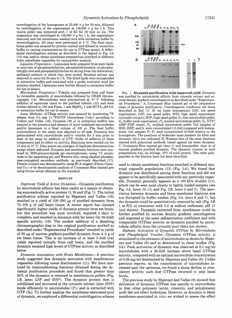
Improved Yield of Active Dynamin-Dynamin purification by microtubule affinity has been useful as a means of obtain- ing enzymatically active protein (1,9) and (Fig. 1). The initial purification scheme reported by Shpetner and Vallee (18) resulted in a yield of 100-200 pg of purified dynamin from 75-100 g of calf brain tissue. A recent report has claimed significantly higher yields of dynamin protein from rat brain, but this procedure was more involved, required 2 days to complete, and resulted in dynamin with far lower (by 30-fold) specific activity (19). The modest addition of a desalting chromatographic step to the original purification method as described under "Experimental Procedures" resulted in yields of 50 pg of sucrose gradient-purified dynamin from 4-5 g of rat brain tissue. This is an increase of at least 5-fold over yields reported initially from calf brain, and the purified dynamin retained high levels of GTPase activity as described below.
Dynamin Association with Brain Membranes-A previous study suggested that dynamin associates with membranous organelles following tissue fractionation (11). We have ana- lyzed by immunoblotting fractions obtained during the dy- namin purification procedure and found that greater than 60% of the dynamin is retained in membranous pellets (Fig. lB, lanes LSP and HSP). The dynamin protein that is solubilized and recovered in the cytosolic extract (lane HSS) binds efficiently to microtubules (PI) and is extracted with GTP (Ss). To further analyze the membrane-associated pool of dynamin, we employed a differential centrifugation scheme
200- *
116- 97- -dyn 66- '
"r .IC
FIG. 1. Dynamin purification with improved yield. Dynamin was purified by microtubule affinity from cytosolic extract and su- crose density gradient centrifugation as described under "Experimen- tal Procedures." A, Coomassie Blue stained gel of the preparative steps of dynamin purification. Centrifugation conditions are those described in Ref. 17. H, rat brain homogenate; LSS, low speed supernatant; LSP, low speed pellet; HSS, high speed supernatant (cytosolic extract); HSP, high speed pellet; SI, first microtubule pellet; Sa, buffer wash supernatant; Pa, washed microtubule pellet; 5'3, GTP/ AMP-PNP eluate; P3, residual microtubule pellet. Gel samples of HSS, HSP, and SI were concentrated 1.5-fold compared with homog- enate. Gel samples P1-P3 were concentrated 10-fold relative to the homogenate. The positions of molecular mass markers (in kDa) and dynamin (dyn) are indicated. €3, Western blot of the same fractions reacted with polyclonal antibody raised against rat brain dynamin. C, Coomassie Blue stained gel ( l a n e 1 ) and immunoblot ( l a n e 2) of sucrose gradient-purified dynamin. The dynamin content in such preparations was, on average, 70% of total protein. The other poly- peptides in the fraction have not been identified.
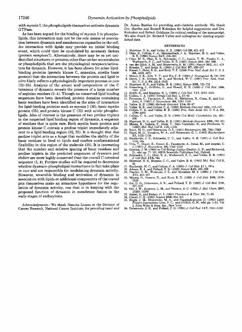
used to obtain membrane fractions enriched in different neu- ronal organelle populations (13) (Fig. 2A). We found that dynamin was distributed among these fractions and did not appear to be specifically associated with any particular organ- elle. Dynamin generally appears as a 100-kDa doublet ( l l ) , which can be seen most clearly in lightly loaded samples (see Fig. 2A, lanes 10-13, and Fig. 2B, lanes 4 and 5). The asso- ciation between dynamin and these membrane fractions was not disrupted by buffer washes (Fig. 2A, lanes 6 and 7), but the dynamin could be quantitatively removed by salt (Fig. 2B 1 M KI) or treatment with 0.2 M sodium carbonate, pH 11 (not shown). Dynamin extracted from membranes could be further purified by sucrose density gradient centrifugation and migrated at the same sedimentation coefficient and with comparable GTPase activity as dynamin purified by micro- tubule affinity from the cytosolic pool (data not shown).
Biphasic Actiuation of Dynamin GTPase by Microtubules and Phospholipid Vesicles-Dynamin GTPase activity is stimulated in the presence of microtubules as shown by Shpet- ner and Vallee (9) and as determined in these studies (Fig. 3A). Peak activation of dynamin was observed at 0.1 mg/ml microtubules with a 26-fold increase above basal GTPase activity, compared with an optimal microtubule concentration of 0.08 mg/ml determined by Shpetner and Vallee (9). Unlike previous reports, as the concentration of microtubules in- creased past the optimum, we found a sharp decline in stim- ulatory activity such that GTPase returned to near basal levels.
The previous study by Shpetner and Vallee (9) showed that activation of dynamin GTPase was specific to microtubules in that other polymers (actin, vimentin, and polyglutamic acid) did not affect hydrolysis. However, as dynamin is likely membrane-associated in vivo, we wished to assess the effect
17242 Dynamin Activation by Phospholipids
of membrane components on dynamin GTPase activity. Fixed concentrations of dynamin were preincubated with
synthetic small unilamellar phospholipid vesicles of varying composition and concentration. These lipid preparations con- tained a homogeneous population of liposomes as viewed by darkfield microscopy (data not shown). In Fig. 3B, a fixed
A 1 2 3 4 5 6 7 8 9 l O 1 1 1213
1 2 3 4 5 6 7 8 9
200- 116-
66- 97-
45-
FIG. 2. Dynamin is a peripheral membrane protein associ- ated with multiple organelle populations. A , Western blot of fractionated rat brain tissue reacted with dynamin polyclonal anti- body. Lane 1, homogenate; lane 2, low speed supernatant; lane 3, nuclear pellet; lane 4, medium speed supernatant; lane 5, synaptoso- mal pellet; lane 6, supernatant from buffer wash of synaptosomes; lane 7, washed synaptosomal pellet; lane 8, cytosol; lane 9, microsomal pellet; lane 10, supernatant following lysis and sedimentation of pellet 5; lane 11, pellet of lysed synaptosomal membranes; lane 12, residual supernatant following sedimentation of supernatant 10; lane 13, syn- aptic vesicle pellet. All samples were resuspended to starting homog- enate volume except for pellets 11 and 13, which were in V3 volume. B, Coomassie Blue stained gel and Western blots of high speed membrane pellet (see “Experimental Procedures”). Lanes 1,4, and 7, unextracted membrane starting material; lanes 2, 5, and 8 KI extract (supernatant); lanes 3,6, and 9, salt-extracted membrane pellet. Lanes 1-3, Coomassie Blue stain for total protein; lanes 4-6, Western blot using anti-dynamin antibody; lanes 7-9, Western blot using anti- clathrin heavy chain antibody.
FIG. 3. Biphasic activation of dy- namin GTP- by microtubules and phospholipid vesicles. Activation of dynamin GTPase by varying concentra- tions of taxol-stabilized pure tubulin mi- crotubules ( A ) or 0.1 mg/ml phospho- lipid vesicles with varying ratios of phos- phatidylserine to phosphatidylcholine ( B ) . Dynamin concentration was 5.8 pg/ ml.
concentration of total lipid was used (0.1 mg/ml), and the ratio of phosphatidylserine to phosphatidylcholine was varied. At 0-5% phosphatidylserine, no activation of GTPase activity was observed. As the ratio of acidic to neutral phospholipid was increased, the activity levels of dynamin also increased, reached a peak at 25% phosphatidylserine, and then returned to basal levels at higher ratios. Experiments designed to cover in more detail the range between 10-30% phosphatidylserine showed a smooth activation profile with reproducible optima a t 25% acidic phospholipid. Similar activation profiles were observed when liposomes of phosphatidylserine alone were used instead of lipid mixtures and when dynamin activity was stimulated 5-fold at the optimal concentration of pure phos- phatidylserine (Table I). This concentration of phosphatidyl- serine (10 pg) is lower than that that gave rise to optimal activation in mixtures with phosphatidylcholine (25 pg/ml). These data indicate that the distribution of negative charges within the lipid vesicles may be important for optimal inter- action with dynamin (20) or may reflect a preference of phosphatidylserine for the inner leaflet of the membrane bilayer (21). Though the percent increase over basal activity was less by using phospholipid than that shown in Fig. 3A and by the studies of others (1,9, 19) for microtubule activa- tion, the difference lies in the somewhat variable basal activity of dynamin GTPase as the activated levels reached with either microtubules or lipids are comparable (see Table I).
We examined the effects of other phospholipids on dynamin GTPase activity to determine if the stimulation was specific for phosphatidylserine or was a more general effect of phos- pholipids. Increasing concentrations of phosphatidylcholine (0.025-0.5 mg/ml) had no effect on dynamin GTPase activity. Addition of liposomes containing varying ratios of phospha- tidylglycerol or phosphatidylinositol to phosphatidylcholine gave rise to activation profiles similar to those observed for phosphatidylserine-containing liposomes (data not shown). Optimal activation of dynamin occurred at 25% phosphati-
8 400 (I)
200
0 I
0 0 . 2 0 . 4 0.6 0.8 1
[Microtubule1 (mg/ml)
B
700
300
- 200
\
: E
loo 0 t 0 20 4 0 60 so 100
Phosphatidylserine Content (%)
Dynamin Activation by Phospholipids 17243
dylglycerol or phosphatidylinositol, representing 5.5- and 7.4- fold activation, respectively (Table I). The similarity in stim- ulation of dynamin by different acidic phospholipids suggests that activation resulted from ionic interactions with the phos- pholipid head group and was not specific for a particular lipid species. However, phosphatidylinositol reproducibly led to greater activity levels than the other lipids tested. Whether this is due to differences in charge distribution in the liposome or is specific to the head group itself has not been determined. The effect of phosphatidylinositol phosphates on dynamin activity has not yet been tested.
Activation by Microtubules and Phospholipids Occurs through a Similar Mechanism-To determine whether the activation of dynamin GTPase by microtubules and acidic phospholipids occurred through a similar mechanism, the effects of these agents in combination were analyzed. In the presence of an optimally activating concentration of micro- tubules, the addition of pure phosphatidylcholine or low ratios of phosphatidylserine to phosphatidylcholine had little effect on GTP hydrolysis (Fig. 4). However, as higher concentra- tions of phosphatidylserine were added along with microtu- bules, inhibition of activity was seen, in keeping with the decrease in GTP hydrolysis observed upon increases in either activator alone. Similar results were obtained upon addition
TABLE I Effect of phospholipids on dynamin GPTase activity
Liposomes containing pure phosphatidylcholine or phosphatidyl- serine, or mixtures of acidic phospholipids with phosphatidylcholine were prepared as described under “Experimental Procedures.” Except for pure phosphatidylserine, lipids were used at a final concentration of 0.1 mg/ml. Values were obtained from individual experiments examining dynamin GTPase activity with increasing acidic lipid content, such as that shown in Fig. 3B, and the basal activities varied somewhat between experiments. The maximally stimulated GTPase activity of dynamin ranged from 747 to 1988 nmol/min/mg (1.2-3.3 s-’) in the presence of acidic phospholipids, compared with from 932 to 1957 nmol/min/mg (1.7-3.3 s-’) in the presence of microtubules.
Additions Maximal Increase activity basal
over
nmol f min f mg -fold 10 pg/ml phosphatidylserine 1063 5.0 25% phosphatidylinositol 1998 7.4 25% phosphatidylglycerol 1473 5.5 100% phosphatidylcholine 197 1.0
of increasing concentrations of microtubules to dynamin ac- tivated by phospholipid (data not shown).
To determine whether ionic interactions between dynamin and microtubules or dynamin and acidic phospholipids were important for activation, the effects of increased ionic strength in the assay buffer were investigated (Fig. 5A). The stimulatory effect of either phospholipid or microtubules was inhibited almost completely at 100 mM salt. Activation of dynamin GTPase by either phospholipids or microtubules was also affected by changes in pH, with a sharp decline in activity to near basal levels above pH 7.2 (data not shown). Basal GTPase activity was much less sensitive to changes in pH between 6.5 and 8.0. Polyglutamic acid had been shown previously to have little effect on dynamin GTPase activity (9). However, as shown in Fig. 5 B , polyglutamic acid inhibited the activation induced by either microtubules or phospholipid, though phospholipid activation was more sensitive to this compound. In addition, GTPase levels showed a reproducible increase of 1.5-fold in the presence of 0.3 mg/ml polygluta- mate alone (not shown). A polyamine, spermine, was also found to inhibit activation induced by microtubules or phos- pholipid at concentrations greater than 2 mM (not shown).
It has been suggested that activation of dynamin by micro- tubules occurs via an interaction of the basic C-terminal domain of dynamin with the acidic C termini of tubulin subunits (9). The results described above indicate that this basic domain of dynamin may be interacting similarly with acidic phospholipids. To test this more directly, we took advantage of the observation that some dynamin preparations were partially proteolyzed during the purification procedure, resulting in the production of an 80-kDa fragment that could be purified by sucrose density gradient centrifugation and that migrated with approximately the same sedimentation coefficient as intact dynamin (Fig. 6). The 80-kDa fragment retained basal GTPase activity (Table 11), indicating that the proteolytic cleavage must occur toward the C terminus of the molecule, as cleavage of 20-kDa from the N terminus would remove two of the three required binding domains for GTP (2, 22). However, the GTPase of the cleaved molecule could not be activated to the same extent as intact dynamin by either microtubules or phospholipid (Table 11). These results are comparable with those obtained by deliberate papain cleavage of the molecule, which resulted in the production of a 90-kDa dynamin fragment exhibiting basal, but not micro-
4 2 5 0 0 aJ rl 400
- -
--
a ‘ 8 3 0 0 -- E-(- ” 200 --
100 -.
0 - I
0 2 0 40 6 0 8 0 100
Phosphatidylserine Content (%) FIG. 4. Addition of both microtubules and phospholipids does not lead to further activation of dynamin GTPase. Phospholipid
vesicles (0.1 mg/ml) of varying ratios of phosphatidylserine to phosphatidylcholine were added to dynamin that had been preincubated with microtubules at 0.1 mg/ml.
17244 Dynamin Activation by Phospholipids
A 100
U sr r‘ 80
P Q 6o
-I JJ 70
a d 50 b 0 4 0 U 9 30 U h 20 PI
10
0 0 50 100 150 200 250 300
[salt] (mn)
B
FIG. 5. Ionic interactions between dynamin and microtubules or phos- pholipids are important for activa- tion. Effect of salt concentration (A) and effect of polyglutamic acid ( B ) on dynamin GTPase activity are shown. GTPase activity was assayed as de- scribed under “Experimental Proce- dures” in the presence of phospholipid (0) or microtubules (O), with additions as indicated. Salt concentration reflects the total contribution of buffer ions and added NaCl.
200-
116- 97- 66-
45-
100
sr r‘ 80
9) 6o
U
-I $ 70 4
PI d 50 b c) 40 U E 30 U l.4 20 a
10
0
-80kDa
FIG. 6. Purification of the 80-kDa fragment of dynamin. Partially proteolyzed dynamin was purified by using sucrose gradient centrifugation as described under “Experimental Procedures.” Pre- parative fractions as shown in Fig. 1A (SI to P3) and sucrose density gradient fractions 11-17 were analyzed by SDS-polyacrylamide gel electrophoresis.
tubule-activated, GTPase (22). The slight activation found for the 80-kDa dynamin fragment (Table 11) was likely due to residual amounts of the intact molecule in the preparation, determined in this experiment to be 18% of total dynamin protein.
Endogenous Brain Vesicles Activate Dynamin GTPase-As the next step in the analysis of activation of dynamin GTP hydrolysis by membrane components, we examined the effects of endogenous brain organelles on activity. Dynamin is pres- ent in a membrane fraction sedimenting a t 138,000 x g (Fig. 1, A and B ) that contains microsomes and plasma membrane- derived vesicles, including coated vesicles (23). When ana- lyzed on blots using equal protein loading (not shown), this fraction (HSP) contained dynamin comparable in amount
TABLE I1 Activation of intact and truncated dynamin by phospholipids and
microtubules Sucrose gradient-purified dynamin or 80-kDa dynamin fragment
was assayed for GTPase activity as described under “Experimental Procedures” and for Table I. Values in parentheses represent -fold increase over basal activity.
with that found in a low speed pellet (see Fig. 1B). The high speed pellet, salt-extracted to remove bound dynamin, clath- rin, and other peripheral proteins (Fig. 2B), activated dyna- min GTPase in a similar manner to that observed for both microtubules and acidic phospholipids (Fig. 7). At low con- centrations of membrane protein, dynamin activity was stim- ulated 2.5-fold. However, as the concentration was increased, dynamin activity dropped back to basal levels. Addition of the salt-stripped membranes to microtubule-stimulated dy- namin resulted in inhibition of GTPase activity, similar to the results described above (Fig. 4). The final level of activa- tion induced by endogenous vesicles alone was lower than
Dynamin Activation by Phospholipids 17245
I
0 10 20 30 40 50
[Vesicle] (ug protein/ml) FIG. 7. Endogenous brain vesicles activate dynamin GTPase. Dynamin GTPase was assayed with increasing concentrations of KI-
extracted vesicles prepared as described under “Experimental Procedures.”
that observed with microtubules or synthetic liposomes. How- ever, peak activation occurred at very low vesicle content. If we assume that the salt-stripped vesicles contain an equal mass of protein and lipid and that the acidic phospholipid content is 15% (24), peak activation occurred at -0.4 pg/ml acidic phospholipid, a 60-fold lower concentration than that seen with synthetic liposomes containing phosphatidylserine and phosphatidylcholine. Further studies will be required to address the possibility that integral protein components of the membrane are also involved in regulation of dynamin GTPase. Preliminary data show that protease-digested mem- branes still activate dynamin to a similar extent as that shown in Fig. 7, but higher concentrations of these membranes are required (data not shown).
DISCUSSION
Aside from the ability of dynamin to coassemble with microtubules, there are no data to suggest that there is a physiologically important interaction between these species in vivo. Dynamin does not localize to microtubules in cells but appears to be membrane-associated (11, 25, 26). The ability of dynamin GTPase to be activated by microtubules in vitro suggests an involvement of these cytoskeletal ele- ments in a regulatory process. However, the genetic evidence for a role for dynamin in coated vesicle formation is incon- sistent with the idea that this protein must interact with microtubules to be functional (reviewed in Ref. 12).
The data presented here show that acidic phospholipids stimulate dynamin GTPase activity severalfold, up to 1988 nmol/min/mg (3.3 s”), an activity comparable with that shown previously for microtubule-stimulated dynamin (9). Experiments examining the sensitivity of activation to salt, pH, polyanions, and cations indicate that activation likely occurs through ionic interactions between dynamin and mi- crotubules or phospholipids. The site on the dynamin mole- cule responsible for the interaction with either activator is in the C-terminal domain, based on the inability of microtubules or phospholipids to activate the 80-kDa truncated form of the enzyme, though basal activity remains unaffected. The C terminus of dynamin is highly basic (2-4), and interactions between this domain and acidic groups of the phospholipids or microtubules are likely involved in the activation process (9). The similarity between effects induced by microtubules and phospholipids indicates that the mechanism of activation
is similar if not identical. In support of this hypothesis, the effects of the two agents on GTPase activity are not additive.
The dynamin molecule likely exists as a homo-oligomer of between two and five subunits, based on the sedimentation coefficient of 10 S calculated from these studies (27), gel permeation chromatography’ (19), and cross-linking studies.’ There may therefore exist at least two microtubule/phospho- lipid binding sites on each dynamin oligomer. The sedimen- tation value of the truncated form of dynamin is not signifi- cantly different from that of the intact molecule, suggesting that the 80-kDa species is able to maintain interactions be- tween subunits. The 80-kDa cleavage product persists (though not efficiently) throughout the microtubule assembly proce- dure, suggesting either that there may be a second microtubule binding domain with weaker affinity in this region of the molecule or that interactions between the 80-kDa fragment and residual intact dynamin mediate the microtubule associ- ation (22). However, the residual intact protein is unable to mediate microtubule or phospholipid activation of the trun- cated form.
Simply neutralizing the basic charge in the C terminus of dynamin is not sufficient for activation of the GTPase. The requirement for a solid phase substrate upon which binding and activation take place (tubulin polymer or liposome) sug- gests that cooperative interactions between dynamin mole- cules and/or cross-linking between adjacent structures (1) may be involved in the activation process, giving rise to the sharp concentration dependence of activation observed. We calculate the stoichiometry of dynamin monomer to micro- tubules under optimal activation conditions to be 1:20, which is far lower than the maximal binding stoichiometry of 1:l obtained by Maeda et al. (19). Future studies will focus on the analysis of the binding properties of dynamin to microtubules and liposomes in order to understand the basis for the biphasic nature of activation.
Lipid activation of dynamin is reminiscent of the effects of acidic phospholipids on myosin I activity. Myosin I binds acidic phospholipid vesicles as well as endogenous membrane preparations (28-30), and this interaction modulates ATP hydrolysis by interfering with actin binding (31). According to our model for dynamin activation, acidic phospholipids interact with the same site as microtubules. Rather than simply inhibiting microtubule activation, however, in analogy
* P. Tuma, unpublished results.
17246 Dynamin Activation by Phospholipids
with myosin I, the phospholipids themselves activate dynamin GTPase.
As has been argued for the binding of myosin I to phospho- lipids, this interaction may not be the sole means of associa- tion between dynamin and membranous organelles in the cell. An interaction with lipids may provide an initial binding event, which could then be modulated by accessory factors (protein receptors?). Alternatively, there may be as yet uni- dentified structures or proteins other than either microtubules or phospholipids that are the physiological receptors/activa- tors for dynamin. However, it has been shown for other lipid- binding proteins (protein kinase C, annexins, myelin basic protein) that the interaction between the protein and lipid in vitro likely reflects a physiologically important process in vivo (32-35). Analysis of the amino acid composition of the C terminus of dynamin reveals the presence of a large number of arginine residues (2-4). Though no conserved lipid binding sequences have been described, protein domains containing basic residues have been identified as the sites of interaction for lipid-binding proteins such as myosin I (36), basic myelin protein (35), and protein kinase C (32) with acidic phospho- lipids. Also of interest is the presence of two proline triplets in the suspected lipid binding region of dynamin, a sequence of residues that is quite rare. Both myelin basic protein and protein kinase C contain a proline triplet immediately adja- cent to a lipid binding region (32, 35). It is thought that this proline triplet acts as a hinge that modifies the ability of the basic residues to bind to lipids and confers conformational flexibility in this region of the molecule (35). It is interesting that the number and relative spacing of basic residues and proline triplets in the predicted sequences of dynamin and shibire are more highly conserved than the overall C-terminal sequence (3, 4). Further studies will be required to determine whether dynamin-phospholipid interactions in fact take place in vivo and are responsible for modulating dynamin activity. However, reversible binding and activation of dynamin in association with lipids or additional components of the coated pits themselves make an attractive hypothesis for the regu- lation of dynamin activity, one that is in keeping with the proposed function of dynamin in membrane fusion in the early stages of endocytosis.
Acknowledgments-We thank Nancita Lomax at the Division of Cancer Research, National Cancer Institute, for providing taxol and
Dr. James Bartles for providing anti-clathrin antibody. We thank Drs. Bartles and Russell Kohnken for helpful suggestions and Drs. Kohnken and Robert Goldman for critical reading of the manuscript. We also thank Dr. Richard Vallee and colleagues for sharing unpub- lished data.
REFERENCES
2. Obar. R.. Collins. C. A.. Hammarback. J. A.. Shoetner. H. S.. and Vallee. 1. Shpetner, H. S., and Vallee, R. B. (1989) Cell 59,421-432
I , . I
R. B . (1990) Nature 347,256-261
Wadsworth. S. C.. and Vallee. R. B. (1991) Nature 351,583-586 3. Chen, M. S., Obar, R. A,, Schroeder, C. C., Austin, T. W., Poodry, C. A.,
4. van der Bliek,'A. M:, and Meyerowitz, E. M. (1991) Nature 351,411-414 5. Kosaka, T., and Ikeda, K. (1983) J. Cell Bid. 97,499-507 6. Kessel, I., Hoist, B. D., and Roth, T. F. (1989) Pmc. Natl. Acad. Sei. U. S. A.
8. 7.
10. 9.
11. 12. 13.
14. 15. 16.
17.
Masur, S. K., Kim, Y.-T., and Wu, C.-F. (1990) J. Neurogenet. 6,191-206 Dever, T. E., Glynias, M. J., and Merrick, W. C. (1987) Proc. Natl. Acad.
Shpetner, H. S., and Vallee, R. B. (1992) Nature 355 , 733-735 Gruenberg, J., Griffths, G., and Howell, K. E. (1989) J. Cell Biol. 108 ,
Scaife, R., and Margolis, R. L. (1990) J. Cell Biol. 11 1,3023-3033 Collins, C. A. (1991) Trends Cell Bid. 1,57-60 Ferrarese, C., Vaccarino, F., Alho, H., Mellstrom, B., Costa, E., and Gui-
Vallee, R. B. (1986) Methods Enzymol. 134,89-97 dotti, A. (1987) J. Neurochem. 48, 1093-1102
Vallee, R. B., and Collins, C. A. (1986) Methods Enzymol. 134,116-127 Collins, C. A,, and Vallee, R. B. (1986) Proc. Natl. Acad. Sci. U. S. A. 8 3 ,
Collins, C. A,, and Vallee, R. B. (1989) Cell Motil. Cytoskeleton 1 4 , 491-
86, 4968-4972
Sci. U. S. A. 8 4 , 1814-1818
1301-1316
4799-4803
6nn
18. ShYpYeutner, H. S., and Vallee, R. B. (1991) Methods Enzymol. 196 , 192-201 19. Maeda, K., Nakata, T., Noda, Y., Sato-Yoshitake, R., and Hirokawa, N.
20. Bazzi, M. D., and Nelsestuen, G. L. (1991) Biochemistry 30 , 7961-7969 21. Bazzi, M. D., Youakim, M. A,, and Nelsestuen, G. L. (1992) Biochemistry
22. Herskovits, J. S., Schroeder, C. C., and Vallee, R. B. (1991) J. Cell Bid.
23. Uriu, T., Omori, K., Omori, K., Yamamoto, A,, Inoue, M., and Inagaki, C.
24. Graham, J. M. (1992) in Cell Biology Labfax (Dealtry, G. B., and Rickwood,
25. Schroeder, 8; C. ,- Obar, R. A., Wadsworth, S. C., and Vallee, R. B. (1991)
26. Shpetner, H. S., Burgess, C. C., and Vallee, R. B. (1992) Mol. Biol. Cell 3 ,
(1992) Mol. Biol. Cell 3 , 1181-1194
31,1125-1134
115 ,34a
J. (1991) J. Neurochem. 5 6 , 1548-1556
D., eds) p 77 101, BIOS Scientific Publishers Ltd., Oxford
J. Cell Biol. 115 ,34a
27. Stachniak, M. C., and Collins, C. A. (1990) J. Cell Bid. 1 1 1 , 291a 28. Adams, R. J., and Pollard, T. D. (1989) Nature 340,565-568 29. Hayden, S. M., Wolenski, J. S., and Mooseker, M. S. (1990) J. cell Biol.
4a
30. Miyata, H., Bowers, B., and Korn, E. D. (1989) J. Cell Bid. 109 , 1519-
31. Zot, H. G., Doberstein, S. K., and Pollard, T. D. (1992) J. Cell Biol. 116,
32. Orr. J. W., Keranen. L. M., and Newton, A. C. (1992) J. Bid. Chem. 267 ,
i 11,443-451
1528
367-376
33. Stabel, S., and Parker, P. J. (1991) Pharmacol. & Ther. 5 1 , 71-95 34. Creutz, C. E. (1992) Science 258,924-931 35. Boggs, J. M., Moscarello, M. A., and Papahadjo oulos, D. (1982) Li id
Protecn Interactcons (Jost, P. C., and Griffth, 8. H., eds) pp. 1-51, $0, 2, John Wiley & Sons, Inc., New York
36. Doberstein, S. K., and Pollard, T. D. (1992) J. Cell Biol. 117,1241-1249