14
The Lymphatic System Chapter 20
| Date post: | 18-Dec-2015 |
| Category: |
Documents |
| Upload: | virginia-butler |
| View: | 234 times |
| Download: | 0 times |
The Lymphatic SystemChapter 20
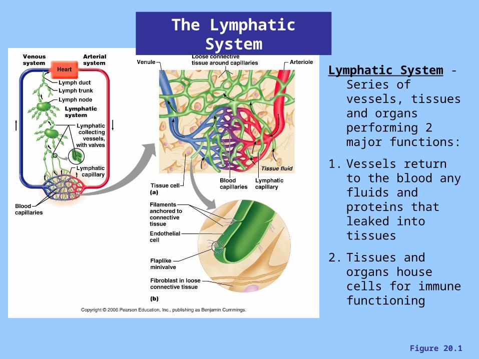
Figure 20.1
The Lymphatic System
Lymphatic System - Series of vessels, tissues and organs performing 2 major functions:
1. Vessels return to the blood any fluids and proteins that leaked into tissues
2. Tissues and organs house cells for immune functioning
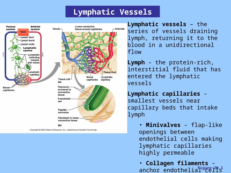
Figure 20.1
Lymphatic Vessels
Lymphatic vessels – the series of vessels draining lymph, returning it to the blood in a unidirectional flow
Lymph – the protein-rich, interstitial fluid that has entered the lymphatic vessels
Lymphatic capillaries – smallest vessels near capillary beds that intake lymph
• Minivalves – flap-like openings between endothelial cells making lymphatic capillaries highly permeable
• Collagen filaments – anchor endothelial cells to surrounding connective tissue, encourages minivalve opening
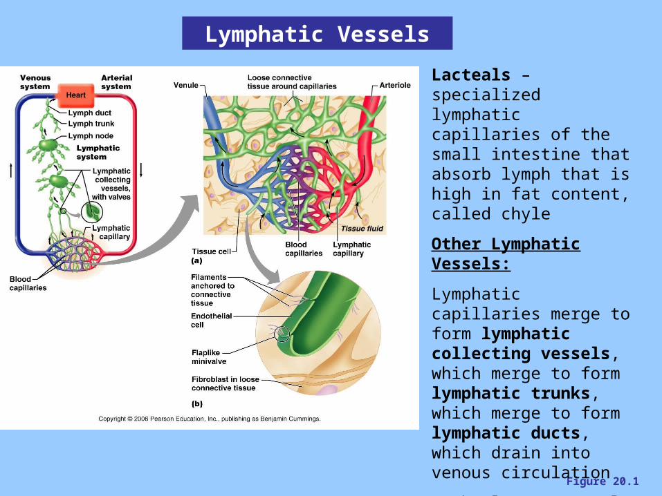
Figure 20.1
Lacteals – specialized lymphatic capillaries of the small intestine that absorb lymph that is high in fat content, called chyle
Other Lymphatic Vessels:
Lymphatic capillaries merge to form lymphatic collecting vessels, which merge to form lymphatic trunks, which merge to form lymphatic ducts, which drain into venous circulation
• The larger vessels have tunics and valves similar to veins
Lymphatic Vessels
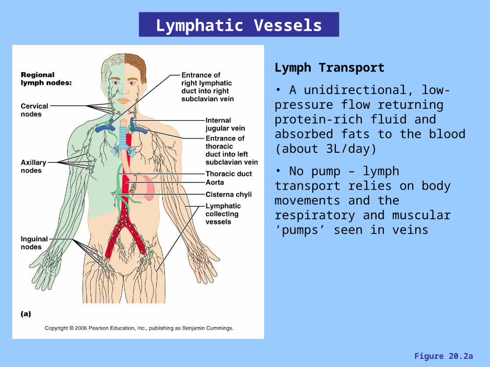
Figure 20.2a
Lymphatic Vessels
Lymph Transport
• A unidirectional, low-pressure flow returning protein-rich fluid and absorbed fats to the blood (about 3L/day)
• No pump – lymph transport relies on body movements and the respiratory and muscular ‘pumps’ seen in veins
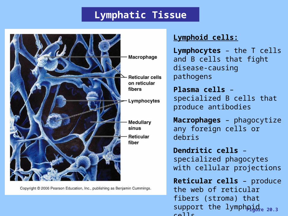
Figure 20.3
Lymphatic Tissue
Lymphoid cells:
Lymphocytes – the T cells and B cells that fight disease-causing pathogens
Plasma cells – specialized B cells that produce antibodies
Macrophages – phagocytize any foreign cells or debris
Dendritic cells – specialized phagocytes with cellular projections
Reticular cells – produce the web of reticular fibers (stroma) that support the lymphoid cells
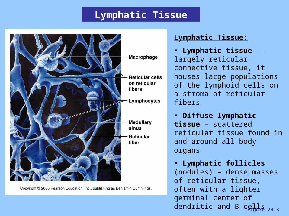
Figure 20.3
Lymphatic Tissue
Lymphatic Tissue:
• Lymphatic tissue - largely reticular connective tissue, it houses large populations of the lymphoid cells on a stroma of reticular fibers
• Diffuse lymphatic tissue – scattered reticular tissue found in and around all body organs
• Lymphatic follicles (nodules) – dense masses of reticular tissue, often with a lighter germinal center of dendritic and B cells
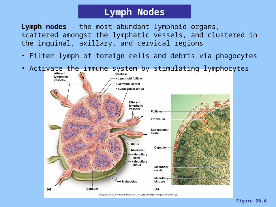
Figure 20.4
Lymph Nodes
Lymph nodes – the most abundant lymphoid organs, scattered amongst the lymphatic vessels, and clustered in the inguinal, axillary, and cervical regions
• Filter lymph of foreign cells and debris via phagocytes
• Activate the immune system by stimulating lymphocytes
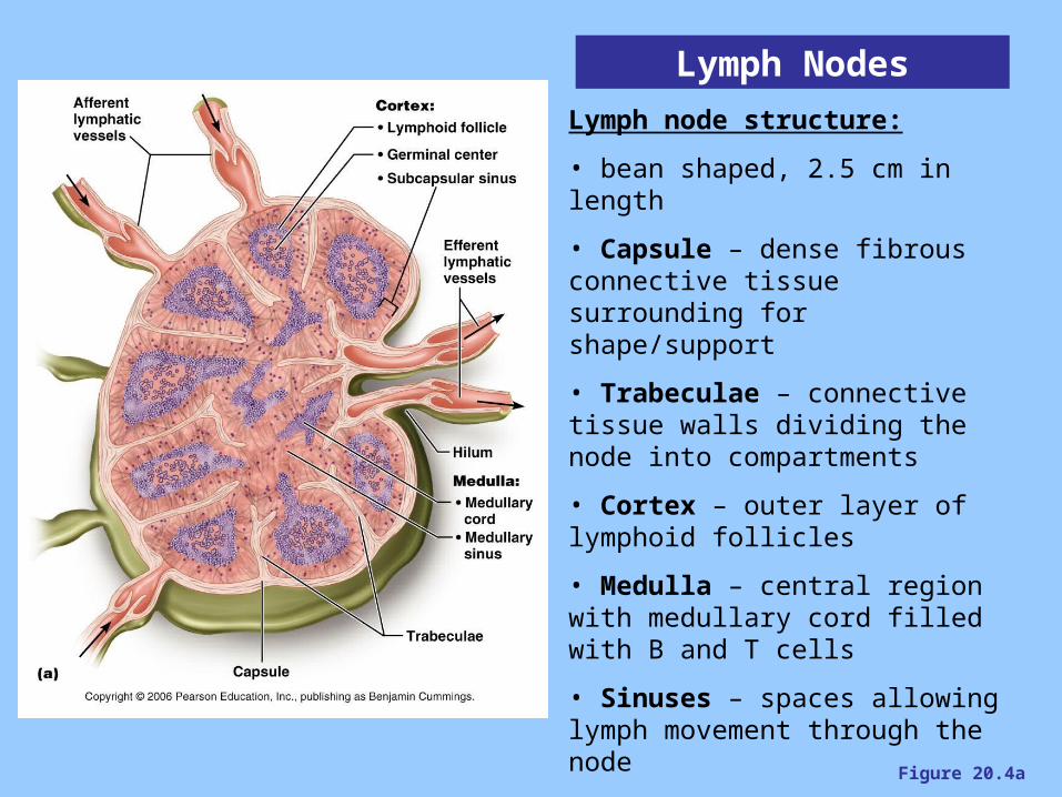
Figure 20.4a
Lymph Nodes
Lymph node structure:
• bean shaped, 2.5 cm in length
• Capsule – dense fibrous connective tissue surrounding for shape/support
• Trabeculae – connective tissue walls dividing the node into compartments
• Cortex – outer layer of lymphoid follicles
• Medulla – central region with medullary cord filled with B and T cells
• Sinuses – spaces allowing lymph movement through the node
• Afferent vessels – bring lymph into the node
• Efferent vessels – drain lymph from the node
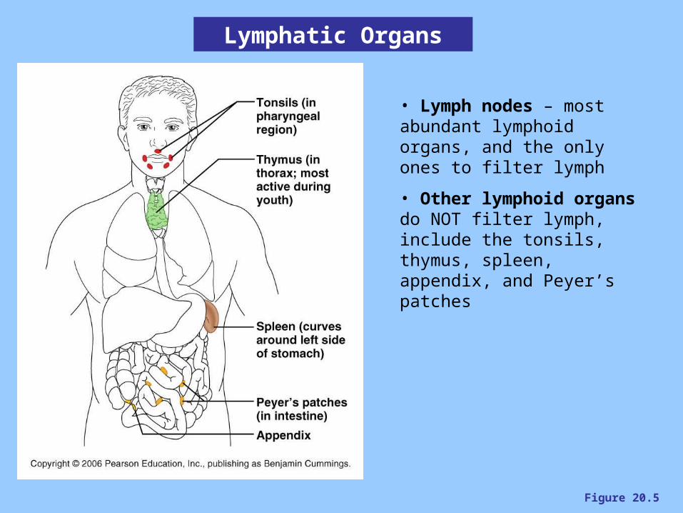
Figure 20.5
Lymphatic Organs
• Lymph nodes – most abundant lymphoid organs, and the only ones to filter lymph
• Other lymphoid organs do NOT filter lymph, include the tonsils, thymus, spleen, appendix, and Peyer’s patches
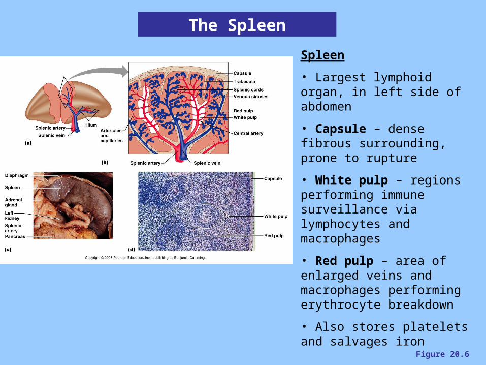
Figure 20.6
The Spleen
Spleen
• Largest lymphoid organ, in left side of abdomen
• Capsule – dense fibrous surrounding, prone to rupture
• White pulp – regions performing immune surveillance via lymphocytes and macrophages
• Red pulp – area of enlarged veins and macrophages performing erythrocyte breakdown
• Also stores platelets and salvages iron
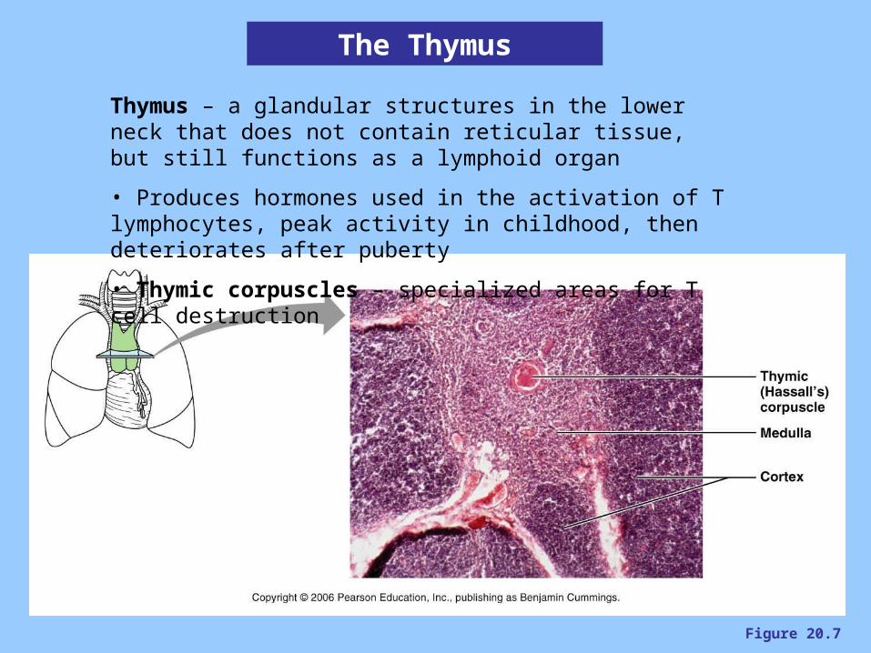
Figure 20.7
The Thymus
Thymus – a glandular structures in the lower neck that does not contain reticular tissue, but still functions as a lymphoid organ
• Produces hormones used in the activation of T lymphocytes, peak activity in childhood, then deteriorates after puberty
• Thymic corpuscles – specialized areas for T cell destruction
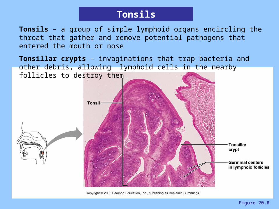
Figure 20.8
Tonsils
Tonsils – a group of simple lymphoid organs encircling the throat that gather and remove potential pathogens that entered the mouth or nose
Tonsillar crypts – invaginations that trap bacteria and other debris, allowing lymphoid cells in the nearby follicles to destroy them
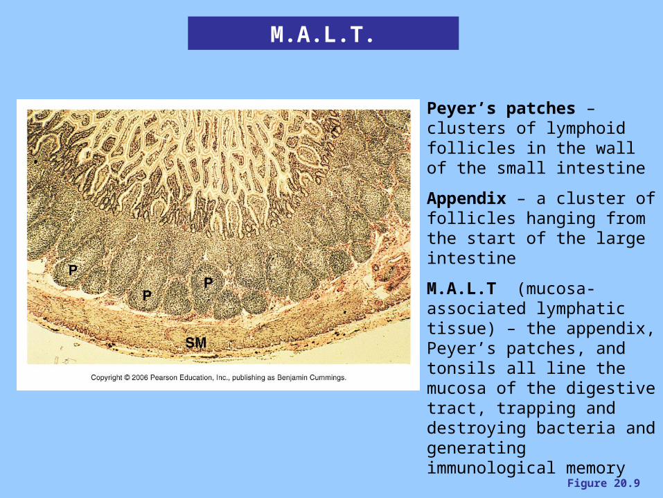
Figure 20.9
M.A.L.T.
Peyer’s patches – clusters of lymphoid follicles in the wall of the small intestine
Appendix – a cluster of follicles hanging from the start of the large intestine
M.A.L.T (mucosa-associated lymphatic tissue) – the appendix, Peyer’s patches, and tonsils all line the mucosa of the digestive tract, trapping and destroying bacteria and generating immunological memory


