136
The Muscular System
| Date post: | 18-Dec-2015 |
| Category: |
Documents |
| Upload: | christina-curtis |
| View: | 227 times |
| Download: | 3 times |

The Muscular System

Muscle Attachment Sites
Muscle exerts force on tendons, which in turn pull on bones or other structures.
When a muscle contracts, it pulls one of the articulating bones toward the other.

Muscle Attachment Sites: Origin & Insertion
Origin – the site of a muscle’s attachment to the more stationary bone.
Insertion – the site of a muscle’s attachment to the more movable bone.
Often, the origin is proximal to the insertion. Belly – the fleshy part of the muscle between
the tendons of the origin and insertion.

Tenosynovitis
Commonly known as tendinitis. Painful inflammation of the tendons, tendon
sheaths, and synovial membranes of the joints.
Trauma, strain, excessive exercise, chronic, repetitive motions can all cause tenosynovitis.
Lever Systems & Leverage
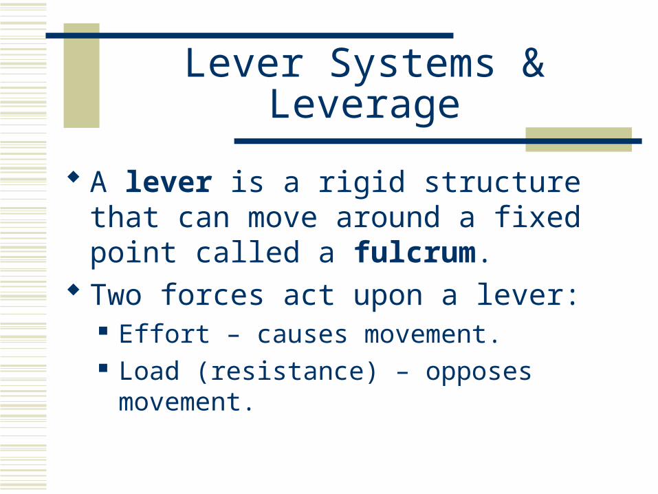
A lever is a rigid structure that can move around a fixed point called a fulcrum.
Two forces act upon a lever: Effort – causes movement. Load (resistance) – opposes movement.

Lever Systems & Leverage
Levers produce trade-offs between effort and the speed and range of motion.
Mechanical advantage (leverage) – a smaller effort can move a heavier load – the effort must move a greater distance.
Mechanical disadvantage – a larger effort moves a lighter load – the effort must move a shorter distance and slower than the load.


Types of Levers
First-class levers. The fulcrum is between the effort and the load. Scissors and seesaws are examples. It can produce either a mechanical advantage or disadvantage
depending upon whether the effort or load is placed closer to the fulcrum.
If an effort is placed farther from the fulcrum than the load, a heavy load can be moved, but not fast or far.
If an effort is placed closer to the fulcrum than the load, only a lighter load can be moved, but it moves far and fast.
Raising the head at the atlanto-occipital joint.
Types of Levers
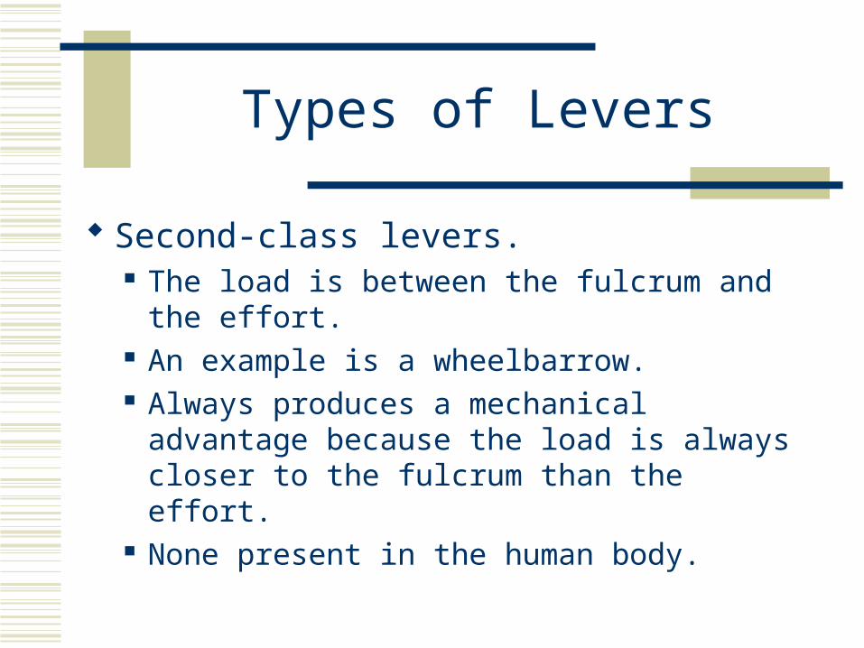
Second-class levers. The load is between the fulcrum and the effort. An example is a wheelbarrow. Always produces a mechanical advantage
because the load is always closer to the fulcrum than the effort.
None present in the human body.

Types of Levers
Third-class levers. The effort is between the fulcrum and the load. An example is a forceps. Always produces a mechanical disadvantage
because the effort is always closer to the fulcrum than the load.
This arrangement favors speed and range of motion over force.


Effects of Fascicle Arrangement
Within a fascicle, all muscles are parallel to one another.
The fascicles may form patterns with respect to the tendon: Parallel. Fusiform (cigar shaped). Circular. Triangular. Pennate (feather shaped).

Effects of Fascicle Arrangement
Fascicular arrangement affects a muscle’s power and range of motion.
The longer the muscles in a fiber, the greater the range of motion they can produce.
The power of a muscle depends upon it’s cross-sectional area (a short muscle fiber can contract as powerfully as a long one).

Coordination Within Muscle Groups
Movements are typically the result of several skeletal muscles acting as a group.
Most skeletal muscles are arranged in opposing (antagonistic) pairs at joints: Flexors – extensors, abductors – adductors, etc.
One muscle is called the prime mover (agonist) which contracts to cause an action and the other muscle is called the antagonist which stretches and yields to the effects of the prime mover.

Coordination Within Muscle Groups
Synergists work with agonists to stabilize intermediate joints and assist the prime mover.
Fixators stabilize the origin of the prime mover so that the prime mover can operate more efficiently.
In limbs, a compartment is a group of skeletal muscles, and their associated blood vessels and nerves, that have a common function. I.E. In the upper limbs with have flexor compartments
and extensor compartments.

Benefits of Stretching
Improved physical performance – greater R.O.M. Decreased risk of injury – decreases the resistance
in tissues. Reduced muscle soreness. Improved posture – stretching helps realign soft
tissues. Muscles should be stretched at a point of slight
discomfort (not pain) for 15-30 seconds.


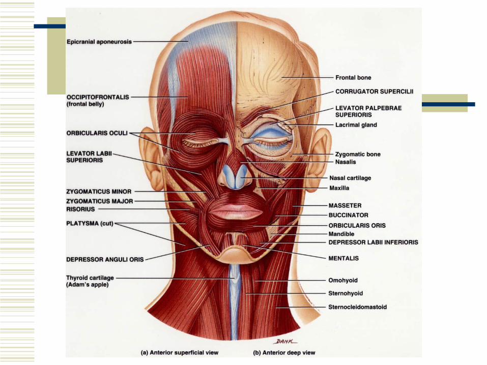
Muscles of Facial Expression
Scalp muscles Occipitofrontalis
Frontal bellyOccipital belly

Muscles of Facial Expression
Mouth muscles Orbicularis oris Zygomaticus major Zygomaticus minor Levator labii superioris Depressor labii inferioris Depressor anguli oris Buccinator

Muscles of Facial Expression
Neck muscle Platysma
Orbit and eyebrow muscles Orbicularis Oculi Corrugator Supercilii Levator palpebrae superioris

Frontalis
Origin – Epicranial aponeurosis Insertion – skin above the eye Action – draws skin anteriorly, raises
eyebrows, and wrinkles skin of the forehead

Occipitalis
Origin – occipital bone and mastoid process of the temporal bone
Insertion – epicranial aponeurosis Action – draws scalp posteriorly
Orbicularis Oris
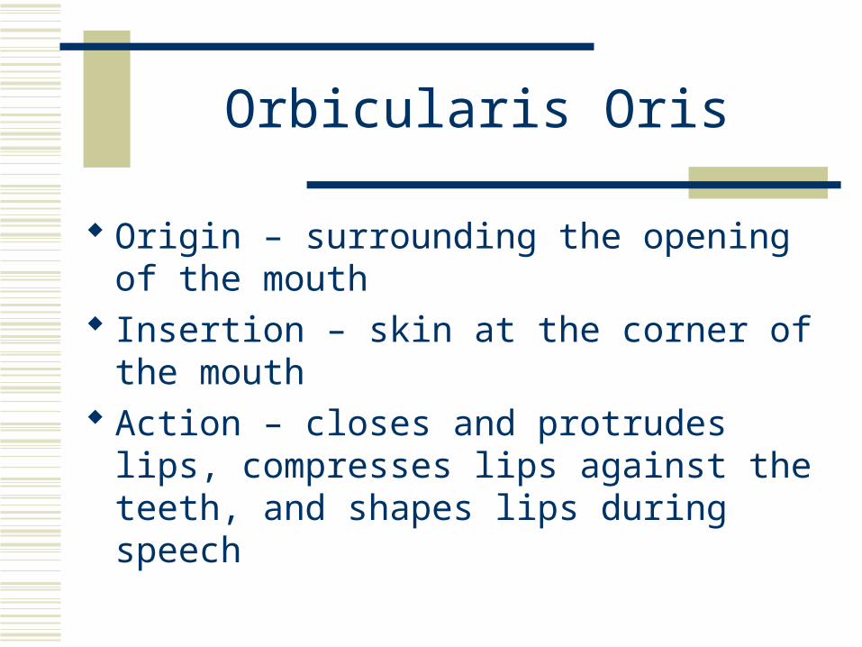
Origin – surrounding the opening of the mouth
Insertion – skin at the corner of the mouth Action – closes and protrudes lips,
compresses lips against the teeth, and shapes lips during speech

Buccinator
Origin – maxilla and mandible Insertion – orbicularis oris Action – draws the angle of the mouth
laterally and inferiorly as in opening the mouth

Platysma
Origin – fascia over deltoid and pectoralis major muscles
Insertion – mandible, muscles around the angle of the mouth and the skin of the lower face
Action – draws the outer part of the lower lip inferiorly and posteriorly as in pouting and depresses the mandible
Orbicularis Oculi
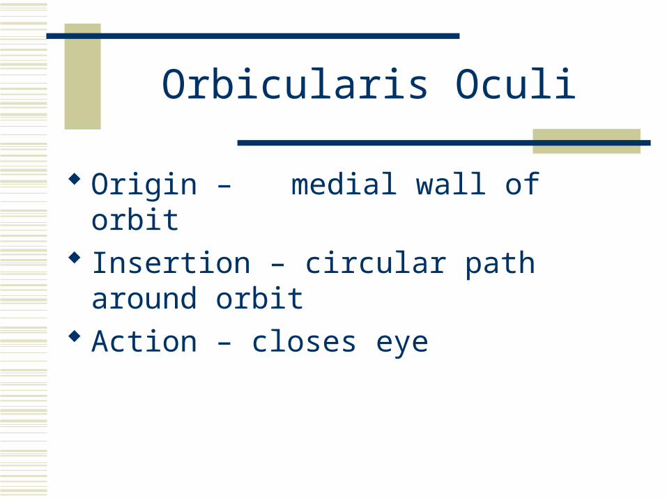
Origin – medial wall of orbit Insertion – circular path around orbit Action – closes eye


Bell’s Palsy (Facial Paralysis)
Unilateral paralysis of the muscles of facial expression.
Due to damage or disease of the facial nerve (cranial nerve VII).
The causes is unknown; However, inflammation of the facial nerve or infection by herpes simplex are suggested causes.

Bell’s Palsy (Facial Paralysis)
The person cannot wrinkle the forehead, close the eye, or pucker the lips on the affected side.
Difficulty in swallowing and drooling often occur.
80% recover within a few weeks to a few months; However, for some the paralysis is permanent.

Muscles That Move the Eyeballs – Extrinsic Eye
Muscles Superior Rectus Inferior Rectus Lateral Rectus Medial Rectus Superior oblique Inferior oblique
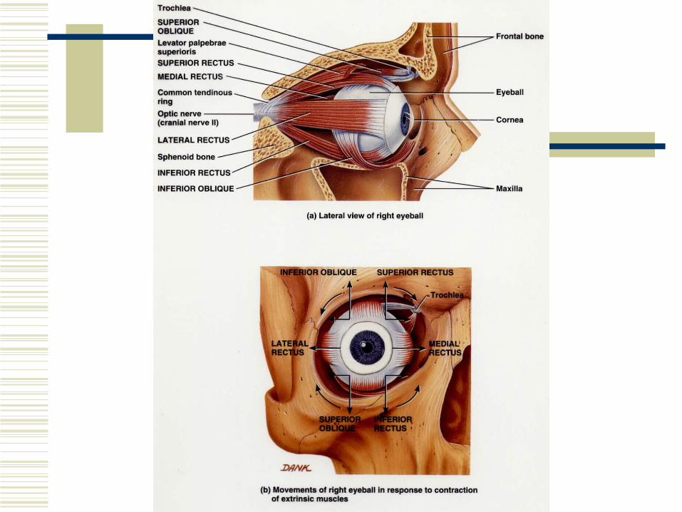

Strabismus
A condition in which the eyes are not properly aligned.
A lesion of the oculomotor nerve (cranial nerve III) or the abducens nerve (cranial nerve VI).

Muscles That Move the Mandible
Masseter Temporalis Medial Pterygoid Lateral Pterygoid

Masseter
Origin – maxilla and zygomatic arch Insertion – angle and ramus of mandible Action – elevates mandible and retracts
mandible (closes mouth)

Temporalis
Origin – temporal bone Insertion – coronoid process and ramus of
mandible Action – elevates and protracts mandible and
moves mandible from side to side


Muscle That Move The Tongue
Genioglossus Styloglossus Palatoglossus Hyoglossus


Muscles of the Anterior Neck
Suprahyoid muscles Digastric Stylohyoid Mylohyoid Geniohyoid
Muscles of the Anterior Neck
Infrahyoid muscles Omohyoid Sternohyoid Sternothyroid Thyrohyoid


Muscles That Move the Head
Sternocleidomastoid Semispinalis Capitis Splenius Capitus Longissimus Capitis
Sternocleidomastoid
Origin – sternum and clavicle Insertion – mastoid process of the temporal bone Action –
Acting together (bilaterally), they flex the cervical portion of the vertebral column, extend the head, and elevate the sternum during forced inhalation
Acting singly (unilaterally), laterally flexes head towards and rotates head away from the side of the contracting muscle


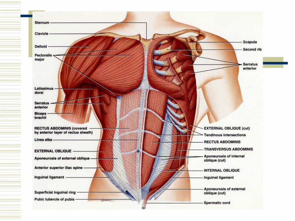
Muscles That Act on the Abdominal Wall
Rectus Abdominus External oblique Internal oblique Transversus Abdominus Quadratus Lumborum
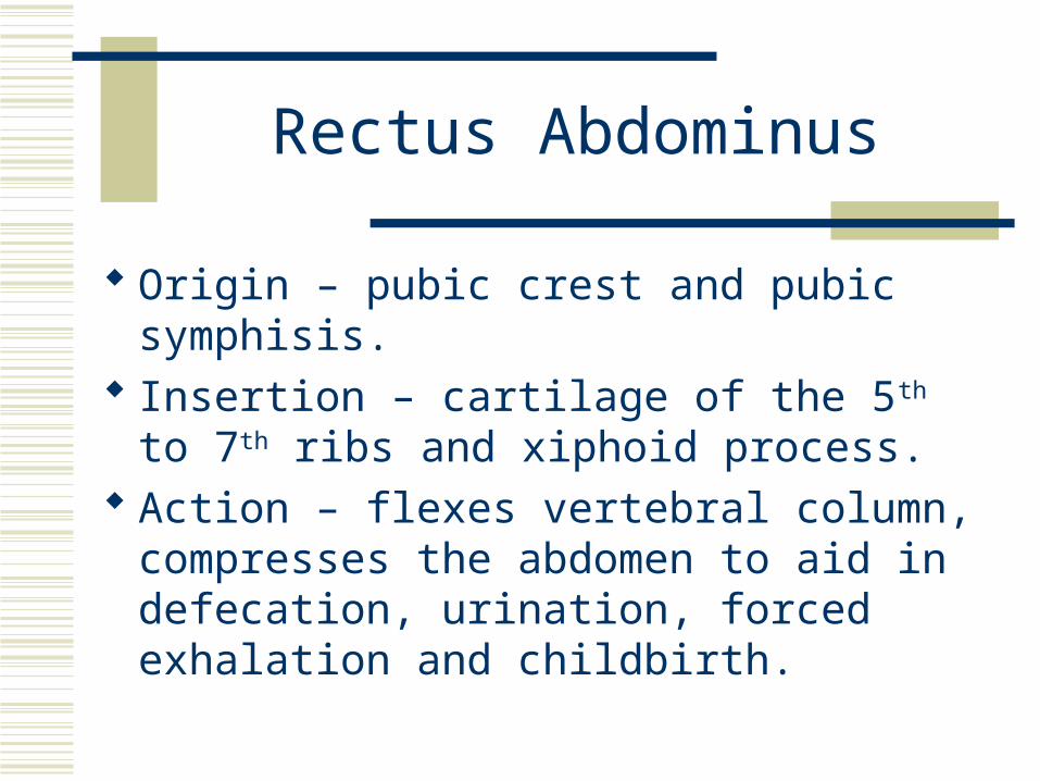
Rectus Abdominus
Origin – pubic crest and pubic symphisis. Insertion – cartilage of the 5th to 7th ribs and
xiphoid process. Action – flexes vertebral column,
compresses the abdomen to aid in defecation, urination, forced exhalation and childbirth.
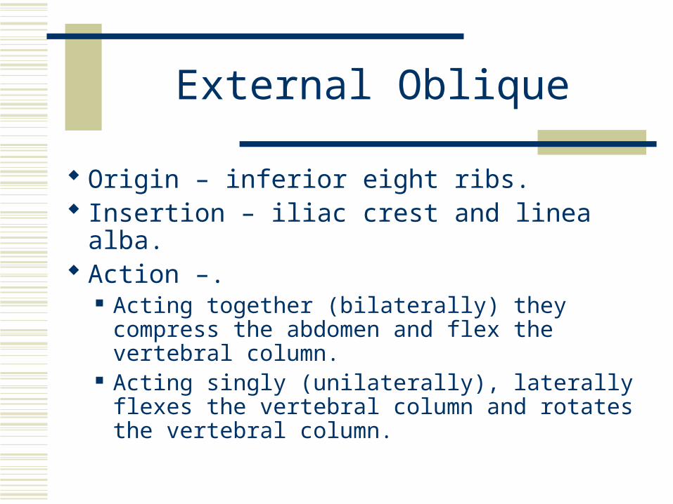
External Oblique
Origin – inferior eight ribs. Insertion – iliac crest and linea alba. Action –.
Acting together (bilaterally) they compress the abdomen and flex the vertebral column.
Acting singly (unilaterally), laterally flexes the vertebral column and rotates the vertebral column.


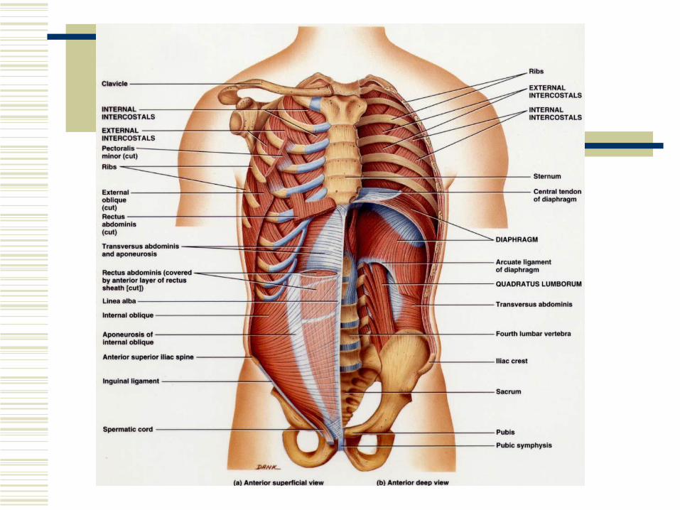
Muscles Used in Breathing
Diaphragm External intercostals Internal intercostals

Muscles of the Pelvic Floor
Levator ani Pubococcygeus Iliococcygeus
Coccygeus

Muscles of the Perineum
Superficial Perineal muscles Superficial transverse perineal Bulbospongiosis
Deep Perineal muscles Deep transverse perineal External urethral sphincter External anal sphincter


Muscles That Move the Pectoral Girdle
Anterior thoracic muscles Subclavius Pectoralis minor Serratus anterior
Posterior thoracic muscles Trapezius Levator scapulae Rhomboid major Rhomboid minor


Muscles That Move the Humerus
Axial muscles that move the Humerus Pectoralis major Latissimus dorsi

Muscles That Move the Humerus
Scapular muscles that move the Humerus Deltoid Subscapularis Supraspinatous Infraspinatous Teres major Teres minor coracobrachialis

Pectoralis Major
Origin – clavicle, sternum, and costal cartilages of 2nd to 6th ribs
Insertion – greater tubercle of the humerus Action –
As a whole, adducts and medially rotates arm at the shoulder joint
Clavicular head alone flexes arm Sternocostal head alone extends the arm

Latissimus Dorsi
Origin – spines of the inferior six thoracic vertebrae, lumbar vertebrae, crests of sacrum and ilum, and inferior four ribs
Insertion – intertubercular sulcus of humerus Action – extends, adducts, and medially
rotates the arm, draws arm inferiorly and posteriorly

Deltoid
Origin – acromial extremity of clavicle (anterior fibers), acromion of scapula (lateral fibers), and spine of scapula (posterior fibers)
Insertion – deltoid tuberosity of humerus Action –
Lateral fibers – abduct arm Anterior fibers – flex and medially rotate arm Posterior fibers – extend and laterally rotate arm


Muscles That Move the Radius and Ulna
Forearm flexors Biceps brachii Brachialis Brachioradialis
Forearm extensors Triceps brachii Anconeus

Muscles That Move the Radius and Ulna
Forearm Pronators Pronator teres Pronator quadratus
Forearm Supinator Supinator

Biceps Brachii
Origin – Long head – tubercle above glenoid cavity of
scapula Short head – coracoid process of scapula
Insertion – radial tuberosity of radius Action – flexes forearm at elbow, supinates
forearm, and flexes arm at shoulder

Brachialis
Origin – distal, anterior surface of humerus Insertion – ulnar tuberosity and coronoid
process of ulna Action – flexes forearm at elbow

Brachioradialis
Origin – lateral border of distal end of humerus
Insertion – superior to styloid process of radius
Action – flexes forearm at elbow, supinates and pronates forearm to neutral position

Triceps Brachii
Origin – Infraglenoid tubercle of scapula, posterior surface of humerus
Insertion – olecranon of ulna Action – extends forearm at elbow, extends
arm at shoulder

Pronator Teres
Origin – medial epicondyle of humerus and coronoid process of ulna
Insertion – midlateral surface of radius Action – pronates forearm at radioulnar
joints and weakly flexes forearm at elbow
Supinator
Origin – lateral epicondyle of humerus and ridge near radial notch of ulna
Insertion – lateral surface of proximal one third radius
Action – supinates forearm at radioulnar joints

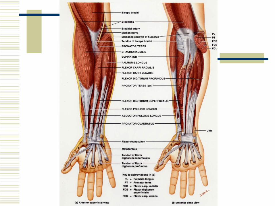
Muscles That Move the Wrist, Hand, Thumb, and Fingers
Superficial anterior (flexor) compartment of the forearm Flexor carpi radialis Palmaris longus Flexor carpi ulnaris Flexor digitorum superficialis
Deep anterior (flexor) compartment of the forearm Flexor pollicus longus Flexor digitorum profundus

Muscles That Move the Wrist, Hand, Thumb, and Fingers
Superficial posterior (extensor) compartment of the forearm Extensor carpi radialis longus Extensor carpi radialis brevis Extensor digitorum Extensor digiti minimi Extensor carpi ulnaris

Muscles That Move the Wrist, Hand, Thumb, and Fingers
Deep posterior (extensor) compartment of the forearm Abductor pollicis brevis Extensor pollicis longus Extensor indicis
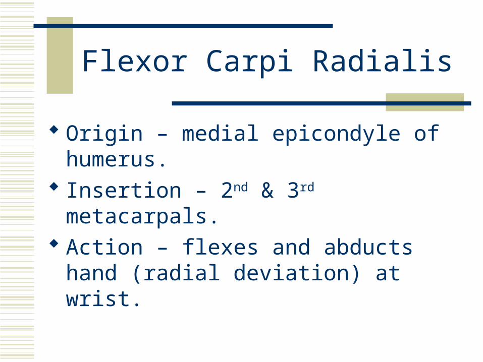
Flexor Carpi Radialis
Origin – medial epicondyle of humerus. Insertion – 2nd & 3rd metacarpals. Action – flexes and abducts hand (radial
deviation) at wrist.

Flexor Carpi Ulnaris
Origin – medial epicondyle of humerus and superior posterior border of ulna
Insertion – pisiform, hamate, and base of 5th metacarpal
Action – flexes and adducts (ulnar deviation) at wrist.
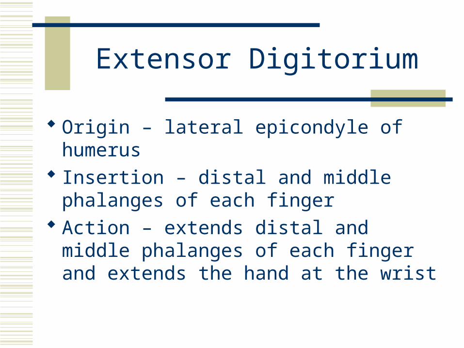
Extensor Digitorium
Origin – lateral epicondyle of humerus Insertion – distal and middle phalanges of
each finger Action – extends distal and middle phalanges
of each finger and extends the hand at the wrist

Extensor Carpi Ulnaris
Origin – lateral epicondyle of humerus and posterior border of ulna
Insertion – 5th metacarpal Action – extends and adducts hand at wrist.
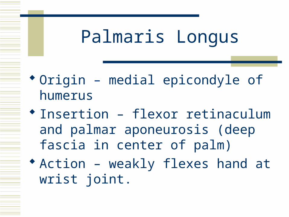
Palmaris Longus
Origin – medial epicondyle of humerus Insertion – flexor retinaculum and palmar
aponeurosis (deep fascia in center of palm) Action – weakly flexes hand at wrist joint.


Intrinsic Muscles of the Hand
Thenar (lateral aspect of the palm) Abductor pollicis Opponens pollicis Flexor pollicis brevis Adductor pollicis

Intrinsic Muscles of the Hand
Hypothenar (medial aspect of the palm) Abductor digiti minimi Flexor digiti minimi brevis Opponens digiti minimi
Intermediate (Midpalmar) Lumbricals Palmar interossei Dorsal interossei


Carpal Tunnel Syndrome
The carpal tunnel is a narrow passageway formed anterior by the flexor retinaculum and posteriorly by the carpal bones.
The median nerve and flexor tendons pass through. They are vulnerable to compression, which results
in pain, numbness, and tingling in the fingers. It is caused by inflammation of the tendon sheaths,
fluid retention, and repetitive activities involving flexion at the wrist.
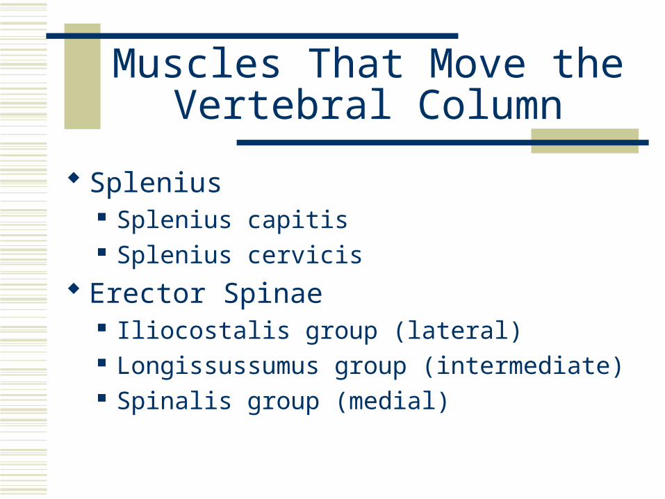
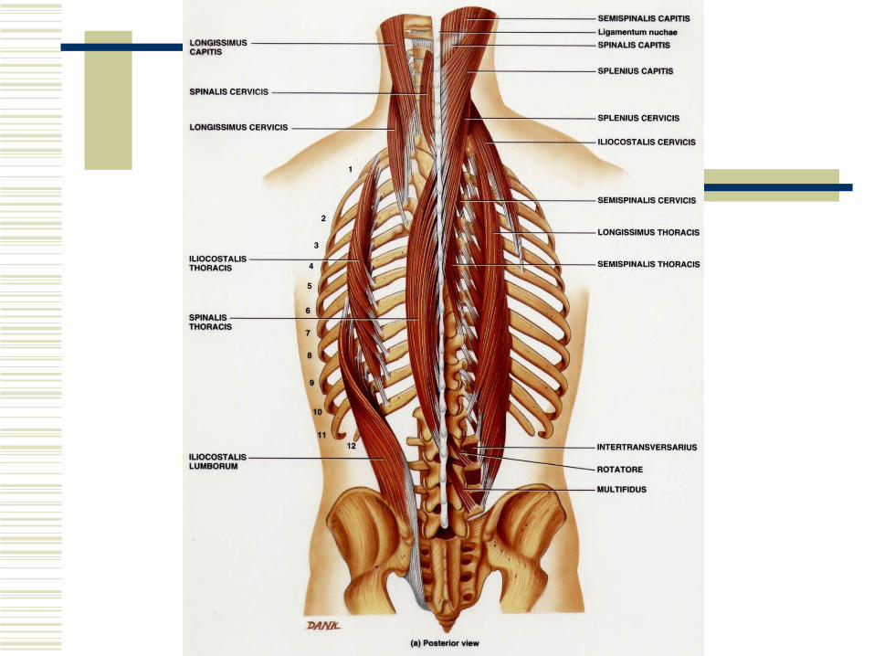
Muscles That Move the Vertebral Column
Splenius Splenius capitis Splenius cervicis
Erector Spinae Iliocostalis group (lateral) Longissussumus group (intermediate) Spinalis group (medial)

Muscles That Move the Vertebral Column
Transversospinales Seminspinalis Multifidus Rotatores
Segmental Interspinales Intertransversarii
Scalenes


Muscles That Move the Femur
Psoas major Iliacus Gluteus maximus Gluteus medius Gluteus minimus Tensor fasciae latae Piriformis

Muscles That Move the Femur
Obturator internus Obturator externus Superior gemellus Quadratus femoris Adductor longus Adductor brevis Adductor magnus Pectineus
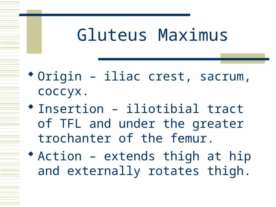
Gluteus Maximus
Origin – iliac crest, sacrum, coccyx. Insertion – iliotibial tract of TFL and under
the greater trochanter of the femur. Action – extends thigh at hip and externally
rotates thigh.

Tensor Fasciae Latae (TFL)
Origin – iliac crest. Insertion – tibia by way of the iliotibial tract. Action – flexes and abducts thigh at hip
joint.

Adductor Longus
Origin – pubic crest and pubic symphysis. Insertion – linea aspera of femur. Action – adducts and flexes thigh at hip joint
and medially rotates thigh.

Adductor Magnus
Origin – inferior ramus of pubis and ischium to ischial tuberosity.
Insertion – linea aspera of femur. Action – adducts thigh at hip and medially
rotates thigh. The anterior part flexes the thigh and the posterior part extends the thigh.


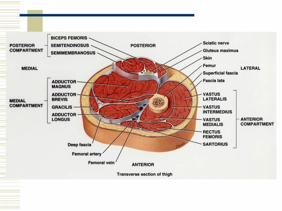
Muscles That Act on the Femur, Tibia, and Fibula
Medial (adductor) compartment of the thigh Adductor magnus Adductor longus Adductor brevis Pectineus Gracilis

Muscles That Act on the Femur, Tibia, and Fibula
Anterior (extensor) compartment of the thigh Quadriceps femoris
Rectus femorisVastus lateralisVastus medialisVastus intermedius
Sartorius
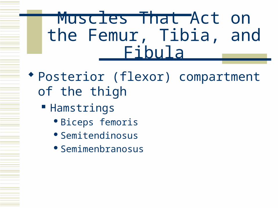
Muscles That Act on the Femur, Tibia, and Fibula
Posterior (flexor) compartment of the thigh Hamstrings
Biceps femorisSemitendinosusSemimenbranosus

Gracilis
Origin – body and inferior ramus of pubis. Insertion – medial surface of body of tibia. Action – adducts thigh at hip, medially
rotates thigh and flexes leg at knee.

Rectus Femoris
Origin – anterior inferior iliac spine (ASIS). Insertion – patella via quadriceps tendon
then tibial tuberosity. Action – extend the leg at the knee joint and
flexes the thigh at the hip.

Vastus Lateralis
Origin – greater trochanter and linea aspera of femur.
Insertion – patella via quadriceps tendon then tibial tuberosity.
Action – extend the leg at the knee joint and flexes the thigh at the hip.
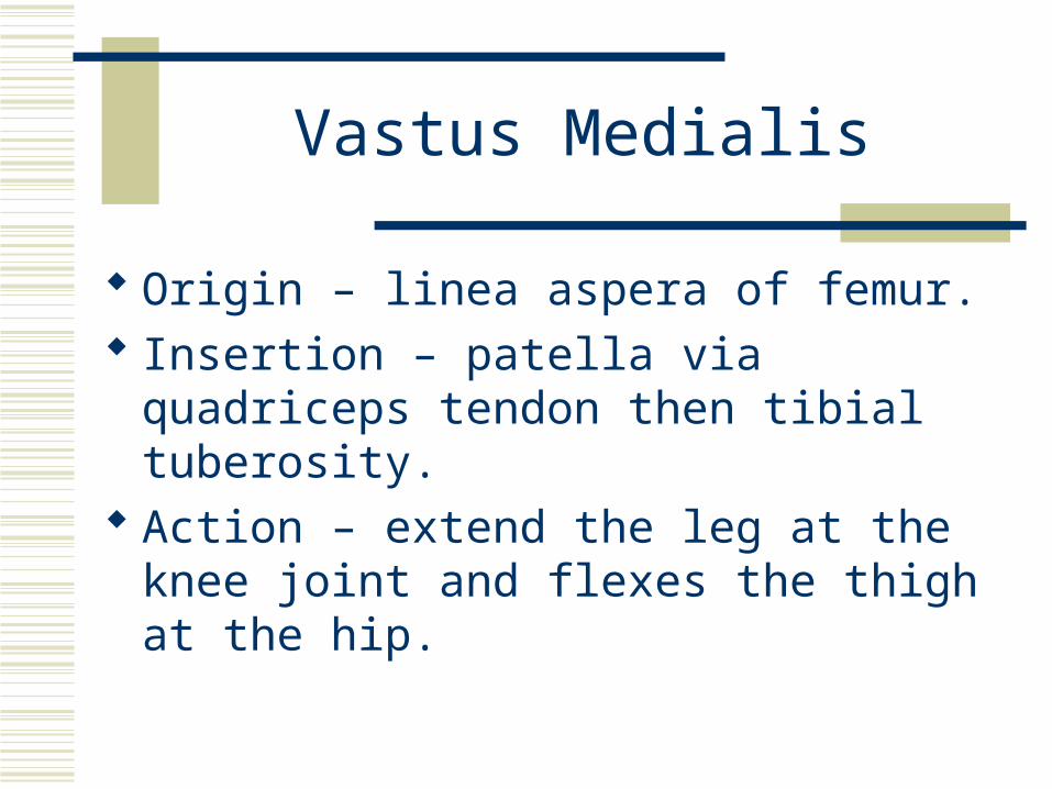
Vastus Medialis
Origin – linea aspera of femur. Insertion – patella via quadriceps tendon
then tibial tuberosity. Action – extend the leg at the knee joint and
flexes the thigh at the hip.

Vastus Intermedius
Origin – anterior and lateral surfaces of body of femur.
Insertion – patella via quadriceps tendon then tibial tuberosity.
Action – extend the leg at the knee joint and flexes the thigh at the hip.

Sartorius
Origin – ASIS. Insertion – medial surface of body of tibia. Action – flexes leg at knee, flexes, abducts,
and externally rotates thigh at hip. “Tailor’s muscle”.

Biceps Femoris
Origin –. Long head – ischial tiberosity. Short head – linea aspera of femur.
Insertion – head of fibula and lateral condyle of tibia.
Action – flexes leg at knee joint and extends thigh at hip.

Semitendinosus
Origin – ischial tiberosity. Insertion – proximal part of medial surface
of shaft of tibia. Action – flexes leg at knee and extends thigh
at hip.

Seminmembranosus
Origin – ischial tiberosity. Insertion – medial condyle of tibia. Action – flexes leg at knee and extends thigh
at hip.
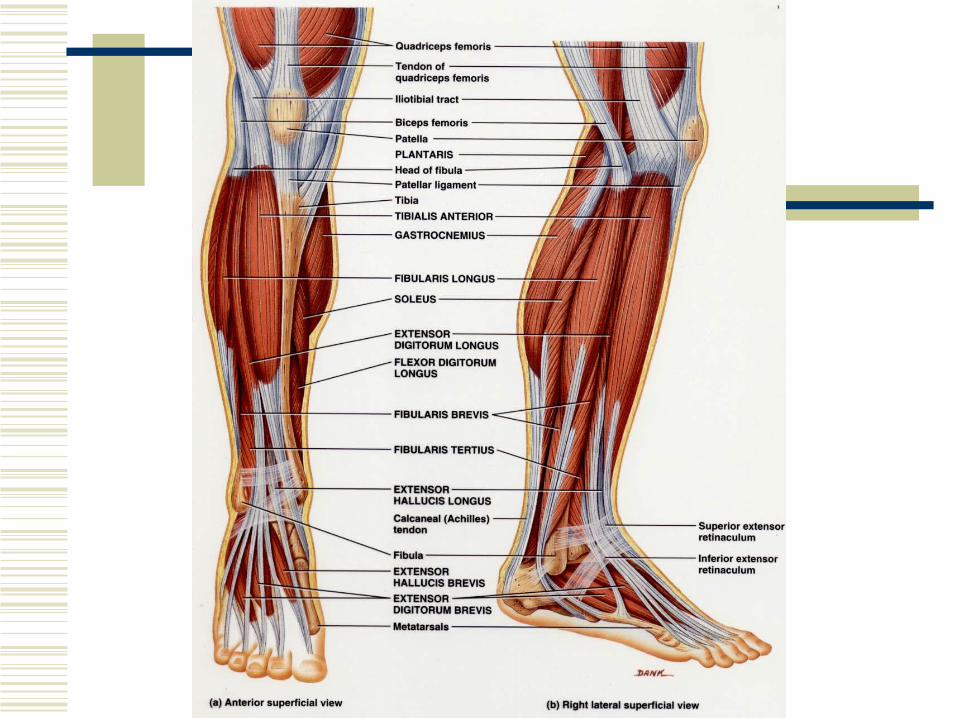
Muscles That Move the Foot and Toes
Anterior compartment of the leg Tibialis anterior Extensor hallucis longus Extensor digitorum longus Fibularis (peroneus) tertius
Lateral (fibular) compartment of the leg Fibularis (peroneus) longus Fibularis (peroneus) brevis

Muscles That Move the Foot and Toes
Superficial posterior compartment of the leg Gastrocnemius Soleus Plantaris
Deep posterior compartment of the leg Popliteus Tibialis posterior Flexor digitorum longus Flexor hallucis longus

Tibialis Anterior
Origin – lateral condyle and body of tibia, interosseous membrane.
Insertion – 1st metatarsal and first (medial) cuneiform.
Action – dorsiflexes foot at ankle and inverts foot at intertarsal joints.

Extensor Digitorium Longus
Origin – lateral condyle of tibia, anterior surface of fibula and interosseous membrane.
Insertion – base of 5th metatarsal. Action – dorsiflexes foot at ankle and everts
foot.
Peronius Longus
Origin – head and body of fibula and lateral condyle of tibia.
Insertion – 1st metatarsal and 1st cuneiform. Action – plantar flexes foot and everts foot.
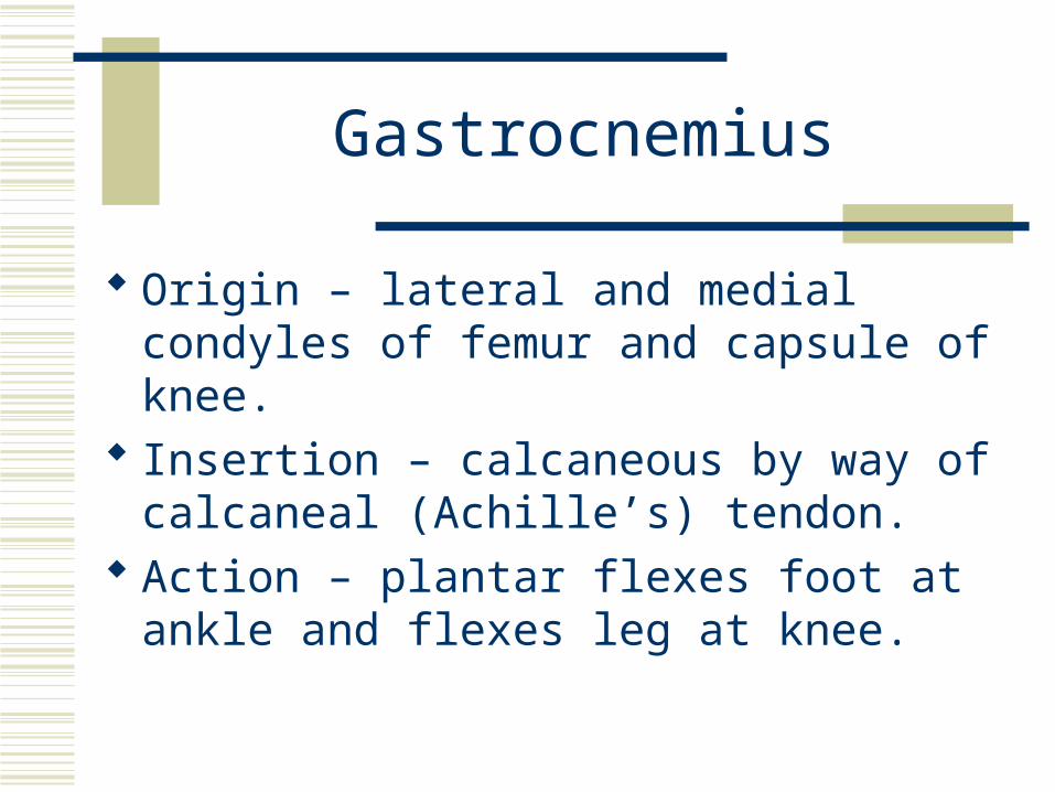
Gastrocnemius
Origin – lateral and medial condyles of femur and capsule of knee.
Insertion – calcaneous by way of calcaneal (Achille’s) tendon.
Action – plantar flexes foot at ankle and flexes leg at knee.

Soleus
Origin – head of fibula and medial border of tibia.
Insertion – calcaneous by way of calcaneal (Achille’s) tendon.
Action – plantar flexes foot at ankle.

Flexor Digitorum Longus
Origin – posterior surface of tibia. Insertion – distal phalanges of toes 2-5. Action – plantar flexes foot and flexes distal
and middle phalanges of toes 2-5.
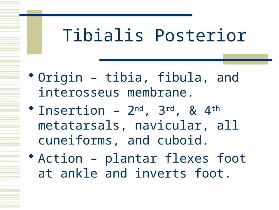
Tibialis Posterior
Origin – tibia, fibula, and interosseus membrane.
Insertion – 2nd, 3rd, & 4th metatarsals, navicular, all cuneiforms, and cuboid.
Action – plantar flexes foot at ankle and inverts foot.

Flexor Hallucis Longus
Origin – inferior two-thirds of fibula. Insertion – distal phalanx of great toe. Action – plantar flexes foot at ankle, flexes
great toe.


Shinsplint Syndrome
Pain or soreness along the tibia, specifically the medial, distal two-thirds.
Caused by tendinitis of the anterior compartment muscles, especially tibialis anterior muscle, inflammation of the periosteum around the tibia or stress fractures of the tibia.
Running on hard surfaces with poorly conditioned muscles, poor support shoes, etc. Contributes to this condition.

Intrinsic Muscles of the Foot
Dorsal Extensor digitorum brevis

Intrinsic Muscles of the Foot
Plantar 1st layer (most superficial)
Abductor hallucisFlexor digitorum brevisAbductor digiti minimi
2nd layerQuadratus plantaeLumbricals

Intrinsic Muscles of the Foot
Plantar 3rd layer
Flexor hallucis brevisAdductor hallucisFlexor digiti minimi
4th layer (deepest)Dorsal interosseiPlantar interossei


Plantar Fasciitis
Otherwise know as painful heel syndrome. Inflammatory reaction due to chronic irritation of
the plantar aponeurosis at its origin on the calcaneous.
The most common cause of heel pain in runners. Tx. - Strip out the plantar aponeurosis with a tennis
ball or golf ball.
Tendons
Patellar tendon & ligament Calcaneal or Achille’s tendon


![MATERIALS AND METHODS · 134 hXQ .ietaO cles, tendons, and ligaments [17]. It can be affected by muscle stiffness or susceptibility to muscle ...](https://static.documents.pub/doc/80x56/61464cc48f9ff81254202d32/materials-and-methods-134-hxq-ietao-cles-tendons-and-ligaments-17-it-can-be.jpg)


![AnOverviewoftheClinicalUseofAntimuscarinicsin ...2 Advances in Urology antimuscarinic action, oxybutynin in high doses exerts muscle-relaxant and local anaesthetic effects [5–8].](https://static.documents.pub/doc/80x56/60c45d5c03186b0ad2131222/anoverviewoftheclinicaluseofantimuscarinicsin-2-advances-in-urology-antimuscarinic.jpg)
