Page 1
1
The Neural Response to Facial Attractiveness
Anjan Chatterjee
Amy Thomas
Sabrina E. Smith
Geoffrey K. Aguirre
From the Department of Neurology and Center for Cognitive Neuroscience, University of
Pennsylvania, Philadelphia, PA.
Correspondence: Anjan Chatterjee, Geoffrey K Aguirre: Department of Neurology,
University of Pennsylvania, 3 West Gates, 3400 Spruce Street, Philadelphia, PA 19104.Tel:
215-662-4265, Fax: 215-349-5579, email [email protected] ;
[email protected]
Running head: facial attractiveness
Page 2
2
Abstract
What are the neural correlates of attractiveness? Using functional magnetic resonance
imaging (fMRI), we addressed this question in the specific context of the apprehension of
faces. When subjects judged facial beauty explicitly, neural activity in a widely distributed
network involving the ventral occipital, anterior insular, dorsal posterior parietal, inferior
dorsolateral and medial prefrontal cortices correlated parametrically with the degree of facial
attractiveness. When subjects were not attending explicitly to attractiveness, but rather were
judging facial identity, the ventral occipital region remained responsive to facial beauty. We
propose that this region, which includes the fusiform face area (FFA), the lateral occipital
cortex (LOC) and medially adjacent regions, is activated automatically by beauty and may
serve as a neural trigger for pervasive effects of attractiveness in social interactions.
Key Words: beauty, neuroaesthetics, face processing, fMRI, reward
Page 3
3
Introduction
Facial attractiveness is likely to be deeply encoded in our biology. Cross-cultural
judgments of facial beauty are quite consistent (Etcoff, 1999; Jones & Hill, 1993; Perrett,
May, & Yoshikawa, 1994). Adults and children within and across cultures show high rates of
agreement in judgments of facial attractiveness (Langlois et al., 2000) suggesting that
universal principles of beauty exist. Further evidence for the view that biologic
underpinnings drive our response to attractiveness comes from infant studies. Infants look
longer at attractive faces within a week of being born, and the effects of attractiveness on
infants’ gaze generalize across race, gender and age by 6 months (J. H. Langlois, Ritter,
Roggman, & Vaughn, 1991; Slater et al., 1998). Thus, the disposition to engage attractive
faces is present in brains that have not been modified greatly by experience. These
observations do not mean that judgments of beauty are not shaped further by cultural factors
(Cunningham, Barbee, & Philhower, 2002), but some components of these judgments are
likely to be universal, components that may have distinct neural underpinnings (Chatterjee,
2004).
Theorists postulate two possible (though not mutually exclusive) evolutionary
mechanisms for why certain faces are considered more attractive than others (Rhodes,
Harwood, Yoshikawa, Miwi, & McLean, 2002). The first possibility is that attractive features
represent phenotypic attributes that are desirable in selecting mates, such as genetic health
and levels of immunocompetence (Etcoff, 1999; Grammer, Fink, Moller, & Thornhill, 2003;
Penton-Voak et al., 2001; Perrett et al., 1998; Symons, 1979; Thornhill & Gangestad, 1999).
On this view, the nervous system has evolved to be attracted to specific configurations of
facial features that signal “good genes”, configurations that we have come to regard as
Page 4
4
beautiful. The second possibility is that preferences arise as a by-product of a general
information-processing mechanism. The leading candidate for such a mechanism is the
extraction of a prototype, or the central exemplar of a category. People prefer prototypes of
different kinds of stimuli, such as color (Martindale & Moore, 1988) and music (Smith &
Melara, 1990). Faces would presumably be another category of stimuli subject to this biased
preference for prototypes (Halberstadt & Rhodes, 2000).
How might the nervous system respond to beauty? Such a response might have at
least three components. These components are the perceptual processing of the object itself,
the emotional response to the object and, when relevant, an explicit judgment about the
object’s beauty. A few studies have reported that attractive faces activate areas within the
orbito-frontal cortex, the nucleus accumbens or the ventral striatum (Aharon et al., 2001;
Ishai, 2007; Kampe, Frith, Dolan, & Frith, 2001; Kranz & Ishai, 2006; O'Doherty et al.,
2003) and that the amygdala has a non-linear relationship to attractiveness (Winston,
O'Doherty, Kilner, Perrett, & Dolan, 2007). These regional activations, within neural
circuitry dedicated to reward systems, are interpreted as reflecting the emotional valence
attached to attractive faces (Senior, 2003). The particular emotional valences are those
involved in the expectation of rewards and the satisfaction of appetites. The idea that
attractive faces are rewarding stimuli, at least for men, is evident behaviorally. Men are
willing to discount higher future rewards for smaller immediate rewards when it comes to
attractive female faces (Wilson & Daly, 2004). Presumably these patterns of neural activation
reflect ways in which attractive faces influence mate selection (Ishai, 2007). The judgment of
beauty, as distinct from its emotional evocations, involves parts of the prefrontal cortex. One
positron emission tomography study showed left frontal activation when subjects assessed
Page 5
5
facial attractiveness (Nakamura et al., 1998). Medial frontal involvement may generalize
beyond faces to responses to beauty of even abstract images as reported by Jacobsen and
colleagues (Jacobsen, Schubotz, Hofel, & v Cramon, 2005).
In contrast to these findings about the emotional response to and judgment of facial
beauty, little is known about the neural underpinnings of the perceptual apprehension of
attractive faces. Winston and colleagues (Winston et al., 2007) found left posterior occipito-
temporal activity was enhanced by facial attractiveness, but did not explore this finding
further. Similarly, Kranz and Ishai (Kranz & Ishai, 2006) found increased activations for
attractive female faces than for unattractive female faces in the lateral fusiform gyrus, but
focused their discussion on activations within reward networks. Perceptual features of faces,
such as averageness, symmetry, the structure of cheek-bones, the relative size of the lower
half of the face and the width of the jaw, influence people’s judgments of facial beauty
(Enquist & Arak, 1994; Karl Grammer & Thornhill, 1994; Penton-Voak et al., 2001). The
influence of such perceptual features suggest that lower-level visual processing that occurs
prior to object processing per se and can affect aesthetic judgments (Chatterjee, 2004) might
play a role in facial beauty perception. With this possibility in mind, we paid special attention
to ventral visual association areas in this study.
Motivated by the logic that facial attractiveness is likely to have biological
underpinnings, we tested two hypotheses using fMRI. First, we tested the hypothesis that
explicit judgment of beauty is associated with a distributed neural response to increasing
levels of beauty, which includes neural structures involved in visual processing. Specifically,
areas of higher visual processing are of interest. We specifically looked at visual association
areas associated with processing of faces, places and objects. Reward circuits, including
Page 6
6
orbitofrontal, insular medial prefrontal and posterior cingulate cortex and the ventral
striatum, might be activated and we would anticipate that dorsolateral prefrontal and parietal
circuits might be involved in the decision making process.
Second, we tested the hypothesis that attractiveness of faces would continue to
modulate neural responses within part of the network engaged in explicit judgments, even
when subjects are not explicitly considering beauty. Individuals with brain damage may
develop prosopagnosia, a deficit in which the ability to recognize faces is impaired. Some
prosopagnosics respond differently (e.g., with different autonomic responses) to familiar than
unfamiliar faces despite not being able to explicitly recognize either (Bauer, 1984; Tranel &
Damasio, 1985). Faces, in general are processed more efficiently than other visual objects
and certain attributes such as emotions conveyed in these faces are processed quite rapidly
(for a review see (Palermo & Rhodes, 2007)). With respect to attractiveness, normal subjects
apprehend facial beauty at a glance (Olson & Marshuetz, 2005). Finally, Winston and
colleagues (Winston et al., 2007) found that parts of medial orbitofrontal cortices responded
to facial attractiveness even when subjects made judgments of age rather than attractiveness
of faces. They reasonably interpret these activations as related to the rewarding properties of
the stimuli that are engaged automatically when viewing attractive faces. However, they did
not pursue the hypothesis that perceptual responses to more attractive faces might trigger the
activation of these reward circuits.
We should also be clear that despite our interest in the neural response in visual
association areas to facial attractiveness, we are not explicitly investigating which visual
properties of faces are producing these responses. Our study focuses on what Fechner
(Fechner, 1860) referred to as an inner psychophysics (the relationship between subjective
Page 7
7
experiences and the physical properties of the nervous system) rather than on an outer
psychophysics (the relationship between subjective experiences and the physical properties
of the stimuli).
Methods
Participants – The study was approved by the Institutional Review Board of the
University of Pennsylvania. All subjects gave informed consent before participating in the
experiments. Thirteen subjects participated in two scanning sessions. There were 7 men and
6 women, age range 18-32 (mean 22.6).
Stimuli – Artificial face stimuli were created using commercial software (GenHead by
Genemation, http://www.genemation.com/) that was modified for use in our lab. The
software allows creation of human faces where the facial identity is determined by settings
on each of 114 parameters, each an eigenvector derived from a principal components analysis
of a large database of face photographs. Additional parameters allow control over ethnicity,
age and gender. Pilot behavioral studies were used to normalize the perceptual salience of
changes in each of the 114 parameters, and to standardize those parameters that had an
obvious effect upon the direction of gaze or facial expression. Therefore, all faces appeared
in the full frontal position with a neutral expression. Faces could then be created with a
normalized measure of distinctiveness, or measured distance from the average face.
We created a set of 100 face sets (50 male, 50 female). Each set initially contained
two faces of clearly different identities. All faces were Caucasian between the ages of 20 and
30 years, with the same distinctiveness score within the parameterized face space (distance to
the average). One face from each pair was arbitrarily designated the “start” face and the other
Page 8
8
labeled the “end” face. These pairs were then “morphed” to create faces at intermediate
points within the parameterized face space. The path between the pairs of faces was
computed so that the intermediate points were also at the same distance from the average
face. The distance of the intermediate faces from the “start” face were expressed in terms of
the % morph towards the “end” face; e.g., 33% morph (2/3 “start” face-1/3 “end” face), 66%
morph (1/3 “start face- 2/3 “end” face), etc. Therefore, each final face set consisted of four
faces: the “start” face, 33% morph, 66% morph, and “end” face (Figure 1). The face stimuli
were full color (32 bits/pixel), and set to be a uniform 288 x 288 pixel size.
Procedure – Each subject participated in two separate scanning sessions, with order
of scanning sessions randomized across subjects. The time between scanning sessions ranged
between 6 and 49 days (mean 27.6).
During both sessions, the stimuli consisted of 500 face pair trials and 200 additional
blank trials during which no stimuli were presented and the subjects did not provide
responses. During each of the 500 trials the subject would view two faces in quick succession
(each stimulus duration 1s, ISI 25 msecs, ITI 975msec). The first face was always a “start”
face from one of the 100 face sets. The second face was either the same as the first face (both
faces the “start” face), completely different (the “start” and “end” face), or a 33% or 66%
“morph” between the “start” and “end” faces. As there were 500 trials and only 400 unique
crossings of face sets with degree of change, 100 randomly selected trials during each
scanning session repeated a particular face pairing. The order of trials was pseudo-random
(determined by use of the OptSeq routine; http://surfer.nmr.mgh.harvard.edu/optseq/).
Page 9
9
During the attractiveness judgment task performed during one scanning session,
subjects were asked to judge whether each face was “more or less attractive than average”.
The subject was explicitly instructed not to judge if they personally found themselves
attracted to the presented face, but instead to judge if the face was better or worse looking
than an average person. Subjects made a judgment and provided a response for each of the
two faces presented in a trial. In the identity judgment task performed during the other
scanning session, subjects were asked to judge if the second face of each pair of faces was
identical to, or in any way different from, the first face. A response was made only after
presentation of the second face. In both tasks, subjects indicated their response by pressing
either a top pair or bottom pair of buttons using both thumbs. Figure 1 illustrates the structure
of the scanning tasks.
In addition to the difference in judgment required by the subject, the tasks also
differed in that participants responded to each face in during the attractiveness rating session
and only to the second of the two faces in the identity judgment. The temporal proximity of
face pairs also requires the modeling of facial attractiveness as the average attractiveness of
each pair of faces (see below). These limitations result from the design of the study to
measure neural adaptation resulting from facial similarity, as opposed to attractiveness; our
finding regarding the effects of attractiveness even when subjects were making identity
judgments was serendipitous. We submit that the inelegance of the design does not itself
invalidate the actual findings regarding the neural effect of facial attractiveness.
MRI scanning – Scanning was performed on a 3 Tesla Siemmens Trio using a
standard quadrature head coil. Echoplanar BOLD fMRI data were collected at a TR of 3
seconds, with 3x3x3 mm isotropic voxels covering the entire brain. Head motion was
Page 10
10
minimized with foam padding, and prospective motion correction (PACE) was performed
during image acquisition. A high-resolution anatomical image (3D MPRAGE) with 1x1x1
mm voxels was also acquired for each subject. Visual stimuli were presented using an Epson
8100 3-LCD projector with Buhl long-throw lenses for rear-projection onto Mylar screens,
which subjects viewed through a mirror mounted on the head coil. Subject responses were
recorded using a fiber-optic response pad (FORP) (http://www.curdes.com/newforp.htm).
A total of 7 BOLD fMRI scanning runs were completed during each scanning session
and each composed of 140 images. The first five scans were dedicated to the attractiveness or
discrimination tasks. The 2 additional BOLD scans were used for definition of functional
regions of interest (ROIs). Categorical functional ROIs were defined for faces (the fusiform
face area or FFA), buildings (the parahippocampal place area or PPA) and general object
forms (the lateral occipital cortex or LOC) using previously described methods (GK Aguirre,
Singh, & D'Esposito, 1999).
Data pre-processing and statistical analysis – BOLD fMRI data were processed
using the VoxBo (http://www.voxbo.org/) software package. After image reconstruction the
data were sinc interpolated in time to correct for the fMRI acquisition sequence (Aguirre,
Zarahn, & D'Esposito, 1997), motion corrected, transformed to a standard spatial frame
(using SPM2; http://www.fil.ion.ucl.ac.uk/spm), and spatially smoothed with a 3 voxel
FWHM 3D Gaussian kernel.
The relative attractiveness of each of the 400 face stimuli was determined by the
proportion of agreement of “better than average” judgments across the thirteen subjects for
each face (Figure 2). The highest possible score for a face was therefore unity if all thirteen
Page 11
11
subjects indicated that the face was better looking than average, and zero if all subjects felt
the subject was worse looking than average. We confirmed that this measure of a
dichotomous judgment produces similar attractiveness ratings as obtained with a Likert scale.
Thirty different subjects (mean age 22.7) rated the faces presented in the fMRI study for the
same duration using a 5 point scale. The averaged Likert judgments of attractiveness for each
face correlated highly (r = 0.85) with the proportion of agreement scores obtained during the
scanning experiment, suggesting that these methods of ascribing levels of attractiveness to
each of the faces in this set are comparable.
Within-subject statistical models of the fMRI data were created as follows. Trials in
which subjects made a correct response (correct in the beauty judgment session defined as
any response within the response-time window) were identified. As the two faces in each
trial were presented in close temporal proximity (preventing measurement of the BOLD
response unique to each face), the average of the attractiveness rating scores of the two faces
was assigned to the trial. An attractiveness covariate was then constructed by modeling a step
function of linear effect of attractiveness score upon neural response for the 3 seconds of the
trial, convolved by a standard hemodynamic response function (Aguirre, Zarahn, &
D'Esposito, 1998). The attractiveness scores were mean centered prior to convolution, to
render the covariate orthogonal to the main effect of stimulus presentation versus the null-
trials. In other words, the attractiveness covariate modeled the variation in neural response to
the presentation of a face that could be linearly related to the attractiveness of the face.
Additional covariates, not of interest here, modeled the main effect of stimulus
presentation versus null-trials, the similarity of pairs of faces, a polynomial expansion of this
similarity covariate, and the average reaction time of subject responses in each trial.
Page 12
12
Nuisance covariates for effects of scan and global signals were also included. Time series
data were subjected to a high-pass (0.0075 Hz) filter, and serial correlation of error terms was
modeled as previously described (Zarahn, Aguirre, & D'Esposito, 1997). Second order
(random effect) analyses were based upon the beta values measured for the particular
covariate of interest. Whole-brain statistical maps were prepared as “effect size maps”, in
which the average beta value attributed to the effect of facial beauty was scaled by the
average beta value attributed to the effect of the presentation of a face versus null-trials. This
permits the assessment of continuous effects across the cortex and the magnitude of the effect
of attractiveness. Map-wise significance was also estimated using permutation testing
(Nichols & Holmes, 2002).
The ROI localization scans were analyzed using a fixed-effects analysis across
subjects. A fixed-effects analysis was felt to be appropriate for this purpose as no hypothesis
was being tested regarding the existence of these well-established functional regions. Instead,
maximal sensitivity was desired for identifying their average location within this set of
subjects. The fusiform face area (FFA) was identified by the voxels within the fusiform gyrus
that demonstrated substantially greater response to faces than to pictures of objects and
buildings, and the parahippocampal place area (PPA) identified with the complementary
contrast. A region was generated for “form responsive cortex” by identifying those voxels
that had a greater response for faces, objects or buildings versus the scrambled stimuli. The
lateral occipital cortex (LOC) was identified as the region with the largest average difference
between the formed stimuli and the phase scrambled stimuli (Figure 3).
Results
Page 13
13
No significant differences according to gender were found for either task. Therefore,
we performed the following analyses with data collapsed across gender. There was a modest
correlation (r=0.270) between the attractiveness scores and reaction times (RT) when
subjects performed explicit judgments of facial beauty. Table 1 provides the average RTs for
the different sessions binned by facial attractiveness. The analysis of the fMRI data included
a covariate that modeled each subject’s average reaction time to each pair of faces, so that
any relationship between the attractiveness covariate and neural activity would not be a first-
order effect of how long the subjects looked at the faces.
ROI analyses revealed neural activity correlated with attractiveness ratings in the
FFA and LOC bilaterally (for all four ROIs, p values < 0.02), but not in the PPA (both p
values > 0.8). An ANOVA showed that the effects of beauty interacted with the ROIs
(F2,72=6.029, p=0.004) but not by hemisphere (F1,72=0.525, p=NS). Post-hoc tests showed
effects in LOC > PPA (p=0.003), a trend towards FFA > PPA (p=0.054) and no difference
between FFA and LOC. Whole brain analyses showed that the ventral activations extended
between and adjacent to FFA and LOC (Figure 3). Additional correlated activity was found
in the dorsal posterior parietal cortex, anterior insula, inferior and medial prefrontal regions
bilaterally (Figure 4). Negatively correlated activity was seen in the anterior and posterior
cingulate cortex. See Table 2 for details of whole brain activation results.
In the identity judgment task, no correlation between the beauty ratings and RT was
found (r=-0.024), reflecting that facial attractiveness was irrelevant to performance of the
task (see also Table 1). Nonetheless, neural activity was correlated with facial attractiveness
within the LOC bilaterally (p < 0.007 in both cases) and the FFA on the left (p < 0.003), but
not the right (p > 0.1). There was no significant effect of facial beauty within the PPA (for
Page 14
14
both, p > 0.5). Again, whole brain analyses revealed that this ventral activation extended
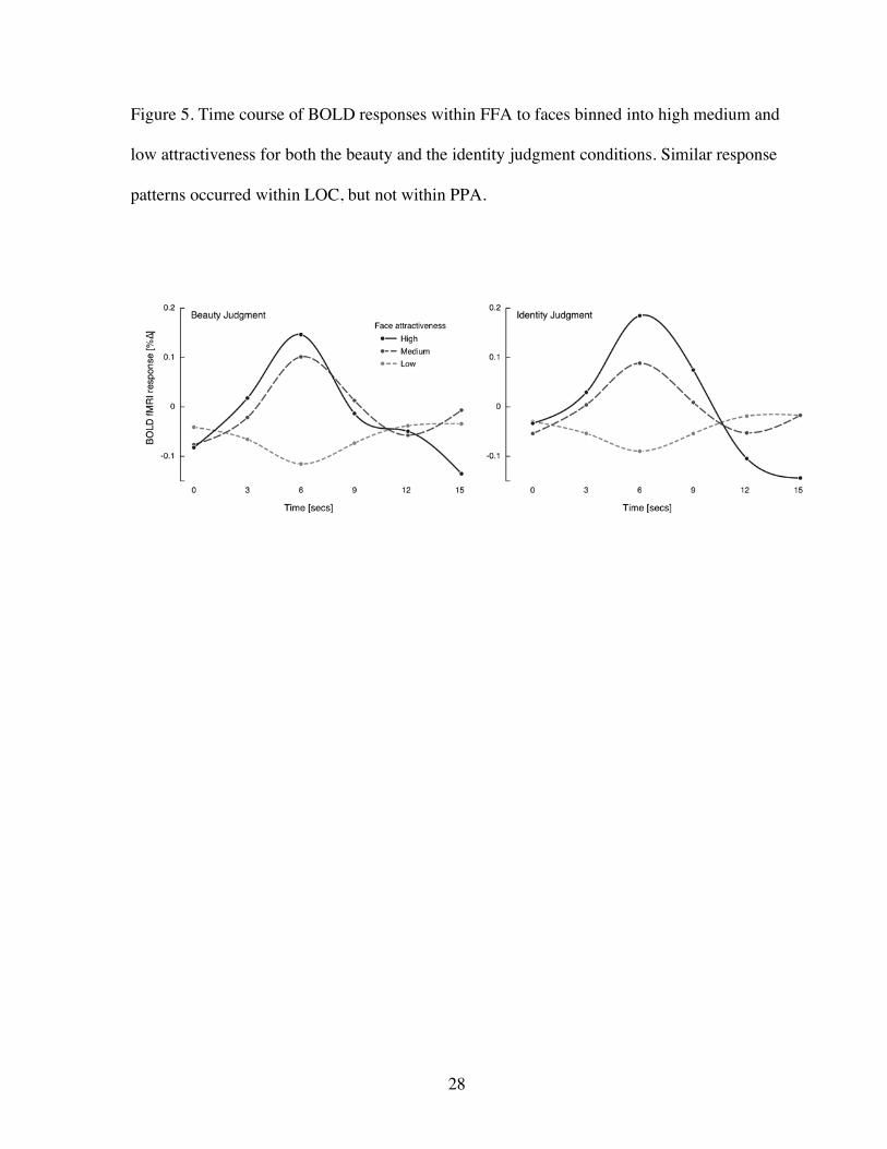
across the FFA (see Figure 5) and LOC as well as in adjacent medial regions, and did not
represent two distinct activation peaks (Figure 3). The distribution of activity was similar to
the pattern seen when subjects made explicit attractiveness judgments. Significant activation
was also seen in the pulvinar bilaterally, but not in parietal or prefrontal regions. We did not
have adequate signal within the orbitofrontal cortex to test the hypothesis that this region was
activated explicitly or automatically by facial attractiveness.
Discussion
Our results confirm that the apprehension of facial beauty is associated with an
identifiable neural response. When subjects make explicit judgments of attractiveness,
neural activity within a distributed network involving ventral visual association cortices and
parts of dorsal posterior parietal and prefrontal cortices varied parametrically with the degree
of attractiveness of the faces viewed. The response to beauty did not represent a general
activation of visual association areas, as the effects were not evident in brain regions that
process buildings and landscapes. We interpret the ventral occipito-temporal activations as
being involved in the visual processing of attractive faces. Recently, Winston and colleagues
(Winston et al., 2007) found a similar region activated more robustly when subjects judged
facial attractiveness as compared to facial age. However, they did not explore the
implications of activation within this region further.
The parietal, medial and dorsolateral frontal activations were present only during
explicit judgments of beauty. We propose that these areas represent neural correlates of the
attention and decision-making components of this task. The positively correlated insular
Page 15
15
activations and negatively correlated anterior and posterior cingulate activations are likely to
represent emotional responses to attractiveness. The frontomedian activation pattern, also
reported by O’Doherty and colleagues in response to attractive faces (O'Doherty et al., 2003)
are similar to activation patterns reported by Jacobsen and colleagues found to beauty
judgments of abstract geometric images (Jacobsen et al., 2005). Jacobsen and colleagues
emphasized that frontomedian activity is probably involved in the evaluative component of
aesthetic judgments, and might turn out to be involved regardless of the domain in which
these judgments are being made. This interpretation is consonant with the view that this
region is involved when one’s evaluation draws on an internally generated and self-
referential processes (Christoff & Gabrielli, 2000), such as “what do I think of the beauty of
this object?” Our observations that activity in posterior cingulate region correlates negatively
with degree of attractiveness, raises the possibility that these neural structures are engaged in
the negative evaluation of the beauty of an object. In that regard, it is of particular interest
that these regions are part of a paralimbic neural system that is dysregulated and overactive
in the resting state in depressed individuals (Mayberg, 1997; Mayberg et al., 1999). These
patients are often anhedonic and do not derive pleasure from objects that others find
pleasurable (Snaith, 1993). Thus, a predisposition to negatively evaluate attractive objects
may be a component of these patients’ anhedonia.
When our participants judged facial identity, specific regions within visual
association cortices continued to respond to facial attractiveness. Despite the irrelevance of
beauty to the task, facial beauty modified evoked neural response to faces by as much as 10%
in some areas (Figure 3). Again, the FFA and LOC and not the PPA, were sensitive to
degrees of facial attractiveness. Whole brain analyses revealed that this activity occurred in a
Page 16
16
contiguous area within and adjacent to the FFA and LOC across the fusiform and inferior
occipital gyrus. Our findings suggest that this ventral occipital region responds to beauty
automatically, regardless of the task in which the subject is engaged. This region may be
involved in visual processing prior to object identification, such as the apprehension of
symmetry and grouping, which also occur automatically and influence aesthetic judgment
(Chatterjee, 2004). The fact that this ventral occipital region of activation extended beyond
parts of cortices especially sensitive to faces raises the possibility that this area may be
responsive to aesthetic objects more generally. Consistent with this possibility, in an fMRI
study Vartanian and Goel (Vartanian & Goel, 2004) found that activity within this area
correlated with preferences for paintings, especially for representational ones and Jacobsen
and colleagues (Jacobsen et al., 2005) found this area to be responsive to symmetry and
aesthetic judgments for novel graphic abstract images.
Could this ventral occipital activation be the neural signature of the extraction of
facial prototypes? On this account the FFA responses to attractiveness would simply be a
reflection of increased activity to facial averageness. This hypothesis is unlikely to be
accurate, since activity within FFA correlates with facial distinctiveness rather than
averageness (Loffler, Yourganov, Wilkinson, & Wilson, 2005). Furthermore, the neural
response to facial beauty was not confined to face processing areas, and extended to area
LOC. Further research will be needed to determine which perceptual attributes (such as
symmetry, or relative sizes of different facial features) drive the increased activity within
LOC.
Is it possible that the ventral-occipital activations reflect greater attention to attractive
faces rather than a response to beauty per se? For two reasons, we think this explanation is
Page 17
17
unlikely. First, regions traditionally associated with attention, such as the posterior parietal
cortex were activated by the explicit beauty judgment conditions and not the identity
judgment condition, suggesting attentional engagement with attractive faces in the former
condition but not the latter. Second, one could test these alternate hypotheses directly by
using faces in which attractiveness and attentional salience are not monotonically correlated.
For example, especially unattractive faces also engage attention. Winston and colleagues
(Winston et al., 2007) did use faces that covered a wide range and found amygdala rather
than the ventral occipital activations were activated by both highly attractive and highly
unattractive faces.
The stimuli used in our experiment on the whole did not contain faces at either
extreme of an attractiveness continuum, super model faces or extremely unattractive faces.
Such faces might be more likely to evoke automatic activity within the amygdala and reward
circuitry than our stimuli as Winston and colleagues found (Winston et al., 2007). We remain
agnostic about orbitofrontal involvement, since we did not have adequate signal within these
regions to test the hypothesis that attractiveness engages these areas.
Attractiveness has pervasive social effects beyond its specific role in mate selection.
Attractive children are considered more intelligent, honest, and pleasant, and are thought to
be natural leaders (Kenealy, Frude, & Shaw, 1988; Lerner, Lerner, Hess, & Schwab, 1991;
Ritts, Patterson, & Tubbs, 1992). Attractive adults are judged to have socially desirable traits,
such as strength and sensitivity (Dion, Berscheid, & Walster, 1972). They are considered
more competent as politicians (Lewis & Bierly, 1990), professors (Romano & Bordiere,
1989) and counselors (Green, 1986). Attractive people are preferred in hiring decisions
(Rynes & Gerhart, 1990), earn more money (Hamermesh & Biddle, 2001) and receive lesser
Page 18
18
punishments for transgressions (Dion, 1972). Thus, a person’s attractiveness influences
social interactions in ways that extends far beyond domains in which attractiveness per se is
directly relevant.
The fact that people are often unaware of the extent to which attractiveness influences
social judgments suggests that facial beauty may be one of a number of facial attributes
apprehended automatically (Palermo & Rhodes, 2007). Facial beauty can be apprehended at
a glance and can bias subsequent cognitive judgments (Olson & Marshuetz, 2005). The
cascade of neural events that result in biases in high-level social decisions is likely to be
triggered by an early perceptual response to attractiveness. We propose that neural activity
within ventral visual cortices in response to facial attractiveness, that occurs even when
subjects are not considering beauty explicitly, serves as the initial trigger for this cascade.
Further along this cascade, medial orbitofrontal mediation (an area in which we had poor
signal detection) may support the emotional valence engendered by attractive faces
automatically (Winston et al., 2007). Senior (Senior, 2003) suggested that the neural
underpinning of face perception has a core system (the inferior occipital gyri, the latyeral
fusiform gyri and the superior temporal sulcus) dedicated to perceptual processing, and an
extended system (the extended amygdala and reward circuitry) dedicated the appraisal of
beauty and its rewarding and aesthetic consequences. This speculation, which he considered
provisional, was motivated by two studies of facial attractiveness (Aharon et al., 2001;
O'Doherty et al., 2003). Our findings suggest that the initial automatic appraisal of facial
beauty occurs earlier than Senior anticipated, within what he referred to as the core system.
In summary, we confirm that facial beauty evokes a widely distributed neural
network involving perceptual, decision-making and reward circuits. In our experiment, the
Page 19
19
perceptual response across FFA and LOC remained present even when subjects were not
attending explicitly to facial beauty. A general and testable hypothesis generated by these
results is that the perceptual response to visual beauty involves patterns of domain specific
and non-specific regional activations. Thus, other objects, such as attractive bodies or
beautiful landscapes might be accompanied by greater activity that extend from domain
specific cortical regions such as the extrastriate body area or the parahippocampal place area
into LOC.
Acknowledgment: We thank Dawn Mechanic for help with statistical analyses. AC
was supported by RO1-DC008779, SES was supported by K12 HD043245, and GKA was
supported by K08 MH 7 2926-01 and a Burroughs-Wellcome Career Development Award.
Page 21
21
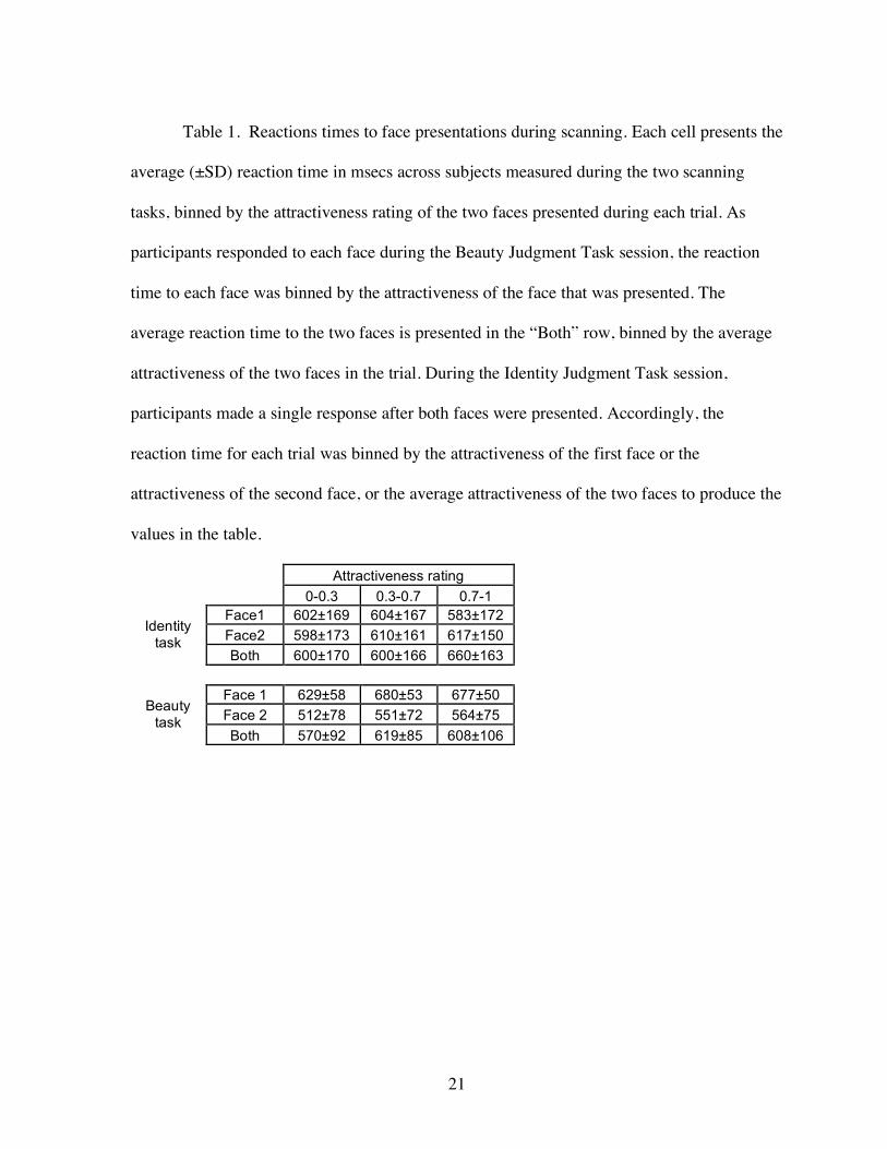
Table 1. Reactions times to face presentations during scanning. Each cell presents the
average (±SD) reaction time in msecs across subjects measured during the two scanning
tasks, binned by the attractiveness rating of the two faces presented during each trial. As
participants responded to each face during the Beauty Judgment Task session, the reaction
time to each face was binned by the attractiveness of the face that was presented. The
average reaction time to the two faces is presented in the “Both” row, binned by the average
attractiveness of the two faces in the trial. During the Identity Judgment Task session,
participants made a single response after both faces were presented. Accordingly, the
reaction time for each trial was binned by the attractiveness of the first face or the
attractiveness of the second face, or the average attractiveness of the two faces to produce the
values in the table.
Attractiveness rating 0-0.3 0.3-0.7 0.7-1
Face1 602±169 604±167 583±172 Face2 598±173 610±161 617±150 Identity
task Both 600±170 600±166 660±163
Face 1 629±58 680±53 677±50 Face 2 512±78 551±72 564±75 Beauty
task Both 570±92 619±85 608±106
Page 22
22
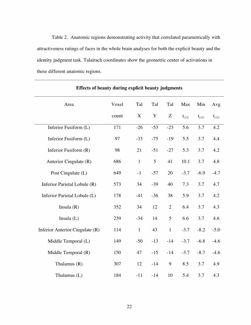
Table 2. Anatomic regions demonstrating activity that correlated parametrically with
attractiveness ratings of faces in the whole brain analyses for both the explicit beauty and the
identity judgment task. Talairach coordinates show the geometric center of activations in
these different anatomic regions.
Effects of beauty during explicit beauty judgments
Area Voxel
count
Tal
X
Tal
Y
Tal
Z
Max
t(12)
Min
t(12)
Avg
t(12)
Inferior Fusiform (L) 171 -26 -53 -23 5.6 3.7 4.2
Inferior Fusiform (L) 97 -33 -75 -19 5.5 3.7 4.4
Inferior Fusiform (R) 98 21 -51 -27 5.3 3.7 4.2
Anterior Cingulate (R) 686 1 5 41 10.1 3.7 4.8
Post Cingulate (L) 649 -1 -57 20 -3.7 -6.9 -4.7
Inferior Parietal Lobule (R) 573 34 -39 40 7.3 3.7 4.7
Inferior Parietal Lobule (L) 178 -41 -36 38 5.9 3.7 4.2
Insula (R) 352 34 12 2 6.4 3.7 4.3
Insula (L) 239 -34 14 5 6.6 3.7 4.6
Inferior Anterior Cingulate (R) 114 1 43 1 -3.7 -8.2 -5.0
Middle Temporal (L) 149 -50 -13 -14 -3.7 -6.8 -4.6
Middle Temporal (R) 150 47 -15 -14 -3.7 -8.7 -4.6
Thalamus (R) 307 12 -14 9 8.5 3.7 4.9
Thalamus (L) 184 -11 -14 10 5.4 3.7 4.3
Page 23
23
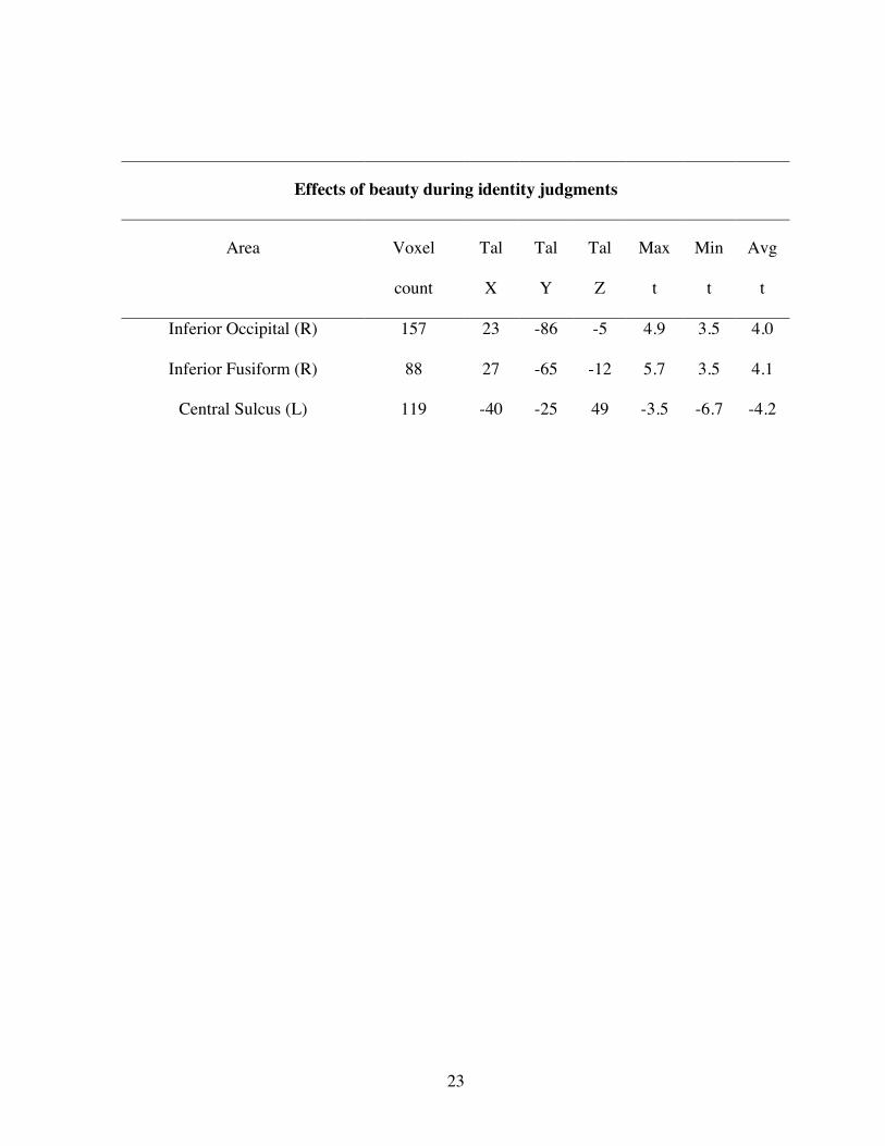
Effects of beauty during identity judgments
Area Voxel
count
Tal
X
Tal
Y
Tal
Z
Max
t
Min
t
Avg
t
Inferior Occipital (R) 157 23 -86 -5 4.9 3.5 4.0
Inferior Fusiform (R) 88 27 -65 -12 5.7 3.5 4.1
Central Sulcus (L) 119 -40 -25 49 -3.5 -6.7 -4.2
Page 24
24
Figure Legends
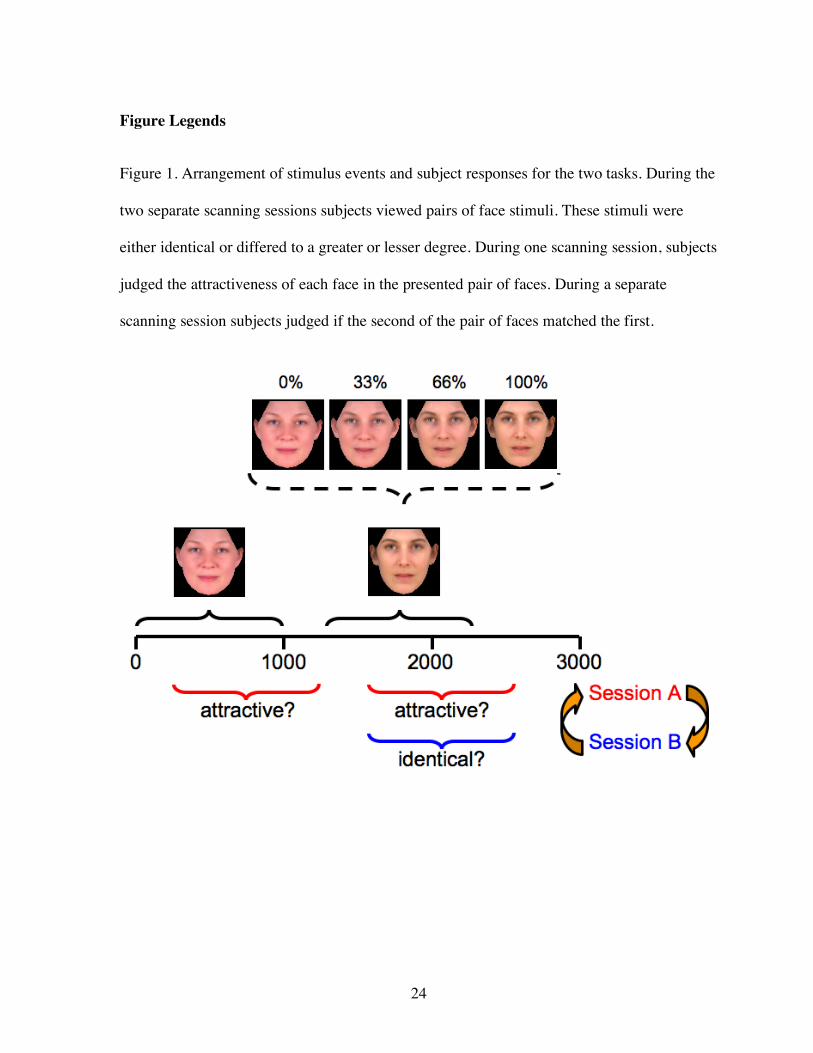
Figure 1. Arrangement of stimulus events and subject responses for the two tasks. During the
two separate scanning sessions subjects viewed pairs of face stimuli. These stimuli were
either identical or differed to a greater or lesser degree. During one scanning session, subjects
judged the attractiveness of each face in the presented pair of faces. During a separate
scanning session subjects judged if the second of the pair of faces matched the first.
Page 25
25
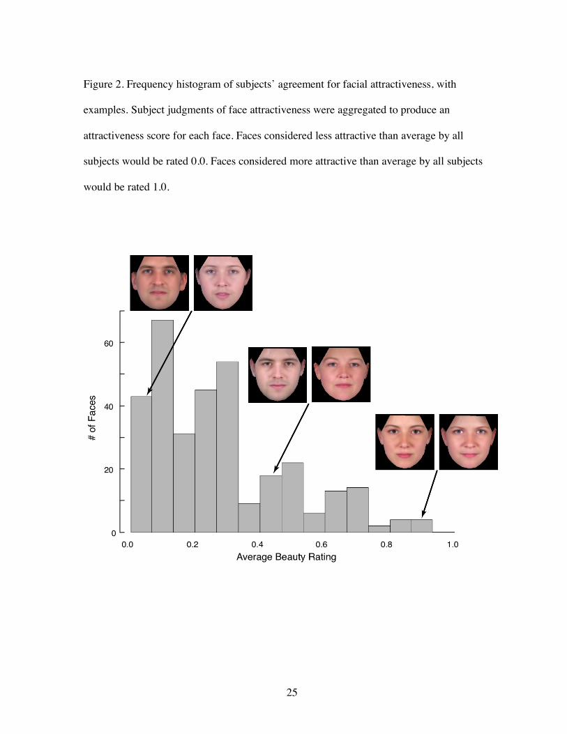
Figure 2. Frequency histogram of subjects’ agreement for facial attractiveness, with
examples. Subject judgments of face attractiveness were aggregated to produce an
attractiveness score for each face. Faces considered less attractive than average by all
subjects would be rated 0.0. Faces considered more attractive than average by all subjects
would be rated 1.0.
Page 26
26
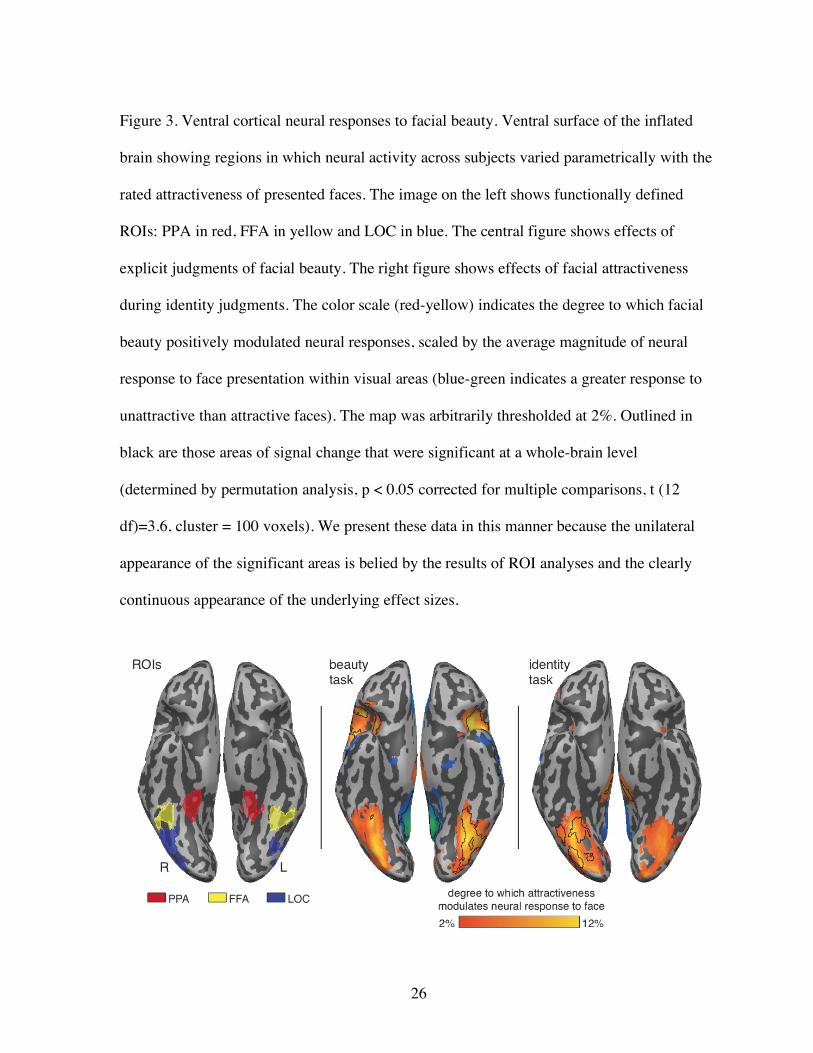
Figure 3. Ventral cortical neural responses to facial beauty. Ventral surface of the inflated
brain showing regions in which neural activity across subjects varied parametrically with the
rated attractiveness of presented faces. The image on the left shows functionally defined
ROIs: PPA in red, FFA in yellow and LOC in blue. The central figure shows effects of
explicit judgments of facial beauty. The right figure shows effects of facial attractiveness
during identity judgments. The color scale (red-yellow) indicates the degree to which facial
beauty positively modulated neural responses, scaled by the average magnitude of neural
response to face presentation within visual areas (blue-green indicates a greater response to
unattractive than attractive faces). The map was arbitrarily thresholded at 2%. Outlined in
black are those areas of signal change that were significant at a whole-brain level
(determined by permutation analysis, p < 0.05 corrected for multiple comparisons, t (12
df)=3.6, cluster = 100 voxels). We present these data in this manner because the unilateral
appearance of the significant areas is belied by the results of ROI analyses and the clearly
continuous appearance of the underlying effect sizes.
Page 27
27
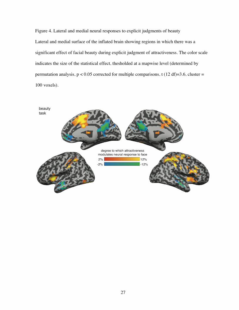
Figure 4. Lateral and medial neural responses to explicit judgments of beauty
Lateral and medial surface of the inflated brain showing regions in which there was a
significant effect of facial beauty during explicit judgment of attractiveness. The color scale
indicates the size of the statistical effect, thesholded at a mapwise level (determined by
permutation analysis, p < 0.05 corrected for multiple comparisons, t (12 df)=3.6, cluster =
100 voxels).
Page 28
28
Figure 5. Time course of BOLD responses within FFA to faces binned into high medium and
low attractiveness for both the beauty and the identity judgment conditions. Similar response
patterns occurred within LOC, but not within PPA.
Page 29
29
References
Aguirre, G., Singh, R., & D'Esposito, M. (1999). Stimulus inversion and the response of face
and object-sensitive cortical areas. Neuroreport, 10(1), 189-194.
Aguirre, G., Zarahn, E., & D'Esposito, M. (1998). The variability of human BOLD
hemodynamic response. Neuroimage, 8, 360-369.
Aguirre, G. K., Zarahn, E., & D'Esposito, M. (1997). Empirical analysis of BOLD fMRI
statistics. II. Spatially smoothed data collected under the null-hypothesis and experimental
conditions. Neuroimage, 5, 199-212.
Aharon, I., Etcoff, N., Ariely, D., Chabris, C., O'Connor, E., & Breiter, H. (2001). Beautiful
faces have variable reward value: fMRI and behavioral evidence. Neuron, 32, 537-551.
Bauer, R. M. (1984). Autonomic recognition of names and faces in prosopagnosia: A
neuropsychological application of the guilty knowledge test. Neuropsychologia, 22, 457-469.
Chatterjee, A. (2004). Prospects for a cognitive neuroscience of visual aesthetics. Bulletin of
Psychology and the Arts, 4, 55-59.
Christoff, K., & Gabrielli, J. (2000). The frontopolar cortex and human cognition: evidence
for a rostrocaudal hierarchical organization within the human prefrontal cortex.
Psychobiology, 28(168-186).
Cunningham, M., Barbee, A., & Philhower, C. (2002). Dimensions of facial physical
attractiveness: The intersection of biology and culture. In G. Rhodes & L. Zebrowitz (Eds.),
Facial Attractiveness. Evolutionary, Cognitive, and Social Perspectives (pp. 193-238).
Westport, CT: Ablex.
Dion, K. (1972). Physical attractiveness and evaluation of children's transgressions. Journal
of Personality and Social Psychology, 24, 207-213.
Page 30
30
Dion, K., Berscheid, E., & Walster, E. (1972). What is beautiful is good. Journal of
Personality and Social Psychology, 24, 285-290.
Enquist, M., & Arak, A. (1994). Symmetry, beauty and evolution. Nature, 372(6502), 169-
172.
Etcoff, N. (1999). Survival of the Prettiest. New York: Anchor Books.
Fechner, G. (1860). Elements of psychophysics (H. Adler, Trans.). New York: Holt, Rinehart
and Winston, Inc.
Grammer, K., Fink, B., Moller, A. P., & Thornhill, R. (2003). Darwinian aesthetics: sexual
selection and the biology of beauty. Biological Review, 78, 385-407.
Grammer, K., & Thornhill, R. (1994). Human (Homo sapiens) facial attractiveness and
sexual selection: the role of symmetry and averageness. Journal of Comparative Psychology,
108(3), 233-242.
Green, C. (1986). Effects of counselor and subject race and counselor physical attractiveness
on impressions and expectations of a female counselor. Journal of Counseling Psychology,
33, 349-352.
Halberstadt, J., & Rhodes, G. (2000). The attractiveness of non-face averages: Implications
for an evolutionary explanation of the attractiveness of average faces. Psychological Science,
11, 285-289.
Hamermesh, D. S., & Biddle, J. E. (2001). Beauty and the labor market. The American
Economic Review, 84(5), 1174-1194.
Ishai, A. (2007). Sex, beauty and the orbitofrontal cortex. International Journal of
Psychophysiology, 63(2), 181-185.
Page 31
31
Jacobsen, T., Schubotz, R., Hofel, L., & v Cramon, D. (2005). Brain correlates of aesthetic
judgments of beauty. Neuroimage, 29, 276-285.
Jones, D., & Hill, K. (1993). Criteria of facial attractiveness in five populations. Human
Nature, 4(3), 271-296.
Kampe, K., Frith, C., Dolan, R., & Frith, U. (2001). Reward value of attractiveness and gaze.
Nature, 413, 589.
Kenealy, P., Frude, N., & Shaw, W. (1988). Influence of children's physical attractiveness on
teacher expectations. The Journal of Social Psychology, 128, 373-383.
Kranz, F., & Ishai, A. (2006). Face perception is modulated by sexual preference. Current
Biology, 16, 63-68.
Langlois, J., Kalakanis, L., Rubenstein, A., Larson, A., Hallam, M., & Smoot, M. (2000).
Maxims or myths of beauty: a meta-analytic and theoretical review. Psychological Bulletin,
126, 390-423.
Langlois, J. H., Ritter, J. M., Roggman, L. A., & Vaughn, L. S. (1991). Facial diversity and
infant preferences for attractive faces. Developmental Psychology, 27(1), 79-84.
Lerner, R., Lerner, J., Hess, L., & Schwab, J. (1991). Physical attractiveness and psychsocial
functioning among early adolescents. Journal of Early Adolescence, 11, 300-320.
Lewis, K., & Bierly, M. (1990). Toward a profile of the female voter: Sex differences in
perceived physical attractiveness and competence of political candidates. Sex-Roles, 22, 1-12.
Loffler, G., Yourganov, G., Wilkinson, F., & Wilson, H. (2005). fMRI evidence for the
neural representation of faces. Nature Neuroscience, 8, 1386-1390.
Martindale, C., & Moore, K. (1988). Priming, prototypicality, and preference. Journal of
Experimental Psychology: Human Perception and Performance, 14, 661-667-.
Page 32
32
Mayberg, H. (1997). Limbic-cortical dysregulation: a proposed model of depression. Journal
of Neuropsychiatry and Clinical Neuroscience, 9, 471-481.
Mayberg, H., Liotti, M., Brannan, S., McGinnis, S., Mahurin, R., & Jerebek, P. (1999).
Reciprocal limbic-cortical function and negative mood: converging PET findings in
depression and normal sadness. American Journal of Psychiatry, 56, 675-682.
Nakamura, K., Kawashima, R., Nagumo, S., Ito, K., Sugiura, M., Kato, T., et al. (1998).
Neuroanatomical correlates of the assessment of facial attractiveness. Neuroreport, 9(4),
753-757.
Nichols, T., & Holmes, A. (2002). Nonparametric tests for functional neuroimaging: a primer
with examples. Human Brain Mapping, 15, 1-25.
O'Doherty, J., Winston, J., Critchley, H., Perret, D., Burt, D., & Dolan, R. (2003). Beauty in
a smile: the role of orbitofrontal cortex in facial attractiveness. Neuropsychologia, 41, 147-
155.
Olson, I., & Marshuetz, C. (2005). Facial attractiveness is appraised in a glance. Emotion, 5,
498-502.
Palermo, R., & Rhodes, G. (2007). Are you always on my mind? A review of how face
perception and attention interact. Neuropsychologia, 45, 75-92.
Penton-Voak, I. S., Jones, B. C., Little, A. C., Baker, S., Tiddeman, B., Burt, D. M., et al.
(2001). Symmetry, sexual dimorphism in facial proportions and male facial attractiveness.
Proceedings of the Royal Society of London: Series B, 268, 1617-1623.
Perrett, D. I., Lee, K. J., Penton-Voak, I., Rowland, D., Yoshikawa, S., Burt, D. M., et al.
(1998). Effects of sexual dimorphism on facial attractiveness. Nature, 394, 884-887.
Page 33
33
Perrett, D. I., May, K. A., & Yoshikawa, S. (1994). Facial shape and judgements of female
attractiveness. Nature, 368, 239-242.
Rhodes, G., Harwood, K., Yoshikawa, S., Miwi, N., & McLean, I. (2002). The attractiveness
of average faces: Cross-cultural evidence and possible biological basis. In G. Rhodes & L.
Zebrowitz (Eds.), Facial Attractiveness. Evolutionary, Cognitive, and Social Perspectives
(pp. 35-58). Westport, CT: Ablex.
Ritts, V., Patterson, M., & Tubbs, M. (1992). Expectations, impressions, and judgments of
physically attractive students: a review. Review of Educational Research, 62, 413-426.
Romano, S., & Bordiere, J. (1989). Physical attractiveness stereotypes and students'
perceptions of college professors. Psychological Reports, 64, 1099-1102.
Rynes, S., & Gerhart, B. (1990). Interviewer assessments of applicant 'fit': An exploratory
investigation. Personnel Psychology, 43, 13-35.
Senior, C. (2003). Beauty in the brain of the beholder. Neuron, 38, 525-528.
Slater, A., Schulenburg, C. V. D., Brown, E., Badenoch, M., Butterworth, G., Parsons, S., et
al. (1998). Newborn infants prefer attractive faces. Infant Behavior and Development, 21(2),
345-354.
Smith, D., & Melara, R. (1990). Aesthetic preference and syntactic prototypicality in music:
'Tis the gift to be simple. Cognition, 34(279-298).
Snaith, P. (1993). Anhedonia: a neglected symptom of psychopathology. Psychological
Medicine, 23, 957-966.
Symons, D. (1979). The Evolution of Human Sexuality. Oxford: Oxford University Press.
Thornhill, R., & Gangestad, S. W. (1999). Facial attractiveness. Trends in Cognitive
Sciences, 3(12), 452-260.
Page 34
34
Tranel, D., & Damasio, A. R. (1985). Knowledge without awareness: An autonomic index
of facial recognition by prosopagnosics. Science, 228, 1453-1454.
Vartanian, O., & Goel, V. (2004). Neuroanatomical correlates of aesthetic preference for
paintings. NeuroReport, 15(5), 893-897.
Wilson, M., & Daly, M. (2004). Do pretty women inspire men to discount the future.
Proceedings of the Royal Society of London, 271, 177-179.
Winston, J., O'Doherty, J., Kilner, J., Perrett, D., & Dolan, R. (2007). Brain systems for
assessing facial attractiveness. Neuropsychologia, 45, 195-206.
Zarahn, E., Aguirre, G., & D'Esposito, M. (1997). Empirical analysis of BOLD fMRI
statistics. II. Spatially smoothed data collected under the null-hypothesis and experimental
conditions. Neuroimage, 5, 179-197.