1521-0111/87/6/996–1005$25.00 http://dx.doi.org/10.1124/mol.114.094987 MOLECULAR PHARMACOLOGY Mol Pharmacol 87:996–1005, June 2015 Copyright ª 2015 by The American Society for Pharmacology and Experimental Therapeutics The Novel Ribonucleotide Reductase Inhibitor COH29 Inhibits DNA Repair In Vitro s Mei-Chuan Chen, Bingsen Zhou, Keqiang Zhang, Yate-Ching Yuan, Frank Un, Shuya Hu, Chih-Ming Chou, Chun-Han Chen, Jun Wu, Yan Wang, Xiyong Liu, D. Lynne Smith, Hongzhi Li, Zheng Liu, Charles D. Warden, Leila Su, Linda H. Malkas, Young Min Chung, Mickey C.-T. Hu, and Yun Yen 1 Departments of Molecular Pharmacology (B.Z., K.Z., F.U., S.H., X.L., D.L.S., Y.Y.), Molecular Medicine (Y.-C.Y., H.L., Z.L., C.D.W., L.S.), Molecular and Cellular Biology (L.H.M.), and Division of Comparative Medicine (J.W., Y.W.), City of Hope National Medical Center, Duarte, California; Department of Obstetrics and Gynecology, Division of Gynecologic Oncology Stanford University School of Medicine, Stanford, California; (Y.M.C., M.C.-T.H.); Ph.D. Program for the Clinical Drug Discovery from Botanical Herbs, College of Pharmacy (M.-C.C.), and Graduate Institute of Pharmacognosy, College of Pharmacy, (M.-C.C), Ph.D. Program for Cancer Biology and Drug Discovery, College of Medical Science and Technology (C.-H.C., Y.Y.), and Department of Biochemistry, School of Medicine, College of Medicine (C.-M.C.), Taipei Medical University, Taipei, Taiwan Received July 25, 2014; accepted March 26, 2015 ABSTRACT COH29 [N-(4-(3,4-dihydroxyphenyl)-5-phenylthiazol-2-yl)-3,4- dihydroxybenzamide], a novel antimetabolite drug developed at City of Hope Cancer Center, has anticancer activity that stems primarily from the inhibition of human ribonucleotide reductase (RNR). This key enzyme in deoxyribonucleotide bio- synthesis is the target of established clinical agents such as hydroxyurea and gemcitabine because of its critical role in DNA replication and repair. Herein we report that BRCA-1–defective human breast cancer cells are more sensitive than wild-type BRCA-1 counterparts to COH29 in vitro and in vivo. Microarray gene expression profiling showed that COH29 reduces the expression of DNA repair pathway genes, suggesting that COH29 interferes with these pathways. It is well established that BRCA1 plays a role in DNA damage repair, especially homolo- gous recombination (HR) repair, to maintain genome integrity. In BRCA1-defective HCC1937 breast cancer cells, COH29 induced more double-strand breaks (DSBs) and DNA-damage response than in HCC1937 1 BRCA1 cells. By EJ5– and DR–green fluorescent protein (GFP) reporter assay, we found that COH29 could inhibit nonhomologous end joining (NHEJ) efficiency and that no HR activity was detected in HCC1937 cells, suggesting that repression of the NHEJ repair pathway may be involved in COH29-induced DSBs in BRCA1-deficient HCC1937 cells. Furthermore, we observed an accumulation of nuclear Rad51 foci in COH29-treated HCC1937 1 BRCA1 cells, suggesting that BRCA1 plays a crucial role in repairing and recovering drug- induced DNA damage by recruiting Rad51 to damage sites. In summary, we describe here additional biologic effects of the RNR inhibitor COH29 that potentially strengthen its use as an anticancer agent. Introduction The prototypic antimetabolite drug hydroxyurea (HU) has been used to treat a variety of human cancers, including chronic myelogenous leukemia, head and neck cancer, and others (Hehlmann, 2003; Shewach and Lawrence, 2007). Its primary anticancer and cellular target is ribonucleotide reductase (RNR), which reduces ribonucleotides to their corresponding deoxy forms to supply dNTPs for DNA replication and repair (Reichard and Ehrenberg, 1983; Xue et al., 2003). The human RNR is composed of the hRRM1 and hRRM2 subunits (Reichard and Ehrenberg, 1983; Xue et al., 2003). After a genotoxic stimulus, an alternate RNR enzyme, which is composed of hRRM1 and p53R2 (a homolog of hRRM2 transactivated by the tumor suppressor protein p53), is induced to supply dNTPs for DNA repair (Shao et al., 2004). Within cells, HU inhibits both types of RNR (Shao et al., 2004) by generating free radicals via oxidative transformation (Young and Hodas, 1964) that quenches free radical–mediated catalysis (Reichard and This research was supported by the National Cancer Institute of the National Institutes of Health [Grants R01-CA127541, R01-CA113859, P30-CA033572]; Taiwan Medical University grant [TMU102-AE1-B43]; and by the Avon Founda- tion for Women grant [02-2013-051]. The content is solely the responsibility of the authors and does not necessarily represent the official views of the National Institutes of Health. M.-C.C. and B.Z. contributed equally to this work. 1 Current affiliation: Ph.D. Program for Cancer Biology and Drug Discovery, College of Medical Science and Technology, Taipei Medical University, Taipei, Taiwan. dx.doi.org/10.1124/mol.114.094987. s This article has supplemental material available at molpharm.aspetjournals. org. ABBREVIATIONS: ATM, ataxia-telangiectasia-mutated; COH29, N-(4-(3,4-dihydroxyphenyl)-5-phenylthiazol-2-yl)-3,4-dihydroxybenzamide; cRNA, complementary RNA; dpf, days post-fertilization; DSB, double-strand breaks; GFP, green fluorescent protein; GO, gene ontology; HR, homologous recombination; HU, hydroxyurea; NHEJ, nonhomologous end joining; PBS, phosphate-buffered saline; RNR, ribonucleotide reductase; siRNA, small interfering RNA. 996 http://molpharm.aspetjournals.org/content/suppl/2015/03/26/mol.114.094987.DC1 Supplemental material to this article can be found at: at ASPET Journals on November 29, 2018 molpharm.aspetjournals.org Downloaded from
Transcript
1521-0111/87/6/996–1005$25.00 http://dx.doi.org/10.1124/mol.114.094987MOLECULAR PHARMACOLOGY Mol Pharmacol 87:996–1005, June 2015Copyright ª 2015 by The American Society for Pharmacology and Experimental Therapeutics
The Novel Ribonucleotide Reductase Inhibitor COH29 InhibitsDNA Repair In Vitro s
Mei-Chuan Chen, Bingsen Zhou, Keqiang Zhang, Yate-Ching Yuan, Frank Un, Shuya Hu,Chih-Ming Chou, Chun-Han Chen, Jun Wu, Yan Wang, Xiyong Liu, D. Lynne Smith,Hongzhi Li, Zheng Liu, Charles D. Warden, Leila Su, Linda H. Malkas, Young Min Chung,Mickey C.-T. Hu, and Yun Yen1
Departments of Molecular Pharmacology (B.Z., K.Z., F.U., S.H., X.L., D.L.S., Y.Y.), Molecular Medicine (Y.-C.Y., H.L., Z.L., C.D.W.,L.S.), Molecular and Cellular Biology (L.H.M.), and Division of Comparative Medicine (J.W., Y.W.), City of Hope National MedicalCenter, Duarte, California; Department of Obstetrics and Gynecology, Division of Gynecologic Oncology Stanford UniversitySchool of Medicine, Stanford, California; (Y.M.C., M.C.-T.H.); Ph.D. Program for the Clinical Drug Discovery from BotanicalHerbs, College of Pharmacy (M.-C.C.), and Graduate Institute of Pharmacognosy, College of Pharmacy, (M.-C.C), Ph.D. Programfor Cancer Biology and Drug Discovery, College of Medical Science and Technology (C.-H.C., Y.Y.), and Department ofBiochemistry, School of Medicine, College of Medicine (C.-M.C.), Taipei Medical University, Taipei, Taiwan
Received July 25, 2014; accepted March 26, 2015
ABSTRACTCOH29 [N-(4-(3,4-dihydroxyphenyl)-5-phenylthiazol-2-yl)-3,4-dihydroxybenzamide], a novel antimetabolite drug developedat City of Hope Cancer Center, has anticancer activity thatstems primarily from the inhibition of human ribonucleotidereductase (RNR). This key enzyme in deoxyribonucleotide bio-synthesis is the target of established clinical agents such ashydroxyurea and gemcitabine because of its critical role in DNAreplication and repair. Herein we report that BRCA-1–defectivehuman breast cancer cells are more sensitive than wild-typeBRCA-1 counterparts to COH29 in vitro and in vivo. Microarraygene expression profiling showed that COH29 reduces theexpression of DNA repair pathway genes, suggesting thatCOH29 interferes with these pathways. It is well established thatBRCA1 plays a role in DNA damage repair, especially homolo-gous recombination (HR) repair, to maintain genome integrity. In
BRCA1-defective HCC1937 breast cancer cells, COH29 inducedmore double-strand breaks (DSBs) and DNA-damage responsethan in HCC1937 1 BRCA1 cells. By EJ5– and DR–greenfluorescent protein (GFP) reporter assay, we found that COH29could inhibit nonhomologous end joining (NHEJ) efficiency andthat no HR activity was detected in HCC1937 cells, suggestingthat repression of the NHEJ repair pathway may be involved inCOH29-induced DSBs in BRCA1-deficient HCC1937 cells.Furthermore, we observed an accumulation of nuclear Rad51foci in COH29-treated HCC19371 BRCA1 cells, suggesting thatBRCA1 plays a crucial role in repairing and recovering drug-induced DNA damage by recruiting Rad51 to damage sites. Insummary, we describe here additional biologic effects of the RNRinhibitor COH29 that potentially strengthen its use as an anticanceragent.
IntroductionThe prototypic antimetabolite drug hydroxyurea (HU) has
been used to treat a variety of human cancers, including chronic
myelogenous leukemia, head and neck cancer, and others(Hehlmann, 2003; Shewach and Lawrence, 2007). Its primaryanticancer and cellular target is ribonucleotide reductase (RNR),which reduces ribonucleotides to their corresponding deoxyforms to supply dNTPs forDNA replication and repair (Reichardand Ehrenberg, 1983; Xue et al., 2003). The human RNR iscomposed of the hRRM1 and hRRM2 subunits (Reichard andEhrenberg, 1983; Xue et al., 2003). After a genotoxic stimulus,an alternate RNR enzyme, which is composed of hRRM1 andp53R2 (a homolog of hRRM2 transactivated by the tumorsuppressor protein p53), is induced to supply dNTPs for DNArepair (Shao et al., 2004). Within cells, HU inhibits bothtypes of RNR (Shao et al., 2004) by generating free radicalsvia oxidative transformation (Young and Hodas, 1964) thatquenches free radical–mediated catalysis (Reichard and
This research was supported by the National Cancer Institute of the NationalInstitutes of Health [Grants R01-CA127541, R01-CA113859, P30-CA033572];Taiwan Medical University grant [TMU102-AE1-B43]; and by the Avon Founda-tion for Women grant [02-2013-051]. The content is solely the responsibility of theauthors and does not necessarily represent the official views of the NationalInstitutes of Health.
M.-C.C. and B.Z. contributed equally to this work.1Current affiliation: Ph.D. Program for Cancer Biology and Drug Discovery,
College of Medical Science and Technology, Taipei Medical University, Taipei,Taiwan.
dx.doi.org/10.1124/mol.114.094987.s This article has supplemental material available at molpharm.aspetjournals.
Ehrenberg, 1983). Blocking this signaling can arrest DNAreplication and reduce cell growth (Shewach and Lawrence,2007); however, therapeutically, HU is limited by its shorthalf-life and problematic side effects, most notably myelosup-pression and gastrointestinal and dermatologic effects (Platt,2008).COH29 [N-(4-(3,4-dihydroxyphenyl)-5-phenylthiazol-2-yl)-
3,4-dihydroxybenzamide] is an RNR inhibitor that demon-strates promise as an anticancer agent and is currently inpreclinical development at City of Hope Cancer Center (Duarte,CA). COH29 is an aromatically substituted thiazole compoundthat occupies a structurally conserved ligand-binding pocket onthe hRRM2 subunit located at the hRRM1/hRRM2 interface,thereby inhibiting hRRM1/hRRM2 assembly and effectively in-hibiting RNR activity (Zhou et al., 2013). In vitro COH29 in-hibits the proliferation of multiple human cancer cell lines withan IC50 less than 10 mM in most cases. Treatment of cancercells with COH29 led to a dose-dependent S-phase arrest,induction of apoptosis, and cell death (Zhou et al., 2013). Onemajor advantage of COH29 over other RNR inhibitors indevelopment, such as 3-aminopyridine-2-carboxaldehyde thio-semicarbazone (Triapine, 3-AP), is that it does not appear to bean iron chelator, thus reducing the potential side effects.In response toDNAdamage, numerousDNA-repair pathway
proteins collectively act to restore DNA continuity and genomicintegrity (Helleday et al., 2008). Among these different repairpathways, base-excision repair and nucleotide-excision repairare both involved in the removal of lesions and their replace-ment with short stretches of DNA. The continuing presence ofsingle-strand breaks during DNA replication will lead tostalled replication forks, whose resolution requires recombina-tion (HR) or nonhomologous end joining (NHEJ) repair, whichis responsible for DNA double-strand breaks (DSBs) (Moelleret al., 2009; Gottipati et al., 2010). The biochemical processes ofHR are mediated by multiple conserved factors, including theessential recombinase RAD51 and tumor suppressors BRCA1and BRCA2 (Curtin 2012).The efficacy of DNA-damaging drugs is highly influenced and
is modulated by cellular DNA repair capacity (Helleday et al.,2008). Indeed, small-molecule inhibitors of DNA repair have beencombined with conventional chemotherapy drugs in preclinicalstudies (Miknyoczki et al., 2003), indicating that the DNA repairmachinery is a promising target for novel cancer treatments.Herein we report that COH29 exhibits enhanced cytotoxicity
in BRCA1-deficient HCC1937 cells compared with HCC19371BRCA1 cells accompanied by significant DNA DSB marker(gH2AX) accumulation in the BRCA1-defective cells, suggest-ing that BRCA1 prevents the prolonged presence of DSBs. Inaddition, we also found that COH29 reduced NHEJ repairefficiency in a concentration-dependent manner. In the settingof BRCA1-defective HCC1937 cells, which we also show areHR-deficient, this inhibition of NHEJ by COH29 dramati-cally reduced repair of DNA lesions. Indicative of this resultis that after COH29 treatment, fewer Rad51 nuclear fociwere observed in HCC1937 than in HCC1937 1 BRCA1cells. In addition, our microarray results revealed thatCOH29 downregulated various DNA repair genes. Thesedata suggest that defective HR and NHEJ DNA repairpathways may contribute to the cytotoxicity of COH29 inBRCA1-deficient cells and, as a corollary, that BRCA1 statusplays a central role in determining the cytotoxicity of COH29in cancer cells.
Materials and MethodsCell Lines. All cell lines were acquired from the American Type
Culture Collection (Manassas, VA), and were maintained in RPMI1640 medium (Mediatech, Inc., Manassas, VA) with 10% fetal bovineserum, 2 mM glutamine, and 100 U of penicillin and 100 mg ofstreptomycin per milliliter of medium (Sigma-Aldrich, St. Louis, MO)at 37°C in 5% CO2. To isolate HCC1937 1 BRCA1 cells, parentalHCC1937 cells were transfected with pcDNA3.1 plasmid expressingfull-length BRCA1 cDNA. Stable transfectant clones were selectedand used for drug sensitivity assays. For stable transfection, cells at30–40% confluence were incubated overnight with 2 mg of plasmidDNA, using FuGENE 6 transfectin reagent (Roche Molecular Bio-chemical, Monza, Italy) according to the manufacturer’s instructions.Cells were then selected in puromycin (1 mg/ml) (Invitrogen LifeTechnologies, La Jolla, CA). After 20–30 days, viable puromycin-resistant colonies from HCC1937 transfections were expanded andscreened. The clones that stably expressed puromycin and retainedgrowth potential were assayed for BRCA1 expression by Western blotanalysis. By Western blot analysis, we evaluated the restoration ofBRCA1 expression in the puromycin-resistant cDNA/transfectantcells. These transfected cells showed an increased expression ofBRCA1 protein, suggesting effective restoration of protein expression.
Reagents. COH29 was synthesized and purified at City of Hope.All other recombinant proteins and antibodies were obtained fromcommercial sources: gH2AX, Cell Signaling Technology (Danvers,MA); Rad51, Novus (Littleton, CO); b-actin, Millipore (Billerica, MA);antibodies specific to FOXO3 (H-144 and N-16, 1:1000), phospho-H2AX serine-139 (g-H2AX, 1:1,000), Rad51 (1:1000), b-tubulin(1:1000), Lamin A/C (1:2000 dilution) PARP, and anti-mouse, andanti-rabbit IgGs were obtained from Santa Cruz Biotechnology(Santa Cruz, CA). Antibodies against FOXO3 (1:1000) and phospho–ataxia-telangiectasia-mutated (ATM) serine-1981 (ATM-pS1981;1:1000 dilution) were obtained from Epitomics (Burlingame, CA) andMillipore, respectively. An antibody against p53-pS15was purchased fromCell Signaling Technology. An anti-p27Kip1 antibodywas purchased fromBDPharMingen (SanDiego, CA). Alexa 488 (green)– andAlexa 594 (red)–conjugated secondary antibodies were obtained from Molecular Probes(Eugene, OR). Anti-rabbit IgG (whole molecule)–fluorescein isothiocya-nate antibody was purchased from Sigma. Rhodamine Red-X goat anti-mouse IgG was purchased from Invitrogen (Carlsbad, CA).
Immunofluorescence. Immunofluorescence experiments onHCC1937 and HCC1937 1 BRCA1 cells were conducted as describedpreviously (Chung et al., 2012; Hu et al., 2014). Briefly, cells weregrown on glass coverslips. After treatment with COH29 (10 mM) for24 hours, cells were fixed with 4% paraformaldehyde for 10 minutesand permeabilized with Triton X-100 (0.5%). The coverslips werewashed with phosphate-buffered saline (PBS) and blocked with PBS-containing 2% bovine serum albumin, incubated with an antibodyspecific to FOXO3 or ATM-pS1981 or g-H2AX or Rad51 (1:50–1:200dilution), followed by Alexa 488–conjugated anti-rabbit or anti-mouse(1:200), Alexa 594–conjugated anti-goat (1:100) secondary antibodies(Molecular Probes). Cells were incubated with 49,6-diamidino-2-phenylindole (Sigma-Aldrich) to stain the nuclei. Specific stainingwas visualized, and images were captured with a Leica SP2 AOBSconfocal laser scanningmicroscope (LeicaMicrosystems, Buffalo Grove,IL). To measure foci-positive cells, we used ∼300 cells randomlycaptured by confocal microscopy. The percentages of foci-positive cellswere calculated from cells containing at least five foci. Each error barpresented is the mean of standard deviation.
Subcellular Fractionation and Immunoblotting. For detailsof the subcellular fractionation, cells were trypsinized and washedwith cold PBS solution twice. After centrifugation at 1200g for5 minutes, cells were incubated in buffer (50 mM HEPES, pH 7.5,150 mM NaCl, 1 mM EDTA) containing 0.2% NP40, supplementedwith protease inhibitors (5 mg/ml each of pepstatin, leupeptin, andaprotinin), and phosphatase inhibitors on ice for 5 minutes. Aftercentrifugation at 1000g for 5 minutes, the supernatant was collected
RNR Inhibitor COH29 Interferes with DNA Repair Pathways 997
(i.e., cytoplasmic fraction), and pellets were washed with the samebuffer twice. Thewashed samples were extracted for 40minutes on icewith fractionation buffer containing 0.5% NP40 for nuclear fraction.All the samples were sonicated and clarified by centrifugation at16,000g for 15 minutes. Protein concentrations of all fractions weredetermined using Bio-Rad Protein Assay (Bio-Rad Laboratories,Hercules, CA). Immunoblotting was performed as described pre-viously (Chung et al., 2012; Hu et al., 2014).
Cytotoxicity and Viability Assays. Cells were seeded into 96-well plates in 100 ml of complete medium at 2000 to 5000 cells perwell, depending on the cell line’s growth rate. After overnightincubation, test compound was added to each well at variousconcentrations in 50 ml of culture medium. After a further incubationfor 96 hours at 37°C, fluorescein diacetate (final concentration: 10mg/ml)and eosin Y [final concentration: 0.1% (w/v)] were added to each well,and the cells were incubated for an additional 20 minutes at 37°C.Cytotoxicity was assessed by Digital Imaging Microscopy Systemdetection (Keshelava et al., 2005).
Viability was assessed using MTS [(3-(4,5-dimethylthiazol-2-yl)-5-(3-carboxymethoxyphenyl)-2-(4-sulfophenyl)-2H-tetrazolium)] as pre-viously described (Zhou et al., 2013).
Orthotopic Tumor Model. Experiments in mice were conductedunder a protocol approved by the Institutional Animal Care and UseCommittee of City of Hope. Female NOD scid g mice with HCC1937and HCC1937 1 BRCA1 cells implanted into the mammary fat padsaround the inguinal area were administered 400 mg/kg COH29 in30% solutol (BASF North America, Florham Park, NJ) or vehicle bydaily gavage for 28 days. Because HCC1937 and HCC1937 1 BRCA1cells form slowly growing tumors, they were implanted usingMatrigel(Becton-Dickinson Biosciences, San Jose, CA). To establish tumors, 4� 106 cells in 200 ml serum-free medium containing 50% Matrigelwere injected into the mammary fat pads around the inguinal area ofa pair of 8-week-old female NOD scid g mice. Once the initial tumorsreached 13 mm in diameter, they were dissected out, minced into 3-mm pieces, and implanted into the inguinal area of the mammary fatpads of the experimental mice. When the transplanted tumorsreached approximately 50 mm2, drug treatment was initiated. Tumordiameters were measured by digital calipers over a 28-day period, andthe tumor volume was calculated using the formula 0.5 � width2 �length for each time point. Mice were euthanized once the tumorsreached approximately 500 mm3, in compliance with City of Hope’sInstitutional Animal Care and Use Committee stopping rules. Student’st test was used to determine the statistical significance between COH29treatment and corresponding vehicle control. P value less than 0.05 (twosides) was considered to indicate statistical significance.
DNA Repair Assays. Reporter cell lines for GFP-based DNAdamage repair assays were established by stable transfection ofHCC1937 and HCC1937 1 BRCA1 cells with the pimEJ5GFPreporter plasmid for NHEJ (Bennardo et al., 2008) and the pHPRT-DRGFP reporter plasmid for HR (Pierce et al., 2001), respectively, andselected with 0.3 mg/ml puromycin. The resultant HCC1937-EJ5GFPand HCC1937 1 BRCA1-DRGFP cells were first pretreated withCOH29 for 24 hours and then transiently transfected with a pre-determinedmixture of pCBA-Scel plasmid to express I-Scel endonucleaseand a plasmid to express DsRed (red fluorescent protein) protein, whichserved as the control for transfection efficiency. After incubation withCOH29 for another 48 hours, 5� 105 cells per transfectionwere analyzedby fluorescence-activated cell sorting to count total GFP and DsRedprotein positive cells. Each assay was performed three times, and datawere presented as the ratio of GFP-positive to DsRed-positive cellsamong whole cells.
Small Interfering RNA Interference Assay. The constructionof the anti-human BRCA1 small interfering RNA (siRNA)–expressingplasmid was performed as described (Un et al., 2006) using previouslypublished anti-human BRCA1 siRNA sequences (59-UCACAGUGUC-CUUUAUGUA-39 and 59-UACAUAAAGGACACUGUGA-39). In eachcase, the annealed oligonucleotide duplex encoding the siRNA wassubcloned into the expression vector psiRNA-hH1zeo (InvivoGen, San
Diego, CA) to express under the control of the RNA polymerase III–dependent H1 RNA promoter. Cells were transfected with theindicated plasmid at equimolar concentrations via electroporation.
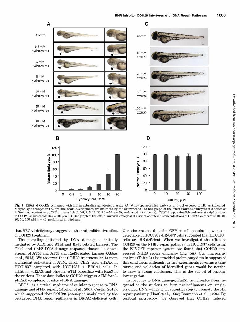
Zebrafish Genotoxicity Assay. Zebrafish (Danio rerio) wereobtained from Zebrafish Core Facility of Taipei Medical Universityand maintained at 28°C on a 14 hours light/10 hours dark cycle.Embryos were incubated at 28°C, and different developmental stageswere determined as described (Westerfield, 1993). Fifteen wild-typeembryos each were treated with concentrations of HU (0, 5, 10, 20,50 mM) or COH29 (0, 10, 20, 50, 100 mM) at 20 hours of postfertilizationto evaluate the mutagenic effect. Treated embryos were observed at2, 3, 4, 5, and 6 days postfertilization (dpf). At 6 dpf, the percentageexhibiting developmental abnormalities and the survival rate weredetermined. Embryos were observed using an Olympus IX70-FLinverted fluorescence microscope (Scientific Solutions AmericasCorp., Waltham, MA). Images were taken using SPOT digital camerasystem (Diagnostic Instruments, Sterling Heights, MI) and assem-bled with ImageJ Software (Schneider et al., 2012).
Microarray Analysis. For microarray analysis, HCC1937 andHCC1937 1 BRCA1 cells were treated with 10 mM COH29 or vehiclefor 24 hours. Total RNA was extracted using TRIzol reagent(Invitrogen). Synthesis and labeling of complementary RNA (cRNA)targets, hybridization of GeneChips, and signal detection were carriedout by the Integrated Genomics Core Facility at City of Hope. TheAffymetrix Human Gene 1.0 ST Array (Affymetrix, Santa Clara, CA)was used for microarray gene expression profiles. The microarray wascarried out using Ambion’s WTExpression kit (Life Technologies) andAffymetrix’s GeneChip Terminal labeling system. Briefly, 100 ng oftotal RNA was used to start the first-strand cDNA synthesis using anengineered random primer plus polyT7 promoter. After the secondstrand cDNA synthesis, the antisense cRNA (in vitro transcription)was generated using T7 RNA polymerase. Then 10 mg of cRNA wasused to start the second cycle of cDNA synthesis using randomprimers plus dUTP and dNTP. The single-strand cDNA was frag-mented and then end-labeled with biotinylated nucleotides in thepresence of terminal deoxynucleotidyl transferase using Affymetrix’sWT Terminal Labeling kit. Labeled single-stranded cDNA (5 mg) washybridizedwith anAffymetrixHumanGene 1.0 ST array, and the arraywas scanned using an Affymetrix GeneChip Scanner 3000 7G. Datahave been deposited into NCBIGeneExpressionOmnibus [GSE55004].
Statistical Processing of Microarray Data. Microarray sam-ples were Robust Multi-Array Analysis normalized (Irizarry et al.,2003) using Partek Genomics Suite (Version 6.6; Partek, Inc.), andgenes were defined as differentially expressed if they showed at leasta 1.2-fold change and a false discovery rate of ,0.05. False discoveryrate values were calculated using the method of Benjamini andHochberg (1995) from the distribution of analysis of variance withLinear Contrast P values. Gene ontology (GO) (Ashburner et al., 2000)enrichment analysis was performed within PartekGenomics Suite, andGO categories were defined significant by Fisher’s exact test P , 0.05.
ResultsCOH29 Targets BRCA1-Defective Human Cancer
Cells. Our previous data showed the broad antitumor activityof COH29 in the NCI-60 cell line panel and that multiplehuman breast cancer cell lines, as well as human ovariancancer cell lines, are sensitive to COH29 (Zhou et al., 2013).Breast and ovarian cancers occur with a greater frequency incarriers of a mutant BRCA1 gene than in the generalpopulation (Wooster and Weber, 2003). We therefore investi-gated the activity of COH29 in several cell lines with differingBRCA1 status, including OV90 (BRCA1 wild-type), UWB1.289(BRCA1-mutant), HCC1937 (BRCA1-mutant), and HCC19371 BRCA1 cells. As shown in Fig. 1A, the UWB1.289 ovariancancer cell line, which expresses truncated BRCA1 protein as
a result of the homozygous 2594delCmutation (DelloRusso et al.,2007), was more sensitive to COH29 (IC50: 12.30 6 1.15 mM)than the OV90 human ovarian cancer cell line that expresseswild-type BRCA1 (IC50: 31.57 6 3.35 mM). We further stablyexpressed BRCA1 in UWB1.289 ovarian cancer cells, whichresulted in a more resistant phenotype in response to COH29treatment comparedwith the parentalUWB1.289 cells (Fig. 1B).Likewise, similar results were observed in an isogenic pair ofhuman breast cancer cell lines. HCC1937 cells were moresensitive to COH29 than their BRCA1 wild type–expressingcounterpart (HCC1937 1 BRCA1) (Fig. 2A). The sensitivity ofBRCA1-deficient cells to COH29 was further tested in anorthotopic tumor explantmodel. The growth ofHCC1937 tumorsimplanted into mouse mammary fat pads was significantly(47.0%; P 5 0.0007) suppressed by daily oral dosing with 400mg/kg COH29 compared with vehicle by day 28 (Fig. 2B). Incontrast, the growth of tumors established with the isogenicHCC1937 1 BRCA1 cells in COH29-treated mice was notsignificantly different from that in vehicle controls at thesame time point (34.3%; P 5 0.1577) (Fig. 2C). As theHCC1937 1 BRCA1–bearing animals were sacrificed perinstitutional guidelines at this time, no further comparisons
between the effect of COH29 on the growth of the HCC1937deficient xenografts and HCC1937 1 BRCA xenografts couldbe made. The HCC1937 xenografts were continued for a totalof 60 days, however, in which the suppression of tumor growthby COH29 continued (data not shown). The in vitro data
Fig. 1. BRCA1 status affects COH29 cytotoxicity in ovarian cancer cells.(A) Dose-response curves for ovarian cancer cells expressing wild-typeBRCA1 (OV90) or mutant BRCA1 (UWB1.289) incubated with COH29 for72 hours. (B) Dose-dependent effects of COH29 on cell viability inUWB1.289 and UWB1.289 + BRCA1 cells. Cell viability was assessed byMTS assay. UWB1.289 + BRCA1 is a stable cell line derived from BRCA1-null UWB1.289 described in Materials and Methods. The points depictedrepresent an average of three independent experiments with error barsindicated.
Fig. 2. BRCA1 status affects COH29 antitumor activity in breast cancercells. (A) Cell viability of COH29 in HCC1937 and HCC1937 + BRCA1cells assessed by MTS assay, and growth of tumor explants wasestablished with HCC1937 (B) and HCC1937 + BRCA1 (C) cells in themammary fat pads of female NOD scid g (NSG) mice. Mice were treateddaily with 400 mg/kg COH29 or vehicle as indicated. Results are the mean6 standard error of tumor measurements from four mice per group.
RNR Inhibitor COH29 Interferes with DNA Repair Pathways 999
indicated COH29 is more potent in BRCA1-defective cells.Among the IC50 values shown in Table 1, COH29 showed 4.8times more potency in HCC1937 cells compared withHCC1937 1 BRCA1. Therefore, we used these two cell linesin the subsequent experiments to investigate the cause of thedifferential sensitivity to COH29.Effect of COH29 on DNA Damage Checkpoints. Next,
we evaluated the effect of COH29 on DNA damage signalingin HCC1937 and HCC1937 1 BRCA1 cells. COH29 inducedsignificant phosphorylation of checkpoint kinase proteinsChk1 and Chk2 and increased the level of the DSB markerg-H2AX in both cell lines (Fig. 3A). Notably, COH29 triggeredmore obvious signaling in HCC1937 cells compared withHCC1937 1 BRCA1 cells in the same concentration range. Asimilar effect was also detected with HU treatment (Fig. 3B). Ithas been reported that foxo3 is necessary for ATM-mediatedapoptotic signaling after DNA damage (Chung et al., 2012). Asshown in Fig. 4A, induction of accumulation of p-ATM, gH2AX,foxo3, and its target protein p27 in the nucleus in response toCOH29was also observed. Furthermore, we found that gH2AXand p-ATM colocalize with foxo3 in the nucleus by confocalimmunofluorescence microscopy (Fig. 4, B and C). These datasuggest COH29 activates DNA damage signaling and re-cruitment of activated-ATM and foxo3 at DNA damage sites.Differential Gene Expression in COH29-Treated
BRCA1-Defective Human Breast Cancer Cells. Wenext performed genome-wide microarray analysis using theAffymetrix GeneChip microarray platform to identify thegene expression profiles and pathways affected by COH29treatment. The RNA expression profile of COH29 treatedHCC1937 breast cancer cells lacking BRCA1 was comparedwith that of COH29 treated HCC1937 1 BRCA1 cells. BothHCC1937 and HCC1937 1 BRCA1 cells showed GOenrichment for DNA repair genes (Table 2; P values rangingfrom 0.0046 to 0.0069) after exposure to 10 mM COH29 for24 hours. These data suggest COH29 may interfere with severalDNA repair pathways. This enrichment remained significantfor the various subsets of DNA repair genes, including DNAligation involved in DNA repair, but not HR repair geneswhen COH29-treated HCC1937 and HCC1937-BRCA1 cellswere compared. This finding is consistent with what weobserved in the NHEJ and HR repair reporter assaysdescribed as follows.Effect of COH29 on DNA Double-Strand Breaks
Repair. DSBs can be repaired either by the HR or NHEJpathways. Previous studies have shown that the EJ5- andDR-GFP reporter assays could be used to measure theability of NHEJ and HR repair respectively (Bennardoet al., 2008). We next sought to elucidate the role of COH29in DSB DNA repair by using integrated EJ5-GFP and DR-GFP reporters in HCC1937 cells. In this system, transient
expression of I-Scel induces DSBs, which if repaired resultsin the generation of GFP1 cells. GFP signal was hardlydetected in HCC1937-DR-GFP cells, whereas red fluores-cent protein signal indicated successful transfection (Fig.5B), consistent with the role of BRCA1 as an essentialcomponent of HR repair and indicating HR deficiency inBRCA1-mutant HCC1937 cells. In addition, we found thatCOH29 suppressed NHEJ repair efficiency in a concentration-dependent manner as shown by reduction in the percent-age of GFP1 cells in HCC1937-EJ5-GFP assay (Fig. 5B).These data suggest that the COH29-suppressed NHEJrepair pathway may also contribute to the accumulation ofDSBs in HR-deficient HCC1937 cells. Recruitment ofRad51 protein at lesions is a well documented step in theHR repair process to facilitate DNA damage repair (Dengand Brodie, 2000). Indeed, we detected obvious Rad51 fociformation in the nucleus in COH29-treated HCC1937/BRCA1 cells compared with COH29-treated HCC1937 cells(Fig. 5C). Similar effects were also observed in HU-treated
TABLE 1Comparison of the effect of COH29 in several cell lines
Fig. 3. COH-29 treatment activates DNA damage checkpoint. (A) Theeffect of COH29 treatment on DNA damage checkpoint proteins inHCC1937 and HCC1937 + BRCA1 human breast cancer cells assessed byWestern blot. (B) The effect of HU on DNA-damage response (DDR)–associated proteins was assessed by Western blot analysis. Cells weretreated with COH29 at the indicated doses for 24 hours, and cell lysateswere subjected to immunoblotting using the indicated antibodies.
cells. Taken together, these data further support ourhypothesis that BRCA1 may be the key player in de-termining the sensitivity of cancer cells to COH29.Genotoxicity of COH29 in Embryos of Zebrafish. We
next assessed the genotoxic effect of COH29 in wild-typezebrafish embryos treated for 1–7 dpf (day postfertilization)with a range of doses of COH29 (0–100 mM). HU (0–50 mM)was included as a positive control because it is known to causedevelopmental defects. As expected, HU caused defects inthe eyes and the heart by 4 dpf (Fig. 6A) and resulted ina dose-dependent increase in the number of mutant embryos(Fig. 6B). It is noteworthy that no developmental defects(Fig. 6C) or decreases in viability (Fig. 6D) were observed in
the presence of COH29, indicating that COH29 exhibitsantitumor activity without causing genotoxicity.
DiscussionIn this study, we sought to further define the biologic effects
of the novel RNR inhibitor COH29, which effectively inhibitsproliferation of various cancer cell lines, especially ovariancancer and leukemic cells, and overcomes resistance to theRNR inhibitor hydroxyurea (Zhou et al., 2013). Building onour initial observation that the BRCA1-deficient ovariancancer cell line UWB1.289 was particularly sensitive toCOH29, we determined that BRCA1 status itself could
Fig. 4. Effect of COH29 on colocalization of DNA-damage response (DDR)–related proteins. (A) The effect of COH29 on DDR-associated proteins wasassessed in cytoplasm and nucleus by Western blot analysis. Cells were treated with COH29 at the indicated doses for 24 hours, and cell lysates weresubjected to immunoblotting (IB) using the indicated antibodies. FOXO3 activity is indicated by the levels of its downstream target p27Kip1.Glyceraldehyde-3-phosphate dehydrogenase (GAPDH) and lamin A/C represent the fractionation and loading controls of cytosolic (C) and nuclear (N)extracts. (B) Phospho-ATM, gH2AX, together with foxo3 in the nucleus, were assessed by indirect immunofluorescence assay. DAPI, 49,6-diamidino-2-phenylindole; DMSO, dimethylsulfoxide.
RNR Inhibitor COH29 Interferes with DNA Repair Pathways 1001
account for this effect. Reconstitution of BRCA1 activity in theHCC1937 human breast cancer cell line, which expressesa truncated, inactive BRCA1 protein (Tomlinson et al., 1998)in the stable transfectant clone HCC1937 1 BRCA1 bluntedresponse to COH29 in vitro and in vivo. In addition, siRNA
knockdown of BRCA1 increased sensitivity to COH29 inA2780 (BRCA1 wild-type) cells. As shown in SupplementalFig. 1, 72-hour treatment with COH29 resulted in lowersurvival in A2780 cells transfected with BRCA1 siRNA thanin those transfected with control siRNA. These data suggested
TABLE 2Gene ontology enrichment of genes downregulated by COH29 treatment
Comparison COH29 effect inBRCA1-deficient cells
COH29 effect inBRCA1 wild-type cells
BRCA1 wild-type vs.BRCA1 deficient cells
Treatment groups HCC1937-COH29 vs.HC1937
HCC1937 1 BRCA1-COH29 vs.HC1937 1 BRCA1
HCC1937 1 BRCA1-COH29 versusHCC1937-COH29
DNA repair, P 0.018 1.6 � 1025 0.001DNA ligation involved in
DNA repair, P0.00065 0.0066 0.06
Double-strand break repair vianonhomologous end joining, P
0.049 0.041 0.04
Double-strand break repair viahomologous recombination, P
0.0069 0.0046 0.26
Double-strand break repair, P 0.0015 6.9 � 1025 0.3
Fig. 5. Effect of COH29 on DNA repair, and expression of DSBmarkers. Flow cytometric analysis of (A) HCC1937-EJ5GFP and (B) HCC1937 + BRCA1-DRGFP cells treated with 0, 0.25, and 0.5 of the IC50 concentration of COH29, with the normalized percentage of GFP-positive cells shown to the right.*P , 0.05, compared with COH-29 untreated cells. (C) The effect of COH29 on RAD51 and g-H2AX foci was assessed by immunofluorescence assaydescribed in Materials and Methods. CTL, control; DAPI, 49,6-diamidino-2-phenylindole; RFP, red fluorescent protein.
that BRCA1 deficiency exaggerates the antiproliferative effectof COH29 treatment.The signaling initiated by DNA damage is initially
mediated by ATM and ATM and Rad3-related kinases. TheChk1 and Chk2 DNA-damage response kinases lie down-stream of ATM and ATM and Rad3-related kinases (Abbaset al., 2013). We observed that COH29 treatment led to moresignificant activation of ATM, Chk1, Chk2, and gH2AX inHCC1937 compared with HCC1937 1 BRCA1 cells. Inaddition, gH2AX and phospho-ATM colocalize with foxo3 inthe nucleus. These data indicate COH29 triggers ATM-foxo3-gH2AX complexes at sites of DNA damage.BRCA1 is a critical mediator of cellular response to DNA
damage and of HR repair, (Moeller et al., 2009; Curtin, 2012),which suggested that COH29 potency is modulated by theperturbed DNA repair pathways in BRCA1-deficient cells.
Our observation that the GFP 1 cell population was un-detectable inHCC1937-DR-GFP cells suggested that HCC1937cells are HR-deficient. When we investigated the effect ofCOH29 on the NHEJ repair pathway in HCC1937 cells usingthe EJ5-GFP reporter system, we found that COH29 sup-pressed NHEJ repair efficiency (Fig. 5A). Our microarrayanalysis (Table 2) also provided preliminary data in support ofthis conclusion, although further experiments covering a timecourse and validation of identified genes would be neededto draw a strong conclusion. This is the subject of ongoinginvestigation.In response to DNA damage, Rad51 translocates from the
cytosol to the nucleus to form nucleofilaments on single-stranded DNA, which is an essential step to promote the HRrepair pathway (Haaf et al., 1995; Baumann et al., 1996). Byconfocal microscopy, we observed that COH29 induced
Fig. 6. Effect of COH29 compared with HU in zebrafish genotoxicity assay. (A) Wild-type zebrafish embryos at 4 dpf exposed to HU as indicated.Morphologic changes in the eye and heart development are indicated by the arrowheads. (B) Bar graph of the effect (mutant embryos) of a series ofdifferent concentrations of HU on zebrafish (0, 0.5, 1, 5, 10, 20, 50 mM; n = 50, performed in triplicate). (C) Wild-type zebrafish embryos at 4 dpf exposedto COH29 as indicated; Bar = 100 mm. (D) Bar graph of the effect (survival embryos) of a series of different concentrations of COH29 on zebrafish (0, 10,20, 50, 100 mM; n = 46, performed in triplicate).
RNR Inhibitor COH29 Interferes with DNA Repair Pathways 1003
significantly more gH2AX foci in HCC1937 cells comparedwith HCC1937 1 BRCA1 cells. In contrast, COH29 inducedRad51 nuclear foci in HCC1937 1 BRCA1 cells (Fig. 5C),suggesting that Rad51 has been recruited at damage sites torepair COH29-triggered DNA damage via the HR repairpathway in these BRCA1 wild-type cells. This effect of COH29on Rad51 is similar to that documented for HU, which isknown to stall replication forks (Petermann et al., 2010), withthe important distinction that COH29 is 20-fold more potentthan HU (Zhou et al., 2013) and is not appreciably genotoxic(Fig. 6). Taken together, these results suggested inhibition ofthe NHEJ repair pathway by COH29 could also contribute toCOH29-induced DSBs in HR-deficient HCC1937 cells.The NHEJ pathway is reported to be the major pathway for
DNA repair of radiation-induced DSBs in mammalian cells(Riballo et al., 2004). Furthermore, a previous report has alsoproposed that quiescent/slowly cycling cancer stem cells aremore likely to use the error prone NHEJ pathway, resulting inoffspring with enhanced chemoresistance and metastaticabilities after replication (Maugeri-Sacca et al., 2012). Thissuggests that targeting the NHEJ pathway may be an effectiveway to kill cancer stem cells. Therefore, NHEJ inhibition mayrepresent a potential strategy in patients with proficient NHEJto increase the response to treatment.The underlying mechanism of the effect of COH29 on DNA
repair needs further evaluation, however. For instance, it isunclear whether this is a direct effect on the DNA repairmachinery or a consequence of depletion of dNTPs resultingfrom RNR inhibition. A handful of reports have indicated thatother RNR inhibitors also affect DNA repair pathways and orcheckpoints. As mentioned, the prototypic RNR inhibitor HU,causes upregulation of Rad51, and formation of both Rad51and gH2AX foci (Petermann et al., 2010). Gemcitabine,a nucleoside analog that also inhibits the RNR large subunit(Shao et al., 2006), has been shown to be more potent inBRCA1 deficient cells, synergize with cisplatin, and induceRad51 and gH2AX foci (Alli et al., 2011). Lin and colleaguesknocked down RRM2 subunit expression and observed resultsconsistent with what we see for COH29, which is a specificRRM2 inhibitor (Zhou et al., 2013), activation of Chk1, andupregulation of gH2AX (Lin et al., 2011). The same group hasrecently shown that the RRM1 inhibitor Triapine (3-AP, 3-aminopyridine-2-carboxaldehyde thiosemicarbazone) causedChk1 activation (Lin et al., 2014).Collectively, our data provide evidence that COH29 is an
RNR inhibitor that can activate DNA damage checkpointsand suppress DNA repair functions without significantgenotoxicity. In summary, this report provides initial evi-dence that COH29 suppresses NHEJ repair, whether directlyor indirectly, and that in the setting of HR deficiency, such asin BRCA1 mutants, this is a particularly effective approach invitro and provides information to guide initial clinicaldevelopment of this compound.
Authorship Contributions
Participated in research design: M.-C. Chen, Zhou, Zhang, Yuan,Un, Wu, Malkas, M.C.-T. Hu, Yen.
Conducted experiments: M.-C. Chen, Zhang, Yuan, S. Hu, Chou,C.-H. Chen, Wu, Wang, Li, Su, Chung.
Performed data analysis: M.-C. Chen, Zhou, Zhang, Yuan, Chou,C.-H. Chen, Wu, X. Liu, Smith, Li, Warden, Z. Liu, Su, Chung.
Wrote or contributed to the writing of the manuscript: M.-C. Chen,Un, Chou, C.-H. Chen, Wu, Smith, Warden, M.C.-T. Hu, Yen.
References
Abbas T, Keaton MA, and Dutta A (2013) Genomic instability in cancer. Cold SpringHarb Perspect Biol 5:a012914.
Alli E, Sharma VB, Hartman AR, Lin PS, McPherson L, and Ford JM (2011) En-hanced sensitivity to cisplatin and gemcitabine in Brca1-deficient murine mam-mary epithelial cells (Abstract). BMC Pharmacol 11:7.
Ashburner M, Ball CA, Blake JA, Botstein D, Butler H, Cherry JM, Davis AP,Dolinski K, Dwight SS, and Eppig JT et al.; The Gene Ontology Consortium (2000)Gene ontology: tool for the unification of biology. Nat Genet 25:25–29.
Baumann P, Benson FE, and West SC (1996) Human Rad51 protein promotes ATP-dependent homologous pairing and strand transfer reactions in vitro. Cell 87:757–766.
Benjamini Y and Hochberg Y (1995) Controlling the false discovery rate: a practicaland powerful approach to multiple testing. J R Stat Soc, B 57:289–300.
Bennardo N, Cheng A, Huang N, and Stark JM (2008) Alternative-NHEJ isa mechanistically distinct pathway of mammalian chromosome break repair. PLoSGenet 4:e1000110.
Chung YM, Park SH, Tsai WB, Wang SY, Ikeda MA, Berek JS, Chen DJ, and Hu MC(2012) FOXO3 signalling links ATM to the p53 apoptotic pathway following DNAdamage (Abstract). Nat Commun 3:1000.
Curtin NJ (2012) DNA repair dysregulation from cancer driver to therapeutic target.Nat Rev Cancer 12:801–817.
DelloRusso C, Welcsh PL, Wang W, Garcia RL, King MC, and Swisher EM (2007)Functional characterization of a novel BRCA1-null ovarian cancer cell line in re-sponse to ionizing radiation. Mol Cancer Res 5:35–45.
Deng CX and Brodie SG (2000) Roles of BRCA1 and its interacting proteins. Bio-Essays 22:728–737.
Gottipati P, Vischioni B, Schultz N, Solomons J, Bryant HE, Djureinovic T, Issaeva N,Sleeth K, Sharma RA, and Helleday T (2010) Poly(ADP-ribose) polymerase ishyperactivated in homologous recombination-defective cells. Cancer Res 70:5389–5398.
Haaf T, Golub EI, Reddy G, Radding CM, and Ward DC (1995) Nuclear foci ofmammalian Rad51 recombination protein in somatic cells after DNA damageand its localization in synaptonemal complexes. Proc Natl Acad Sci USA 92:2298–2302.
Hehlmann R (2003) Current CML therapy: progress and dilemma. Leukemia 17:1010–1012.
Helleday T, Petermann E, Lundin C, Hodgson B, and Sharma RA (2008) DNA repairpathways as targets for cancer therapy. Nat Rev Cancer 8:193–204.
Hu T, Chung YM, Guan M, Ma M, Ma J, Berek JS, and Hu MC (2014)Reprogramming ovarian and breast cancer cells into non-cancerous cells bylow-dose metformin or SN-38 through FOXO3 activation (Abstract). Sci Rep4:5810.
Irizarry RA, Hobbs B, Collin F, Beazer-Barclay YD, Antonellis KJ, Scherf U,and Speed TP (2003) Exploration, normalization, and summaries of high densityoligonucleotide array probe level data. Biostatistics 4:249–264.
Keshelava N, Frgala T, Krejsa J, Kalous O, and Reynolds CP (2005) DIMSCAN:a microcomputer fluorescence-based cytotoxicity assay for preclinical testing ofcombination chemotherapy. Methods Mol Med 110:139–153.
Lin ZP, Lee Y, Lin F, Belcourt MF, Li P, Cory JG, Glazer PM, and Sartorelli AC(2011) Reduced level of ribonucleotide reductase R2 subunits increases dependenceon homologous recombination repair of cisplatin-induced DNA damage. MolPharmacol 80:1000–1012.
Lin ZP, Ratner ES, Whicker ME, Lee Y, and Sartorelli AC (2014) Triapine disruptsCtIP-mediated homologous recombination repair and sensitizes ovarian cancercells to PARP and topoisomerase inhibitors. Mol Cancer Res 12:381–393.
Maugeri-Saccà M, Bartucci M, and De Maria R (2012) DNA damage repair pathwaysin cancer stem cells. Mol Cancer Ther 11:1627–1636.
Miknyoczki SJ, Jones-Bolin S, Pritchard S, Hunter K, Zhao H, Wan W, Ator M,Bihovsky R, Hudkins R, and Chatterjee S et al. (2003) Chemopotentiation oftemozolomide, irinotecan, and cisplatin activity by CEP-6800, a poly(ADP-ribose)polymerase inhibitor. Mol Cancer Ther 2:371–382.
Moeller BJ, Pasqualini R, and Arap W (2009) Targeting cancer-specific syntheticlethality in double-strand DNA break repair. Cell Cycle 8:1872–1876.
Petermann E, Orta ML, Issaeva N, Schultz N, and Helleday T (2010)Hydroxyurea-stalled replication forks become progressively inactivated andrequire two different RAD51-mediated pathways for restart and repair. MolCell 37:492–502.
Pierce AJ, Hu P, Han M, Ellis N, and Jasin M (2001) Ku DNA end-binding proteinmodulates homologous repair of double-strand breaks in mammalian cells. GenesDev 15:3237–3242.
Platt OS (2008) Hydroxyurea for the treatment of sickle cell anemia. N Engl J Med358:1362–1369.
Reichard P and Ehrenberg A (1983) Ribonucleotide reductase: a radical enzyme.Science 221:514–519.
Riballo E, Kühne M, Rief N, Doherty A, Smith GC, Recio MJ, Reis C, Dahm K, FrickeA, and Krempler A et al. (2004) A pathway of double-strand break rejoining de-pendent upon ATM, Artemis, and proteins locating to gamma-H2AX foci. Mol Cell16:715–724.
Schneider CA, Rasband WS, and Eliceiri KW (2012) NIH Image to ImageJ: 25 yearsof image analysis. Nat Methods 9:671–675.
Shao J, Zhou B, Chu B, and Yen Y (2006) Ribonucleotide reductase inhibitors andfuture drug design. Curr Cancer Drug Targets 6:409–431.
Shao J, Zhou B, Zhu L, Qiu W, Yuan YC, Xi B, and Yen Y (2004) In vitro charac-terization of enzymatic properties and inhibition of the p53R2 subunit of humanribonucleotide reductase. Cancer Res 64:1–6.
Shewach DS and Lawrence TS (2007) Antimetabolite radiosensitizers. J Clin Oncol25:4043–4050.
Tomlinson GE, Chen TT, Stastny VA, Virmani AK, Spillman MA, Tonk V, Blum JL,Schneider NR, Wistuba II, and Shay JW et al. (1998) Characterization of a breast
cancer cell line derived from a germ-line BRCA1 mutation carrier. Cancer Res 58:3237–3242.
Un F, Qi C, Prosser M, Wang N, Zhou B, Bronner C, and Yen Y (2006) ModulatingICBP90 to suppress human ribonucleotide reductase M2 induction restores sen-sitivity to hydroxyurea cytotoxicity. Anticancer Res 26 (4B):2761–2767.
Westerfield M (1993) The Zebrafish Book: A Guide for the Laboratory Use of Zebrafish(Brachydanio rerio), M. Westerfield, Eugene, OR.
Wooster R and Weber BL (2003) Breast and ovarian cancer. N Engl J Med 348:2339–2347.
Xue L, Zhou B, Liu X, Qiu W, Jin Z, and Yen Y (2003) Wild-type p53 regulates humanribonucleotide reductase by protein-protein interaction with p53R2 as well ashRRM2 subunits. Cancer Res 63:980–986.
Young CW and Hodas S (1964) Hydroxyurea: Inhibitory Effect on DNA Metab-olism. Science 146:1172–1174.
Zhou B, Su L, Hu S, Hu W, Yip ML, Wu J, Gaur S, Smith DL, Yuan YC, and SynoldTW et al. (2013) A small-molecule blocking ribonucleotide reductase holoenzymeformation inhibits cancer cell growth and overcomes drug resistance. Cancer Res73:6484–6493.
Address correspondence to: Yun Yen, Ph.D Program for Cancer Biologyand Drug Discovery, Taipei Medical University, 250 Wuxing Street, Taipei11031, Taiwan. E-mail: [email protected]
RNR Inhibitor COH29 Interferes with DNA Repair Pathways 1005