The role of macrophages in interstitiallung diseases
Giulio Rossi1, Alberto Cavazza2, Paolo Spagnolo3,4, Salvatore Bellafiore2,Elisabetta Kuhn2, Pierpaolo Carassai1, Laura Caramanico1, Gloria Montanari5,Gaia Cappiello6, Alessandro Andreani7, Francesca Bono8 andNazarena Nannini9
Number 3 in the Series “Pathology for the clinician”Edited by Peter Dorfmüller and Alberto Cavazza
Affiliations: 1Unit of Pathologic Anatomy, Azienda USL Valle d’Aosta, Regional Hospital “Parini”, Aosta, Italy.2Unit of Pathologic Anatomy, Azienda Arcispedale S. Maria Nuova/IRCCS, Reggio Emilia, Italy. 3MedicalUniversity Clinic, Canton Hospital Baselland University of Basel, Basel, Switzerland. 4Section of RespiratoryDiseases, Dept of Cardiac, Thoracic and Vascular Sciences, University of Padua, Padua, Italy. 5Unit ofPneumology, Azienda Arcispedale S. Maria Nuova/IRCCS, Reggio Emilia, Italy. 6Unit of Pneumology, AziendaUSL Modena, Civic Hospital of Mirandola, Mirandola, Italy. 7Unit of Pneumology, Azienda Ospedaliero-Universitaria Policlinico, Modena, Italy. 8Unit of Pathologic Anatomy, San Gerardo Hospital, IRCCS, Monza,Italy. 9Unit of Pathologic Anatomy, Azienda ULSS 13, Hospital of Dolo, Dolo, Italy.
Correspondence: Giulio Rossi, Unit of Pathologic Anatomy, Azienda USL Valle d’Aosta, Regional Hospital“Parini”, Via Ginevra 3, 11100 Aosta, Italy. E-mail: [email protected]
@ERSpublicationsMorphology and localisation of macrophages in the lungs is helpful in the diagnosis of interstitiallung disease http://ow.ly/7bci30bBwmQ
Cite this article as: Rossi G, Cavazza A, Spagnolo P, et al. The role of macrophages in interstitial lungdiseases. Eur Respir Rev 2017; 26: 170009 [https://doi.org/10.1183/16000617.0009-2017].
ABSTRACT The finding of collections of macrophages/histiocytes in lung biopsy and bronchoalveolarlavage is relatively common in routine practice. This morphological feature in itself is pathological, but theexact clinical significance and underlying disease should be evaluated together with clinical data,functional respiratory and laboratory tests and imaging studies.
Morphological characteristics of macrophages and their distribution along the different pulmonarystructures should be examined carefully by pathologists. Indeed, haemosiderin-laden macrophages areassociated with smoking-related diseases when pigment is fine and distribution is bronchiolocentric, whilealveolar haemorrhage or pneumoconiosis are the main concerns when pigment is chunky or coarse andthe macrophages show an intra-alveolar or perilymphatic location, respectively. In the same way,pulmonary accumulation of macrophages with foamy cytoplasm is generally associated with pathologiesleading to broncho-bronchiolar obstruction (e.g. diffuse panbronchiolitis, hypersensitivity pneumonia orcryptogenic organising pneumonia) or alternatively to exogenous lipoid pneumonia, some drug toxicity(e.g. amiodarone exposure or toxicity) and metabolic disorders (e.g. type B Niemann–Pick disease).
This pathology-based perspectives article is aimed at concisely describing the diagnostic possibilitieswhen faced with collection of macrophages in lung biopsy and cytology.
IntroductionIn the lung, macrophages and histiocytes play a key role in inflammatory and phagocytic processesthrough complex production of cytokines and cellular interaction [1, 2].
Recent studies have demonstrated dynamic changes of circulating monocytes, lung alveolar macrophagesand interstitial macrophages with M1 (pro-inflammatory) and M2 (anti-inflammatory) phenotypes inpreclinical models of interstitial lung diseases (ILDs) [3].
Nevertheless, the presence of collections of macrophages is considered a pathologic feature, although theexact clinical significance may range in a single case. Indeed, the occurrence of several macrophages in thelung often represent an incidental finding, while in other cases macrophages are indeed the problem.
Among ILDs, the careful evaluation of the morphological characteristics and the exact localisation of themacrophages disclosed in a lung biopsy may be fundamental diagnostic clues for pathologists. Pathologistshave different diagnostic methodologies to identify macrophage-based lung lesions with various diagnosticyields, including bronchoalveolar lavage (BAL), transbronchial biopsy, transbronchial cryobiopsy andsurgical lung biopsy (table 1) [4, 5].
Several histochemical stains and immunohistochemical markers are used to recognise histiocytes incytology and biopsy. Among histochemical stains oil red O or Sudan black are useful in recognisinglipid-rich histiocytes on fresh material (alcohol-based fixation used for conventional haematoxylin–eosinstain digest the lipids that are not visible in formalin-fixed paraffin-embedded samples), while Perls’Prussian blue stains the iron-laden macrophages. Periodic acid–Schiff (PAS) and May–Giemsa stains arehelpful in differentiating some metabolic diseases. Among immunohistochemical markers, CD68, S-100protein, CD1a, langerin, CD56, CD163, CD4, lysozyme, α1-antitrypsin and factor XIIIa are the mostcommon primary antibodies used to recognise different histiocytic disorders [6]. Of note, there is evidencethat human alveolar macrophages from donors are maintained in the lung parenchyma for several yearsafter lung transplantation, possibly resulting in an important mechanism for the development of chronicgraft rejection [7].
This article summarises the most important features of lung pathologies characterised by the accumulationof histiocytes (with the exclusion of granulomatous processes) and provides a practical approach to thediagnosis of ILDs from the “viewpoint” of the macrophages.
Pathologies consisting of accumulation of macrophages containing finehaemosiderin pigmentThe accumulation of yellow-brown, haemosiderin-laden, pigmented macrophages (“smokers’macrophages”) within the lumens of respiratory bronchioles and into the peribronchiolar alveolar spaces(figure 1) is seen very frequently and in the great majority of cases secondary to a cigarette smoking habit[8–11]. Macrophages initially fill the lumen of the respiratory bronchioles and peribronchiolar alveolileading to a specific type of bronchiolitis, namely respiratory bronchiolitis. This histological finding isassociated with a patchy submucosal and peribronchiolar chronic inflammation with or without mucostasisand occurs in virtually all cigarette smokers; patients are generally asymptomatic or paucisymptomatic andhistological stigmata of respiratory bronchiolitis may persist for decades after smoking cessation [9].
In some cases, particularly in heavy smokers, alveolar accumulation of pigmented macrophages extendsfrom peribronchiolar zones to the periphery of the lobules, determining an ILD affecting the upper lunglobes with centrilobular nodules and ground-glass opacities at high-resolution computed tomography(HRCT) [12–14].
This phenomenon is defined as RB-ILD when the subpleural alveoli are not involved in the process, whilethe misnomer “desquamative interstitial pneumonia” (DIP) [15] is used when the entire lobule is involvedby accumulation of smokers’ macrophages (although the differential diagnosis between RB-ILD and DIP issometimes arbitrary and has no clinical significance). Respiratory bronchiolitis, RB-ILD and DIP dorepresent a continuous spectrum of histological lesions secondary to their accumulation of macrophages
Previous articles in this series: No. 1: Ghigna MR, Mooi WJ, Grünberg K. Pulmonary hypertensive vasculopathy inparenchymal lung diseases and/or hypoxia. Eur Respir Rev 2017; 26: 170003. No. 2: Bubendorf L, Lantuejoul S, deLangen AJ, et al. Nonsmall cell lung carcinoma: diagnostic difficulties in small biopsies and cytological specimens. EurRespir Rev 2017; 26: 170007.
Received: Jan 15 2017 | Accepted after revision: April 26 2017
Conflict of interest: None declared.
Provenance: Commissioned article, peer reviewed.
https://doi.org/10.1183/16000617.0009-2017 2
PATHOLOGY FOR THE CLINICIAN | G. ROSSI ET AL.
into the peripheral zone of the lungs and secondary to cigarette smoking in the great majority of cases(also collected under the umbrella term “smoking-related ILDs”).
A subset of patients with RB-ILD and DIP have an insidious presentation with exertional dyspnoea,symptomatic wheezing, persistent cough and concomitant functional impairment and imagingabnormalities at chest radiography and HRCT [13–15]. In most cases, RB-ILD and DIP show a relativelystable clinical course with long-term outcomes and good prognosis.
a) b)
c) d)
FIGURE 1 a) Smokers’ macrophages show a fine, golden haemosiderin cytoplasmic pigment; b) in respiratorybronchiolitis (RB), smokers’ macrophages are located in the bronchiolar lumen and peribronchiolar alveoli; inc) RB-associated interstitial lung disease and d) desquamative interstitial pneumonia, smokers’ macrophagesprogressively expand to the peripheral alveoli.
TABLE 1 Diagnostic yield of the different sampling techniques in macrophage-rich pulmonary diseases
–: almost never diagnostic; +: weakly diagnostic; ++: moderately diagnostic; +++: almost always diagnostic. #: bronchoalveolar lavage is helpfulin supporting the diagnosis once clinically suspected, but rarely diagnostic per se.
https://doi.org/10.1183/16000617.0009-2017 3
PATHOLOGY FOR THE CLINICIAN | G. ROSSI ET AL.
From the pathologist’s viewpoint, it is important to underline that respiratory bronchiolitis, RB/ILD andDIP represent diagnoses of exclusion in all cases [13–15]. In other words, since the accumulation ofsmokers’ macrophages in a lung biopsy or BAL are nonspecific morphological features, it is mandatory toascertain that they represent the real and unique problem of the patient. Indeed, respiratory bronchiolitis,RB-ILD and DIP may be observed concurrently in other more aggressive diseases, namely lung cancer andidiopathic pulmonary fibrosis. Keeping this in mind, a diagnosis of RB-ILD or DIP on small biopsy orBAL may be considered only in an adequate clinicoradiologic context.
In addition, both primary and metastatic lung tumours are rarely associated with a peculiar discohesivegrowth pattern resulting in airspace filling by macrophage-like tumour cells, closely simulating RB-ILDand DIP. The malignant cytology of the tumour nuclei and immunohistochemical stains are the clues indifferential diagnosis [16].
However, a subset of DIP occurs in nonsmokers and is related to occupational inhaled toxins, drugtoxicity and connective tissue diseases [17]. In addition, the DIP pattern has been associated with geneticdiseases, such as ABCA3 mutations [18].
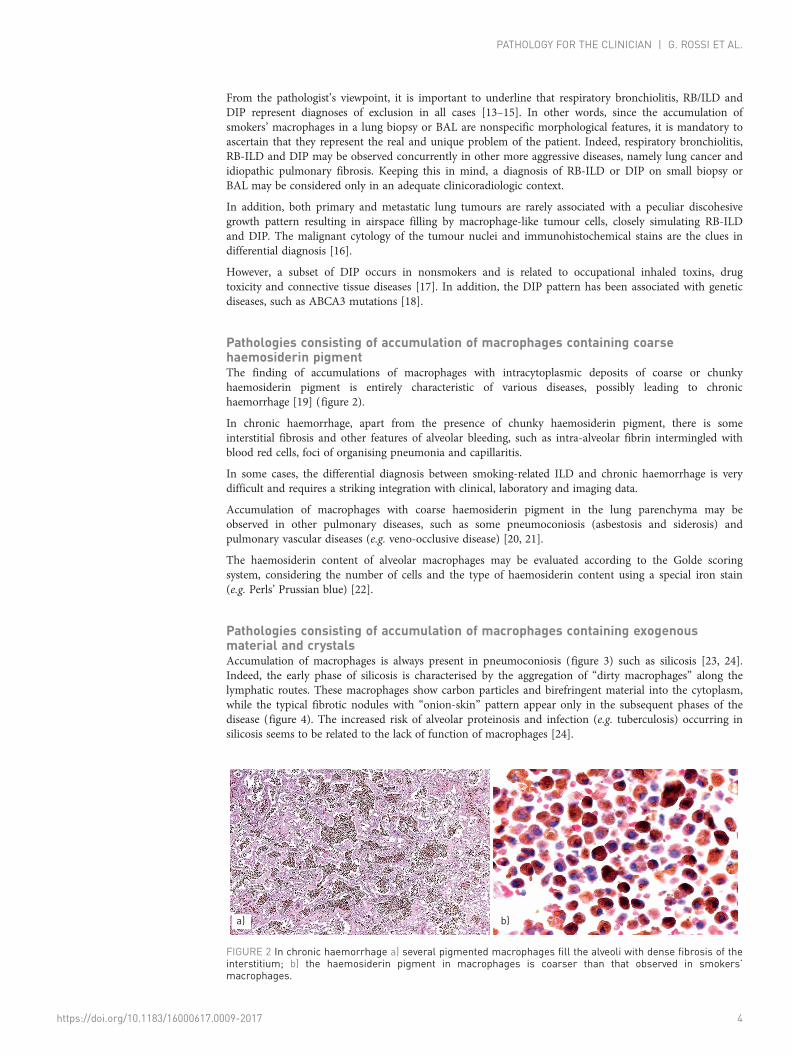
Pathologies consisting of accumulation of macrophages containing coarsehaemosiderin pigmentThe finding of accumulations of macrophages with intracytoplasmic deposits of coarse or chunkyhaemosiderin pigment is entirely characteristic of various diseases, possibly leading to chronichaemorrhage [19] (figure 2).
In chronic haemorrhage, apart from the presence of chunky haemosiderin pigment, there is someinterstitial fibrosis and other features of alveolar bleeding, such as intra-alveolar fibrin intermingled withblood red cells, foci of organising pneumonia and capillaritis.
In some cases, the differential diagnosis between smoking-related ILD and chronic haemorrhage is verydifficult and requires a striking integration with clinical, laboratory and imaging data.
Accumulation of macrophages with coarse haemosiderin pigment in the lung parenchyma may beobserved in other pulmonary diseases, such as some pneumoconiosis (asbestosis and siderosis) andpulmonary vascular diseases (e.g. veno-occlusive disease) [20, 21].
The haemosiderin content of alveolar macrophages may be evaluated according to the Golde scoringsystem, considering the number of cells and the type of haemosiderin content using a special iron stain(e.g. Perls’ Prussian blue) [22].
Pathologies consisting of accumulation of macrophages containing exogenousmaterial and crystalsAccumulation of macrophages is always present in pneumoconiosis (figure 3) such as silicosis [23, 24].Indeed, the early phase of silicosis is characterised by the aggregation of “dirty macrophages” along thelymphatic routes. These macrophages show carbon particles and birefringent material into the cytoplasm,while the typical fibrotic nodules with “onion-skin” pattern appear only in the subsequent phases of thedisease (figure 4). The increased risk of alveolar proteinosis and infection (e.g. tuberculosis) occurring insilicosis seems to be related to the lack of function of macrophages [24].
a) b)
FIGURE 2 In chronic haemorrhage a) several pigmented macrophages fill the alveoli with dense fibrosis of theinterstitium; b) the haemosiderin pigment in macrophages is coarser than that observed in smokers’macrophages.
https://doi.org/10.1183/16000617.0009-2017 4
PATHOLOGY FOR THE CLINICIAN | G. ROSSI ET AL.
In different forms of pneumoconiosis, the exogenous material is covered by iron leading to ferruginousbodies. The asbestos body is a peculiar type of ferruginous body in which the exogenous material isrepresented by the asbestos corpuscles. This is unique and consists of a transparent, not birefringent andlinear corpuscle [8].
Intravenous talcosis is another form of pneumoconiosis caused by the injection of material containingsilicates and characterised by the intravascular accumulation of exogenous material [25].
Chronic aspiration pneumonitis is a relatively common, but often underrated pathology characterised bythe finding of mono-/multinucleated macrophages with intracytoplasmic alimentary digested debris or
a)
b) c)
FIGURE 3 a) The accumulation of haemosiderin pigment may be observed in some cases of pneumoconiosis,such as b) asbestosis with the presence of asbestos bodies showing a linear non-birifringent “core” attractingferrous material or c) pneumosiderosis (welder’s lung) with bronchiolitis and sheets of macrophages havingcytoplasmic iron pigment.
a) b)
FIGURE 4 Silicosis is characterised by a) irregular, stellate-shaped nodules along the lymphatic routes, withb) “dirty” macrophages and comparison of dense fibrosis in chronic phases.
https://doi.org/10.1183/16000617.0009-2017 5
PATHOLOGY FOR THE CLINICIAN | G. ROSSI ET AL.
other exogenous material in the background of chronic bronchiolocentric damage with bronchiolarmetaplasia and inflammatory reaction. Exogenous lipoid pneumonitis is a peculiar variant of aspirationpneumonitis due to prolonged aspiration of mineral oils leading to a pulmonary histological lesionidentical to the subcutaneous paraffinoma with areas of dense fibrosis including clear vacuoles of differentsize, often surrounded by foamy macrophages (figure 5). Examination of BAL fluid may be used todiagnose exogenous lipoid pneumonitis when numerous lipid-laden macrophages are disclosed in thecorrect clinicoradiological setting [26].
The finding of multinucleated macrophages with intracytoplasmic cholesterol crystals/clefts is a frequentand nonspecific histological finding in lung biopsies. Nevertheless, their presence in the background of acellular (lymphocyte and plasma cell-rich infiltrate) interstitial pneumonitis is an important morphologicalcircumstantial proof favouring hypersensitivity pneumonitis (allergic extrinsic alveolitis), particularly whenthe inflammatory process is accentuated in the centrolobular regions [27, 28].
The finding of numerous macrophages with intracytoplasmic cholesterol clefts under the pleura or in theperipheral regions of the lung should raise the possibility of a pulmonary alveolar proteinosis in thehealing phase. In this case, a meticulous search for focal areas including alveoli containing thecharacteristic eosinophilic, PAS-positive material is mandatory to confirm the diagnosis [29].
Rarely, primary pulmonary lymphomas (e.g. marginal zone or mucosa-associated lymphoid tissuelymphoma and lymphoplasmacytic lymphoma) or lung involvement by plasma cell proliferations (multiplemyeloma or plasma cell disorders) and lymphomas are associated with aggregates of macrophagescontaining intracytoplasmic eosinophilic, refractile, rod-shaped crystals due to intralysosomal accumulationof immunoglobulin light chain crystals (figure 6). This condition is called crystal-storing histiocytosis [30]and has been described in some reactive processes secondary to non-neoplastic immune-mediated diseases(i.e. rheumatoid arthritis) or the use of clofazimine for the treatment of leprosy [31].
Chronic eosinophilic pneumonitis [32] is characterised by the accumulation of intra-alveolar eosinophilicgranulocytes, macrophages and fibrin. Macrophages may contain eosinophilic granules leading to abrilliant pink staining of the cytoplasm. The finding of these macrophages, particularly when associatedwith fibrin deposits and organising pneumonia, is an important clue in suspecting a chronic eosinophilicpneumonia, even when eosinophilic granulocytes are very few or absent (possibly due to steroidtreatment).
Pathologies consisting of accumulation of foamy macrophagesIn routine practice, the presence of foamy histiocytes in BAL fluid and lung biopsies is a very frequentfinding (figure 7). Foamy macrophages represent an incidental and poorly significant sign in the greatmajority of cases. Indeed, it is very common to observe foamy macrophages akin to necrotic processes (e.g.resected lung tumours after neoadjuvant chemotherapy) peripherally to an obstructive pathology of thebronchial tree, along various levels and due to different causes, or in the setting of areas of parenchymalconsolidation (“golden pneumonia”) at the periphery of a lung tumour occluding the bronchial lumen.
However, in some circumstances foamy macrophages are an important “spy” of an underlying subtleobstructive respiratory pathology, possibly not considered at a first glance by the pathologist.
For example, endoalveolar foamy macrophages are frequently noted in bronchiolitis and small airwaydiseases, a complex group of diseases characterised by a clinically and functional obstructive picture
a) b)
FIGURE 5 a) Lipoid exogenous pneumonia on bronchoalveolar lavage is characterised by macrophages withseveral intracytoplasmic vacuoles of various sizes; b) in chronic phases, the lesion is partially substituted byfibrosis and chronic inflammation, leading to a nodular mass known as paraffinoma.
https://doi.org/10.1183/16000617.0009-2017 6
PATHOLOGY FOR THE CLINICIAN | G. ROSSI ET AL.
coupled to subtle-to-evident inflammation and fibrosis of the small airways at histology [33, 34]. Inaddition, in the context of a diffuse infiltrative pneumonia, the finding of intra-alveolar foamymacrophages should lead to the consideration of hypersensitivity pneumonia [27, 28] and organisingpneumonia (bronchiolitis obliterans organising pneumonia or cryptogenic organising pneumonia) [33–35], the ILDs most commonly associated with a prominent accentuation of peribronchiolar inflammation(bronchiolitis).
A peculiar variant of small airway disease is “diffuse panbronchiolitis”, a clinicopathological entityfrequently observed in Japan and very rare in Western countries [36]. Patients present with productivecough, exertional dyspnoea, chronic sinusitis and elevated titres of cold hemagglutinins. At HRCT, diffusepanbronchiolitis shows bilateral diffuse centrolobular nodules with bronchiectasis and bronchiolectasis.Lung biopsy reveals accumulation of foamy macrophages in the lumen of the alveoli and peribronchiolarinterstitium.
These histological features are incorrectly considered to be pathognomonic of diffuse panbronchiolitis, butrarely, interstitial aggregates of foamy histiocytes may be observed in other diseases, as in the peripherallung portion of patients with cystic fibrosis, in lung involvement by idiopathic chronic inflammatory boweldiseases, some vasculitides and connective tissue diseases [37, 38]. As in many non-neoplastic lungpathologies, the diagnosis of diffuse panbronchiolitis requires a careful correlation between histology,clinical, laboratory and imaging data.
Apart from small-airway diseases, another group of pathologies characterised by the presence of foamymacrophages is represented by drug toxicities [8]. This is another poorly recognised and underrated rubricof lung diseases by clinicians and pathologists.
In the great majority of cases, drug toxicity in the lung consists of nonspecific histological features, rangingfrom acute (diffuse alveolar damage or haemorrhage) to chronic interstitial pneumonitis morphologicallyidentical to nonspecific interstitial pneumonia or even idiopathic pulmonary fibrosis (www.pneumotox.com).
The definitive recognition of a pulmonary drug toxicity requires a close correlation of histological clueswith clinical data and exclusion of other diseases justifying the lung injury. Despite this, it is quite true
a) b)
FIGURE 6 a) Crystal-storing histiocytosis with sheets of macrophages containing intracytoplasmic eosinophiliccrystal of immunoglobulin in a plasmacytoma and b) mucosa-associated lymphoid tissue-type lymphoma.
a) b)
FIGURE 7 Endoalveolar foamy histiocytes in a) hypersensitivity pneumonia and b) amiodarone-related toxicity.Images courtesy of T.V. Colby, from the Charles B. Carrington Collection, sited at the Dept of LaboratoryMedicine and Pathology, Mayo Clinic, Scottsdale, AZ, USA.
that drug toxicity should always be taken into account when pathologists face with an intra-alveolaraccumulation of foamy macrophages with scattered, sparse eosinophils and foci of organising pneumonia.
Virtually all drugs may cause accumulation of foamy macrophages in the lung, but amiodarone toxicityis by far the most commonly encountered [39]. Amiodarone is an anti-arrhythmic agent leading tolung toxicity in ∼5–10% of cases. Amiodarone is often associated with foamy changes either in themacrophages and type II pneumocytes as a sign of drug exposure, not necessarily indicatingdrug toxicity. The real significance of these histological features should be evaluated in the light ofclinical data.
Finally, the accumulation of foamy histiocytes is observed in exogenous lipoid pneumonia and in somehistiocytic disorders, some infections and metabolic diseases.
Infections characterised by the accumulation of macrophagesAccumulation of pulmonary macrophages may be the morphological clue to some infections, particularlywhen associated with granulomas, necrosis and aggregates of neutrophils or in immunocompromisedpatients.
In all these situations, it is mandatory to rigorously exclude an infection using of specific stains (e.g.methenamine silver stain, Gram stain or Ziehl–Neelsen) and possibly microbiological cultures.
In this article, we focus on two unusual infections that occasionally involve the lung, namely Whipple’sdisease and malakoplakia.
Whipple’s disease [40] is a rare infection involving several organs which is sustained by the Gram-positivebacterium Tropheryma whippelii. The disease mainly affects adult males and presents with systemicsymptoms (arthralgia and weight loss) together with gastrointestinal and neurological manifestations. Thepulmonary involvement is exceedingly rare and characterised by bronchiolocentric accumulation ofintensely PAS-positive macrophages.
Malakoplakia is an uncommon inflammatory condition characterised by the accumulation of macrophagesdue to defective lysosomal activity and microtubular assembly leading to impairment of the normalphagocytic process with accumulation of benign macrophagesis. This rare condition occurs mostcommonly in the urinary tract and is due to the accumulation of macrophages unable to destroyphagocytosed bacteria. In the lung, malakoplakia [41, 42] is often secondary to Rhodococcus equi infection,Gram-positive coccobacilli common in the environment, which may cause fatal pneumonia in horses.Occasionally, other micro-organisms are responsible for pulmonary malakoplakia.
Malakoplakia may occur in immunocompromised and immunocompetent individuals concurrently toextrapulmonary involvement.
Chest radiography shows an opacity of the upper lobes tending to cavitate and the disease is often resistantto common antibiotic therapy. The histological features (figure 8) are characterised by abscessescontaining the Gram-positive bacilli associated with dense accumulation of macrophages. Diseaseprogression is characterised by a decrease in neutrophils and the number of bacilli coupled with anincrease in macrophages. These peculiar macrophages tend to create confluent nodular tumour-like massesand are characterised by an ample eosinophilic cytoplasm (von Hansemann histiocytes) containingintracytoplasmic coccobacilli and scattered rounded von Kossa-positive inclusions consistent withMichaelis–Gutmann bodies.
Langerhans cell histiocytosis and other histiocytic disordersLangerhans cell histiocytosisLangerhans cell histiocytosis (LCH) may involve the lung as localised disease or, more rarely, as part of asystemic process [43]. Systemic disease is generally considered a clonal pathology [44, 45], while thepulmonary isolated form is generally considered a polyclonal reactive process secondary to active orpassive cigarette smoking in virtually all cases [46, 47].
Pulmonary LCH is relatively rare and generally occurs in young smoking adults with a male prevalence.∼25% of patients with LCH are asymptomatic, while cough, dyspnoea, chest pain, fever and weight lossare the most common and nonspecific symptoms. Pneumothorax is present in a quarter of cases.Respiratory function tests may return entirely normal results or show mild-to-moderate restrictive/obstructive abnormalities.
Imaging studies (chest radiographs and computed tomography scans) generally show pulmonaryalterations characterised by multiple, tiny, peribronchiolar nodules (early phase) and cysts (chronic,advanced phase) of varying and irregular shapes. These lesions are preferentially located at the upper
https://doi.org/10.1183/16000617.0009-2017 8
PATHOLOGY FOR THE CLINICIAN | G. ROSSI ET AL.
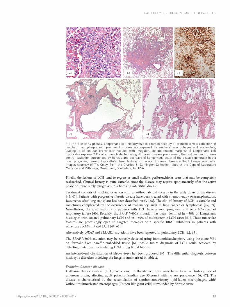
lobes, sparing the costophrenic angles [48, 49]. Ground-glass opacities and mediastinal lymphadenopathymay be observed [40], while solitary parenchymal or endobronchial nodules are very rare [50, 51]. Despiteentirely characteristic imaging findings at computed tomography, the diagnosis usually requires atransbronchial or even surgical biopsy, particularly in atypical cases. BAL fluid analysis is very helpfulwhen ⩾5% of CD1a-positive Langerhans cells are found [52, 53] (figure 9).
At histology, the early phases of LCH are characterised by collections of Langerhans histiocytes in theperibronchiolar interstitial tissue; they tend to aggregate in small nests. In contrast, the presence ofscattered and isolated Langerhans cells is a very common and nonspecific finding in smokers [51, 54].Langerhans histiocytes show a peculiar morphology (discrete amounts of pale to slightly eosinophiliccytoplasm devoid of any pigment, irregular nuclei with prominent grooves and a delicate nuclearmembrane) and a characteristic phenotype at immunohistochemistry (expression of S100 protein, CD1a,CD68 and Langerin and factor XIIIa-negative).
Langerhans cells proliferate along the bronchiolar wall and the alveolar septa leading to irregular,stellate-shaped, multiple nodules accompanied by a discrete quote of eosinophils and pigmented smokers’macrophages into the alveolar lumen and septa. With time, the central zone of the nodule develops a densefibrosis and central cavitations due to progressive destruction of the bronchiolar wall [55].
In the chronic phase, the Langerhans cells are often visible only at the periphery of the lesions and absentin the central fibrotic areas. In this mature phase of LCH, the disease shows multiple stellate nodules,some of which are cavitated and characteristically separated by normal lung parenchyma. Thesehistological alterations are easily recognisable at low magnification, and it is often possible to observeseveral lesions at difference stages of evolution (active CD1a-rich cellular nodules akin to “burnt-out”fibrotic and cavitated nodules) in the same histological slide. Occasionally, the nodules grow into thevascular walls, provoking pulmonary hypertension, which may be severe [56], or extend to the pleura,leading to pneumothorax and simulating an eosinophilic pleuritis [57].
a)
b) c)
FIGURE 8 Dense clusters of a) eosinophilic macrophages (von Hansemann cells) containing b) vonKossa-positive, rounded bodies (Michaelis–Gutmann bodies) (circled) and c) Gram-positive coccobacilli(circled).
https://doi.org/10.1183/16000617.0009-2017 9
PATHOLOGY FOR THE CLINICIAN | G. ROSSI ET AL.
Finally, the lesions of LCH tend to regress as small stellate, peribronchiolar scars that may be completelyreabsorbed. Clinical history is quite variable, since the disease may regress spontaneously after the activephase or, more rarely, progresses to a fibrosing interstitial disease.
Treatment consists of smoking cessation with or without steroid therapy in the early phase of the disease[43, 47]. Patients with progressive fibrotic disease have been treated with chemotherapy or transplantation.Recurrence after lung transplant has been described rarely [58]. The clinical history of LCH is variable andsometimes complicated by the occurrence of malignancy, such as lung cancer or lymphomas [47, 59].Nevertheless, the great majority of patients with LCH have a good prognosis, and only 10% died ofrespiratory failure [60]. Recently, the BRAF V600E mutation has been identified in ∼30% of Langerhanshistiocytes with isolated pulmonary LCH and in ∼60% of multisystemic LCH cases [61]. These molecularfeatures are promisingly open to targeted therapies with specific BRAF inhibitors in patients withrefractory BRAF-mutated LCH [47, 61].
Alternatively, NRAS and MAP2K1 mutations have been reported in pulmonary LCH [62, 63].
The BRAF V600E mutation may be robustly detected using immunohistochemistry using the clone VE1on formalin-fixed paraffin-embedded tissue [64], while future diagnosis of LCH could achieved bydetecting mutations in circulating DNA using liquid biopsy.
An international classification of histiocytoses has been proposed [65]. The differential diagnosis betweenhistiocytic disorders involving the lungs is summarised in table 2.
Erdheim–Chester diseaseErdheim–Chester disease (ECD) is a rare, multisystemic, non-Langerhans form of histiocytosis ofunknown origin, affecting adult patients (median age 53 years) with no sex prevalence [66, 67]. Thedisease is characterised by the accumulation of xanthomatous/foamy lipid-laden macrophages, with/without multinucleated macrophages (Touton-like giant cells) surrounded by fibrotic tissue.
a)
d) e)
b) c)
FIGURE 9 In early phases, Langerhans cell histiocytosis is characterised by a) bronchiocentric collection ofpeculiar macrophages with prominent grooves accompanied by smokers’ macrophages and eosinophils,leading to b) cellular bronchiolar nodules with irregular, stellate-shaped margins; c) Langerhans cellhistiocytes express CD1a at immunohistochemistry; d) during disease progression, the nodules tend to formcentral cavitation surrounded by fibrosis and decrease of Langerhans cells; e) the disease generally has agood prognosis, leaving hypocellular bronchiolocentric scars of dense fibrosis without Langerhans cells.Images courtesy of T.V. Colby, from the Charles B. Carrington Collection, sited at the Dept of LaboratoryMedicine and Pathology, Mayo Clinic, Scottsdale, AZ, USA.
https://doi.org/10.1183/16000617.0009-2017 10
PATHOLOGY FOR THE CLINICIAN | G. ROSSI ET AL.
These histiocytes show immunohistochemical expression of CD68, factor XIIIa, CD163 and occasionallyS100 protein, whereas CD1a and Langerin are entirely negative. It is still unclear whether ECD is a clonalprocess [68, 69].
The disease generally presents with bilateral and symmetric cortical osteosclerosis of the diaphyseal andmetaphyseal regions in the long bones, but it may involve central nervous system, orbit, pituitary glandand hypothalamic areas, kidney, retroperitoneum, skin (often misdiagnosed as xanthoma) and lung (14%of cases) [66, 70].
Interestingly, patients concurrently or metachronously affected by ECD and LCH are reported in literature,thus suggesting a relationship between these diseases [65]. Pulmonary involvement was not found to be amajor prognostic factor in ECD and an overall mortality rate of 59% has been reported [66]. Pulmonaryinvolvement is characterised by dyspnoea [70–75] and thickness of the interlobular septa and pleura withcentrolobular nodular and ground-glass opacities.
At histology, lung biopsy shows a diffuse infiltration and expansion of the lymphatic bundles (pleura,interlobular septa and peribronchiolar connective tissue) by xanthomatous/foamy histiocytes surroundedby dense fibrosis and a lymphoplasmacytic inflammatory infiltrate (figure 10).
At low magnification, the marked fibroinflammatory dilatation of the lymphatic bundles in absence of“dirty macrophages” (pathognomonic of silicosis) and granulomas (suggestive of sarcoidosis) should alertthe pathologist to the possibility of pulmonary involvement of ECD.
The BRAF V600E mutation is reported in ⩽60% of patients with ECD. Significant, rapid clinical andbiological improvements with the BRAF inhibitor vemurafenib have been reported in patients withrefractory and severe forms of ECD [65, 70].
TABLE 2 Main characteristics of Langerhans cell histiocytosis (LCH), Erdheim–Chester disease (ECD) and Rosai–Dorfmandisease (RDD)
Smokinghabit
Histology Chest CT Special techniques Molecularabnormalities
LCH Yes Bronchiolocentric nodules with Langerhanscells, eosinophils and other inflammatory
cells;multiple stellate-shaped nodules or fibroticcysts separated by normal lung parenchyma
Bronchiolocentric nodulesand/or fibrotic cysts with
upper lobes involvement andsparing of costophrenic
angles
S100+, CD1a+,Langerin+ factor XIIIa-
Birbeck granules atelectron microscopy
BRAF (V600E)mutations in ∼30%
ECD No Expansion of pleura, interlobular septa andperibronchiolar tissue with xanthomatous/
foamy histiocytes and interspersedinflammatory cells, multinucleateTouton-like giant cells in a fibrotic
background
Thickening of pleura,interlobular septa and
centrilobular nodules with/without pleural effusion and
ground-glass opacities;mediastinal involvement
CD68+, CD163+, factorXIIIa+ CD1a-, S100-,
Langerin-
BRAF (V600E)mutations in ⩽60%
RDD No Expansion of pleura, interlobular septa andperibronchiolar tissue by histiocytic cellswith mixed inflammatory infiltrates and
emperipolesis phenomenon
Diffuse lymphadenopathies;expansion of pleura,interlobular septa andbronchiolocentric tissue
CD68+, S100+, CD14+
and CD11c1 CD1a-,Langerin-, factor XIIIa-
No BRAF V600Emutation
CT: computed tomography.
a) b)
FIGURE 10 Erdheim–Chester disease is characterised by a) thickening of the pleura and interlobular septawith b) dense fibrosis and a proliferation of lymphocytes and macrophages.
https://doi.org/10.1183/16000617.0009-2017 11
PATHOLOGY FOR THE CLINICIAN | G. ROSSI ET AL.
Rosai–Dorfman diseaseSinus histiocytosis with massive lymphadenopathy (commonly known as Rosai–Dorfman disease (RDD))is a histiocytic disorder of unknown cause, primarily characterised by lymphadenopathy (particularly ofthe cervical region) and, less frequently, involvement of skin, nasal sinuses, soft tissue, orbit, bones,salivary glands and central nervous system. Among 423 patients included in an international registry [76],only nine (2%) cases (six females and three males; median age 14 years) had tracheal, bronchial orpulmonary involvement, while the lung parenchyma was affected in only three cases. Concurrentlymphadenopathy and nasal involvement was observed in all nine cases.
Given the rarity of lung involvement, the morphological characteristics of pulmonary RDD are poorlyrecognized. In our single case, there was a marked expansion of the pleural, peribronchiolar andconnective tissue of interlobular septa. In these locations, the characteristic histiocytes (macrophages withlarge and pale cytoplasm with emperipolesis phenomenon, expression of S100 protein and CD68 withnegativity for CD1a, factor XIIIa and CD163) were intermingled with a mixed inflammatory infiltrate anddense fibrosis [76]. The differential diagnosis between ECD, RDD and other histiocytic disorders nototherwise specified is often problematic, requiring a close correlation with clinical data. No BRAF V600Emutation has been reported in RDD [65, 70].
Metabolic diseasesGaucher diseaseGaucher disease is an autosomal recessive lysosomal storage disease caused by mutations in GBA1 withresultant defective function of the expressed enzyme, acid β-glucosidase (glucocerebrosidase or GCase)leading to excess accumulation of glucosylceramide and glucosylsphingosine in macrophages and otherlineage cells with multiorgan involvement [77, 78]. Three types of Gaucher disease are described, basedupon the presence and severity of neurological involvement. Type 1, lacking central nervous systeminvolvement, accounts for ∼90% of cases, with features usually limited to the visceral organs (e.g.hepatosplenomegaly). Types 2 and 3 are neuronopathic variants distinguished by the age of onset andbiological progression of neurological disease: type 2 is the acute variant with early onset at age 6 monthsand severe, progressive neurological and visceral involvement leading to death in the first 1–2 years of life;and type 3 is the subacute neuronopathic variant associated with later-onset neurological and visceraldisease of varying severity [79].
Lung involvement is frequent, particularly in infantile forms and patients with L444P homozygosis [80],with accumulations of Gaucher cells into the alveoli, interstitial space and pleura, around arteries and inthe alveolar capillaries. Collection of Gaucher macrophages in pulmonary capillaries and arteries maycause pulmonary hypertension [81]. Gaucher cells may show various patterns, including intracapillaryinvolvement, lymphangitic pattern with patchy aggregated into the alveolar interstitium, diffuse thickeningor alveolar septa and intra-alveolar accumulation. Gaucher cells are CD68-positive and have an eccentricnucleus and abundant granular or fibrillary blue-grey cytoplasm with a wrinkled tissue paper-likeappearance with abundant lightly PAS-positive and iron-stained fibrillary material in the cytoplasm [82].Diagnosis is confirmed by the levels of serum glucocerebrosidase.
Niemann–Pick diseaseNiemann–Pick disease is a rare autosomal recessive lysosomal storage disease with three subtypes andaccumulation of sphingomyelin-rich macrophages. Types A and B result from a deficiency of acidsphingomyelinase activity, while type C is a complex lipid storage disorder. Type A is a fatalneurodegenerative disorder of infancy; type B is a less severe form. Pulmonary involvement occurs in allthree types [83, 84], but most frequently in type B.
Imaging findings consist of ground-glass opacities and smooth interlobular septal thickening, andintralobular lines involving mainly the lower lobes [84]. Histology is quite characteristic [85, 86] withfinely vacuolated foamy macrophages (Niemann–Pick cells) filling the alveolar spaces, alveolar septa andpleura. The ciliated bronchiolar epithelium presents a cytoplasm with fine vacuolation or clear change(figure 11). May–Giemsa stain and the Schmorl reaction evidences intensely blue-stained Niemann–Pickcells (“sea-blue histiocytes”) [82]. BAL is helpful in the diagnosis of Niemann–Pick’s disease, showing thepresence of the characteristic clear macrophages [87]. The diagnosis is confirmed by the analysis ofsphingomyelinase in the serum and in vitro fibroblasts.
Fabry diseaseFabry disease is an X-linked lysosomal storage recessive disorder caused by deficiency of α-galactosidase A,which leads to storage of neutral glycosphingolipids in virtually all tissues and cells [88]. Cutaneous andmucosal angiokeratomas, hypohydrosis and paraesthaesia of the extremities are usually present. The
https://doi.org/10.1183/16000617.0009-2017 12
PATHOLOGY FOR THE CLINICIAN | G. ROSSI ET AL.
progressive involvement of vascular system may lead to renal and cardiac failure. The obstructive lungdisease is the main pulmonary abnormality in these patients (36% of cases) [89], secondary toaccumulation of neutral glycosphingolipids (leading to dense granular inclusion bodies) along thebronchial tree with progressive narrowing of the airways by accumulated glycosphingolipids in thebronchial cells and smooth muscle cells.
Hermansky–Pudlak diseaseHermansky–Pudlak disease is an autosomal recessive disorder characterised by oculocutaneous albinism,defects of platelet aggregation with bleeding diathesis, granulomatous colitis and systemic accumulation ofmacrophages containing ceroid pigment (amorphous lipid–protein complexes) [90, 91]. Pulmonaryinvolvement (the most important complication of this disease) at chest computed tomography showsincreased reticular opacities, thickened interlobular septa, and ground-glass infiltrates in addition tofibrotic changes, including traction bronchiectasis and honeycombing [92].
Histological lesions in the lung reveal an interstitial pneumonia with a nonspecific interstitialpneumonia-like or usual interstitial pneumonia-like pattern. Collection of macrophages withintracytoplasmic ceroid pigment are present in lung biopsy and upon BAL [93], as well as in other organs(e.g. intestine) [94]. The presence of surfactant engulfment at electron microscopy with secondary markedvacuolisation of pneumocytes has been noted [95].
MiscellaneaSeveral other exceedingly rare disorders characterised by the accumulation of macrophages may involve thelungs. Patients with mucopolysaccharidoses often show a pulmonary involvement with obstruction of theairways and recurrent infections. Other rare metabolic diseases may show pulmonary alterations, such asinfantile gangliosidosis GM1 [96], leukodystrophies (Krabbe disease) [97], glicogenosis (Pompe disease)[98], Farber disease [99], cystinosis, Marfan syndrome and Ehlers–Danlos syndrome [100, 101].
Final remarksA careful morphological evaluation of the features and localisation of macrophages in the lung is oftenhelpful in reducing the diagnostic possibilities and differential diagnosis. Obviously, these findings need astriking correlation with clinical, laboratory and imaging data.
Faced with accumulation of haemosiderin-laden macrophages, the first concern should be forsmoking-related lesions (when pigment is fine) or alveolar haemorrhage and some pneumoconiosis (whenpigment is chunky and coarse).
Faced with collection of foamy macrophages, pathologists first should take into account all the pathologiesassociated with broncho-bronchiolar obstruction, or alternatively exogenous lipoid pneumonia, drugtoxicity and metabolic disorders. When foamy macrophages are sited in the alveoli, encrusted in theinterstitium and coupled to bronchiolitis, a “diffuse panbronchiolitis” should be considered. If the diseasepresents clinically as an interstitial pneumonia, organising pneumonia (cryptogenic organising pneumoniaor secondary organising pneumonia), hypersensitivity pneumonia, drug toxicity and some metabolicdiseases are at the top of the diagnostic list. Collections of PAS-positive macrophages in the pleura andinterlobular septa associated with cytoplasmic clear changes of bronchiolar epithelium (particularly whenalveolar accumulation of foamy macrophages is diffuse without inflammation and fibrosis) are quiteconsistent with the possibility of type B Niemann–Pick disease.
a) b)
FIGURE 11 Niemann–Pick disease, type B, with a) diffuse accumulation of intra-alveolar foamy histiocytes andb) clarification of the bronchiolar epithelium.
https://doi.org/10.1183/16000617.0009-2017 13
PATHOLOGY FOR THE CLINICIAN | G. ROSSI ET AL.
Multiple and stellate nodules separated by normal lung is quite suggestive of LCH. Expansion of thepleural and peri-broncho-vascular connective tissue associated with fibrosis and mixed inflammatory cellswith histiocytes in the interlobular septa is strongly suspicious of ECD or RDD. Finally, any unclearaccumulation of macrophages in the lung raises the possibility of a systemic metabolic disorder, even inadulthood.
References1 Coalson JJ. The adult lung: structure and function. In: Saldana MJ, ed. Pathology of Pulmonary Disease.
Philadelphia, Lippincott, 1994; pp. 3–14.2 Marten K, Hansell DM. Imaging of macrophage-related lung diseases. Eur Radiol 2005; 15: 727–741.3 Xiang G, Zhang Y, Su C, et al. Dynamic changes of mononuclear phagocytes in circulating, pulmonary alveolar
and interstitial compartments in a mouse model of experimental silicosis. Inhal Toxicol 2016; 28: 393–402.4 Poletti V, Chilosi M, Olivieri D. Diagnostic invasive procedures in diffuse infiltrative lung diseases. Respiration
2004; 71: 107–119.5 Poletti V, Ravaglia C, Gurioli C, et al. Invasive diagnostic techniques in idiopathic interstitial pneumonias.
Respirology 2016; 21: 44–50.6 Wang CW, Colby TV. Histiocytic lesions and proliferations in the lung. Semin Diagn Pathol 2007; 24: 162–182.7 Eguíluz-Gracia I, Schultz HH, Sikkeland LI, et al. Long-term persistence of human donor alveolar macrophages
in lung transplant recipients. Thorax 2016; 71: 1006–1011.8 Hasleton P, Flieder DB. Spencer’s Pathology of the Lung. 6th Edn. Cambridge, Cambridge University Press,
2013.9 Fraig M, Shreesha U, Savici D, et al. Respiratory bronchiolitis: a clinicopathologic study in current smokers,
ex-smokers, and never-smokers. Am J Surg Pathol 2002; 26: 647–653.10 Ryu JH, Myers JL, Capizzi S, et al. Desquamative interstitial pneumonia and respiratory bronchiolitis-associated
interstitial lung disease. Chest 2005; 127: 178–184.11 Travis WD, King TE, Bateman ED, et al. American Thoracic Society/European Respiratory Society international
multidisciplinary consensus classification of idiopathic interstitial pneumonias. Am J Respir Crit Care Med 2002;165: 277–304.
12 Sieminska A, Kuziemski K. Respiratory bronchiolitis-interstitial lung disease. Orphanet J Rare Dis 2014; 9: 106.13 Katzenstein AA. Smoking-related interstitial fibrosis (SRIF), pathogenesis and treatment of usual interstitial
pneumonia (UIP), and transbronchial biopsy in UIP. Mod Pathol 2012; 25: Suppl. 1, S68–S78.14 Pipavath SJ, Lynch DA, Cool C, et al. Radiologic and pathologic features of bronchiolitis. AJR Am J Roentgenol
(DIP): report of 7 cases and review of the literature. Am J Surg Pathol 2014; 38: 921–924.17 Godbert B, Wissler MP, Vignaud JM. Desquamative interstitial pneumonia: an analytic review with an emphasis
on aetiology. Eur Respir J 2013; 22: 117–123.18 Doan ML, Guillerman RP, Dishop MK, et al. Clinical, radiological and pathological features of ABCA3
mutations in children. Thorax 2008; 63: 366–373.19 Cavazza A, Rossi G, Barbareschi M, et al. Emorragia polmonare. Malattie associate, alterazioni morfologiche e
criteri diagnostici. [Pulmonary haemorrhage. A review of the literature from the pathologist’s perspective.]Pathologica 2003; 95: 422–435.
20 Churg A, Green FH. Pathology of Occupational Lung Disease. Baltimore, Williams and Wilkins, 1998.21 Lederer H, Muggli B, Speich R, et al. Haemosiderin-laden sputum macrophages for diagnosis in pulmonary
veno-occlusive disease. PLoS One 2014; 9: e115219.22 Golde DW, Drew WL, Klein HZ, et al. Occult pulmonary haemorrhage in leukaemia. Br Med J 1975; 2: 166–168.23 Crouch E, Churg A. Ferruginous bodies and the histologic evaluation of dust exposure. Am J Surg Pathol 1984; 8:
109–116.24 Travis WD, Colby TV, Koss MN, et al. Non-neoplastic Disorders of the Lower Respiratory Tract. Atlas of
Nontumor Pathology. Washington, AFIP/ARP, 2002.25 Crouch E, Churg A. Progressive massive fibrosis of the lung secondary to intravenous injection of talc. A
pathologic and mineralogic analysis. Am J Clin Pathol 1983; 80: 520–526.26 Corwin RW, Irwin RS. The lipid-laden alveolar macrophage as a marker of aspiration in parenchymal lung
disease. Am Rev Respir Dis 1985; 132: 576–581.27 Coleman A, Colby TV. Histologic diagnosis of extrinsic allergic alveolitis. Am J Surg Pathol 1988; 12: 514–518.28 Colby TV, Coleman A. The histologic diagnosis of extrinsic allergic alveolitis and its differential diagnosis. In:
Fenoglio-Preiser M, Wolff M, Rilke F, eds. Progress in Surgical Pathology. Berlin, Springer, 1989; pp. 11–25.29 Suzuki T, Trapnell BC. Pulmonary alveolar proteinosis syndrome. Clin Chest Med 2016; 37: 431–440.30 Rossi G, De Rosa N, Cavazza A, et al. Localized pleuropulmonary crystal-storing histiocytosis: 5 cases of a rare
histiocytic disorder with variable clinicoradiologic features. Am J Surg Pathol 2013; 37: 906–912.31 Ionescu DN, Pierson DM, Qing G, et al. Pulmonary crystal-storing histiocytoma. Arch Pathol Lab Med 2005;
129: 1159–1163.32 Marchand E, Reynaud-Gaubert M, Lauque D, et al. Idiopathic chronic eosinophilic pneumonia. A clinical and
follow-up study of 62 patients. Medicine 1998; 77: 299–312.33 Colby TV. Bronchiolitis. Pathologic considerations. Am J Clin Pathol 1998; 109: 101–109.34 Ryu JH, Myers JL, Swensen SJ. Bronchiolar disorders. Am J Respir Crit Care Med 2003; 168: 1277–1292.35 Colby TV. Pathologic aspects of bronchiolitis obliterans organizing pneumonia. Chest 1992; 102: Suppl. 1,
38S–43S.36 Poletti V, Chilosi M, Casoni G, et al. Diffuse panbronchiolitis. Sarcoidosis Vasc Diffuse Lung Dis 2004; 21:
94–104.37 Iwata M, Colby TV, Kitaichi M. Diffuse panbronchiolitis: diagnosis and distinction from various pulmonary
38 Camus P, Piard F, Ashcroft T, et al. The lung in inflammatory bowel disease. Medicine 1993; 72: 151–183.39 Malhotra A, Muse VV, Mark EJ. Case records of the Massachusetts General Hospital. Weekly clinicopathological
exercises. Case 12–2003. An 82-year-old man with dyspnea and pulmonary abnormalities. N Engl J Med 2003;348: 1574–1585.
40 Durand DV, Lecomte C, Cathebras P, et al. Whipple disease. Clinical review of 52 cases. Medicine 1997; 76:170–184.
41 Kwon KY, Colby TV. Rhodococcus equi pneumonia and pulmonary malakoplakia in acquired immunodeficiencysyndrome. Pathologic features. Arch Pathol Lab Med 1994; 118: 744–748.
42 Colby TV, Kwon KY. Rhodococcus equi – an old bug with a new name; relevance for the anatomic pathologist?Adv Anat Pathol 1995; 4: 263–269.
43 Vassallo R, Ryu JH, Colby TV, et al. Pulmonary Langerhans’-cell histiocytosis. N Engl J Med 2000; 342:1969–1978.
44 Willman CL, Busque L, Griffith BB, et al. Langerhans’-cell histiocytosis (histiocytosis X) – a clonal proliferativedisease. N Engl J Med 1994; 331: 154–160.
45 Yu RC, Chu C, Buluwela L, et al. Clonal proliferation of Langerhans cells in Langerhans cell histiocytosis. Lancet1994; 343: 767–768.
46 Yousem SA, Colby TV, Chen YY, et al. Pulmonary Langerhans’ cell histiocytosis: molecular analysis of clonality.Am J Surg Pathol 2001; 25: 630–636.
47 DeMartino E, Go RS, Vassallo R. Langerhans cell histiocytosis and other histiocytic diseases of the lung. ClinChest Med 2016; 37: 421–430.
48 Moore DA, Godwin JD, Müller NL, et al. Pulmonary histiocytosis X: comparison of radiographic and CTfindings. Radiology 1989; 172: 249–254.
49 Brauner MW, Grenier P, Mouelhi MM, et al. Pulmonary histiocytosis X: evaluation with high-resolution CT.Radiology 1989; 172: 255–258.
50 Khoor A, Myers JL, Tazelaar HD, et al. Pulmonary Langerhans cell histiocytosis presenting as a solitary nodule.Mayo Clin Proc 2001; 76: 209–211.
51 O’Donnell AE, Tsou E, Awh C, et al. Endobronchial eosinophilic granuloma: a rare cause of total lungatelectasis. Am Rev Respir Dis 1987; 136: 1478–1480.
52 Colby TV, Lombard C. Histiocytosis X in the lung. Hum Pathol 1983; 14: 847–856.53 Travis WD, Borok Z, Roum JH, et al. Pulmonary Langerhans cell granulomatosis (histiocytosis X). A
clinicopathologic study of 48 cases. Am J Surg Pathol 1993; 17: 971–986.54 Casolaro MA, Bernaudin JF, Saltini C, et al. Accumulation of Langerhans’ cells on the epithelial surface of the
lower respiratory tract in normal subjects in association with cigarette smoking. Am Rev Respir Dis 1988; 137:406–411.
55 Kambouchner M, Basset F, Marchal J, et al. Three-dimensional characterization of pathologic lesions inpulmonary Langerhans cell histiocytosis. Am J Respir Crit Care Med 2002; 166: 1483–1490.
56 Hamada K, Teramoto S, Narita N, et al. Pulmonary veno-occlusive disease in pulmonary Langerhans’ cellgranulomatosis. Eur Respir J 2000; 15: 421–423.
57 Askin FB, McCann BG, Kuhn C. Reactive eosinophilic pleuritis: a lesion to be distinguished from pulmonaryeosinophilic granuloma. Arch Pathol Lab Med 1977; 101: 187–191.
58 Etienne B, Bertocchi M, Gamondes JP, et al. Relapsing pulmonary Langerhans cell histiocytosis after lungtransplantation. Am J Respir Crit Care Med 1998; 157: 288–291.
59 Lombard CM, Medeiros J, Colby TV. Pulmonary histiocytosis X and carcinoma. Arch Pathol Lab Med 1987; 111:339–341.
60 Vassallo R, Ryu JH, Schroeder DR, et al. Clinical outcomes of pulmonary Langerhans’-cell histiocytosis in adults.N Engl J Med 2002; 346: 484–490.
61 Haroche J, Charlotte F, Arnaud L, et al. High prevalence of BRAF V600E mutations in Erdheim–Chester diseasebut not in other non-Langerhans cell histiocytoses. Blood 2012; 120: 2700–2703.
62 Mourah S, How-Kit A, Meignin V, et al. Recurrent NRAS mutations in pulmonary Langerhans cell histiocytosis.Eur Respir J 2016; 47: 1785–1796.
63 Zeng K, Ohshima K, Liu Y, et al. BRAFV600E and MAP2K1 mutations in Langerhans cell histiocytosis occurpredominantly in children. Hematol Oncol 2016; in press [https://doi.org/10.1002/hon.2344].
64 Ballester LY, Cantu MD, Lim KPH, et al. The use of BRAF V600E mutation-specific immunohistochemistry inpediatric Langerhans cell histiocytosis. Hematol Oncol 2017; in press [https://doi.org/10.1002/hon.2388].
65 Emile JF, Abla O, Fraitag S, et al. Revised classification of histiocytoses and neoplasms of the macrophage-dendritic cell lineages. Blood 2016; 127: 2672–2681.
66 Veyssier-Belot C, Cacoub P, Caparros-Lefebvre D, et al. Erdheim–Chester disease. Clinical and radiologiccharacteristics of 59 cases. Medicine 1996; 75: 157–169.
67 Bisceglia M, Cammisa M, Suster S, et al. Erdheim–Chester disease: clinical and pathologic spectrum of four casesfrom the Arkadi M. Rywlin slide seminars. Adv Anat Pathol 2003; 10: 160–171.
68 Chettritt J, Paradis V, Dargere D, et al. Chester–Erdheim disease: a neoplastic disorder. Hum Pathol 1999; 30:1093–1096.
69 Al-Quran S, Reith J, Bradley J, et al. Erdheim–Chester disease: case report, PCR-based analysis of clonality, andreview of literature. Mod Pathol 2002; 15: 666–672.
70 Haroche J, Arnaud L, Cohen-Aubart F, et al. Erdheim–Chester Disease. Rheum Dis Clin North Am 2013; 39:299–311.
72 Devouassoux G, Lantuejoul S, Chatelain P, et al. Erdheim–Chester disease: a primary macrophage cell disorder.Am J Respir Crit Care Med 1998; 157: 650–653.
73 Egan AJM, Boardman LA, Tazelaar HD, et al. Erdheim–Chester disease: clinical, radiologic, and histopathologicfindings in five patients with interstitial lung disease. Am J Surg Pathol 1999; 23: 17–26.
74 Rush WL, Andriko JAW, Galateau-Salle F, et al. Pulmonary pathology of Erdheim–Chester disease. Mod Pathol2000; 13: 747–754.
75 Allen CT, Chevez-Barrios P, Shetlar DJ, et al. Pulmonary and ophthalmic involvement with Erdheim–Chesterdisease: a case report and review of the literature. Arch Pathol Lab Med 2004; 128: 1428–1431.
76 Foucar E, Rosai J, Dorfman R. Sinus histiocytosis with massive lymphadenopathy (Rosai–Dorfman disease):review of the entity. Semin Diagn Pathol 1990; 7: 19–73.
77 Adachi M. Pulmonary involvement in some inborn errors of metabolism. In: Saldana MJ, ed. Pathology ofPulmonary Disease. Philadelphia, Lippincott, 1994; pp. 105–113.
78 Amir G, Ron N. Pulmonary pathology in Gaucher’s disease. Hum Pathol 1999; 30: 666–670.79 Burrow TA, Sun Y, Prada CE, et al. CNS, lung, and lymph node involvement in Gaucher disease type 3 after 11
years of therapy: clinical, histopathologic, and biochemical findings. Mol Genet Metab 2015; 114: 233–241.80 Santamaria F, Parenti G, Guidi G, et al. Pulmonary manifestations of Gaucher disease: an increased risk for
L444P homozygotes? Am J Respir Crit Care Med 1998; 157: 985–989.81 Harats D, Pauzner R, Elstein D, et al. Pulmonary hypertension in two patients with type I Gaucher disease while
on alglucerase therapy. Acta Haematol 1997; 98: 47–50.82 Chen M, Wang J. Gaucher disease: review of the literature. Arch Pathol Lab Med 2008; 132: 851–853.83 Mendelson DS, Wasserstein MP, Desnick RJ, et al. Type B Niemann–Pick disease: findings at chest radiography,
thin-section CT, and pulmonary function testing. Radiology 2006; 238: 339–345.84 von Ranke FM, Pereira Freitas HM, Mançano AD, et al. Pulmonary involvement in Niemann–Pick disease: a
state-of-the-art review. Lung 2016; 194: 511–518.85 Falco F, Bembi B, Cavazza A, et al. Pulmonary involvement in an adult female affected by type B Niemann Pick
disease. Sarcoidosis Vasc Diffuse Lung Dis 2005; 22: 229–233.86 Nicholson AG, Florio R, Hansell DM, et al. Pulmonary involvement by Niemann–Pick disease. A report of six
cases. Histopathology 2006; 48: 596–603.87 Nicholson AG, Wells AU, Hooper J, et al. Successful treatment of endogenous lipoid pneumonia due to
Niemann–Pick type B disease with whole-lung lavage. Am J Respir Crit Care Med 2002; 165: 128–131.88 Franzen D, Krayenbuehl PA, Lidove O, et al. Pulmonary involvement in Fabry disease: overview and
perspectives. Eur J Intern Med 2013; 24: 707–713.89 Brown LK, Miller A, Bhuptani A, et al. Pulmonary involvement in Fabry disease. Am J Respir Crit Care Med
1997; 155: 1004–1010.90 El-Chemaly S, Young LR. Hermansky–Pudlak syndrome. Clin Chest Med 2016; 37: 505–511.91 Schinella RA, Greco MA, Garay SM, et al. Hermansky–Pudlak syndrome: a clinicopathologic study. Hum Pathol
1985; 16: 366–376.92 Vicary GW, Vergne Y, Santiago-Cornier A, et al. Pulmonary fibrosis in Hermansky–Pudlak syndrome. Ann Am
Thorac Soc 2016; 13: 1839–1846.93 White DA, Walker Smith GJ, Cooper JA, et al. Hermansky–Pudlak syndrome and interstitial lung disease: report
of a case with lavage findings. Am Rev Respir Dis 1984; 138–141.94 Schinella RA, Greco MA, Cobert BL, et al. Hermansky–Pudlak syndrome with granulomatous colitis. Ann Intern
Med 1980; 92: 20–23.95 Nakatani Y, Nakamura N, Sano J, et al. Interstitial pneumonia in Hermansky–Pudlak syndrome: significance of
florid foamy swelling/degeneration (giant lamellar body degeneration) of type-2 pneumocytes. Virchows Arch2000; 437: 304–313.
96 Matsumoto T, Matsumori H, Taki T, et al. Infantile GM1-gangliosidosis with marked manifestation of lungs.Acta Pathol Jpn 1979; 29: 269–276.
97 Clarke JTR, Ozere RL, Krause VW. Early infantile variant of Krabbe globoid cell leucodystrophy with lunginvolvement. Arch Dis Child 1981; 56: 640–642.
98 Lightman NI, Schooley RT. Adult-onset acid maltase deficiency: case report of an adult with severe respiratorydifficulty. Chest 1977; 72: 250–252.
99 Rutsaert J, Tondeur M, Vamos-Hurwitz E, et al. The cellular lesions of Farber’s disease and their experimentalreproduction in tissue culture. Lab Invest 1977; 36: 474–480.
100 Murray RA, Poulton TB, Saltarelli MG, et al. Rare pulmonary manifestation of Ehlers–Danlos syndrome. JThorac Imaging 1995; 10: 138–141.
101 Yost BA, Vogelsang JP, Lie JT. Fatal hemoptysis in Ehlers–Danlos syndrome. Old malady with a new curse. Chest1995; 107: 1465–1467.