Page 1
Retrospective Theses and Dissertations Iowa State University Capstones, Theses andDissertations
2003
The role of Rev-SR protein interactions in theregulation of equine infectious anemia virusreplicationGregory Saang ParkIowa State University
Follow this and additional works at: https://lib.dr.iastate.edu/rtd
Part of the Microbiology Commons, Molecular Biology Commons, and the Veterinary MedicineCommons
This Dissertation is brought to you for free and open access by the Iowa State University Capstones, Theses and Dissertations at Iowa State UniversityDigital Repository. It has been accepted for inclusion in Retrospective Theses and Dissertations by an authorized administrator of Iowa State UniversityDigital Repository. For more information, please contact [email protected] .
Recommended CitationPark, Gregory Saang, "The role of Rev-SR protein interactions in the regulation of equine infectious anemia virus replication " (2003).Retrospective Theses and Dissertations. 611.https://lib.dr.iastate.edu/rtd/611
Page 2
The role of Rev-SR protein interactions in the regulation of equine infectious anemia virus replication
by
Gregory Saang Park
A dissertation submitted to the graduate faculty
in partial fulfillment of the requirements for the degree of
DOCTOR OF PHILOSOPHY
Major: Genetics
Program of Study Committee: Susan L. Carpenter, Major Professor
Norman F. Cheville W. Allen Miller Chris K. Tuggle Daniel F.Voytas
Iowa State University
Ames, Iowa
2003
Copyright © Gregory Saang Park, 2003. All rights reserved.
Page 3
UMI Number: 3085936
Copyright 2003 by
Park, Gregory Saang
All rights reserved.
®
UMI UMI Microform 3085936
Copyright 2003 by ProQuest Information and Learning Company.
All rights reserved. This microform edition is protected against
unauthorized copying under Title 17, United States Code.
ProQuest Information and Learning Company 300 North Zeeb Road
P.O. Box 1346 Ann Arbor, Ml 48106-1346
Page 4
ii
Graduate College Iowa State University
This is to certify that the doctoral dissertation of
Gregory Saang Park
has met the dissertation requirements of Iowa State University
Maf r Professor
For t Major Program
Signature was redacted for privacy.
Signature was redacted for privacy.
Page 5
iii
Dedication
For my father, Dr. Yong Ho Park
For my mother, Sharon Park
You gave me life and shaped my mind.
You did the best with what you had to work with.
Thank you.
Page 6
iv
TABLE OF CONTENTS
ACKNOWLEDGMENTS vi
ABSTRACT vii
CHAPTER 1. GENERAL INTRODUCTION 1 Dissertation Organization 1 Introduction 2 Overall Goal 19 References 19
CHAPTER 2. FUNCTIONAL CHARACTERIZATION OF TAT ACTIVITY FROM EIAV ALTERNATIVELY SPLICED MESSENGER RNAS 34
Abstract 34 Introduction 35 Materials and Methods 37 Results and Discussion 38 References 41
CHAPTER 3. SF2/ASF INHIBITS EQUINE INFECTIOUS ANEMIA VIRUS REV ACTIVITY AND VIRAL REPLICATION 53
Abstract 53 Introduction 54 Materials and Methods 56 Results 62 Discussion 66 References 70
CHAPTER 4. GENERAL CONCLUSION 91 Alternatively Spliced Tat Transcripts 91 SF2/ASF Inhibits EIAV Rev-mediated Nuclear Export and EIAV Replication 92 Future Studies 93 References 95
APPENDIX A. THE PURIFICATION OF EIAV REV 97 Introduction 97 Methods and Results 99 Acknowledgements 105 References 105
Page 7
V
APPENDIX B. BINDING OF EQUINE INFECTIOUS ANEMIA VIRUS REV TO AN EXON SPLICING ENHANCER MEDIATES ALTERNATIVE SPLICING AND NUCLEAR EXPORT OF VIRAL MRNAS 110
Abstract 110 Introduction 111 Materials and Methods 113 Results 118 Discussion 123 Acknowledgements 127 References 127
Page 8
VI
ACKNOWLEDGEMENTS
First of all, I thank my major professor Dr. Susan L. Carpenter for the many years of
time, effort, and money that she has sunk into my education and development to become a
scientist. It is comforting to know that almost 1/5* of my current lifespan has been spent in
an office and laboratory under the guidance of a truly good scientist. In addition, she has
provided me with her friendship and a mutual interest in outside activities, which proved
invaluable during my years in Iowa.
I thank the current and former members of the Carpenter laboratory for their insight,
discussions, knowledge, technical expertise, and personal friendships: Michael Belshan,
Prasith Baccam, Pam Bruelman, Amanda Johnson, Yuxing Li, Sean Murphy, Mariah Porter,
Brett Sponseller, Robert J. Thompson, Nick Wills, and Wendy Wood. I especially thank
Mike and Yvonne Wannemuehler, who are dear to me. You have been so kind, you have
shared so much, and you have helped me when I was in need.
I must thank my many friends who have offered advice and played such an important
role in my life. You know who you are. I cannot name you all. There would not be enough
room. Special thanks to a dear friend, Carissa Steelman.
Finally, I thank my program of study committee members:
Norman Cheville, W. Allen Miller, Chris Tuggle, and Daniel Voytas.
Page 9
vii
ABSTRACT
Equine infectious anemia virus (EIAV) is a member of the lentivirus subfamily of
retroviruses that produces a variable clinical disease course characterized as acute, chronic,
and inapparent. The clinical signs can vary according to the stage of disease, and generally
correlate with levels of virus replication. As with other retroviruses, EIAV utilizes both
RNA and proteins to produce alternatively spliced transcripts required for virus replication.
EIAV encodes a protein called Rev, which functions by binding unspliced and singly spliced
viral mRNAs in the nucleus at a sequence called the Rev responsive element (RRE) and
exporting them into the cytoplasm. Rev is absolutely required for virus replication, and
factors that inhibit Rev function would be expected to inhibit virus replication. EIAV Rev is
encoded in exons 3 and 4 of a bicistronic, four-exon mRNA, which also encodes the protein
Tat in exons 1 and 2. The presence of Rev results in the expression of an alternatively
spliced viral mRNA that differs from the four-exon mRNA by lacking exon 3. Exon 3
contains cis-acting sequences that function as both an exon splicing enhancer (ESE) and a
RRE. ESEs bind cellular SR proteins to assist in the recognition and inclusion of exons.
Therefore, the EIAV ESE/RRE sequences bind both SR proteins and Rev. The goal of this
research is to characterize the interactions between EIAV and cellular SR proteins that
modulate virus replication. I first show that Rev-mediated alternative splicing of exon 3 is
not a mechanism to up-regulate Tat activity. I demonstrate that SF2/ASF inhibits Rev-
mediated nuclear export activity and EIAV replication in vitro. I show that the RNA binding
domain of SF2/ASF is necessary and sufficient for the inhibition of Rev nuclear export
activity and EIAV replication. Further, the inhibition of both Rev activity and virus
Page 10
viii
replication correlated with the SR protein RNA binding specificity. These results suggest
that SR proteins and Rev compete for binding viral RNAs at the ESE/RRE. Therefore,
factors that modulate intracellular concentrations of SR proteins may play a role in regulating
Rev nuclear export activity and EIAV replication.
Page 11
1
CHAPTER 1. GENERAL INTRODUCTION
Dissertation Organization
This dissertation biologically characterizes the interactions of virus and host cell
factors in equine infectious anemia virus (EIAV) alternative splicing. This dissertation is in
the alternative format and has four chapters and two appendices. The first chapter provides a
general background of retroviruses, the importance of splicing in retroviral gene expression,
and introduces the retrovirus EIAV. There is a description of splicing and the SR protein
family of splicing regulators, with an emphasis on the SR protein SF2/ASF. Finally, there is
a detailed description of SF2/ASF and EIAV Rev interactions with EIA viral RNAs. Chapter
2 is a paper to be submitted for publication in the journal Virus Genes that is a functional
characterization of the monocistronic Tat transcripts of EIAV. This work was done in
collaboration with Michael Belshan and Susan Schommer. Susan Schommer and Michael
performed the cloning of the EIAV cDNAs. The monocistronic Tat transcript is an
alternatively spliced variant of a bicistronic transcript that encodes Tat and Rev. The
presented data indicates that Tat activity from either monocistronic or bicistronic viral
mRNAs was not significantly different, suggesting that RNA of the downstream rev gene
does not affect translation initiation at the CUG start codon of the upstream tat gene.
Chapter 3 is a paper to be submitted for publication in the journal Molecular and Cellular
Biology that characterizes SF2/ASF inhibition of both EIAV Rev activity and EIAV
replication. All of the work in this manuscript was performed by myself. The RNA binding
domain of SF2/ASF was found to be necessary and sufficient for the inhibition of Rev
activity and virus replication. The results suggest that SF2/ASF competes with Rev for
Page 12
2
binding viral RNAs, inhibiting Rev activity, and subsequently, EIAV replication. Chapter 4
includes the general conclusions of my dissertation research as well as my recommendations
for future studies. Appendix A is a detailed description of the methods used and the progress
made towards purifying the protein EIAV Rev, which will be used in RNA binding
competition studies with SF2/ASF, and in mapping the Rev RNA binding domain. Appendix
B is a paper published in the journal Molecular and Cellular Biology by Michael Belshan
that is a molecular characterization of Rev-mediated alternative splicing. Whereas the
majority of the paper was written and performed by Michael Belshan, I performed the protein
purification of Rev and the in vitro assays in which SF2/ASF inhibited Rev-mediated nuclear
export. The purification of Rev was essential to much of the work in the paper. The protein
was used in the in vitro binding assays for the identification of the RRE, and in the in vitro
splicing assays to show that Rev inhibits exon 3 splicing.
Introduction
Retroviruses are a family of enveloped, ssRNA viruses, whose hallmark is the use of
a viral encoded RNA-dependent DNA polymerase, or reverse transcriptase, to produce a
linear, dsDNA copy from a ssRNA genome (reviewed in 19). In retroviral replication, a
retrovirus attaches to a host-cell, fuses with the lipid bi-layer, and the viral core enters the
cytoplasm (Figure 1). The viral core goes through a process of uncoating and the packaged
reverse transcriptase creates the intermediate dsDNA genome, which then translocates into
the nucleus and integrates into the host-cell genome. From the integrated viral DNA copy
called the provirus, transcription at the 5'-long terminal repeat (LTR) produces full-length
transcripts that are the source of all viral mRNAs necessary for replication. All retroviruses
Page 13
3
Fusion
Reverse Transcription
Uncoating Attachment Nuclear Translocation
Proteolytic maturation BZ> •—
Progeny RNA
Translation
Assembly
Budding
Figure 1. General retroviral replication cycle. After attachment, the retroviral and cellular
membranes fuse, releasing the core into the cytoplasm. The core goes through a process of
uncoating, and the ssRNA genome is reverse transcribed into dsDNA with the packaged
reverse transcriptase. The gray/black/white boxes indicate the U3 (gray), R (black), and U5
(white) sequences that make up the long terminal repeats (LTRs) of the retrovirus. The
dsDNA copy of the viral genome translocates into the nucleus, integrates into the host cell
genome, and transcription from the single viral promoter in the 5' LTR produces unspliced
transcripts. The unspliced transcripts encode gag and pol and serve as new virion genomes.
For retroviral replication, some unspliced transcripts must be spliced to produce the singly
spliced transcripts encoding env. Virion proteins and genomes are assembled at the cell
membrane and new infectious viruses are produced by budding and proteolytic maturation.
Figure adapted from Coffin et al. 1997 (19).
produce mRNAs that encode three major coding regions for the polyproteins gag, pol, and
env. The group-specific antigen gene, gag, encodes the major internal core structural
proteins: matrix, capsid, and nucleocapsid. The pol gene encodes minimally the reverse
Page 14
4
transcriptase and integrase enzymes, and env encodes the envelope glycoproteins made up of
surface and transmembrane components, which have roles in cell attachment and virus entry.
In addition, all retroviruses encode a protease in the gag and/or pol ORFs, which plays a role
in virion maturation. Because retroviruses have a limited genome size and a single promoter,
they utilize the host-cell splicing machinery that acts on unspliced viral transcripts to produce
multiply spliced transcripts that are necessary for replication.
Figure 2. Molony murine leukemia virus genome and transcripts. The boxes indicate the open
reading frames, the horizontal dark lines indicate exon sequences, and the diagonal lines
indicate the spliced out RNA sequence.
For some retroviruses, such as Moloney murine leukemia virus, inefficient splicing of
the unspliced transcript produces a second transcript that encodes the env (Figure 2). For
other retroviruses, virally encoded proteins regulate RNA synthesis and expression. For
example, the Lentiviridae subfamily of retroviruses encodes two proteins Tat and Rev
(reviewed in 26,85). Tat functions by entering the nucleus and binding to a bulged, stem-
loop structure found at the 5' terminus of all viral mRNAs called the Tat activation region
(TAR) (22,28,42,74,75). Tat binds TAR in association with cyclin T1 (91) and cyclin-
dependent kinase 9 (Cdk9) (7). In this complex called P-TEFb, Cdk9 phosphorylates the C-
LTR
env
Page 15
5
terminal domain of RNA polymerase H (RNAPII) (59,94,98), which shifts transcription from
initiation to elongation. Therefore, Tat up-regulates transcription.
Figure 3. Mechanism of Rev function. Rev enters the nucleus, binds singly spliced and
unspliced viral mRNAs, multimerizes, and exports the mRNAs into the cytoplasm. The
singly spliced and unspliced viral mRNAs encode the structural proteins of the virus and the
unspliced mRNA serves as the genomes for new virions. The cytoplasmic expression of the
structural proteins is dependent on Rev, and therefore, Rev is absolutely required for virus
replication.
A second lentiviral regulatory protein is Rev. The prototypical and most-
characterized Rev is human immunodeficiency virus (HIV) Rev (reviewed in 26,48,72). Rev
functions by entering the nucleus, binding singly and unspliced viral mRNAs at a sequence
in the env called the Rev responsive element (RRE) (20,97), multimerizing (71,96), and
exporting the mRNAs into the cytoplasm through the exportin 1 (Crml) nuclear export
pathway (Figure 3) (29,30). Because Rev is necessary for the cytoplasmic expression of the
unspliced and incompletely spliced viral mRNAs, Rev is absolutely required for virus
replication.
Provirus
•AAAA pre mRNA
AAA
AAAA exportin 1
Structural Proteins Structural Proteins and
Progeny RNA genome importin (3
Page 16
An in vitro assay has been developed to measure Rev nuclear export activity (4,5,44),
which uses a chloramphenicol acetyltransferase (CAT) reporter plasmid (Figure 4). When
transacted into cells, the CAT-based Rev reporter expresses mRNAs that, from 5' to 3',
consist of a splice donor, a CAT gene, a RRE, and a splice acceptor. Therefore, the CAT
gene and RRE are within RNA sequence that is recognized as an intron. In the absence of
Rev, the CAT gene and RRE are spliced out of the transcript and no CAT is expressed.
However, if the reporter is in the presence of Rev, Rev binds the RRE and exports the
unspliced reporter transcript into the cytoplasm, and CAT is expressed. CAT can then be
assayed as a quantification of Rev nuclear export activity.
SD SA r — i l - . ; I
- - - • CMV -pi CAT —! HREi—r
+
+
= —| CAT —
Figure 4. In vitro Rev nuclear export reporter. The Rev reporter plasmid produces transcripts
that contain a CAT gene and a RRE flanked by a splice donor (SD) and a splice acceptor
(SA). In the absence of Rev, the CAT gene and RRE are spliced out of mRNAs, whereas in
the presence of Rev, Rev binds the RRE, exports reporter transcripts into the cytoplasm, and
CAT is expressed.
Equine infectious anemia virus
Equine infectious anemia virus (EIAV) is a lentivirus that encodes both Rev and Tat
proteins. EIAV is closely related to caprine arthritis-encephalitis virus as well as the human,
simian, and feline immunodeficiency viruses (HIV-1 and -2, SIV, FIV). Infection with
Page 17
7
EIAV produces a persistent, lifelong infection in horses and other members of the family
Equidae (reviewed in 77,78). Most EIAV-infected horses show little to no sign of infection
(45), but the disease course may vary to include periods of acute, chronic, and/or inapparent
disease. Clinical signs include cycles of high fever, thrombocytopenia and/or anemia, and
each febrile episode is associated with viremia. Interestingly, there is no single immune
parameter that correlates to the control of virus replication or the clinical disease in EIAV
infection (36-38). EIAV is transmitted through blood or blood products, most commonly by
the large biting insects of the Tabanidae family (horseflies); another common method of
transmission is through the use of contaminated blood transfusions, needles, or surgical
equipment. EIAV is tropic to cells of the monocyte/macrophage lineage, with the majority of
virus replication occurring in the tissue macrophages (70,79), and the highest titers of virus
found in the serum, liver, spleen, bone marrow, lung and kidney (79).
There are five major transcripts that have been identified in EIAV replication (68). A
fully spliced, four-exon, bicistronic mRNA encodes both Tat and Rev (mRNAl, Figure 5)
(67,81). Tat is translated from exons 1 and 2, and Rev is translated from exons 3 and 4
(23,24,67,69,81), though the known functional domains of Rev are wholly encoded in exon 4
(31,40,60). In the absence of Rev, only the bicistronic mRNA is expressed in the cytoplasm
(62,76), and thus, Rev mediates the cytoplasmic expression of the other EIAV mRNAs. One
of the four mRNAs is an alternatively spliced mRNA identical to the fully spliced, four-exon
mRNA, but lacking rev exon 1 and encoding only Tat (mRNA 2, Figure 5) (62). As with
other lentiviral Rev's, EIAV Rev binds and exports the unspliced and singly spliced viral
mRNAs, which encode the structural genes (mRNA 4 and mRNA 5, Figure 5).
Page 18
8
po! f757!
Til gp9Q 1 gp45 M gag
mRNA
I ttm
gp9Q gp45
gag
Protein
Tat, Rev
Tat
Ttm
Env
Gag, Pol
Figure 5. The EIAV genome, major transcripts, and the proteins encoded. Messenger RNA I
encodes the regulatory proteins Tat and Rev. In the presence of Rev, mRNA2 is produced,
which is identical to mRNAl, but lacks rev exon I. Messenger RNA3 encodes a truncated
transmembrane protein (Ttm), which has not been well characterized. Rev also mediates the
cytoplasmic expression of the mRNAs encoding structural proteins and serving as the
progeny genome (mRNA4 and mRNA5).
EIAV Rev is a 165 amino acid protein that is functionally homologous to HIV Rev,
yet shares little amino acid homology. EIAV Rev is not as well characterized as HIV Rev,
but some functional domains have been identified, including the nuclear localization signal
(NLS)(amino acids 160-165) (65) and the nuclear export signal (NES) (amino acids 32-55)
(31). The RNA binding domain (RED) has not been mapped, but recent studies suggested
that a region of the protein that mediates alternative splicing (amino acids 75-127) may be
the RNA binding domain (65). Finally, some studies suggest that EIAV Rev also
multimerizes, but the domain has not been mapped (88). Interestingly, the EIAV functional
domains differ in their structural organization compared to HIV Rev (Figure 6). The residues
of the HIV Rev RBD/NLS are located toward the amino end of the protein and are flanked
Page 19
9
by the residues of the multimerization domain, and the residues of the HIV Rev NES are
located toward the carboxy end.
EIAV Rev Alternative Splicing/
NES RNA binding? NLS
NLS/RBD
HIV Rev i r " 1
NES
t t MULTIMERIZATION
Figure 6. The domain organization of both EIAV Rev and HIV Rev. Though they are
functionally homologous, the domain organization is different between EIAV Rev and HIV
Rev. Both proteins contain nuclear export signals (NES) and nuclear localization signals
(NLS), though the HIV Rev NLS is also the RNA binding domain (RBD). In contrast, the
RBD of EIAV Rev has not been well characterized, but may be the identified alternative
splicing domain. Finally, the multimerization domain of HIV Rev flanks the NLS/RBD,
whereas EIAV Rev multimerization has not been well characterized.
Pre-mRNA Splicing
There is growing evidence that many of the major processes within the nucleus
including transcription, splicing, and nuclear export, are not separate, but intricately
connected (reviewed in 6,25,33). Splicing is a post-transcriptional modification performed
on pre-messenger RNA (pre-mRNA) to remove introns and join segments of RNA as exons
(reviewed in 33,41,52). The splicing reaction is carried out by the splicing machinery, and
involves many interactions between a number of cis-acting pre-mRNA sequences and a
larger number of trans-acting factors.
Three cis-RNA sequences are necessary in splicing. These are the 5' splice site, the
branch point, and the 3' splice site. The 5' splice site is also called the splice donor, and it has
Page 20
the consensus RNA sequence ^AGjGUPuAGLP6, where the line between the AG and GUPu
marks the border between the upstream exon and downstream intron, and the Pu represents
either purine nucleotide (G or A). The branch point is downstream of the 5' splice site and
has a consensus RNA sequence CUPuAPy, where Py denotes either C or U, and also is the
beginning of a polypyrimidine tract. The 3' splice site is also called the splice acceptor, and
it has the consensus RNA sequence "4NPyAG|PuN+2. The line between the AG and PuN
marks the border between the upstream intron and the downstream exon, and the N
represents any RNA nucleotide. Thus, the 5' splice donor defines the downstream boundary
of one exon, and the 3' splice acceptor defines the upstream border of another exon.
The trans-acting splicing machinery includes, amongst other proteins, a multi-protein,
catalytic RNA-protein complex called the spliceosome. The spliceosome is made up of a
large number of proteins that are divided into two groups: the small nuclear
ribonucleoprotein complexes (snRNPs), which are RNA-protein complexes, and the non-
snRNPs, which are also called general splicing factors (GSFs). Spliceosome assembly is
initiated through snRNP components at the cis-KNA sequences. During assembly, major
complex formations are made by an ordered interaction of other snRNPs (Figure 7). In the
commitment or early (E) complex, the pre-mRNA is bound at the 5' splice site by the U1
snRNP, at the branch point by splicing factor 1 and 65 kDa subunit of the heterodimeric
factor U2AF, and at the 3' splice site by the 35 kDa subunit of U2AF. It is thought that the E
complex defines exons such that the formation of the E complex commits the pre-mRNA to a
particular splicing choice. Complex A is formed when the U2 snRNP displaces splicing
factor 1 and U2AF. Complex B1 and then Complex B2, the mature spliceosome, follow
Complex A when the U5/U4-U6 tri-snRNP and a number of GSFs interact with the pre-
Page 21
11
mRNA. During the interaction, the RNA component of U6 base pairs with the 5' splice
donor, and the protein component of U6 associates with the U2 snRNP. The U5 snRNP and
GSFs remain associated with the spliceosome, and both the U4 and U1 snRNPs are released.
Many of the GSFs involved in spliceosome assembly belong to a family of proteins called
the SR proteins.
FUT
U4
(BP)
UI
I U2AF/ 1
ISF1 / s s t
U2AF 3
U1
I I til 15'E7i l?y
pa AGI
ACAGA" U6
ÀCÂGÂ' U6
~U2~]
Early (E) Complex
t Complex A
• Complex B2
Figure 7. Spliceosomal assembly. The commitment complex, or E complex, forms when the
pre-mRNA is bound by the Ul snRNP at the 5' splice donor, by both splicing factor 1 (SF1)
and the 65 kDa subunit of U2AF at the branch point (BP), and by the 35 kDa subunit of
U2AF at the 3' splice acceptor. The U2 snRNP binds the branch point and displaces SF1 and
U2AF to form Complex A. The mature spliceosome, Complex B2, is formed when the
U5/U4-U6 tri-snRNP interacts with the pre-mRNA, releasing the U4 and Ul snRNPs. The
figure was adapted from Murray and Jarrell, 1999 (66).
Page 22
12
SR Proteins
SR proteins are a family of proteins that are involved in the process of splicing during
pre-mRNA processing, and are also involved in other stages of gene expression including
transcription, 5' capping, polyadenylation, and nucleocytoplasmic transport (reviewed in
8,9,14,33,58,84). SR proteins function in both exon-independent and exon-dependent
splicing (43). In exon-independent splicing, SR proteins assist in protein-protein interactions
during the splicing reaction, but they do not directly interact with the pre-mRNA. In exon-
dependent splicing, SR proteins bind pre-mRNA to aid splice site recognition by the
components of the splicing machinery. The necessity for exon-dependent splicing is
typically conditional on the strength of the splice site, which is judged by the splice site's
conformity to a consensus sequence.
Members of the SR protein family are generally characterized by the presence of one
or two amino-terminal RNA recognition motifs (RRMs) and a carboxy-terminal RS domain.
The RRM conforms to a consensus RNP-type RNA binding domain, whereas the RS domain
consists of a number of arginine-serine dipeptide repeats. A second defining characteristic of
SR proteins is the ability to activate splicing in a SR protein-depleted in vitro splicing assay.
To date, 10 human SR proteins have been identified (SRp20, SF2/ASF, SC35, SRp30c, 9G8,
SRp40, SRp46, SRp55, SRp75, p54), and numerous highly conserved homologs, orthologs,
and other SR family members have been identified in a wide variety of species of both plants
and animals including: Arabidopsis thaliana, Chironomas tentons, Caenorhabditis elegans,
Mus musculus, and Drosophila melanogaster (1,2,13,56,57). The two prototypical SR
proteins that separate the one and two RRM-containing protein members are SC35 and
SF2/ASF (99).
Page 23
The SR Protein Functional Domains
The main function of the SR protein RRM is in determining substrate-specificity by
binding to RNA (17,46,55,63,64,84,87). The consensus RNA binding sequence of specific
SR protein RRMs has been determined using a procedure called SELEX (sequential
evolution of ligands by exponential enrichment) (86). The results of SELEX analyses shows
that each RRM can bind a number of different RNA sequences, but there are specific RNA
sequences that distinct SR proteins will bind. In addition, SR proteins that have two RRMs
require both to bind a specific RNA sequence (99). Therefore, sequence-specific RNA
binding depends not only on the identity of the RRM, but also on the number of RRMs (83).
The other functional domain of the SR proteins is the RS domain. The major
functional role of the RS domain is in mediating the protein-protein interactions among SR
proteins and the components of the splicing machinery (49,92). These interactions include,
but are not limited to, phosphorylation, nuclear localization, and direct interactions with other
SR proteins as well as other splicing proteins. RS domains may also have some minor role in
determining substrate specificity, but they contribute little to RNA binding (17,63,83). RS
domains are somewhat conserved at the amino acid level, and for some SR proteins, are
functionally interchangeable (17,90). Phosphorylation and dephosphorylation of the RS
domain modulates the activity of SR proteins, and phosphorylation is mediated by the
interaction of the RS domain with two SR protein kinases (SRPK1 and SRPK2) (50). The
phosphorylation state of the RS domain also affects SR protein intracellular movement and
subcellular localization. RS domains function as sufficient nuclear localization signals, and
interact with two importin g family nuclear import proteins called transportin-SR (TRN-SR)
and transportin-SR2 (TRN-SR2) (47,53). For some SR proteins, the RS domain also confers
Page 24
an ability of the SR protein to shuttle from the nucleus back into the cytoplasm (11,16). In
addition, RS domains are required for SR protein movement between the nuclear speckles
(storage sites) and sites of active transcription (50,64).
SR Protein Regulated Alternative Splicing
SR proteins play a role in both constitutive and alternative splicing (reviewed in
14,35,61). Constitutive splicing is simply the removal of an intron between two exons,
whereas alternative splicing is the differential use of multiple splice sites in a pre-mRNA to
construct different mRNAs consisting of various exons. The four most common modes of
alternative splicing are: exon exclusion/inclusion, use of alternative 3' splice acceptors, use
of alternative 5' splice donors, and mutually exclusive exons (Figure 8). SR proteins
function in a parallel and concentration-dependent manner in splicing. For example, a
particular SR protein may be sufficient but not necessary to assist in the splicing of a
substrate, as it can be replaced by a different SR protein. In addition, SR proteins may assist
in exon inclusion during alternative splicing at a certain concentration, but an increase or
decrease in that concentration may change the choice of splice site utilization and result in a
different mRNA. SR protein expression varies in a number of tissues and cellular activation
states (39,54,95), and it has been shown that alternative splicing occurs in a tissue-specific
manner (80).
It is generally agreed that SR proteins function in alternative splicing by binding to
RNA sequences within pre-mRNAs and assisting in the recognition of non-consensus splice
sites, which leads to the inclusion of exons (reviewed in 8,14,84). In particular, SR proteins
are involved during E complex formation, which is the complex that commits the pre-mRNA
Page 25
15
to a splicing pattern. For example, the SR proteins SF2/ASF and SC35 interact with the Ul-
70kDa protein subunit of the Ul snRNP, and assist in stabilizing the Ul snRNP interaction
with the 5' splice donor (12,27,49,93). In addition, SR proteins can bind U2AF and assist in
the recognition of 3' splice acceptor (92).
Exon exclusion/inclusion
Alternative 3' splice acceptors —[
Alternative 5' splice donors
Mutually exclusive exons
Figure 8. Four common modes of alternative splicing. Each mode shows two possible
splicing choices, and are represented by either the top diagonal lines or bottom diagonal lines.
For example, in exon exclusion/inclusion, the mRNA may be spliced to include the exon (top
diagonal lines), or may be spliced to exclude the exon (bottome diagonal lines). The boxes
indicate the exon sequences, the horizontal lines indicate the intron sequences, and the
diagonal lines indicate the spliced intron. Adapted from Cartegni et al., 2002 (14).
In alternative splicing, the prevalent theory of exon exclusion/inclusion is called exon
definition, which proposes that the exon, not the intron, is the unit recognized by the splicing
machinery (73). Thus, there is communication between the 3' splice acceptor and the 5'
splice donor that flank an exon in order for the spliceosome to recognize the exon and
include it during pre-mRNA processing (73). Because SR proteins can interact with both the
Ul-70k and U2AF proteins simultaneously (92), it is thought that SR proteins assist in the
recognition of the splice sites as well as bridge the interactions between the proteins at the
Page 26
upstream 3' and downstream 5' splice sites. Indeed, many intraexonic RNA binding sites
have been discovered, and have an active role in regulating alternative splicing.
In addition to the 5' splice donor, branch point, and 3' splice acceptor, there are two
groups of CLV-RNA elements that also modulate spicing. Those that mediate the inclusion of
an exon during constitutive or alternative splicing are referred to as exon splicing enhancers
(ESEs). Those that mediate the exclusion of an exon are referred to as exon splicing
silencers (ESSs) (3,82). SR proteins function by binding ESEs and recruiting the splicing
machinery to non-consensus (weak) splicing signals through RS domain-mediated protein-
protein interactions. SR proteins may also function by binding RNA and antagonizing the
action of nearby ESSs. Investigation has identified a large number ESEs that are purine-rich
and intraexonic, but ESEs are not strictly purine-rich (15,21).
The SR Protein SF2/ASF
One particular SR protein that is extensively studied in the process of splicing is the
protein Splicing Factor 2/Alternative Splicing Factor (SF2/ASF). SF2/ASF is also referred to
as ASF/SF2 (32,51). SF2/ASF is highly conserved among mammals, and numerous
homologs and orthologs have been discovered in other species including birds, plants,
worms, and insects (1,2,13,56,57,95). For example, between humans and mice, the amino
acid sequence of SF2/ASF is 100% identical. SF2/ASF is also an essential gene for cell
viability (89). Some of the major effects of SF2/ASF in splicing are to (i) assist the Ul
snRNP bind the 5' splice donor, (ii) to assist the U2AF bind the 3' splice acceptor, and (iii) to
play a role in the first part of the splicing reaction (12,49,92).
Page 27
17
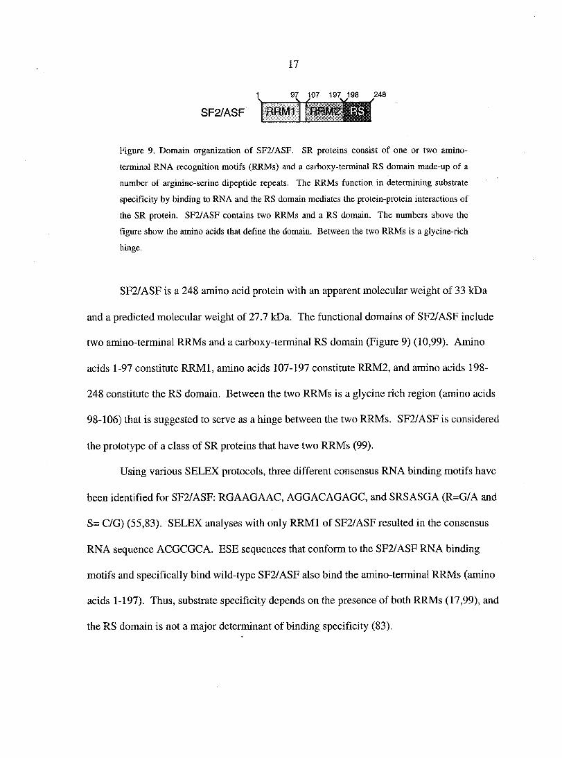
SF2/ASF
97 107 197 198 248 -W
RRM1 RRM2 m Figure 9. Domain organization of SF2/ASF. SR proteins consist of one or two amino-
terminal RNA recognition motifs (RRMs) and a carboxy-terminal RS domain made-up of a
number of arginine-serine dipeptide repeats. The RRMs function in determining substrate
specificity by binding to RNA and the RS domain mediates the protein-protein interactions of
the SR protein. SF2/ASF contains two RRMs and a RS domain. The numbers above the
figure show the amino acids that define the domain. Between the two RRMs is a glycine-rich
hinge.
SF2/ASF is a 248 amino acid protein with an apparent molecular weight of 33 kDa
and a predicted molecular weight of 27.7 kDa. The functional domains of SF2/ASF include
two amino-terminal RRMs and a carboxy-terminal RS domain (Figure 9) (10,99). Amino
acids 1-97 constitute RRMl, amino acids 107-197 constitute RRM2, and amino acids 198-
248 constitute the RS domain. Between the two RRMs is a glycine rich region (amino acids
98-106) that is suggested to serve as a hinge between the two RRMs. SF2/ASF is considered
the prototype of a class of SR proteins that have two RRMs (99).
Using various SELEX protocols, three different consensus RNA binding motifs have
been identified for SF2/ASF: RG A AG A AC, AGGACAGAGC, and SRSASGA (R=G/A and
S= C/G) (55,83). SELEX analyses with only RRMl of SF2/ASF resulted in the consensus
RNA sequence ACGCGCA. ESE sequences that conform to the SF2/ASF RNA binding
motifs and specifically bind wild-type SF2/ASF also bind the amino-terminal RRMs (amino
acids 1-197). Thus, substrate specificity depends on the presence of both RRMs (17,99), and
the RS domain is not a major determinant of binding specificity (83).
Page 28
SF2/ASF and EIAV Rev
As with other retroviruses, EIAV utilizes the splicing machinery to produce the viral
RNAs necessary for replication. EIAV is also a lentivirus and produces the regulatory
protein Rev, which is necessary for the cytoplasmic expression of the unspliced and singly
spliced viral mRNAs. Interestingly, exon 3 of the four-exon, bicistronic viral mRNA
(mRNAl, Figure 4) contains a purine-rich sequence that functions as both an ESE and as an
EIAV RRE (ESE/RRE) (5,34). This ESE/RRE was sufficient for SR protein binding (18),
and was necessary for exon inclusion, Rev binding, and Rev-dependent nuclear export (5).
The SR protein that specifically binds the ESE/RRE is SF2/ASF (18,34), and the ESE/RRE
contains multiple sequences that conform to the SF2/ASF consensus RNA binding motif
RG A AG A AC.
A mutational analysis of the ESE/RRE has shown that the sequences that bind Rev
also strongly assist in ESE-mediated splicing, and mutations that inhibit ESE function also
inhibit RRE function (3). However, mutation of the ESE/RRE does not exclusively knock
out the function of both the ESE and RRE. Thus, it is thought that the RNA binding sites for
SF2/ASF and Rev on the ESE/RRE overlap, but are not the same. It is clear that Rev can
inhibit exon 3 inclusion in the bicistronic viral mRNA (5,62), and SF2/ASF can inhibit Rev-
mediated nuclear export activity (5). These results suggest that Rev and SF2/ASF
competitively bind the ESE/RRE, which regulates splicing and viral mRNA expression.
Because Rev is absolutely required for virus replication, SF2/ASF may also inhibit EIAV
replication. Therefore, the interaction of Rev and SF2/ASF with the ESE/RRE may
modulate EIAV replication, and ultimately contribute to viral persistence and pathogenesis.
Page 29
19
Overall Goal
The lentiviral protein Rev is absolutely required for EIAV replication, and factors that
inhibit Rev function would be expected to inhibit virus replication. The cellular SR protein
SF2/ASF inhibits EIAV Rev function (5). Rev and SF2/ASF are regulators of viral mRNA
splicing and expression, and they both play a role in EIAV alternative splicing. The goal of
this research is to characterize the interactions between EIAV and cellular SR proteins that
modulate virus replication. It is our hypothesis that SF2/ASF inhibits Rev nuclear export
activity and virus replication. To test this hypothesis, we have undertaken the following
specific aims:
Specific Aims:
1. Determine the effect of Rev-mediated alternative splicing on EIAV Tat activity.
(Chapter 2)
2. Determine the effect of SF2/ASF expression on Rev-dependent nuclear export.
(Chapter 3)
3. Determine the effect of SF2/ASF expression on EIAV replication.
(Chapter 3)
References
1. Allemand, E., R. Gattoni, H.M. Bourbon, J. Stevenin, J.F. Caceres, J. Soret, and
J. Tazi. 2001. Distinctive features of Drosophila alternative splicing factor RS
domain: implication for specific phosphorylation, shuttling, and splicing activation.
Mol. Cell. Biol. 21:1345-1359.
Page 30
20
2. Alzhanova-Ericsson, A.T., X. Sun, N. Visa, E. Kiseleva, T. Wurtz, and B.
Daneholt. 1996. A protein of the SR family of splicing factors binds extensively to
exonic Balbiani ring pre-mRNA and accompanies the RNA from the gene to the
nuclear pore. Genes Dev. 10:2881-2893.
3. Amendt, B.A., D. Hesslein, L.-J. Chang, and C M. Stoltzfus. 1994. Presence of
negative and positive cis-acting RNA splicing elements within and flanking the first
tat coding exon of human immunodeficiency virus type 1. Mol. Cell. Biol. 14:3960-
3970.
4. Belshan, M., M.E. Harris, A.E. Shoemaker, T.J. Hope, and S. Carpenter. 1998.
Biological characterization of Rev variation in equine infectious anemia virus. J.
Virol. 72:4421-4426.
5. Belshan, M., G.S. Park, P. Bilodeau, C M. Stoltzfus, and S. Carpenter. 2000.
Binding of equine infectious anemia virus Rev to an exon splicing enhancer mediates
alternative splicing and nuclear export of viral mRNAs. Mol. Cell. Biol. 20:3550-
3557.
6. Bentley, D. 1999. Coupling RNA polymerase II transcription with pre-mRNA
processing. Current Opinion in Cell Biology 11:347-31.
7. Bieniasz, P.D., T.A. Grdina, H P. Bogerd, and B.R. Cullen. 1999. Recruitment of
cyclin Tl/P-TEFb to an HIV type 1 terminal repeat promoter proximal RNA target if
both necessary and sufficient for full activation of transcription. PNAS 96:7791-
7796.
Page 31
21
8. Bïencowe, B.J., J.A.L. Bowman, S. McCracken, and E. Rosonina. 1999. SR-
related proteins and the processing of messenger RNA precursors. Biochem. Cell
Biol. 77:277-291.
9. Caceres, J.F. and A.R. Kornblihtt. 2002. Alternative splicing: multiple control
mechanisms and involvement in human disease. Trends Genet. 18:186-193.
10. Caceres, J.F. and A.R. Kramer . 1993. Functional analysis of pre-mRNA splicing
factor SF2/ASF structural domains. EMBO-J. 12:4715-4726.
11. Caceres, J.F., G.R. Screaton, and A.R. Krainer. 1998. A specific subset of SR
proteins shuttles continuously between the nucleus and the cytoplasm. Genes &
Develop. 12:55-66.
12. Cao, W., S.F. Jamison, and M.A. Garcia-Blanco. 1997. Both phosphorylation and
dephosphorylation of ASF/SF2 are required for pre-mRNA splicing in vitro. RNA
3:1456-1467.
13. Carninci, P. and Y. Hayashizaki. 1999. High-efficiency full-length cDNA clonging.
Methods Enzymol. 303:19-44.
14. Cartegni, L., S.L. Chew, and A.R. Krainer. 2002. Listening to silence and
understanding nonsense: exonic mutations that affect splicing. Nat Rev Genet. 3:285-
298.
Page 32
22
15. Cavaloc, Y., C F. Bourgeois, L. Kister, and J. Stevenin. 1999. The slicing factors
9G8 and SRp20 transactivate splicing through different and specific enhancers. RNA
3:468^83.
16. Cazalla, D., J. Zhu, L. Manche, E. Huber, A.R. Krainer, and J.F. Caceres. 2002.
Nuclear export and retention signals in the RS domain of SR proteins. Mol. Cell.
Biol. 22:6871-6882.
17. Chandler, S.D., A. Mayeda, J.M. Yeakley, A.R. Krainer, and X.-D. Fu. 1997.
RNA splicing specificity determined by the coordinated action of RNA recognition
motifs in SR proteins. Proc. Natl. Acad. Sci. USA 94:3596-3601.
18. Chung, H.-K. and D. Derse. 2001. Binding sites for Rev and ASF/SF2 map to a 55-
nucleotide purine-rich exonic element in equine infectious anemia virus RNA. J.
Biol. Chem. 276:18960-18967.
19. Coffin, J.M., S.H. Hughes, and H E. Varmus. 1997. Retroviruses. Cold Spring
Harbor Laboratory Press, New York.
20. Cook, K.S., G.J. Fisk, J. Hauber, N. Usman, T.J. Daly, and J R. Rusche. 1991.
Characterization of HIV-1 REV protein: binding stoichiometry and minimal RNA
substrate. Nucleic Acids Reseach 19:1577-1583.
21. Coulter, L.R., M.A. Landree, and T.A. Cooper. 1997. Identification by a new class
of exonic splicing enhancers by in vivo selection. Mol. Cell. Biol. 17:2143-2150.
Page 33
22. Dingwall, C., I. Ernberg, M.J. Gait, S.M. Green, S. Heaphy, J. Karn, A.D. Lowe,
M. Singh, M.A. Skinner, and R. Valerio. 1989. Human immunodeficiency virus 1
tat protein binds trans-activation-responsive region (TAR) RNA in vitro. Proc. Natl.
Acad. Sci. USA 86:6925-6929.
23. Dorn, P., L. DaSilva, L. Martarano, and D. Derse. 1990. Equine infectious anemia
virus tat: insights into the structure, function, and evolution of lentivirus trans-
activator proteins. J. Virol. 64:1616-1624.
24. Dorn, P L. and D. Derse. 1988. cis- and trans-acting regulation of gene expression
of equine infectious anemia virus. J. Virol. 62:3522-3626.
25. Dreyfuss, G., V.N. Kim, and N. Kataoka. 2002. Messenger-RNA-binding proteins
and the messages they carry. Nat Rev Mol Cell Biol 3:195-205.
26. Emerman, M. and M.H. Malim. 1998. HIv-1 regulatory/accessory genes: keys to
unraveling viral and host cell biology. Science 280:1180-1184.
27. Eperon, I.C., O.V. Makarova, A. Mayeda, S.H. Munroe, J.F. Caceres, D.G.
Hayward, and A.R. Krainer. 2000. Selection of alternative 5' splice sites: role of U1
snRNP and models for the antagonistic effects of SF2/ASF and hnRNP Al. Mol.
Cell. Biol. 20:8303-8318.
28. Feng, S. and E C. Holland. 1988. HIV-1 Tat frares-activation requires the loop
sequence within TAR. Nature (London) 334:165
Page 34
24
29. Fischer, U., J. Huber, W.C. Boelens, I.W. Mattal, and R. Luhrmann. 1995. The
HIV-l rev activation domain is a nuclear export signal that acceses an export pathway
used by specific cellular RNAs. Cell 82:475-483.
30. Fridell, R.A., H P. Bogerd, and B.R. Cullen. 1996. Nuclear export of late HIV-l
mRNAs occurs via a cellular protein export pathway. Proc. Natl. Acad. Sci. USA
93:4421-4424.
31. Fridell, R.A., K M. Partin, S. Carpenter, and B.R. Cullen. 1993. Identification of
the activation domain of equine infectious anemia virus rev. J. Virol. 67:7317-7323.
32. Ge, H. and J.L. Manley. 1990. A protein factor, ASF, controls cell-specific
alternative splicing of SV40 early pre-mRNA in vitro. Cell 62:25-34.
33. Goldstrohm, A C., A.L. Greenleaf, and M.A. Garcia-Blanco. 2001. Co-
transcriptional splicing in pre-messenger RNAs: considerations for the mechanism of
alternative splicing. Gene 277:31-47.
34. Gontarek, R.R. and D. Derse. 1996. Interactions among SR proteins, an exonic
splicing enhancer, and a lentivirus rev protein regulate alternative splicing. Mol. Cell.
Biol. 16:2325-2331.
35. Graveley, B.R. 2000. Sorting out the complexity of SR protein functions. RNA
6:1197-1211.
Page 35
36. Hammond, S.A., S.J. Cook, D.L. Lichtenstein, C.J. Issel, and R.C. Montelaro.
1997. Maturation of the cellular and humoral immune responses to persistant
infection in horses by equine infectious anemia virus is a complex and lengthy
process. J. Virol. 71:3840-3852.
37. Hammond, S.A., F. Li, B.M. McKeon, S.J. Cook, C.J. Issel, and R.C. Montelaro.
2000. Immune responses and viral replication in long-term inapparent carrier ponies
inoculated with equine infectious anemia virus. J. Virol. 74:5968-5981.
38. Hammond, S.A., M L. Raabe, C.J. Issel, and R.C. Montelaro. 1999. Evaluation of
antibody parameters as potential correlates of protection or enhancement by
experimental vaccines to equine infectious anemia virus. Virology 262:416-430.
39. Hanamura, A., J.F. Caceres, A. Mayeda, B.R.Jr. Franza, and A.R. Krainer.
1998. Regulated tissue-specific expression of antagonistic pre-mRNA splicing
factors. RNA 4:430-444.
40. Harris, M.E., R.R. Gontarek, D. Derse, and T.J. Hope. 1998. Differential
requirements for alternative splicing and nuclear export functions of equine infectious
anemia virus Rev protein. Mol. Cell. Biol. 18:3889-3899.
41. Hastings, M L. and A.R. Krainer. 2001. Pre-mRNA splicing in the new
millennium. Cell Biol 13:302-309.
Page 36
26
42. Hauber, J. and B.R. Cullen. 1988. Mutational analysis of the trans-activation-
responsive region of the human immunodeficiency virus type 1 long terminal repeat.
J. Virol. 62:673-679.
43. Hertel, K.J. and T. Maniatis. 1999. Serine-arginine (SR)-rich splicing factors have
an exon-independent function in pre-mRNA splicing. Proc. Natl. Acad. Sci. USA
96:2651-2655.
44. Huang, X., T.J. Hope, B.L. Bond, D. McDonald, K. Grahl, and T.G. Parslow.
1991. Minimal rev-response element for type 1 human immunodeficiency virus. J.
Virol. 65:2131-2134.
45. Issel, C.J. and W.V. Adams. 1979. Serologic survey for equine infectious anemia
virus in Louisiana. JAVMA 174:286-288.
46. Jumma, H. and P.J. Nielsen. 2000. Regulation of SRp20 exon 4 splicing. Biochica
et Biophysica Acta 1-2:137-143.
47. Kataoka, N., J.L. Bachorik, and G. Dreyfuss. 1999. Transportin-SR, a nuclear
import receptor for SR proteins. J. Cell Biol. 145:1145-1152.
48. Kjems, J. and P. Askjaer. 2000. Rev protein and its cellular partners. Advances in
Pharmacology 48:251-298.
49. Kohtz, J.D., S.F. Jamison, C.L. Will, P. Zuo, R. Luhrmann, M.A. Garcia-Blanco,
and J.L. Manley. 1994. Protein-protein interactions and 5'-splice-site recognition in
mammalian mRNA precursors. Nature 368:119-124.
Page 37
27
50. Koizumi, J., Y. Okamoto, H. Onogi, A. Mayeda, A.R. Krainer, and M.
Hagiwara. 1999. The subcellular localization of SF2/ASF is regulated by direct
interaction with SR protein Kinases (SRPKs). J. Biol. Chemistry 274:1112-11131.
51. Krainer, A.R., G.C. Conway, and D. Kozak. 1990. The essential pre-mRNA
splicing factor SF2 influences 5' splice site selection by activating proximal sites.
Cell 62:35-42.
52. Kramer, A. 1996. The structure and function of proteins involved in mammalian pre-
mRNA splicing. Annu. Rev. Biochem. 65:367-409.
53. lai, M.C., R.I. Lin, and W.Y. Tarn. 2001. Transportin-SR2 mediates nuclear import
of phosphorylated SR proteins. PNAS U.S.A. 98:10154-10159.
54. Lemaire, R., A. Winne, M. Sarkissian, and R. Lafyatis. 1999. SF2 and SRp55
regulation of CD45 exon 4 skipping during T cell activation. Eur. J. Immunol.
29:823-837.
55. Liu, H.-X., M. Zhang, and A. Krainer. 1998. Identification of functional exonic
splicing enhancer motifs recognized by individual SR proteins. Genes and
Development 12:1998-2012.
56. Longman, D., I.L. Johnstone, and J.F. Carceres. 2000. Functional characterization
of SR and SR-related genes in Caenorhabditis. EMBO J. 19:1625-1637.
Page 38
28
57. Lopato, S., M. Kalyna, S. Borner, R. Kobayashi, A.R. Kràiner, and A. Barta.
1999. atSRp30, one of two SF2/AFS-like proteins from Arabidopsis thaliana,
regulates splicing of specific plant genes. Genes & Development 13:987-1001.
58. Lopez, A.J. 1998. Alternative splicing of pre-mRNA: developmental consequences
and mechanisms of regulation. Ann. Rev. Genet. 32:279-395.
59. Mancebo, H.S., G. Lee, J. Flygare, J. Tomassini, P. Luu, Y. Zhu, J. Peng, C.
Blau, D. Hazuda, D. Price, and O. Flores. 1997. P-TEFb kinase is required for HIV
Tat transcriptional activation in vivo and in vitro. Genes Dev. 11:2633-2644.
60. Mancuso, V.A., T.J. Hope, L. Zhu, D. Derse, T. Phillips, and T.G. Parslow. 1994.
Posttranscriptional effector domains in the rev proteins of feline immunodeficiency
virus and equine infectious anemia virus. J. Virol. 68:1998-2001.
61. Manley, J.L. and R. Tacke. 1996. SR proteins and splicing control. Genes Dev.
10:1569-1579.
62. Martarano, L., R. Stephens, N. Rice, and D. Derse. 1994. Equine infectious anemia
virus frans-regulatory protein rev controls viral mRNA stability, accumulation, and
alternative splicing. J. Virol. 68:3102-3111.
63. Mayeda, A., G.R. Screaton, S.D. Chandler, X.D. Fu, and A.R. Krainer. 1999.
Substrate specificities of SR proteins in constitutive splicing are determined by their
RNA recognition motifs and composite pre-mRNA exonic elements. Mol. and Cell.
Biol. 19:1853-1863.
Page 39
29
64. Misteli, T., J.F. Caceres, J.Q. Clement, A.R. Kramer, and M.F. Wilkinson. 1998.
Serine phosphorylation of SR proteins is required for their recruitment to sites of
transcription in vivo. J. Cell Biol. 143:297-307.
65. Murphy, S.M., M. Belshan, P. Bruellman, Y. Li, T.J. Hope, and S. Carpenter. In
preparation. Functional domains of equine infectious anemia virus Rev.
66. Murray, H.L. and K.A. Jarrell. 1999. Flipping the switch to an active spliceosome.
Cell 96:599-602.
67. Noiman, S., A. Gazit, O. Tori, L. Sherman, T. Miki, S R. Tronick, and A. Yaniv.
1990. Identification of sequences encoding the equine infectious anemia virus tat
gene. Virology 176:280-288.
68. Noiman, S., A. Yaniv, L. Sherman, S R. Tronick, and A. Gazit. 1990. Pattern of
transcription of the genome of equine infectious anemia virus. J. Virol. 64:1839-
1843.
69. Noiman, S., A. Yaniv, T. Tsach, T. Miki, S R. Tronick, and A. Gazit. 1991. The
tat protein of equine infectious anemia virus is encoded by at least three types of
transcripts. Virology 184:521-530.
70. Oaks, J.L., T.C. McGuire, C. Ulibarri, and T.B. Crawford. 1998. Equine
infectious anemia virus is found in tissue macrophages during subclinical infection.
J. Virol. 72:7263-7269.
Page 40
71. Olsen, H., A. Cochrane, P. Dillon, C. Nalin, and C. Rosen. 1990. Interaction of the
human immunodeficiency virus type 1 rev protein with a structured region in env
mRNA is dependent on multimer formation mediated through a basic stretch of
amino acids. Genes Dev. 4:1357-1364.
72. Pollard, V.W. and M.H. Malim. 1998. The HIV-1 REV protein. Annu. Rev.
Microbiol. 52 :491-532.
73. Robberson, B.L., G.J. Cote, and S.M. Berget. 1990. Exon definition may facilitate
splice site selection in RNAs with multiple exons. Mol. Cell. Biol. 10:84-94.
74. Roy, S., U. Delling, C.H. Chen, C.A. Rosen, and N. Sonenberg. 1990. A bulge
structure in HIV-1 TAR RNA is required for Tat binding and Tat-mediated
transactivation. Genes Dev. 4:1365-1373.
75. Roy, S., N.T. Parkin, C. Rosen, J. Itovitch, and N. Sonenberg. 1990. Structural
requirements for transactivation of human immunodeficiency virus type 1 long
terminal repeat-directed gene expression by tat: importance of base pairing, loop
sequence, and bulges in the tat-responsive sequence. J. Virol. 64:1402-1406.
76. Schiltz, R.L., D.S. Shih, S. Rasty, R.C. Montelaro, and K.E. Rushlow. 1992.
Equine infectious anemia virus gene expression: characterization of the RNA splicing
pattern and the protein products encoded by open reading frames SI and S2. J. Virol.
66:3455-3465.
Page 41
31
77. Sellon, D.C. 1993. Equine infectious anemia. Vet. Clin. North Am. Equine Pract.
9:321-336.
78. Sellon, DC., F.J. Fuller, and T.C. McGuire. 1994. The immunopathogenesis of
equine infectious anemia virus. Virus Res. 32:111-138.
79. Sellon, D C., S T. Perry, L. Coggins, and F.J. Fuller. 1992. Wild-type equine
infectious anemia virus replicates in vivo predominantly in tissue macrophages, not in
peripheral blood monocytes. J. Virol. 66:5906-5913.
80. Smith, C.W.J., J.G. Patton, and B. Nadal-Ginard. 1989. Alternative splicing in the
control of gene expression. Annu.Rev.Genet. 23:527-577.
81. Stephens, R.M., D. Derse, and N R. Rice. 1990. Cloning and characterization of
cDNAs encoding equine infectious anemia Tat and putative Rev proteins. J. Virol.
64:3716-3725.
82. Stoltzfus, C M. and S.J. Fogarty. 1989. Multiple regions in the Rous sarcoma virus
src gene intron act in cis to affect the accumulation of unspliced RNA. J. Virol.
63:1669-1676.
83. Tacke, R. and J.L. Manley. 1995. The human splicing factors ASF/SF2 and SC35
possess distinct, functionally significant RNA binding specificities. EMBO 14:3540-
3551.
84. Tackle, R. and J.L. Manley. 1999. Determinants of SR protein specificity. Curr.
Opin. in Cell Biol. 11:358-362.
Page 42
32
85. Tang, H., K L. Kuhen, and F. Wong-Staal. 1999. Lentivirus replication and
regulation. Annu. Rev. Genet 33:133-170.
86. Tuerk, C. and L. Gold. 1990. Systematic evoluton of ligands by exponential
enrichment: RNA ligands to bacteriophage T4 DNA polymerase. Science 249:505-
510.
87. van der Houven van Oordt, W., K. Newton, G.R. Screaton, and J.F. Caceres.
2000. Role of SR protein modular domains in alternative splicing specificity in vivo.
Nucleic Acids Research 28:4822-4831.
88. Vogt, V.M. 2000. Ubiquitin in retrovirus assembly: Actor or bystander? PNAS
97:12945-12947.
89. Wang, J., Y. Takagaki, and J.L. Manley. 1994. Targeted disruption of an essential
vertebrate gene. ASF/SF2 is required for cell viability. Genes Dev. 10:2588-2599.
90. Wang, J., S. Xioa, and J.L. Manley. 1998. Genetic analysis of the SR protein
ASF/SF2: interchangeabilityof RS domains and negative control splicing. Genes &
Development 12:2222-2233.
91. Wei, P., M.E. Garber, S. Fang, W.H. Fischer, and K.A. Jones. 1998. A novel
CDK9-associated c-type cyclin interacts directly with HIV-1 Tat and mediates its
high-affinity, loop-specific binding to TAR RNA. Cell 92:451-462.
92. Wu, J.Y. and T. Maniatis. 1993. Specific interactions between proteins implicated
in splice site selection and regulated alternative splicing. Cell 75:1061-1070.
Page 43
93. Xiao, S.H. and J.L. Manley. 1997. Phosphorylation of the ASF/SF2 RS domain
affects both protein-protein and protein—RNA interactions and is necessary for
splicing. Genes Dev. 11:334-344.
94. Yang, X., C.H. Herrmann, and A.P. Rice. 1996. The human immunodeficiency
virus Tat proteins specifically associate with TAK in vivo and require the carboxyl-
terminal domain of the RNA polymerase II for function. J. of Virology 70:4576-
4584.
95. Zahler, A.M., K M. Neugebauer, W.S. Lane, and M B. Roth. 1993. Distinct
functions of SR proteins in alternative pre-mRNA splicing. Science 260:219-222.
96. Zapp, M., T. Hope, T. Parslow, and M. Green. 1988. Oligomerization and RNA
binding domains of the type 1 human immunodeficiency virus rev protein: a dual
function for an arginine-rich motif. Proc. Natl. Acad. Sci. USA 88:7734-7738.
97. Zapp, M L. and M R. Green. 1989. Sequence-specific RNA binding by the HIV-1
Rev protein. Nature 342:714-716.
98. Zhu, Y., T. Pe'ery, J. Peng, Y. Ramanathan, N. Marshall, T. Marshall, B.
Amendt, M B. Mathews, and D.H. Price. 1997. Transcription elongation factor P-
TEFb is required for HIV-1 tat transactivation in vitro. Genes Dev. 11:2622-2632.
99. Zuo, P. and J.L. Manley. 1993. Functional domains of the human splicing factor
ASF/SF2. EMBO-J. 12:4727-4737.
Page 44
34
CHAPTER 2. FUNCTIONAL CHARACTERIZATION OF TAT ACTIVITY FROM
EIAV ALTERNATIVELY SPLICED MESSENGER RNAS
A paper to be submitted to the journal Virus Genes
Gregory Park, Michael Belshan, Susan Schommer, and Susan Carpenter
Abstract
Similar to other Antiviruses, equine infectious anemia virus (EIAV) encodes the
regulatory proteins Tat and Rev, which regulate virus gene expression. Tat up-regulates
transcription and Rev binds at a region of viral RNA called the Rev responsive element
(RRE), and exports incompletely spliced viral transcripts into the cytoplasm. Tat and Rev
are translated from a four-exon bicistronic mRNA, which is the predominate mRNA
expressed early after infection. Tat translation is initiated at a CUG in exon 1, and only
through a leaky scanning mechanism is Rev translation initiated at the first AUG located in
exon 3. Exon 3 also contains the EIAV RRE. Rev mediates the expression of a three-exon,
alternatively spliced mRNA that lacks exon 3 and encodes only Tat. To determine the effect
of Rev-mediated alternative splicing on Tat expression, we used transient assays to
functionally analyze alternative spliced mRNAs. There was no significant difference in Tat
activity between monocistronic and bicistronic cDNAs, suggesting that Tat expression is not
different between the three-exon and four-exon EIAV mRNAs. Thus, the presence or
absence of Exon 3 did not affect Tat activity, suggesting that neither the RRE in rev exon 1
nor translation initiation of Rev affect Tat expression.
Page 45
35
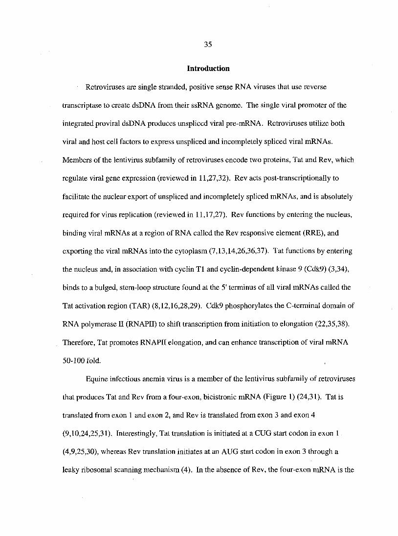
Introduction
Retroviruses are single stranded, positive sense RNA viruses that use reverse
transcriptase to create dsDNA from their ssRNA genome. The single viral promoter of the
integrated proviral dsDNA produces unspliced viral pre-mRNA. Retroviruses utilize both
viral and host cell factors to express unspliced and incompletely spliced viral mRNAs.
Members of the lenti virus subfamily of retroviruses encode two proteins, Tat and Rev, which
regulate viral gene expression (reviewed in 11,27,32). Rev acts post-transcriptionally to
facilitate the nuclear export of unspliced and incompletely spliced mRNAs, and is absolutely
required for virus replication (reviewed in 11,17,27). Rev functions by entering the nucleus,
binding viral mRNAs at a region of RNA called the Rev responsive element (RRE), and
exporting the viral mRNAs into the cytoplasm (7,13,14,26,36,37). Tat functions by entering
the nucleus and, in association with cyclin T1 and cyclin-dependent kinase 9 (Cdk9) (3,34),
binds to a bulged, stem-loop structure found at the 5' terminus of all viral mRNAs called the
Tat activation region (TAR) (8,12,16,28,29). Cdk9 phosphorylates the C-terminal domain of
RNA polymerase II (RNAPII) to shift transcription from initiation to elongation (22,35,38).
Therefore, Tat promotes RNAPII elongation, and can enhance transcription of viral mRNA
50-100 fold.
Equine infectious anemia virus is a member of the lenti virus subfamily of retroviruses
that produces Tat and Rev from a four-exon, bicistronic mRNA (Figure 1) (24,31). Tat is
translated from exon 1 and exon 2, and Rev is translated from exon 3 and exon 4
(9,10,24,25,31). Interestingly, Tat translation is initiated at a CUG start codon in exon 1
(4,9,25,30), whereas Rev translation initiates at an AUG start codon in exon 3 through a
leaky ribosomal scanning mechanism (4). In the absence of Rev, the four-exon mRNA is the
Page 46
only cytoplasmically expressed mRNA (mRNAl, Figure 1) (23,30). The presence of Rev
results in the expression of the other EIAV mRNAs (mRNA3, mRNA4, mRNA5) including a
monocistronic, three-exon mRNA, encoding only Tat (mRNA 2) (23). The monocistronic
mRNA is identical to the bicistronic mRNA, except it lacks exon 3.
Leaky ribosomal scanning proposes that only some ribosomes stop to initiate
translation at non-AUG start codons or AUG start codons in a weak context (A or G not at
position -3 and G not at +4), while most continue scanning downstream (20). There are
factors that affect leaky scanning translation other than the context of the start codon. RNA
secondary structures can affect translation initiation in bicistronic messages, depending on
their location. RNA secondary structures located downstream of the first initiation site can
enhance translation of the first cistron by slowing ribosomes enough for codon/anti-codon
base pairing to occur (19).
Exon 3 of the four-exon bicistronic mRNA contains not only the translation initiation
site for Rev, but also contains the EIAV RRE, which is suggested to have secondary structure
(6,15). Secondary structure would be consistent with other RREs, such as HIV's RRE, which
is a large secondary structure (5,18,21). Thus, the three-exon alternatively spliced RNA may
differ from the four-exon mRNA in Tat expression due to the absence of exon 3, which may
contain RNA factors that affect translation of the upstream cistron. To determine the effects
of EIAV Rev-mediated alternative splicing on Tat expression, transient expression assays
were used to compare the functional activity of Tat among alternatively spliced EIAV
cDNAs. There was no significant difference in Tat activity in cells transfected with the
monocistronic or the bicistronic cDNAs, suggesting that Tat expression is not different
between the monocistronic and bicistronic EIAV mRNAs that encode Tat. Therefore, the
Page 47
37
results suggest that neither the RRE nor translation initiation at the Rev AUG affect Tat
translation.
Materials and Methods
Plasmids. EIAV MA-1 cDNAs (4x+, 3x+, 4x-) were amplified by RT-PCR of total
RNA isolated from Cf2Th cells transfected with Rev+ or Rev- proviral DNA using the 5'
primer, CGCAGACCCTACCTGTTG and the 3* primer, TAGCCTGCTATGCGTCCTAC
(Figure 2). The cDNA products were TA-cloned into pCR3.1 (Invitrogen, Carlsbad, Calif.)
and confirmed by sequence analysis (DNA Sequencing and Synthesis Facility, Iowa State
University). Transcription of the cDNAs in pCR3.1 is under control of the CMV promoter.
ETat-M was constructed by David Derse in pRSPA-S (pRSETAT-M) (9). Exon 1 of the Tat
cDNA in pRSETAT-M is missing the first 38 nucleotides and starts at an engineered AUG
initiation site. Transcription of pRSETAT-M is under control of the RSV promoter. The
plasmid pCHl 10 (Amersham Pharmacia, Buckinghamshire, UK) produces (3-galactosidase.
The LTR-CAT reporter plasmid pCATLTREIAV-1 contains the EIAV LTR upstream of a
chloramphenicol acetyltransferase (CAT) gene. The plasmid pcDNA3 (Invitrogen) was used
as a negative control and produces no Tat.
Transient transaction assays. Transient transfections were performed in canine
fetal thymus (Cf2Th) cells, which support both EIAV Tat activity and EIAV replication (33).
Cf2Th cells were maintained in Dulbecco's modified Eagle media supplemented with 10%
fetal calf serum and penicillin/streptomycin. All transfections were performed with the
transfection reagent LipofectAMINE (Life Technologies/Invitrogen, Carlsbad, CA)
according to reagent protocols. Briefly, 0.2 \xg of individual cDNA plasmids were co-
Page 48
transfected into cells with 0.2 |xg pCHl 10,1.0 jig of pCATLTREIAV-1, and an amount of
pUC19 to equalize the amount of DNA used in all transfections. Two days post-transfection,
cells were harvested, lysed by freeze/thaw, and clarified lysates were assayed for transfection
efficiency by their (3-galactosidase activity. Normalized amounts of cell lysate were then
assayed for CAT expression with a commercially available CAT enzyme-linked
immunosorbent assay (ELIS A) kit (Roche Molecular Biochemicals, Indianapolis, IN). Pilot
assays quantified CAT from pRSETAT-M transfected cells, and results were used to
determine the parameters to assay all the transfected cell lysates. Initial assays determined a
range of Tat activity from co-transfection of increasing amounts of pRSETAT-M or p3x+
with pCATLTREIAV -1 (data not shown). Results were used to determine the working
amount of Tat cDNA plasmid.
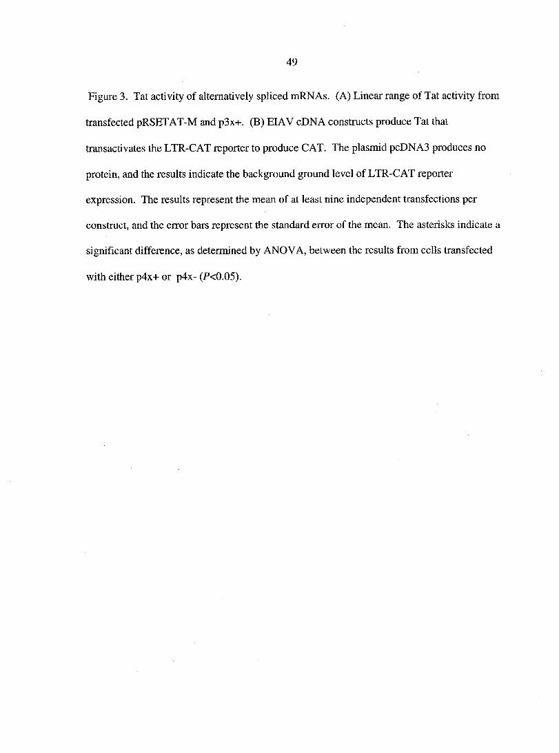
Results and Discussion
EIAV Tat translation is initiated at a non-standard CUG in exon 1 of both the three-
exon and four-exon EIAV mRNAs. Rev translation initiates in exon 3 at the first AUG of the
EIAV four-exon, bicistronic viral mRNA. Thus, leaky scanning of the CUG initiation codon
permits translation of Rev (4). Interestingly, Rev mediates the expression of the three-exon
mRNAs that lack exon 3 and encode only Tat. Exon 3 contains a purine-rich sequence that
functions as both an ESE and an RRE. Typical lenti viral RRE's are highly structured RNA
elements. It is possible that initiation at the Tat CUG is altered depending on the presence or
absence of the AUG initiation site and/or RNA secondary structure in exon 3. If so, Rev-
mediated alternative splicing may be a novel mechanism to increase Tat activity. To
determine if EIAV alternative splicing results in mRNAs that differ in Tat activity, cDNAs
Page 49
were constructed and tested for Tat activity in an in vitro transient expression assay. EIAV
Tat constructs, 3x+ and 4x+ (Figure 2), represent the three-exon and four-exon mRNAs,
respectively, and differ in the absence and presence of exon 3. Thus, 3x+ encodes only Tat,
and 4x+ encodes both Tat and Rev. We also constructed a cDNA that contain exon 3, but
produce no Rev due to a stop codon in exon 4. This would allow us to differentiate any
effect due to Rev or exon 3. Thus, 4x- encodes a truncated Rev protein (amino acids 1-79)
that contains only the identified nuclear export signal. The truncated Rev protein lacks
amino acids 80-165, which includes the domains required for RNA binding and nuclear
localization, and the protein is expected to be non-functional. The ETat-M construct has an
engineered AUG start codon for Tat instead of the wild-type CUG, and was used as a
positive control for Tat activity.
Less than 2-fold differences in levels of CAT were observed among the cDNA
constructs. There was no significant difference in Tat activity between the 3x+ and 4x+
constructs (P>0.05) (Figure 3), which indicates that Tat activity is the same between the
monocistronic and bicistronic EIAV Tat mRNAs. Co-transfection of the CAT reporter with
the 4x- construct resulted in levels of CAT not significantly different from 3x+ (P>0.05).
Together, these data suggest that the presence of exon 3 has little to no effect on Tat activity
(Figure 3). Interestingly, the 4x- construct had significantly higher Tat activity than the 4x+
construct (P<0.05). While this suggests that Rev inhibited Tat activity, there is no significant
difference in Tat activity between the 3x+ and 4x+ constructs. In addition, trans-
complementation experiments showed that Rev does not significantly affect Tat activity (data
not shown). It is possible that the presence of a truncated Rev protein may affect Tat
activity. Together, the data indicate that there is no difference in Tat activity between the
Page 50
40
monocistronic and bicistronic EIAV mRNAs, and suggest that exon 3 does not affect Tat
expression. Thus, EIAV Rev-mediated alternative splicing may be a mechanism to express
mRNAs that produce Tat without producing Rev, which may play a role in regulating both
viral protein and mRNA expression.
Rev mediates exclusion of exon 3, and the subsequent expression of the alternatively
spliced, three-exon mRNAs that encode only Tat. Recently, an exon splicing enhancer has
been mapped to exon 3 (2,6,15). The ESE sequence also functions as a RRE (ESE/RRE) and
the sequences of the ESE/RRE bind Rev. The cellular splicing protein SF2/ASF also binds
the ESE/RRE and assists in exon 3 inclusion (2,15). The mechanism that results in
expression of the alternatively spliced, three-exon mRNA is not clear, but current models
suggest that exon 3 exclusion is the result of the inhibition of splicing, due to the binding of
Rev at the ESE/RRE. The biological significance of the three-exon mRNA in virus
replication is not known, but our data indicate that exon 3 had no effect on Tat activity. This
does not necessarily mean that production of a second mRNA species encoding Tat plays no
role in EIAV replication. Indeed, Tat is produced at very low levels, yet has profound affects
on viral transcription (3). Our data suggests that Rev-mediated alternative splicing is not a
mechanism to increase Tat expression. Thus, further investigations are necessary to better
understand if the generation of the alternatively spliced mRNA is important in EIAV
replication.
Page 51
41
References
1. Beisel, C.E., J.F. Edwards, L.L. Dunn, and N.R. Rice. 1993. Analysis of multiple
mRNAs from pathogenic equine infectious anemia virus (EIAV) in an acutely infected
horse reveals a novel protein, ttm, derived from the carboxy terminus of the EIAV
transmembrane protein. J. Virol. 67:832-842.
2. Belshan, M., G.S. Park, P. Bilodeau, C M. Stoltzfus, and S. Carpenter. 2000.
Binding of equine infectious anemia virus Rev to an exon splicing enhancer mediates
alternative splicing and nuclear export of viral mRNAs. Mol. Cell. Biol. 20:3550-3557.
3. Bieniasz, P.D., T.A. Grdina, H P. Bogerd, and B.R. Cullen. 1999. Recruitment of
cyclin Tl/P-TEFb to an HIV type 1 terminal repeat promoter proximal RNA target if
both necessary and sufficient for full activation of transcription. PNAS 96:7791-7796.
4. Carroll, R. and D. Derse. 1993. Translation of equine infectious anemia virus
bicistronic tat-rev mRNA requires leaky ribosome scanning of the tat CTG initiation
codon. J. Virol. 67:1433-1440.
5. Charpentier, B., F. Stutz, and M. Rosbash. 1997. A dynamic in vivo view of the
HIV-1 Rev-RRE interaction. J Mol. Biol. 266:950-962.
Page 52
42
6. Chung, H.-K. and D. Derse. 2001. Binding sites for Rev and ASF/SF2 map to a 55-
nucleotide purine-rich exonic element in equine infectious anemia virus RNA. J. Biol.
Chem. 276:18960-18967.
7. Cook, K.S., G.J. Fisk, J. Hauber, N. Usman, T.J. Daly, and J R. Rusche. 1991.
Characterization of HIV-1 REV protein: binding stoichiometry and minimal RNA
substrate. Nucleic Acids Reseach 19:1577-1583.
8. Dingwall, C., I. Ernberg, M.J. Gait, S.M. Green, S. Heaphy, J. Kara, A.D. Lowe,
M. Singh, M.A. Skinner, and R. Valerio. 1989. Human immunodeficiency virus 1 tat
protein binds trans-activation-responsive region (TAR) RNA in vitro. Proc. Natl. Acad.
Sci. USA 86:6925-6929.
9. Dorn, P., L. DaSilva, L. Martarano, and D. Derse. 1990. Equine infectious anemia
virus tat: insights into the structure, function, and evolution of lentivirus Zraru-activator
proteins. J. Virol. 64:1616-1624.
10. Dorn, P L. and D. Derse. 1988. cis- and trans-acting regulation of gene expression of
equine infectious anemia virus. J. Virol. 62:3522-3626.
11. Emerman, M. and M.H. Malim. 1998. HIv-1 regulatory/accessory genes: keys to
unraveling viral and host cell biology. Science 280:1180-1184.
Page 53
43
12. Feng, S. and B.C. Holland. 1988. HTV-1 Tat îran.s-activation requires the loop
sequence within TAR. Nature (London) 334:165
13. Fischer, U., J. Ruber, W.C. Boelens, I.W. Mattal, and R. Luhrmann. 1995. The
HIV-1 rev activation domain is a nuclear export signal that acceses an export pathway
used by specific cellular RNAs. Cell 82:475-483.
14. Fridell, R.A., H P. Bogerd, and B.R. Cullen. 1996. Nuclear export of late HIV-1
mRNAs occurs via a cellular protein export pathway. Proc. Natl. Acad. Sci. USA
93:4421-4424.
15. Gontarek, R.R. and D. Derse. 1996. Interactions among SR proteins, an exonic
splicing enhancer, and a lentivirus rev protein regulate alternative splicing. Mol. Cell.
Biol. 16:2325-2331.
16. Hauber, J. and B.R. Cullen. 1988. Mutational analysis of the trans-activation-
responsive region of the human immunodeficiency virus type 1 long terminal repeat. J.
Virol. 62:673-679.
17. Kjems, J. and P. Askjaer. 2000. Rev protein and its cellular partners. Advances in
Pharmacology 48:251-298.
Page 54
44
18. Kjems, J., M. Brown, D.D. Chang, and P.A. Sharp. 1991. Structural analysis of the
interaction between the human immunodeficiency virus Rev protein and the Rev
response element. Proc. Natl. Acad. Sci. USA 88:683-687.
19. Kozak, M. 1990. Downstream secondary structure facilitates recognition of initiator
codons by eukaryotic ribosomes. Proc. Natl. Acad. Sci. USA 87:8301-8305.
20. Kozak, M. 2002. Pushing the limits of the scanning mechanism for initiation of
translation. Gene 299:1-34.
21. Malim, M.H., J. Hauber, S.-Y. Le, J.V. Maizel, and B.R. Cullen. 1989. The HIV-1
rev trans-activator acts through a structured target sequence to activate nuclear export
of unspliced viral mRNA. Nature 338:254-256.
22. Mancebo, H.S., G. Lee, J. Flygare, J. Tomassini, P. Luu, Y. Zhu, J. Peng, C. Blau,
D. Hazuda, D. Price, and O. Flores. 1997. P-TEFb kinase is required for HIV Tat
transcriptional activation in vivo and in vitro. Genes Dev. 11:2633-2644.
23. Martarano, L., R. Stephens, N. Rice, and D. Derse. 1994. Equine infectious anemia
virus frans-regulatory protein rev controls viral mRNA stability, accumulation, and
alternative splicing. J. Virol. 68:3102-3 111.
Page 55
45
24. Noiman, S., A. Gazit, O. Tori, L. Sherman, T. Miki, S.R. Tronick, and A. Yaniv.
1990. Identification of sequences encoding the equine infectious anemia virus tat gene.
Virology 176:280-288.
25. Noiman, S., A. Yaniv, T. Tsach, T. Miki, S.R. Tronick, and A. Gazit. 1991. The tat
protein of equine infectious anemia virus is encoded by at least three types of
transcripts. Virology 184:521-530.
26. Olsen, H., A. Cochrane, P. Dillon, C. Nalin, and C. Rosen. 1990. Interaction of the
human immunodeficiency virus type 1 rev protein with a structured region in env
mRNA is dependent on multimer formation mediated through a basic stretch of amino
acids. Genes Dev. 4:1357-1364.
27. Pollard, V.W. and M.H. Malim. 1998. The HIV-1 REV protein. Annu. Rev.
Microbiol. 52:491-532.
28. Roy, S., U. Delling, C.H. Chen, C.A. Rosen, and N. Sonenberg. 1990. A bulge
structure in HIV-1 TAR RNA is required for Tat binding and Tat-mediated
transactivation. Genes Dev. 4:1365-1373.
Page 56
46
29. Roy, S., N.T. Parkin, C. Rosen, J. Itovitch, and N. Sonenberg. 1990. Structural
requirements for transactivation of human immunodeficiency virus type 1 long terminal
repeat-directed gene expression by tat: importance of base pairing, loop sequence, and
bulges in the tat-responsive sequence. J. Virol. 64:1402-1406.
30. Schiltz, R.L., D.S. Shih, S. Rasty, R.C. Montelaro, and K.E. Rushlow. 1992. Equine
infectious anemia virus gene expression: characterization of the RNA splicing pattern
and the protein products encoded by open reading frames SI and S2. J. Virol. 66:3455-
3465.
31. Stephens, R.M., D. Derse, and N R. Rice. 1990. Cloning and characterization of
cDNAs encoding equine infectious anemia Tat and putative Rev proteins. J. Virol.
64:3716-3725.
32. Tang, H., K.L. Kuhen, and F. Wong-Staal. 1999. Lentivirus replication and
regulation. Annu. Rev. Genet 33:133-170.
33. Taube, R., K. Fujinaga, D. Irwin, J. Wimmer, M. Geyer, and B.M. Peterlin. 2000.
Interactions between equine eye lin Tl, Tat, and TAR are disrupted by a leucine-to-
valine substitution found in human cyclin Tl. J. Virol. 74:892-898.
Page 57
47
34. Wei, P., M.E. Garber, S. Fang, W.H. Fischer, and K.A. Jones. 1998. A novel
CDK9-associated c-type cyclin interacts directly with HIV-1 Tat and mediates its high-
affinity, loop-specific binding to TAR RNA. Cell 92:451-462.
35. Yang, X., C.H. Herrmann, and A.P. Rice. 1996. The human immunodefinciency
virus Tat proteins specifically associate with TAK in vivo and require the carboxyl-
terminal domain of the RNA polymerase II for function. J. of Virology 70:4576-4584.
36. Zapp, M., T. Hope, T. Parslow, and M. Green. 1988. Oligomerization and RNA
binding domains of the type 1 human immunodeficiency virus rev protein: a dual
function for an arginine-rich motif. Proc. Natl. Acad. Sci. USA 88:7734-7738.
37. Zapp, M L. and M R. Green. 1989. Sequence-specific RNA binding by the HIV-1
Rev protein. Nature 342:714-716.
38. Zhu, Y., T. Pe'ery, J. Peng, Y. Ramanathan, N. Marshall, T. Marshall, B. Amendt,
M B. Mathews, and D.H. Price. 1997. Transcription elongation factor P-TEFb is
required for HIV-1 tat transactivation in vitro. Genes Dev. 11:2622-2632.
Page 58
48
Figure Legends
Figure 1. The five major mRNAs of EIAV and their encoded proteins. Messenger RNA1 and
mRNA2 encode the regulatory proteins Tat and Rev. Messenger RNA3 encodes a truncated
transmembrane protein (Ttm) (1). Rev facilitates the nuclear export of incompletely spliced
mRNA4 and unspliced mRNA5, which encode the structural proteins of the virus. The
unspliced mRNA5 also serves as the genome for new virions. The boxes of the genome
indicate the open reading frames. The thick, horizontal lines of the mRNAs indicate the exon
sequences of the mRNAs, whereas the diagonal lines indicate the intron sequences that are
spliced out. The proteins the mRNAs encode are listed.
Figures 2. EIAV genome and cDNA constructs. (A) EIAV genome and four-exon mRNA
that encodes both Tat and Rev. Exons 1 and 2 encode Tat and exons 3 and 4 encode Rev.
The boxes of the genome indicate the open reading frames, whereas the boxes of the RNA
indicate the exons. (B) Expression plasmids used to evaluate the effect of alternative splicing
on Tat activity. Promoters and protein products produced from the constructs created are
shown. The 4x+ construct produces both Tat and Rev. The 3x+ construct is the same as the
4x+ construct except that it lacks exon 3, and produces only Tat. The diagonal lines of 3x+
indicate exon 3 exclusion in the cDNA. The 4x- construct contains a stop codon in exon 4
and encodes a truncated Rev protein that contains only the nuclear export signal. The ETat-
M construct produces Tat from an engineered AUG start codon instead of the wild-type
CUG, and transcription is under control of the RSV promoter. The boxes of the genome
indicate the open reading frames, whereas the boxes of the constructs indicate the exons
included.
Page 59
49
Figure 3. Tat activity of alternatively spliced mRNAs. (A) Linear range of Tat activity from
transfected pRSETAT-M and p3x+. (B) EIAV cDNA constructs produce Tat that
transactivates the LTR-CAT reporter to produce CAT. The plasmid pcDNA3 produces no
protein, and the results indicate the background ground level of LTR-CAT reporter
expression. The results represent the mean of at least nine independent transfections per
construct, and the error bars represent the standard error of the mean. The asterisks indicate a
significant difference, as determined by ANOVA, between the results from cells transfected
with either p4x+ or p4x- (P<0.05).
Page 60
50
0 poI
mRNA
gag gp9Q | gp45~\
Protein
Tat, Rev
Tat
Ttm
gp9Q gp45 Env
gag Gag, Pol
Figure 1.
Page 61
51
Expression Piasmids
4x+
3x+
4x-
ETat-M
Promoter Exons CUG AUG
CMV -[ggg;
CMV H3H C CMV
RSV
yUAG
h|2|3| | „
HHB
Proteins
Tat, Rev
Tat
Tat
Tat
Figure 2.
Page 62
52
pcDNAS |
ETat-M 4"
3x+ +
4x+ 4 ' *
4x- -h * i i i i •
0 20 40 60 80 100
pg CAT
Figure 3.
Page 63
53
CHAPTER 3. SF2/ASF INHIBITS EQUINE INFECTIOUS ANEMIA VIRUS
REV ACTIVITY AND VIRAL REPLICATION
A paper to be submitted to the journal Molecular and Cellular Biology
Gregory Park and Susan Carpenter
Abstract
Complex retroviruses encode the essential regulatory protein Rev/Rex, which
functions by entering the nucleus, binding viral mRNAs at the Rev/Rex responsive element
(RRE), and exporting the singly spliced and unspliced viral mRNAs necessary for virus
replication into the cytoplasm. Equine infectious anemia virus (EIAV) encodes Rev in exons
3 and 4 of a four exon, bicistronic viral mRNA. A purine-rich sequence in exon 3 functions
as an exon splicing enhancer (ESE), and binds the cellular SR protein SF2/ASF. The ESE
also binds Rev and functions as the EIAV RRE (ESE/RRE). Here, we show that SF2/ASF
inhibits Rev-dependent nuclear export activity and EIAV replication. Inhibition mapped to
the RNA binding domain of SF2/ASF and correlated with the RNA binding specificity.
These results indicate that SF2/ASF binding viral mRNAs inhibits Rev-dependent nuclear
export activity and EIAV replication, and suggest that the SF2/ASF and Rev compete for
binding the ESE/RRE.
Page 64
54
Introduction
Retroviruses have a limited genome size and employ a variety of mechanisms to
express unspliced or differentially spliced viral mRNAs transcribed from a single promoter.
One such mechanism involves the use of non-consensus splice sites and czj-regulatory
sequences to produce unspliced and singly spliced viral mRNAs by inefficient splicing.
Other mechanisms utilize viral and cellular proteins that act in trans to regulate the
cytoplasmic expression of incompletely spliced viral mRNAs. Members of the lentivirus
subfamily of retroviruses encode the protein Rev to assist in the cytoplasmic expression of
the singly and unspliced viral mRNAs. The prototypical Rev is human immunodeficiency
virus type 1 (HIV-1) Rev, which functions by entering the nucleus, binding singly and
unspliced viral mRNAs at a sequence in the env called the Rev responsive element (RRE)
(14,55), multimerizing (37,54), and exporting the mRNAs into the cytoplasm through the
exportin 1 (Crml) nuclear export pathway (16,18). The RRE-containing viral mRNAs
encode the structural proteins necessary for replication, and the unspliced mRNAs also serve
as the genome for new virions. The cytoplasmic expression of these viral RNAs is dependent
on Rev and, therefore, Rev is absolutely required for virus replication.
Equine infectious anemia virus (EIAV) is a lenti virus similar to HIV, and also
encodes a Rev that it is necessary in replication. Early in infection, EIAV expresses a
multiply spliced, four-exon, bicistronic mRNA (mRNAl, Figure 1). The first and second
exons encode the transcriptional activator protein Tat, and the third and fourth exons encode
Rev (46). EIAV Rev (ERev) is a 165 amino acid protein with little amino acid homology to
other lentiviral Rev proteins. It is functionally homologous* to HIV-1 Rev, yet not as well
characterized (19,32). As with other Rev's, ERev enters the nucleus, binds RRE-containing
Page 65
mRNAs, and is necessary for the nuclear export of the singly and unspliced transcripts
(mRNAs 3&4, Figure 1) through an exportin 1-mediated pathway (16-18,45). In addition,
the presence of ERev results in the expression of an alternatively spliced viral mRNA that
lacks exon 3. The rev AUG start codon is located in exon three and the alternatively spliced
mRNA2 (Figure 1) encodes only Tat.
Exon 3 contains a purine-rich sequence that functions as both an exon splicing
enhancer (ESE) and as a RRE (ESE/RRE). Exon splicing enhancers are c à-acting,
intraexonic sequences that assist in exon recognition and inclusion during splicing by
interacting with a family of cellular proteins, called the SR proteins (reviewed in 5,20,33).
Members of the SR protein family share a modular, functional domain structure of one or
two amino-terminal RNA recognition motifs (RRMs) and a carboxy-terminal RS domain that
consists of a number of arginine-serine dipeptide repeats (12). SR proteins function by
binding ESEs and assisting components of the splicing machinery to recognize non-
consensus splice sites through protein-protein interactions (reviewed in 5,10,49). SR protein
activity is both substrate and concentration-dependent, and their intracellular concentration
has been shown to vary in a number or tissues and cellular activation states (22,25,53).
Many SR proteins have been identified in a variety of both plants and animals. One SR
protein, SF2/ASF, is highly conserved in mammals and has numerous homologs in other
diverse species including birds, plants, worms, and insects (1,2,8,29,30,53). SF2/ASF binds
the ESE/RRE in EIAV rev exon 1, which suggests it is the SR protein partner to the ESE
(13,21).
In vitro splicing assays demonstrated that the purine-rich ESE motif was required for
exon three splicing, and that splicing was inhibited by GST-ERev fusion protein (4). The
Page 66
current model for alternative splicing of mRNA2 is that binding of ERev to the ESE/RRE
inhibits SF2/ASF-dependent exon 3 inclusion (4,13). Interestingly, our previous results
suggested that SF2/ASF can inhibit Rev-dependent nuclear export (4). Here, we further
investigate the effects of SF2/ASF on EIAV Rev-dependent nuclear export. SF2/ASF did not
affect the expression of Rev, but inhibited Rev-dependent nuclear export activity and EIAV
replication in a dose-dependent manner. Further, the inhibition mapped to the RNA binding
domain and correlated with RNA substrate specificity. These data indicate that SF2/ASF
binding the viral RNA inhibits both Rev activity and EIAV replication, and suggest that Rev
and SF2/ASF are functionally competitive. If so, the intracellular concentrations of SR
proteins may be an important factor in the regulation of EIAV replication.
Materials and Methods
PGR and Plasmid Construction. All plasmids and constructs were confirmed by
sequence analysis (Iowa State University DNA Synthesis and Sequencing Facility). PCRs
for the construction of plasmids were carried out using 1 jaM primers and standard protocols
as per the manufacturer (Perkin Elmer, Foster City, CA). Unless otherwise indicated, PCRs
consisted of 30 cycles of 2 min of denaturation at 94°C, 1 min of annealing at 55°C, and 1
min of extension at 72°C, followed by an additional cycle with a 7 min extension.
The expression plasmid pERev contains a Rev cDNA derived from a MA-1 isolate of
EIAV previously described (9). pAERev was constructed by the digestion and ligation of
two Rev fragments to produce a cDNA that encodes ERev without amino acids 19-23. The
upstream fragment, which encodes amino acids 1-18, was PCR amplified using the 5' primer
A AT ACG ACTC ACT ATAG and the 3' primer GAGTCTAGATTCTTCTTTCAG. The
Page 67
57
downstream fragment, which encodes amino acids 24-165, was PGR amplified using the 5'
primer GAGTCTAGAGACTGGTGGAAA and the 3' primer
GAGGAATTCTCATAAATGTTTCCTCCT. The underlined nucleotides indicate an
introduced Xbal site at the end and beginning of the two respective fragments. The resulting
PCR products were digested with Xbal, ligated together using standard protocols, and
amplified with the 5' primer TAAGAACAGCATGGCAGAATCGAAG and the 3' primer
GAGGAATTCTCATAAATGTTTCCTCCT. The resulting cDNA was TA cloned into the
eukaryotic expression vector pCR3.1 (Invitrogen, Carlsbad, CA).
A recombinant baculovirus that expresses SF2/ASF was generously provided by Tom
Maniatis (Harvard University). The SF2/ASF cDNA was amplified from purified
baculovirus DNA using the 5' primer ACCGCCATGTCGGGAGGT and the 3' primer
ATCTTATGTACGAGAGCG, and TA cloned into pCR3.1. The SF2/ASF deletion mutants
pSF2ARS and pSF2ARRMS were also constructed by PCR amplification from recombinant
baculovirus DNA and TA cloning into pCR3.1. The SF2ARS cDNA was constructed by
amplifying a SF2/ASF fragment that encodes amino acids 1-195 using the 5' primer
ACCGCCATGTCGGGAGGT and the 3' primer GGGCTAATCAACTTTAACCCGG. The
underlined codon of the 3' primer indicates the anti-sense introduced stop codon after amino
acid 195. The SF2ARRMS cDNA was constructed by amplifying a SF2/ASF fragment that
encodes amino acids 196-248 using the 5' primer
ACCGCC ATGCCC AG A AGTCC AAGTT AT and the 3' primer
ATCTTATGTACGAGAGCG. The underlined codon in the 5' primer indicates a start codon
introduced before amino acid 196.
Page 68
58
Both pSF2ARRMl and pSF2ARRM2 were constructed by PCR amplification from
pSF2/ASF and TA cloning into pCR3.1. The SF2ARRM1 cDNA was constructed by
amplifying a SF2/ASF fragment that encodes amino acids 107-248 using the 5' primer
ACCGCCATGGCTCCCCGAGGTCGC and the 3' primer
GGATCCATCTTATGTACGAGAGCG. The SF2/ASF deletion mutant SF2ARRM2 was
constructed by amplifying the RRM1 and RS domain cDNAs with primers that introduce a
Pvul restriction site into both. The RRM1 cDNA encoding amino acids 1-97 was amplified
with the 5' primer ACCGCCATGTCGGGAGGT and 3' primer
ATCGATCGTCGGCCTGTTCC. The RS domain cDNA encoding amino acids 198-248
was amplified with the 5' primer TACGATCGCCAAGTTATGGA and 3' primer
GGATCCATCTTATGTACGAGAGCG. The underlined nucleotides in the primers indicate
the Pvul restriction site. The PCR products were digested with Pvul and ligated together
using standard protocols. The full length SF2ARRM2 cDNA was then amplified using the 5'
primer ACCGCCATGTCGGGAGGT and the 3' primer
GGATCCATCTTATGTACGAGAGCG, and the resulting cDNA was cloned into pCR3.1.
Subcellular localization of SF2/ASF and the SF2/ASF deletion mutants were assessed
using GFP fusion proteins. Restriction digestion of the flanking EcoRl and BamHl sites in
the multiple cloning site of pCR3.1 produced cDNA fragments that were cloned into the
multiple cloning site of an EcoRl and BamHl digested GFP vector, pEGFP-C2 (BD
Biosciences Clontech, Palo Alto, CA). One (Xg of each GFP fusion construct was transfected
into Cf2Th cells, and 48 hours post-transfection, nuclear localization was observed by light
microscopy using an inverted Nikon Diaphot fluorescence microscope with a 40X objective,
a 100W high-pressure mercury lamp, and epifluorescence filters.
Page 69
59
A plasmid containing the cDNA of SC35 was obtained from ATCC (ATCC#95691),
and the cDNA was amplified and TA cloned into pCR3.1 using the 5' primer
GATTTAGGTGACACTATAG and the 3' primer TAATACGACTCACTATAGGG.
Transfections. Transfections were performed according to the reagent protocol with
TransIT-LTl transfection reagent (Minis, Madison, WI) and canine fetal thymus (Cf2Th)
cells. Cells were maintained in Dulbecco's modified Eagle's medium (DMEM)
supplemented with 10% fetal calf serum and penicillin-streptomycin. In all transfections,
pcDNA3 (Invitrogen, Carlsbad, CA) was used to equalize the amount of DNA per
transfection, and TransIT-LTl was used at a ratio of 2 jLtl:l|Xg DNA.
Western blot analyses. Cf2Th cells were transfected with 100 ng of pAERev, 200
ng of the pCHllO, increasing concentrations (50,100, or 500 ng) of pSF2/ASF, and a
varying concentration of pcDNA3. Two days post-transfection, cells were harvested,
resuspended in a 100 fxl of 0.25 M Tris-HCl (pH 7.5), and 50 (xl was lysed by freeze/thawing
and assayed for (3-galactosidase activity. The remaining volume was lysed by adding 2X
lysis buffer (63 mM Tris-HCl, pH 6.8,1% (w/v) SDS, 10% (v/v) |3-mercaptoethanol, 0.004%
(w/v) bromophenol blue). Samples were normalized for (3-galactosidase activity, and
electrophoresed in a 12% SDS-PAGE gel. Duplicate gels were blotted, blocked with a 1%
gelatin solution, and reacted with either EIAV convalescent horse serum or a monoclonal
anti-SF2/ASF antibody (Zymed Laboratories, San Francisco, CA). Blots were then reacted
with 125I-protein G (Amersham Biosciences, Allington Heights, IL) or 125I-anti-mouse Ig
(Amersham Biosciences, Allington Heights, IL) and analyzed with a Personal Molecular
Imager FX Phosphorlmager (Biorad, Hercules, CA) using Quantity One (Biorad, Hercules,
CA) imaging and analysis software. For Western blot analyses of SC35, a commercially
Page 70
60
available anti-SC35 monoclonal antibody (Sigma, St. Louis, MO) was used. One fig of
pSC35 was transfected into Cf2Th cells, and two days post-transfection Western blot
analyses of transfected cell lysates showed protein expression of SC35 higher than
background levels of non-transfected cell lysates.
CAT assays. The Rev nuclear export reporter pERRE-1 A has been previously
described (4). Briefly, pERRE-1 A contains EIAV nucleotides 5281 to 5795 downstream of a
CAT gene. Thus, pERRE-1 A includes the identified EIAV RRE (nucleotides 5485 to 5540),
(Figure IB). The CAT gene and RRE sequences are flanked by an upstream splice donor
and a downstream splice acceptor. In the presence of Rev, reporter transcripts are bound by
Rev, exported into the cytoplasm, and CAT is expressed. In the absence of Rev, reporter
transcripts are spliced, removing the CAT gene and RRE. Thus, no CAT is expressed.
Cf2Th cells were transfected with 1 jxg of pERRE-1 A, 0.2 pig of pCHl 10 and the required
expression plasmids. Two days post-transfection cells were harvested and resuspended in 0.5
ml of 0.25 M Tris-HCl (pH 7.5). Cells were lysed by freeze/thawing, and lysates were
assayed for p-galactosidase activity to normalize for transfection efficiency. Normalized
lysates were assayed for CAT expression with a commercially available CAT enzyme-linked
immunosorbent assay (ELIS A) kit (Roche Molecular Biochemicals, Indianapolis, IN). Pilot
assays were performed with pAERev and pERRE-1 A co-transfected cell lysates to determine
assay conditions within the linear range of the CAT ELIS A. The results of pilot assays were
used to adjust the normalized amounts of cell lysates to assay conditions, and all sample
lysates were subsequently assayed for CAT.
Virus replication assays. The EIAV infectious molecular clone pSPEIAV-19R was
kindly provided by Susan Payne (University of Texas, Arlington) and has been previously
Page 71
described (38). Cf2Th cells were co-transfected with 2 fig of pSPEIAV-19R, 0.2 jig of
pCHl 10, and the various expression plasmids. Supernatant was collected and cells were
harvested, lysed, and assayed for P-galactosidase activity. Cell-free, infectious virus was
titered on equine dermal (ED) cells with a focal immunoassay using a chromogenic method
of detection as previously described (44). Briefly, harvested culture supernatant samples
were clarified by centrifugation at 2000 x g for 5 min, serially diluted in DMEM containing
polybrene (8 jig/ml), and added to cultures of ED cells. Five days post-inoculation, ED cells
were washed with TNE buffer (10 mM Tris-HCl, 150 mM NaCl, 1 mM EDTA, pH 7.4), and
fixed with 100% methanol. Cells were incubated with an EIAV convalescent horse serum,
followed by incubation with a horseradish peroxidase-conj ugated goat anti-horse IgG
polyclonal antibody, and incubation with the AEC substrate (0.2 mg/ml 3-amino-9-
ethylcarbazole, 0.01% H202, 0.05 M NaOAc, pH 5.0). Focus forming units (FFUs) were
counted and results were normalized to P-galactosidase activity.
Reverse transcriptase (RT) assays were performed as previously described (9).
Briefly, 10 |ll of clarified culture supernatant was mixed with 30 (il RT cocktail (50 mM
Tris-HCl, pH 7.9,75 mM KC1, 2 mM dithiothreitol, 5 mM MgClz, 0.05% Nonidet P-40,4
|ig/ml oligo(dT), 5 jug/ml polyriboadenylic acid, 20 jiCi/ml [a-32P]dTTP), incubated at 37°C
for 2 hours, and 8 fil was spotted onto DE81 paper. After washing, the DE81 paper was
analyzed using a phosphorlmager and software as described above. Results were normalized
to P-galactosidase activity.
Page 72
62
Results
SF2/ASF inhibits EIAV Rev-dependent nuclear export. In previous studies, we
determined that increasing concentrations of the SR protein SF2/ASF resulted in a dose-
dependent decrease in Rev nuclear export activity (4). The first exon of the Rev cDNA
contains the EIAV RRE which binds SF2/ASF (13) and functions as an ESE (ESE/RRE) (4).
Therefore, it is possible that SF2/ASF binding to rev mRNA could affect Rev expression in
transient assays. The transient assays are done in the linear range of Rev activity, in which
the levels of Rev protein are below the limits of our detection. To rule out the possibility that
the observed decrease in activity was due to SF2/ASF inhibition of Rev protein expression
rather than an inhibition of Rev function, we constructed a Rev cDNA expression plasmid in
which we deleted the poly-purine stretch in the ESE/RRE, thus eliminating the SF2/ASF
binding site (AERev). Cf2Th cells were co-transfected with pAERev in the presence of
increasing amounts of pSF2/ASF, and cell lysates were analyzed by Western blot analysis.
Increasing concentrations of pSF2/ASF did not decrease levels of AERev protein (Figure
2A). The nuclear export activity of AERev was comparable to wild-type Rev (Figure 2B),
although it was less efficient in the lower range of concentrations tested. Therefore, AERev
was used to further examine the effect of SF2/ASF on Rev function.
Increasing concentrations of pSF2/ASF resulted in a significant, dose-dependent
decrease in Rev nuclear export activity (P<0.05) (Figure 3A). In the absence of AERev,
there was little change in the expression of CAT over the range of pSF2/ASF concentrations
tested. Therefore, the inhibition of nuclear export by SF2/ASF was not due to an
independent effect on the CAT reporter plasmid, but to a specific inhibition of Rev function.
CAT levels were restored when increasing amounts of pAERev were co-transfected with an
Page 73
inhibitory amount of pSF2/ASF (Figure 3B). This suggested that AERev and SF2/ASF
compete for a factor in the Rev export pathway.
The amino-terminal RNA recognition motifs of SF2/ASF are necessary and
sufficient to inhibit Rev-dependent nuclear export. SR proteins are comprised of
modular, functional domains, including one or two amino-terminal RRMs and a carboxy-
terminal RS domain (12). The RRMs function typically in RNA binding, whereas the RS
domain mediates protein-protein interactions. SELEX analyses of individual members of the
SR protein family have shown differences in RNA binding specificity, depending on the
identity and number of RRM domains (Figure 4). For example, the SR protein SF2/ASF
contains two RRMs, and the RNA binding specificity differs depending on whether one or
both RRMs are present (48). The ESE/RRE in EIAV rev exon 1 contains multiple copies of
a near perfect binding site for SF2/ASF. To determine if binding to the ESE/RRE plays a
role in the inhibition of Rev dependent nuclear export activity, we constructed a series of
SF2/ASF deletion mutants that lack either the RS domain or one or both of the RRMs (Figure
5A). Nuclear localization of each construct was confirmed using GFP-fusion proteins (data
not shown).
Each SF2/ASF mutant was tested for effect on Rev nuclear export activity in transient
expression assays (Figure 5B). In the absence of AERev, none of the constructs had an
inhibitory effect on CAT expression from the reporter plasmid (data not shown). Deletion of
the RS domain (SF2ARS) resulted in a significant inhibition of Rev activity to levels
equivalent to that of wild-type SF2/ASF (Figure 5B). However, deletion of one or both of
the amino-terminal RRMs (SF2ARRMS, SF2ARRM1, SF2ARRM2) had no significant effect
on Rev activity (P>0.05). To rule out that these results reflected differences in levels of
Page 74
protein expression, assays were done using increasing concentrations of deletion plasmids.
Co-transfection of up to 10 fold more of either pSF2ARRMlor pSF2ARRM2 resulted in no
significant inhibition of Rev activity (data not shown). Therefore, the presence of
RRM1/RRM2 was both necessary and sufficient for SF2/ASF inhibition of Rev activity. The
RNA binding specificity of both SF2/ASF and SF2ARS is conferred by RRM1/RRM2, and
distinct from the RNA consensus binding specificity conferred by RRM1 alone (Figure 4).
Moreover, the consensus RRM1/RRM2 binding site is nearly identical to multiple parts of
the EIAV ESE/RRE. The correlation between RNA binding specificity and Rev inhibition
suggests that binding to the viral RNAs at the ESE/RRE is required for SF2/ASF inhibition
of Rev activity.
The amino-terminal RNA recognition motifs of SF2/ASF are necessary and
sufficient to inhibit EIAV replication. Rev is absolutely required for virus replication, and
factors that inhibit Rev would be predicted to inhibit EIAV replication. To determine if
SF2/ASF could inhibit production of infectious virus, Cf2Th cells were transfected with an
infectious molecular clone in the presence or absence of pSF2/ASF. In cells transfected with
the proviral clone and pcDNA3, the supernatant reverse transcriptase (RT) activity increased
over 48 hours. In the presence of pSF2/ASF, RT activity was only slightly higher than
background levels over the same time period (Figure 6A), such that by 48 hours, there was
15-fold less RT activity in pSF2/ASF-transfected cells as compared to cells transfected with
pcDNA3. When increasing amounts of pSF2/ASF were co-transfected, we observed a dose-
dependent decrease in virus titer, with nearly a 2-log reduction in virus titer in the presence
of 1 jig of transfected pSF2/ASF (Figure 6B). To map the domains of SF2/ASF that inhibit
EIAV replication, each SF2/ASF deletion construct was co-transfected with the proviral
Page 75
clone. SF2ARS inhibited virus replication similar to wild-type SF2/ASF, whereas no
significant inhibition was observed with SF2ARRMS, SF2ARRM1, or SF2ARRM2 (P>0.05)
(Figure 6C). Therefore, the presence of RRM1/RRM2 was necessary and sufficient to inhibit
virus replication, indicating that the inhibition of EIAV replication correlated with SR protein
RNA binding specificity.
Results using SF2/ASF deletion constructs indicated that inhibition of EIAV Rev
activity correlated with RNA binding specificity of the SR protein. SC35 is another member
of the SR protein family, but SC35 has only one RRM and differs from SF2/ASF in RNA
binding specificity (Figure 4) (7,12,23,34,47,51,52). To rule out that the SR protein
inhibition of EIAV replication was due to an excess of SR proteins, we co-transfected the
EIAV molecular clone with either pSF2/ASF or pSC35. As before, co-transfection with
pSF2/ASF resulted in a significant reduction in virus titer (Figure 7A). SC35 did inhibit
EIAV replication, but the reduction was less than 10% of the inhibition observed with
SF2/ASF (6-fold verses 68-fold) (Figure 7A). Western blot analyses of cells transfected with
pSF2/ASF or pSC35 showed increased expression of both SR proteins above constitutive
levels (data not shown). Thus, while some inhibition of virus replication might be attributed
to excess SR protein, it appeared that the primary effect of SF2/ASF on EIAV replication
was specific to SF2/ASF and correlated with the RNA binding specificity of the SR protein.
To determine if the inhibition of EIAV replication was due to the inhibition of Rev
activity, we used an in vitro system that requires the presence of Rev in trans to produce
virus. Cf2Th/51 is a stably transfected cell line containing a Rev-defective pro virus, and RT
activity is produced only in the presence of Rev (3). When Cf2Th/51 cells were trans-
complemented with AERev and SF2/ASF, there was nearly a 3-fold decrease in supernatant
Page 76
RT activity (Figure 7B). In contrast, when AERev was expressed in Cf2Th/51 cells with
SC35, there was only a modest 20% reduction in supernatant RT activity. Thus, the marked
inhibition of EIAV replication appeared to be due to a SF2/ASF-specific inhibition of Rev
activity. Together, the results further support a model wherein SF2/ASF inhibits EIAV
replication by competing with Rev for binding viral mRNAs and inhibiting Rev function.
Discussion
One function of SR proteins is to bind exon splicing enhancers (ESEs) and enhance
splice site recognition and exon inclusion in pre-mRNA splicing. The lenti viral protein Rev
binds the Rev responsive element (RRE) and mediates the nuclear export of unspliced and
incompletely spliced viral mRNAs, and is necessary for viral replication. In EIAV, the first
exon of rev contains a purine-rich sequence that functions as both a ESE and as a RRE
(ESE/RRE) (4,21). The ESE/RRE binds both SF2/ASF and ERev (4,13,21), and ERev
inhibits exon inclusion in in vitro splicing assays (4). In the present study, we examined
SF2/ASF inhibition of ERev-dependent nuclear export activity. SF2/ASF did not inhibit the
expression of Rev protein, but caused a dose-dependent decrease in Rev-dependent nuclear
export activity and EIAV replication. Rev activity could be restored by increasing
concentrations of pAERev, suggesting that SF2/ASF and Rev compete for some limiting
factor in the Rev-dependent nuclear export pathway. Deletion analyses indicated that the
RRM1/RRM2 of SF2/ASF was both necessary and sufficient to inhibit Rev activity and
EIAV replication. There was correlation between RNA binding specificity of SF2/ASF and
Rev inhibition, which suggested that SF2/ASF binding viral mRNAs inhibits both Rev
activity and virus replication. Together, these results support a model wherein SF2/ASF and
Page 77
Rev compete for binding to viral mRNAs at the ESE/RRE. Therefore, factors that modulate
intracellular concentrations of SF2/ASF may be important in the regulation of EIAV Rev-
dependent nuclear export activity and virus replication.
The primary function of an SR protein RRM is to determine substrate specificity by
binding RNA in a sequence specific manner (12,34,49,51). Our data indicate that SF2/ASF
binding the ESE/RRE is required for the inhibition of Rev-dependent nuclear export activity
and EIAV replication, and suggest that SF2/ASF competes with Rev in Rev-dependent
nuclear export. Together, this supports a model wherein SF2/ASF competes with Rev for
binding the ESE/RRE. However, it remains unclear if the binding of SF2/ASF inhibits the
binding of Rev, or just Rev-dependent nuclear export activity. Rev binding to the ESE/RRE
inhibits ESE-dependent EIAV splicing (4,21). Therefore, it appears that binding the
ESE/RRE by either Rev or SF2/ASF inhibits the function of the other protein. Thus, it
remains to be determined if the SF2/ASF and Rev functional competition is the result of the
binding competition. Further studies must be done to elucidate the mechanism by which
SF2/ASF and Rev bind the ESE/RRE and to better understand the role of SF2/ASF in rev
exon 1 inclusion during splicing.
There are three, nearly consecutive AAAGAAGAA sequence motifs in the ESE/RRE
that mediate Rev binding, Rev-dependent nuclear export, SF2/ASF binding, and in vitro
splicing (Table 2) (4,13). SF2/ASF can bind RNA that contains either one, two, or three
motifs (13), whereas the two 3' motifs of the ESE/RRE appear to be necessary to promote
exon inclusion in an in vitro splicing assay (Table 2) (4). Interestingly, Rev binds RNA
containing either or both of the two 3' motifs, yet wild-type Rev-dependent nuclear export
activity depends on the presence of all three motifs (4). While it is not clear if both SF2/ASF
Page 78
and Rev bind the exact same sequences, it is apparent that binding of one inhibits function of
the other. Chung and Derse have suggested that the ESE and RRE sequences are distinct, yet
overlap, and they have shown that Rev and SF2/ASF can bind the ESE/RRE simultaneously
(13). This suggests that the requirements for binding may be less stringent than the
requirements for functional activity, as data presented here suggested that Rev and SF2/ASF
are functionally competitive. If Rev nuclear export activity is dependent on presence of all
three motifs, it is possible that SF2/ASF binding at any one of the ESE/RRE motifs would
inhibit Rev nuclear export activity, but not binding. Further investigations are necessary to
characterize the specific interactions among SF2/ASF, Rev and the ESE/RRE that are
required for exon inclusion and nuclear export.
The SR proteins play a significant role in the regulation of gene expression in a
number of viruses. In adenovirus replication, the early and late phases of transcript
expression are regulated by the phosphorylation state of the SR proteins in infected cells
(24,26,36). Early transcripts express the viral protein E4-ORF4, which preferentially
interacts with hyperphosphorylated SR proteins as well as the cellular protein phosphatase
PP2A (24). The interactions among E4-ORF4, SR proteins, and PP2A induce the
dephosphorylation and inactivation of the SR proteins (6,36), which shifts the phase of
transcript expression from early to late. Thus, exogenous, hyperphosphorylated SR proteins
added during the early phase of mRNA expression inhibit the shift to the late phase and
subsequently, inhibit virus replication (35). In the lentivirus HIV, SF2/ASF has been
reported to bind the HIV RRE in a Rev-dependent manner and inhibit both Rev nuclear
export activity and replication (41). The HTV RRE is neither an ESE nor is it located with
the Rev gene, however, the RRM1/RRM2 of SF2/ASF was sufficient to inhibit HIV Rev
Page 79
69
activity (40,41). Other HIV studies suggest that there may be a number of other proteins that
interact with SF2/ASF, HIV Rev, and the HIV RNA to regulate Rev activity and virus
replication (31,39,40,50). The finding that SF2/ASF can modulate EIAV Rev activity
provides an additional example of how viruses utilize complex cellular mechanisms of gene
regulation to regulate viral gene expression.
SR protein expression varies in different tissues and cellular activation states, which
can result in the expression of alternatively spliced mRNAs in a tissue or cell-specific
manner (22,43,53). For example, SF2/ASF expression increases in T lymphocytes in
response to activation, resulting in the alternative splicing of CD45 mRNAs (25). Our results
show that SF2/ASF inhibits EIAV replication in a dose-dependent manner. EIAV replicates
in monocytes/macrophages, and we have previously shown that activated and infected
macrophages impair viral replication through an, as yet, unidentified post-transcriptional
mechanism (44). It is possible that the activation of macrophages increases the cellular
expression of SF2/ASF, thereby decreasing viral replication by binding viral RNAs at the
ESE/RRE and inhibiting Rev activity. Indeed, SR protein expression varies during
development, differentiation, and proliferation (15). Further investigations to examine
intracellular concentrations of SF2/ASF during EIAV infection may identify a role for
SF2/ASF in regulating EIAV replication in vivo.
Page 80
70
References
1. Allemand, E., R. Gattoni, H.M. Bourbon, J. Stevenin, J.F. Caceres, J. Soret, and
J. Tazi. 2001. Distinctive features of Drosophila alternative splicing factor RS
domain: implication for specific phosphorylation, shuttling, and splicing activation.
Mol. Cell. Biol. 21:1345-1359.
2. Alzhanova-Ericsson, A.T., X. Sun, N. Visa, E. Kiseleva, T. Wurtz, and B.
Daneholt. 1996. A protein of the SR family of splicing factors binds extensively to
exonic Balbiani ring pre-mRNA and accompanies the RNA from the gene to the
nuclear pore. Genes Dev. 10:2881-2893.
3. Belshan, M., M.E. Harris, A.E. Shoemaker, T.J. Hope, and S. Carpenter. 1998.
Biological characterization of Rev variation in equine infectious anemia virus. J.
Virol. 72:4421-4426.
4. Belshan, M., G.S. Park, P. Bilodeau, C M. Stoltzfus, and S. Carpenter. 2000.
Binding of equine infectious anemia virus Rev to an exon splicing enhancer mediates
alternative splicing and nuclear export of viral mRNAs. Mol. Cell. Biol. 20:3550-
3557.
5. Blencowe, B.J., J.A.L. Bowman, S. McCracken, and E. Rosonina. 1999. SR-
related proteins and the processing of messenger RNA precursors. Biochem. Cell
Biol. 77:277-291.
Page 81
71
6. Cao, W., S.F. Jamison, and M.A. Garcia-Blanco. 1997. Both phosphorylation and
dephosphorylation of ASF/SF2 are required for pre-mRNA splicing in vitro. RNA
3:1456-1467.
7. Caputi, M., A. Mayeda, A.R. Kramer, and A.M. Zahler. 1999. hnRNP A/B
proteins are required for inhibition of HIV-1 pre-mRNA splicing. EMBO J. 18:4060-
4067.
8. Carninci, P. and Y. Hayashizaki. 1999. High-efficiency full-length cDNA clonging.
Methods Enzymol. 303:19-44.
9. Carpenter, S. and B. Chesebro. 1989. Change in host cell tropism associated with in
vitro replication of equine infectious anemia virus. J. Virol. 63:2492-2496.
10. Cartegni, L., S.L. Chew, and A.R. Krainer. 2002. Listening to silence and
understanding nonsense: exonic mutations that affect splicing. Nat Rev Genet. 3:285-
298.
11. Cavaloc, Y., C.F. Bourgeois, L. Kister, and J. Stevenin. 1999. The slicing factors
9G8 and SRp20 transactivate splicing through different and specific enhancers. RNA
3:468-483.
Page 82
72
12. Chandler, S.D., A. Mayeda, J.M. Yeakley, A.R. Krainer, and X.-D. Fu. 1997.
RNA splicing specificity determined by the coordinated action of RNA recognition
motifs in SR proteins. Proc. Natl. Acad. Sci. USA 94:3596-3601.
13. Chung, H.-K. and D. Derse. 2001. Binding sites for Rev and ASF/SF2 map to a 55-
nucleotide purine-rich exonic element in equine infectious anemia virus RNA. J.
Biol. Chem. 276:18960-18967.
14. Cook, K.S., G J. Fisk, J. Hauber, N. Usman, T.J. Daly, and JR. Rusche. 1991.
Characterization of HIV-1 REV protein: binding stoichiometry and minimal RNA
substrate. Nucleic Acids Reseach 19:1577-1583.
15. Du, K., J.I. Leu, Y. Peng, and R. Taub. 1998. Transcriptional up-regulation of the
delayed early gene HRS/SRp40 during liver regeneration. J. Biol. Chem. 273:35208-
35215.
16. Fischer, U., J. Huber, W.C. Boelens, I.W. Mattal, and R. Luhrmann. 1995. The
HIV-1 Rev activation domain is a nuclear export signal that accesses an export
pathway used by specific cellular RNAs. Cell 82:475-483.
17. Fornerod, M., M. Ohno, M. Yoshida, and I.W. Mattaj. 1997. CRM1 is an export
receptor for leucine-rich nuclear export signals. Cell 90:1051-1060.
Page 83
73
18. Fridell, R.A., H.P. Bogerd, and B.R. Cullen. 1996. Nuclear export of late HIV-1
mRNAs occurs via a cellular protein export pathway. Proc. Natl. Acad. Sci. USA
93:4421-4424.
19. Fridell, R.A., K M. Partin, S. Carpenter, and B.R. Cullen. 1993. Identification of
the activation domain of equine infectious anemia virus rev. J. Virol. 67:7317-7323.
20. Fu, X.-D. 1995. The superfamily of arginine/serine-rich splicing factors. RNA
1:663-680.
21. Gontarek, R.R. and D. Derse. 1996. Interactions among SR proteins, an exonic
splicing enhancer, and a lentivirus rev protein regulate alternative splicing. Mol. Cell.
Biol. 16:2325-2331.
22. Hanamura, A., J.F. Caceres, A. Mayeda, B.R.Jr. Franza, and A.R. Krainer.
1998. Regulated tissue-specific expression of antagonistic pre-mRNA splicing
factors. RNA 4 :430-444.
23. Jumma, H. and P.J. Nielsen. 2000. Regulation of SRp20 exon 4 splicing.
Biochemica et Biophysica Acta 1-2:137-143.
24. Kanopka, A., O. Muhlemann, S. Petersen-Mahrt, C. Estmer, C. Ohrmalm, and
G. Akusjarvi. 1998. Regulation of adenovirus alternative RNA splicing by
dephosphorylation of SR proteins. Nature 393:185-187.
Page 84
74
25. Lemaire, R., A. Winne, M. Sarkissian, and R. Lafyatis. 1999. SF2 and SRp55
regulation of CD45 exon 4 skipping during T cell activation. Eur. J. Immunol.
29:823-837.
26. Leppard, K.N. 1998. Regulated RNA processing and RNA transport during
adenovirus infection. Seminars in Virology 8:301-307.
27. Liu, H.X., S.L. Chew, L. Cartegni, M.Q. Zhang, and A.R. Krainer. 1999. Exonic
splicing enhancer motif recognized by human SC35 under splicing conditions. Mol.
Cell. Biol. 20:1063-1071.
28. Liu, H.-X., M. Zhang, and A. Krainer. 1998. Identification of functional exonic
splicing enhancer motifs recognized by individual SR proteins. Genes and
Development 12:1998-2012.
29. Longman, D., I.L. Johnstone, and J.F. Carceres. 2000. Functional characterization
of SR and SR-related genes in Caenorhabditis. EMBO J. 19:1625-1637.
30. Lopato, S., M. Kalyna, S. Dorner, R. Kobayashi, A.R. Krainer, and A. Barta.
1999. atSRp30, one of two SF2/AFS-like proteins from Arabidopsis thaliana,
regulates splicing of specific plant genes. Genes Dev. 13:987-1001.
31. Luo, Y., H. Yu, and B.M. Peterlin. 1994. Cellular protein modulates effects of
human immunodeficiency virus type 1 rev. J. Virol. 68:3850-3856.
Page 85
75
32. Mancuso, V.A., T.J. Hope, L. Zhu, D. Derse, T. Phillips, and T.G. Parslow. 1994.
Posttranscriptional effector domains in the rev proteins of feline immunodeficiency
virus and equine infectious anemia virus. J. Virol. 68:1998-2001.
33. Manley, J.L. and R. Tacke. 1996. SR proteins and splicing control. Genes Dev.
10:1569-1579.
34. Mayeda, A., G.R. Screaton, S.D. Chandler, X.D. Fu, and A.R. Krainer. 1999.
Substrate specificities of SR proteins in constitutive splicing are determined by their
RNA recognition motifs and composite pre-mRNA exonic elements. Mol. Cell. Biol.
19:1853-1863.
35. Molin, M. and G. Akusjarvi. 2000. Overexpression of essential splicing factor
ASF/SF2 blocks the temporal shift in adenovirus pre-mRNA splicing and reduces
virus progeny formation. J. of Virology 74:9002-9009.
36. Nilsson, C.E., S. Perterson-Mahrt, A.R. Krainer, T. Kleinberger, and G.
Akusjarvi. 2001. The adenovirus E4-ORF4 splicing enhancer protein interacts with a
subset of phosphorylated SR proteins. EMBO J. 20:864-871.
Page 86
76
37. Olsen, H., A. Cochrane, P. Dillon, C. Nalin, and C. Rosen. 1990. Interaction of the
human immunodeficiency virus type 1 rev protein with a structured region in env
mRNA is dependent on multimer formation mediated through a basic stretch of
amino acids. Genes Dev. 4:1357-1364.
38. Payne, S.L., J. Rausch, K. Rushlow, R.C. Montelaro, C. Issel, M. Flaherty, S.
Perry, D. Sellon, and F. Fuller. 1994. Characterization of infectious molecular
clones of equine infectious anaemia virus. J. Gen. Virol. 75:425-429.
39. Petersen-Mohart, S.K., C. Estmer, C. Ohrmalm, D.A. Matthews, W.C. Russell,
and G. Akusjarvi. 1999. The splicing factor-associated protein, p32, regulates RNA
splicing by inhibiting ASF/SF2 RNA binding and phosphorylation. EMBO 18:1014-
1024.
40. Pongoski, J., K. Asai, and A. Cochrane. 2002. Positive and negitive modulation of
Human Immunodeficiency Virus type 1 rev function by cis and trans regulators of
viral RNA splicing. J. Virol. 76:5108-5120.
41. Powell, D M., M.C. Amaral, J.Y. Wu, T. Maniatis, and W.C. Greene. 1997. HIV
Rev-dependent binding of SF2/ASF to the Rev response element: Possible role in
Rev-mediated inhibition of HIV RNA splicing. Proc. Natl. Acad. Sci. USA 94:973-
978.
Page 87
77
42. Schaal, T.D. and T. Maniatis. 1999. Multiple distinct splicing enhancers in the
protein-coding sequences of a constitutively spliced pre-mRNA. Mol. Cell. Biol.
19:261-273.
43. Smith, C.W.J., J.G. Patton, and B. Nadal-Ginard. 1989. Alternative splicing in the
control of gene expression. Annu. Rev. Genet. 23:527-577.
44. Smith, T.A., E. Davis, and S. Carpenter. 1998. Endotoxin treatment of EIAV-
infected horse macrophage cultures decreases production of infectious virus. J. Gen.
Virol. 79:747-755.
45. Stade, K., C.S. Ford, C. Guthrie, and K. Weis. 1997. Exportin 1 (Crmlp) is an
essential nuclear export factor. Cell 90:1041-1050.
46. Stephens, R.M., D. Derse, and N R. Rice. 1990. Cloning and characterization of
cDNAs encoding equine infectious anemia Tat and putative Rev proteins. J. Virol.
64:3716-3725.
47. Sureau, A., R. Gattoni, Y. Dooghe, J. Stevenin, and J. Soret. 2001. SC35
autoregulates its expression by promoting splicing events that destabilize its mRNAs.
The EMBO J. 20:1785-1796.
Page 88
78
48. Tacke, R. and J.L. Manley. 1995. The human splicing factors ASF/SF2 and SC35
possess distinct, functionally significant RNA binding specificities. EMBO 14:3540-
3551.
49. Tackle, R. and J.L. Manley. 1999. Determinants of SR protein specificity. Curr.
Opin. Cell Biol. 11:358-362.
50. Tange, T O., T.H. Jensen, and J. Kjems. 1996. In vitro interaction between human
immunodeficiency virus type 1 Rev protein and splicing factor ASF/SF2-associated
protein, p32. J. Biol. Chem. 271:10066-10072.
51. van der Houven van Oordt, W., K. Newton, G.R. Screaton, and J.F. Caceres.
2000. Role of SR protein modular domains in alternative splicing specificity in vivo.
Nuc. Acids Res. 28:4822-4831.
52. Wang, J. and J.L. Manley. 1995. Overexpression of the SR proteins ASF/SF2 and
SC35 influences alternative splicing in vivo in diverse ways. RNA 1:335-346.
53. Zahler, A.M., K M. Neugebauer, W.S. Lane, and M B. Roth. 1993. Distinct
functions of SR proteins in alternative pre-mRNA splicing. Science 260:219-222.
54. Zapp, M., T. Hope, T. Parslow, and M. Green. 1988. Oligomerization and RNA
binding domains of the type 1 human immunodeficiency virus rev protein: a dual
function for an arginine-rich motif. Proc. Natl. Acad. Sci. USA 88:7734-7738.
Page 89
79
55. Zapp, M L. and M R. Green. 1989. Sequence-specific RNA binding by the HIV-1
Rev protein. Nature 342:714-716.
Table 1. Summary of Rev. SF2/ASF. and ESE/RRE interactions.
RNA Motifs of the ESE/RRE RNA Functional Activities
1 2 3 Reva
Binding Nuclearb
Export SF2/ASF0
Binding In Vitrod
Splicing
-AAAGAAGAATCTAAAGAAGAAAAAAGAAGAA +++e +++ +++ +++
AAAGAAGAA +/- ++ ++ +/-
AAAGAAGAAAAAAGAAGAA +++ - +++ ++
AAAGAAGAA AAAGAAGAA ++ + ND +
a RNA binding studies were performed using Rev and the ESE/RRE as a substrate in
previous studies (4). The symbols indicate the binding activity of the motif(s).
b Transient Rev-dependent nuclear export assays were performed with Rev reporter plasmids
containing the ESE/RRE in previous studies (4). The symbols indicate the Rev-dependent
nuclear export activity of the motif(s).
c RNA binding studies were performed in previous studies using SF2/ASF and the RNA
sequences listed as substrates (13). The symbols indicate the ability of the motif(s) to bind
SF2/ASF. ND indicates not determined.
d In vitro splicing assays were performed in previous studies with RNA substrates containing
the motif listed (4). The symbols indicate the effect of the motif(s) on splicing of the
substrate.
e The activity of the RNA motifs on protein binding, nuclear export, or in vitro splicing
activity of the RNA motifs are indicated by symbols representing a gradation of effects from
no activity (-) to highest activity (+++).
Page 90
80
Figure Legends
Figure 1. The EIAV genome, mRNAs, proteins, and rev exon 1 sequence. (A) The EIAV
genome, the mRNAs produced during replication, and the proteins they encode. The boxes
indicate the open reading frames. The horizontal, thick lines indicate the exon sequences of
the mRNA, and the diagonal thin lines indicate the spliced out RNA sequence. Messenger
RNA1 is a bicistronic, four exon mRNA that encodes both Tat (?) and Rev (r). Messenger
RNA2 is similar to mRNAl, but lacks rev exon 1 and encodes only Tat. The structural genes
of EIAV, including gag, pol, and env, are encoded by the singly spliced mRNA3 and the
unspliced mRNA4. (B) The sequence of rev exon 1. The Rev start codon is underlined, and
the light grey box indicates the identified EIAV RRE (light grey box)(4). The dark gray
boxes indicate the purine-rich sequences that function in both Rev binding, SF2/ASF
binding, Rev-mediated nuclear export, and ESE function. SA indicates the upstream splice
acceptor and SD indicates the downstream splice donor of the exon.
Figure 2. Characterization of AERev expression and activity. (A) Western blot analysis of
AERev expression in the presence of increasing concentrations of SF2/ASF. The bar graph
represents the counts of AERev obtained from phosphorimager analysis of the blot shown.
(B) Comparison of Rev nuclear export activity between wild-type Rev (white bars) and
AERev (gray bars). Results represent the mean of two independent transfections for wild-
type pERev and five independent transfections for pAERev. The error bars of the figure and
subsequent figures represent the standard error of the mean. Asterisks indicate the
concentration at which there is a significant difference in nuclear export activity between
wild-type Rev and AERev (PcO.Ol).
Page 91
Figure 3. SF2/ASF inhibits Rev-dependent nuclear export activity. (A) Cf2Th cells were co-
transfected with 1 |lg pERRE-lA and increasing concentrations of pSF2/ASF in the presence
(—) or absence (—) of 50 ng pAERev. The results represent the mean value of at least six
independent transfections in the presence of pAERev and three independent transfections in
the absence of pAERev. The asterisk indicates the concentration at which there was no
significant reduction of Rev nuclear export activity as compared to cells transfected in the
absence of pSF2/ASF (P>0.05). All other concentrations of pSF2/ASF above 1 ng resulted
in a significant reduction of Rev nuclear export activity as compared to cells transfected in
the absence of pSF2/ASF (P<0.05). (B) Cf2Th cells were co-transfected with 1 |Lig pERRE-
1 A, 50 ng pSF2/ASF, and increasing concentrations of pAERev. The results represent the
mean of at least three independent transfections. All concentrations above 0 ng pAERev
resulted in a significant difference from cells transfected in the absence of pAERev (P<0.05).
Figure 4. Consensus RNA sequences bound by SR protein RRMs. SELEX analyses of the
individual RRMs of SF2/ASF and SC35 result in a number of RNA sequences that both SR
proteins will bind. The consensus sequence motifs of those RNA sequences are listed. The
symbols for alternative bases are: R=A/G, S=C/G, M=A/C, W=A/U, Y=C/U, and K=G/U.
The SF2/ASF consensus RNA binding motif RGAAGAAC nearly matches the three
AGAAGAA motifs in the ESE/RRE.
Figure 5. RRM1/RRM2 of SF2/ASF is necessary and sufficient to inhibit Rev-dependent
nuclear export activity. (A) SF2/ASF consists of two amino-terminal RRMs and a carboxy-
terminal RS domain. SF2/ASF deletion mutants were constructed and lack the RS domain or
Page 92
one or both of the RRMs. (B) Cf2Th cells were co-transfected with 1 fig of pERRE-lA, 50
ng of p AERev (if indicated) and 50 ng of SF2/ASF construct. Results represent the mean
value of at least six independent transfections and are expressed as a percentage of the
AERev activity. The asterisks indicate significant differences from cells co-transfected with
only pERRE-lA and pAERev (f<0.001). There is no significant difference between values
from transfections with either pSF2/ASF or pSF2/ARS (P>0.05).
Figure 6. SF2/ASF and RRM1/RRM2 inhibit EIAV replication. (A) 2 jig of either
pSF2/ASF (—) or pcDNA3 (—) were co-transfected into cells with the EIAV infectious
molecular clone pSPEIAV-19R. (A) Supernatant RT activity over 48 hours. The results
represent the mean of six independent transfections as determined by phosphorimager
analyses of RT assays. (B) Infectious virus collected at 48 hours after co-transfection with 2
fig of pSPEIAV-19R and increasing concentrations of pSF2/ASF was titrated in equine
dermal (ED) cells. The results represent the mean of three independent transfections from
one of two experiments and are reported as focus forming units per mL of clarified
supernatant (FFU/mL). Values from all three concentrations of pSF2/ASF were significantly
different from control transfections with only pSPEIAV-19R (PcO.OOOl). (C) 1 fig of each
SF2/ASF expression construct was co-transfected into cells with 2 fig of pSPEIAV-19R, and
48 hours post-transfection clarified supematants were titrated for infectious virus on ED
cells. The results represent the mean of three independent transfections from one of two
experiments. The asterisks represent values significantly different from cells transfected with
only pSPEIAV-19R (P<108). There is no significant difference between values from
transfections with either pSF2/ASF or pSF2ARS (P>0.05).
Page 93
83
Figure 7. SF2/ASF inhibition of EIAV replication is due to a specific inhibition of Rev
activity. (A) Cf2Th cells were co-transfected with 2 (ig of pSPEIAV-19R and 1 jxg of either
pcDNA3, pSF2/ASF, or pSC35. At 48 hours post-transfection, clarified supematants were
assayed for infectious virus. The results represent the mean of three independent
transfections from one of two experiments, and the error bars represent the standard error of
the mean. The asterisks represent values significantly different from cells transfected with
only pSPEIAV-19R (PcO.OOl) (B) Cf2Th/51 cells were co-transfected with 100 ng of
pAERev and 1 |ig of either pcDNA3, pSF2/ASF, or pSC35. 48 hours post-transfection,
clarified supematants were assayed for RT activity, and quantified by phosphorimager
analysis. The results represent the mean of at least six independent transfections. The
asterisks indicate a significant difference in values from cell transfected with pcDNA3
(f<0.01).
Page 94
84
A. pot
\mvi Til gp9Q I gp4s IH
mRNA Protein
£ Tat, Rev
gag DOI Gag, Pol
B.
T 1 ITAAGAACAGCATGGCAGAATCGAAGGAAGCAAGAGACCAAGAAAH ,.W,C1 CiAAAGMGAATC rAAASAAQAAAAAAUAAfiAAAl GA01GG1GGAAAAT A J
Figure 1.
Page 95
85
AERev
100 100 100 ng pAERev
ng Rev plasmid
Figure 2.
Page 96
86
A.
fe o CD CL
-pAERev
20 +50 ng pAERev
100 1000 0 1 10
ng pSF2/ASF
B.
50
40 +50 ng pSF2/ASF
30
20
100 1000 0 10
ngpAERev
Figure 3.
Page 97
87
SR Protein Domain(s) RNA Binding Motif
SF2/ASF RRM1/RRM2 RGAAGAAC AGGACRRAGC CRSMSGW
RRM1 ACGCGCA
RRM2 ND
SC35 RRM AGSAGAGUA GUUCGAGUA UGUUCSAGWU GWUWCCUGCUA GGGUAUGCUG GAGCAGUAGKS AGGAGAU GRYYMCYR UGCYGYY
Figure 4.
Page 98
88
SF2/ASF RRM1 rrm|FF}
SF2ARRMS
RRM1 RRM2
RRM2
SF2ARRM2 RRM1 kg-
120
100
£ 80
I
rh
til
60 *
r$i
40
20
AERev: + +
1 a 8.
1 I
U_
I S i 03
+ + + cz) 04 S 2 5 GC tr CC CC cc DC <1 < < CVJ ou CM Li li- li-ez) to CO
Construct
Figure 5.
Page 99
p
FFU/mL
°co
î ON
pcDNAS
SF2/ASF
SF2ARS
SF2ARRMS
SF2ARRM1
SF2ARRM2
a
3- * ...Jjgri *
£ S | g m P ^ Si
Counts/mL (X105) FFU/mL
Page 100
3
I -j
> FFU/mL
°co
pcDNA3
SF2/ASF
SC35
°m
pcDNA3
m
Counts/mL (X105) i\i ro co
« 8 8 8 8 8 8
8
SF2/ASF
SC35
Page 101
91
CHAPTER 4. GENERAL CONCLUSIONS
Retroviruses utilize the post-transcriptional mechanism of splicing to produce the
necessary viral mRNAs for replication. Splicing during replication of simple retroviruses
produces only a singly spliced viral mRNA. Splicing during replication of complex
retroviruses, such as HTV, may produce more than 30 multiply spliced viral mRNAs (6).
Splicing in EIAV replication results in four major mRNA transcripts. As with other
lentiviral Rev proteins, EIAV Rev binds and exports the unspliced and singly spliced viral
mRNAs into the cytoplasm. Because Rev is absolutely required for virus replication, factors
that can inhibit Rev function would be expected to inhibit virus replication. EIAV produces
a persistent, life-long infection, and most infected animals show no sign of infection. Unlike
other lentiviral infections, which result in a chronic disease course, EIAV infection results in
a variable disease course, including periods of acute, chronic and/or inapparent disease. The
acute and chronic disease courses are characterized by periods of high virus replication
associated with cycles of fever, thrombocytopenia, and/or anemia (7,8). Therefore, virus and
host cell factors that affect virus replication, may also play a role in EIAV pathogenesis.
Alternatively Spliced Tat Transcripts
Splicing of the EIAV primary transcript produces, among other transcripts, a four-
exon, bicistronic mRNA that encodes Tat and Rev. The presence of Rev results in exon
exclusion and the subsequent expression of an alternatively spliced mRNA similar to four-
exon mRNA, but lacking exon 3. Exon 3 is also rev exon 1, and thus, Rev mediates
production of mRNAs that only encode Tat. Interestingly, the Tat start codon is a CUG, and
Page 102
only by leaky scanning through the CUG is translation initiated at the Rev start codon, which
is the first AUG of the bicistronic message (2). Though the presence of a non-AUG start
codon upstream of an AUG start codon of a bicistronic message is not uncommon, the fact
that the start codon is a CUG and the fact that is the transcript is viral makes the bicistronic
EIAV mRNA uncommon.
We have found that the bicistronic and monocistronic Tat transcripts have the same
Tat activity, suggesting that the RNA sequences of exon 3 do not affect translation initiation
at the Tat CUG (Chapter 2). Our results also suggest that initiation at the Rev AUG and the
RRE of exon 3 do not have an effect on Tat translation. This is consistent with the
mechanism of leaky scanning, as Kozak states that translation initiation of the first start
codon is not influenced by a better initiation site downstream (5). Thus, the findings from
this work indicate that exclusion of exon 3 is not a mechanism to directly regulate Tat
expression, but may be a mechanism to produce Tat without producing more Rev.
SF2/ASF Inhibits EIAV Rev-mediated Nuclear Export and EIAV Replication
Interestingly, rev exon 1 contains a sequence that is both an ESE and an RRE
(ESE/RRE). This is unique among the lentiviruses as other lentiviral RREs are not ESEs and
are not located within RNA that encodes Rev. Though the ESE/RRE binds the SR protein
called SF2/ASF as well as Rev, the role of each in exon 3 inclusion/exclusion is not fully
described (1,3). Earlier work established that Rev inhibits ESE activity (1,4). We have
found that SF2/ASF inhibits Rev nuclear export activity in a dose-dependent manner, but not
by inhibiting Rev expression (Chapter 3) (1). Further, Rev and SF2/ASF appear to
functionally compete with one another.
Page 103
As might be expected of a protein that inhibits Rev function, SF2/ASF also inhibited
EIAV replication. In fact, SF2/ASF potently inhibited EIAV replication. The inhibition of
both Rev nuclear export activity and EIAV replication mapped to the RNA binding domain,
made up of both RNA recognition motifs of SF2/ASF. These results suggest that SF2/ASF
and Rev may compete for binding the ESE/RRE. Because functional activity is dependent
on binding for Rev and typically so for SF2/ASF, a model of competitive binding would
support the in vitro functional competition observed.
Viruses do interact with SR proteins in many ways, and can exploit many of their
functions, which may influence both virus replication and host cells. Interestingly, the
impact of viruses on host cell gene expression through the exploitation of the SR proteins has
not been studied and may be an important area to investigate. Our results suggest that Rev
and SF2/ASF compete for binding the ESE/RRE, and it is possible that Rev and SF2/ASF
bind the same sequences. If so, Rev may bind cellular RNA sequences that are normally
bound by SF2/ASF, and could potentially affect cellular gene expression.
Future studies
The studies of this dissertation biologically characterize the effects of the interactions
of EIAV with Rev and SF2/ASF. The identification of SF2/ASF as a potent inhibitor of
virus replication in vitro suggests that the protein may be a cellular factor involved in EIAV
pathogenesis. There are a number of questions that need to be answered as to the role of
SF2/ASF in EIAV replication. Though we show that SF2/ASF inhibited both Rev nuclear
export activity and EIAV replication, we have not directly tested if the inhibition of EIAV
replication is due to an inhibition of Rev nuclear export activity. Therefore, future studies
Page 104
should address this through Northern analysis of cytoplasmic and nuclear viral RNAs in the
presence of SF2/ASF.
From the research of this dissertation, the most logical, next step is to characterize the
specific interactions of Rev and SF2/ASF with the ESE/RRE. Only a few mutations have
been made in the ESE/RRE to investigate the RNA requirements for protein binding.
Therefore, purification of both Rev and SF2/ASF and their use in RNA gel shift assays will
determine if Rev and SF2/ASF compete for binding the ESE/RRE. A variety of RNA
substrates should also be used to determine if the ESE/RRE has important secondary
structure for protein binding. The results of these assays will assist in determining the
mechanism behind the functional competition apparent between Rev and SF2/ASF. I and
others have put a major effort into the purification of Rev protein for use in the assays, and
the protocol to purify Rev is nearly complete. Therefore, the purification of Rev should also
be pursued in the future, and the successful purification of functional Rev will be necessary
to directly test the hypothesis that Rev competes with SF2/ASF for binding the ESE/RRE.
In addition, the exact role of SF2/ASF in the activity of the ESE of exon 3 is
unknown, and future work should determine the role of SF2/ASF in exon 3 inclusion.
SF2/ASF binds the ESE in in vitro binding assays, but purified SF2/ASF has never been
used. The protein used in binding assays was from purified, total SR proteins (3). In
addition, in vitro splicing assays that have shown the Rev inhibition of ESE function have
always used purified, total SR proteins (1). Therefore, it will be important to use purified
SF2/ASF in in vitro splicing assays to determine if SF2/ASF is sufficient to promote exon 3
inclusion, and if purified Rev can inhibit exon inclusion.
Page 105
95
It will also be important to determine if the SF2/ASF inhibition of EIAV replication is
biologically significant in in vivo virus replication. Though SF2/ASF expression has been
shown to vary among different tissues and cellular states, the levels of cellular expression
during replication and/or during different stages of disease are not known. Therefore, the
examination of SF2/ASF expression in cells before and after virus infection will assist in
determining if endogenous levels of SF2/ASF affect EIAV replication.
References
1. Belshan, M., G.S. Park, P. Bilodeau, C M. Stoltzfus, and S. Carpenter. 2000.
Binding of equine infectious anemia virus Rev to an exon splicing enhancer mediates
alternative splicing and nuclear export of viral mRNAs. Mol. Cell. Biol. 20:3550-
3557.
2. Carroll, R. and D. Derse. 1993. Translation of equine infectious anemia virus
bicistronic tat-rev mRNA requires leaky ribosome scanning of the tat CTG initiation
codon. J. Virol. 67:1433-1440.
3. Chung, H.-K. and D. Derse. 2001. Binding sites for Rev and ASF/SF2 map to a 55-
nucleotide purine-rich exonic element in equine infectious anemia virus RNA. J.
BioLChem. 276:18960-18967.
4. Gontarek, R.R. and D. Derse. 1996. Interactions among SR proteins, an exonic
splicing enhancer, and a lentivirus rev protein regulate alternative splicing. Mol. Cell.
Biol. 16:2325-2331.
Page 106
96
5. Kozak, M. 2002. Pushing the limits of the scanning mechanism for initiation of
translation. Gene 299:1-34.
6. Pollard, V.W. and M.H. Malim. 1998. The HtV-1 REV protein. Annu. Rev.
Microbiol. 52:491-532.
7. Sellon, D.C. 1993. Equine infectious anemia. Vet. Clin. North Am. Equine Pract.
9:321-336.
8. Sellon, D.C., F.J. Fuller, and T.C. McGuire. 1994. The immunopathogenesis of
equine infectious anemia virus. Virus Res. 32:111-138.
Page 107
97
APPENDIX A. THE PURIFICATION OF EIAV REV
Introduction
Members of the lentivirus subfamily of retroviruses produce viral proteins during
replication that function in regulating the expression of viral mRNAs. The two most
common regulatory proteins are called Tat and Rev. Where Tat assists RNA polymerase II
in elongation to up-regulate viral transcription, Rev is an essential protein that functions by
localizing in the nucleus, binding unspliced or incompletely spliced viral mRNAs at a
nucleotide sequence called the Rev responsive element (RRE), and exporting the RNAs to
the cytoplasm through the exportin 1 pathway of nuclear export (reviewed in 4). The
structural proteins necessary for replication are encoded by the unspliced and incompletely
spliced viral mRNAs, and in the absence of Rev, these mRNAs are not expressed in the
cytoplasm. Thus, Rev is absolutely required for virus replication.
The lenti virus equine infectious anemia virus (EIAV) produces a Rev protein from
the third and fourth exons of a bicistronic, four-exon mRNA. Interestingly, rev exon 1
contains an EIAV RRE and binds Rev. The sequences of the RRE also function as an exon
splicing enhancer (ESE), which is necessary in the recognition and inclusion of rev exon 1
during splicing. In the presence of Rev, an alternatively spliced mRNA is produced identical
to the four-exon mRNA except lacking rev exon 1. The mechanism that results in Rev-
mediated alternative splicing has not been fully elucidated. Previous studies suggest that the
exclusion of rev exon 1 is due to competition between Rev and the cellular splicing protein
SF2/ASF for binding the sequences of the ESE and RRE. However, the exact sequences
bound by either protein are not known, and it appears they are not the same, but overlap
Page 108
(ESE/RRE). Therefore, it was of interest to directly test the hypothesis that SF2/ASF and
Rev compete for binding the sequences of the ESE/RRE.
The 165 amino acid EIAV Rev protein (Figure 1) is functionally homologous to other
lenti viral Revs, but has little amino acid homology to them. Many of the functional domains
of EIAV Rev have yet to be fully defined, and those that have been mapped, are in different
locations of the protein than in other lenti viral Revs. For example, the amino acids
responsible for the nuclear export activity of the Rev (NES) are located toward the amino-
terminus of EIAV Rev (amino acids 32-55), whereas in other lenti viral Revs, the NES is
located near the carboxy-terminus. In addition, in vitro studies in our laboratory describe a
role for EIAV Rev amino acids 75-127 in alternative splicing (3), as scanning deletion
mutagenesis within amino acids 75-127 reduce Rev-mediated alternative splicing. Because
Rev and the SF2/ASF bind the ESE/RRE region, we hypothesize that the Rev alternative
splicing domain may be the RNA binding domain. To further define the functional domains
of EIAV Rev, it was of interest to map the RNA binding domain. By better understanding
how Rev and SF2/ASF bind the ESE/RRE, and by mapping the of the Rev RNA binding
domain, the role of Rev in EIAV alternative splicing will be better understood. Therefore, to
further characterize the functional role of Rev in EIAV alternative splicing, the first specific
aim was to purify functional EIAV Rev. Purification of Rev would result in a protocol for
use in the additional purification of a number of Rev deletion mutants, which could be used
in RNA binding assays to map the RNA binding domain.
Page 109
99
Methods and Results
Glutathione S-transferase (GST) Gene Fusion System. Initial studies to purify
Rev utilized a prokaryotic GST-fusion protein expression system from Pharmacia Biotech.
cDNA fragments of an EIAV Rev clone (H21) (1) and a H21 Rev-derived mutant lacking
amino acids 32-55 (RevANES) were cloned into two vectors, pGEX-2T and pGEX-3X
(Amersham Biosciences), using the flanking 5' BamHl and 3' EcoRl sites. Each resulting
vector was transformed into both BL21 (Novagen, Madison, WI) and JM109 (New England
Biolabs, Beverly, MA) E. coli strains. The pGEX-2T vector encodes a GST protein upstream
of Rev with a six amino acid thrombin protease cleavage site immediately upstream of the
multiple cloning site of the vector. The pGEX~3X vector encodes a GST protein upstream of
Rev with four amino acids between the two proteins that are a Factor Xa protease cleavage
site. GST-Rev RNA transcription is induced from the tac promoter by addition of IPTG to a
bacterial culture. In protein purification, after inducing expression of GST-Rev, the bacteria
are harvested and lysed, and the lysate is added to a column containing a fixed glutathione
substrate, which binds to the GST portion of GST-Rev. Multiple washes remove the
unwanted proteins, and the purified fusion protein is eluted from the column with a reduced
glutathione buffer.
Initial studies optimized protein expression from the GST-H21 Rev plasmid;
examining a number of factors including the IPTG concentration, induction time, and the
method of lysis. Concentrations from 0.1 mM to 1 mM IPTG, induction times from one to
five hours, and lysis including sonication and the commercial buffer Bacterial Protein
Extraction Reagent (B-PER) (Pierce, Rockford, IL) were tested. Two different methods
using glutathione-fixed substrates were tested for binding, including a pre-packed column
Page 110
100
(Pierce) and a batch purification (Amersham Biosciences). These experiments determined
that the optimal induction of GST-Rev was with 1 mM IPTG in log growth cells for 5 hours.
The best method for lysis was resuspension of the harvested pellet in B-PER. The best
purification was obtained by using the Glutathione Sepharose 4B bulk matrix. After
washing, GST-Rev protein was eluted with a 15 mM reduced glutathione buffer (50 mM
Tns-HCl, pH 8.0), dialyzed with TN buffer (50 mM Tris-HCl, 100 mM NaCl, pH 8.0), and
quantified with a BCA assay (Pierce). Protein purification from bacteria consistently
resulted in low yields (-15 ng/ml bacterial culture) and degradation products. The GST-Rev
degradation products were used successfully in RNA binding assays and in in vitro splicing
assays to identify the EIAV RRE and ESE (Appendix B). Figure 2 shows a Western blot of
the protein products from the purification that were used in the experiments of the Appendix
B(2).
Because the goal of these studies was to purify Rev for use in RNA competitive
binding assays, a higher yield of pure protein was required. A variety of methods were
investigated to increase yield and decrease Rev degradation products. The first included the
use of the Rev ANES protein, which would eliminate a number of charged amino acids from
the protein that may have effect on induced GST-Rev expression (as Rev has always
empirically seemed poisonous to cells). A second variable tested was the bacterial cell type,
therefore, the protease deficient BL21 strain was used. The results indicated that the use of
Rev ANES protein did not improve yield, and that changing the host strain to BL21 did not
decrease the degradation.
In addition to other products, Rev appeared to cleave from GST-Rev in earlier studies
(Figure 2), and therefore, a number of studies were initiated to purify Rev from the degraded
Page 111
101
sample. Initial studies with a size exclusion column did not result in purified Rev. However,
the results could have been due to the low yield, as no protein was detected by either protein
quantification assays or Western blot analysis. Therefore, to improve the yield of Rev from
the degraded preparation, protease cleavage of GST-Rev was investigated using the specific
protease sites between GST and Rev. Protease cleavage was performed on GST-Rev while
on the column, as well as on the eluted GST-Rev preparation. Results of protease cleavage
assays showed that Thrombin did not cleave GST-Rev, and Factor Xa cleaved GST-Rev at a
number of sites within Rev.
Rev purification using a baculovirus expression system. The goal of Rev protein
purification was to prepare purified Rev and Rev deletion constructs for use in RNA binding
assays. Studies of protein expression in a prokaryotic system resulted in low-yield and
degraded protein products. Protein purification was switched to a eukaryotic, baculovirus
expression system in hopes of increasing yield and purity, with an added benefit of expressed
protein that is post-translationally modified.
The MaxBac 2.0 Transfection and Expression kit was utilized (Invitrogen, Carlsbad,
CA). All kit protocols were used with the provided reagents, unless otherwise stated.
Briefly, a desired gene is cloned into the multiple cloning site of a baculovirus transfer vector
downstream of a sequence encoding six tandem histidine residues (6-His) and an
enterokinase cleavage (Xpress) tag. The transfer vector containing the desired gene is
transfected into insect cells with linearized Autographa californica multiple nuclear
polyhedrosis virus (AcMNPV) DNA. Through recombination, the transfer vector provides
the essential sequence needed for replication, including the desired gene. Recombinant virus
is selected, amplified, and when the virus infects cells, the desired protein is expressed.
Page 112
102
The H21 Rev cDNA was cloned into the baculovirus transfer vector pBlueBacHis2-C
(Invitrogen) using flanking 5' BamHl and 3' EcoRl restriction enzyme sites (pBBH-Rev21).
Standard kit protocols were followed to create a recombinant baculovirus that expresses Rev.
Linearized viral DNA (Bac-N-Blue DNA) was transfected with pBBH-Rev21 into the
Spodoptera frugiperda derived insect cell line Sf9. At 72 hours post-transfection,
supernatant was harvested and assayed for budded, recombinant virus by a viral plaque
assay. During the assay, 5-bromo-4-chloro-3-indolyl-f3-D-gaIactoside was added to the
growth media, and recombinant viral plaques appeared blue in color. Individual plaques
from the plaque assay were removed and placed in separate wells of a 12 well microliter
plate with 5 X 10⁵ Sf9 cells/well resulting in a P-l stock. After 72 hours, viral DNA was
isolated, and PGR was performed with primers flanking the Rev gene to confirm recombinant
virus contained Rev. From individual PGR positive wells, 20 (0,1 of the P-l viral stock was
used to infect a 25 cm2 flask with 2X10⁶ Sf9 cells to generate a high-titer P-2 viral stock.
From the P-2 stock a high-titer, large-volume P-3 stock was generated and titered for use in
Rev expression and purification studies.
Initial studies determined the optimal multiplicity of infection (MOI), expression time
post-inoculation, and preparation of cell lysate. In addition, Rev expression was confirmed
by Western blot analysis of infected cell lysates using polyclonal rabbit anti-Rev sera and a
mouse monoclonal antibody to the 6-His tag portion of the protein. The optimal MOI was 1
and the optimal expression time post-infection was 4 days. The 6-His Tag allowed protein
purification to be done in either native or denaturing conditions, and the Xpress System
Protein Purification kit (Invitrogen) was used. In both native and denaturing conditions, the
lysate was added to a column containing the nickel affinity resin ProBond (Invitrogen).
Page 113
After multiple washes, the protein is eluted using either an imidazole buffer (3M imidazole,
500 mM NaCl, 20 mM NaP04, pH 6.0) for native elution, or a low pH buffer (8M Urea, 20
mM Na2HP04, 500 mM NaCl, pH 4.0) for denaturing elution.
Initial analyses of Rev purification using freeze/thaw for lysis in native conditions
showed that freeze/thaw did not release Rev into the lysate, and the majority of Rev was
located in an insoluble fraction. Therefore, we investigated other lysis methods to improve
the soluble fraction of Rev. Results of the studies showed that the addition of mild-
detergents could increase the amount of soluble Rev in the lysate, but the 6-His Rev did not
bind the column. Therefore, we next investigated purification of Rev in denaturing
conditions.
As per the denaturing conditions purification protocol, the cell pellets were
resuspended in 10 mis of Guanidinium Lysis Buffer. Cell lysates were passed through an 18
gauge needle four times and then centrifuged at 20,000 X g for 10 minutes. Supematants
were added to a pre-equilibrated ProBond resin column in two, 5 mis aliquots. After addition
of each aliquot, the resin was resuspended and the column was rocked for 10 minutes to
allow the protein to bind. The resin was then settled by centrifugation at 800 X g for 2
minutes, and the supernatant was removed. The column was washed twice with 4 mis of
Denaturing Binding Buffer, five times with 4 mis Denaturing Wash Buffer 6, and four times
with 4 mis of Denaturing Wash Buffer 5.3. Each wash cycle consists of resuspending the
resin in the wash solution, rocking the column for 2 minutes and settling the resin by
centrifugation at 800 Xg for 2 minutes. To confirm the efficacy of each wash, absorbance
values at 260 nm were monitored by a spectrophotometer. After washing, the protein was
eluted from the column, by adding 7 mis of Denaturing Elution Buffer and collecting 1 ml
Page 114
104
aliquots. Protein aliquots were snap frozen in N2 (1) and stored at -80°C. Protein elution was
again monitored by spectrophotometer with absorbance values at 260 nm. Fractions that
contained protein were analyzed by Western blot analysis, and results showed that the
fractions contained pure Rev.
Large amounts (1 mg/ 1.0X10⁶ cells) of pure Rev were successfully purified under
denaturing conditions (Figure 3). Because the protein was denatured, it is expected to have
no functional activity. Denatured Rev functional activity could not be tested, because the
denaturing buffer inhibited in vitro functional assays. Therefore, a number of studies were
performed to renature the protein, using both gradient and step-dialysis. Briefly, 8 M Urea
was added to a base buffer containing 20 mM Hepes, pH 7.9, 2.5 mM MgCl2, 1 M KC1, 1
mM EDTA. During the renaturation, protein dialysis occurred in either buffers with half
molar decreasing steps in Urea or in the base buffer containing 8M urea, and increasing
amounts of base buffer over time. In either gradient or step dialysis, the results showed that
Rev precipitated when the Urea concentration decreased below 3 M. To improve the
solubilization of Rev, further studies used buffer containing the base reagents and varying
concentrations of other reagents to reduce protein-protein interactions. These included: NaCl
(< 1 M), MgCl2 (< 10 mM), P-mercaptoethanol (BME)(<6 mM), DTT (0.5 mM), and tRNA
(-100 |ig/|il). The results showed some success, and the best result in which Rev stayed
soluble to 1 M Urea resulted from renaturation by step dialysis in a buffer containing a
variable concentration of Urea (6M to 1M) in 20 mM Hepes, pH 7.9, 1 M KC1, 10 mM
MgC12,1 mM EDTA, and 6 mM BME. The same result was also observed in other studies
using a low pH buffer (50 mM NaOAc, 1 M KC1, pH 5.0).
Page 115
Recent studies have investigated the possibility of renaturation while on the nickel
column, and those studies showed that the protein was released from the column as the Urea
concentration decreased. Finally, studies have shown that renaturation of a Rev mutant
expressed by the baculovirus system in which a large number of charged amino acids were
deleted (A144-165), also precipitates at 1 M Urea. Future studies will be in purification of
Rev amino acids 75-127, as it our hypothesis that these amino acids contain the RNA binding
domain.
Acknowledgements
I would like to thank Michael Belshan and Sean Murphy, who initiated these studies
and cloned the Rev cDNA into pGEX-2T. In addition, I thank Yuanbin Ru for the most
recent studies of protein renaturation on the column and the renaturation attempt of the
RevA144-165.
References
1. Belshan, M., M.E. Harris, A.E. Shoemaker, T.J. Hope, and S. Carpenter. 1998.
Biological characterization of Rev variation in equine infectious anemia virus. J. Virol.
72:4421-4426.
2. Belshan, M., G.S. Park, P. Bilodeau, C M. Stoltzfus, and S. Carpenter. 2000.
Binding of equine infectious anemia virus Rev to an exon splicing enhancer mediates
alternative splicing and nuclear export of viral mRNAs. Mol. Cell. Biol. 20:3550-3557.
Page 116
106
3. Murphy, S.M., M. Belshan, P. Bruellman, Y. Li, T.J. Hope, and S. Carpenter. In
preparation. Functional domains of equine infectious anemia virus Rev.
4. Pollard, V.W. and M.H. Malim. 1998. The HTV-1 REV protein. Annu. Rev.
Microbiol. 52:491-532.
Figure Legends
Figure 1. EIAV Rev amino acid sequence. The amino acid sequences of a cDNA isolated
from Th-1 infected horse macrophage cultures (1). NES indicates the identified nuclear
export signal (amino acids 32-55). RBD/ASD indicates the putative RNA binding
domain/alternative splicing domain (amino acids 75-125). NLS indicates the nuclear
localization signal (amino acids 160-165).
Figure 2. Bacterial expressed GST-Rev. GST-Rev has an estimated molecular weight of -46
kDa. GST has a estimated molecular weight of -29 kDa, and Rev has an estimated
molecular weight of -18 kDa. Convalescent horse serum was used for the detection of Rev,
and a monoclonal antibody against GST was used for the detection of GST. Uninduced cell
lysate and GST-induced cell lysate were run a separate blot and are not shown.
Figure 3. Baculovirus expressed 6-His Rev. Rev was nickel affinity purified. Anti-Xpress is
a mouse monoclonal antibody that recognizes an epitope within the enterokinase cleavage
site. The tagged Rev protein has an estimated molecular weight of 24 kDa. The primary Rev
detection reagent is an anti-Rev polyclonal sera made from Rev-peptide injected rabbits.
Page 117
107
1 40 MAESKEARDQEMNLKEESKEEKRRNDWWKIDPQGPLESDQ
NES
41 80 WCRVLRQSLPEEKIPSQTCIARRHLGPGPTQHTPSRRDRW
NES RBD/ASD
81 120 IRGQILQAEVLQERLEWRIRGVQQVAKELGEVNRGIWREL
RBD/ÀSD 121 160
YFREDQRGDFSAWGGYQRAQERLWGEQSSPRVLRPGDSKR RBD ZASD NLS
161 165 RRKHL
NLS
Figure 1
Page 118
108
KDa
" - 43.9- GST-Rev
- -32.1- *
Rev—• • -18.1-
anti-EIAV anti-GST
Figure 2
GST-Rev
Degraded / GST-Rev \
Page 119
109
AcMNPV infected Sf9 lysate
wt Rev Rev wt kDa
- 32.5
6-His Rev —• on»
• 18.4
anti-Xpress anti-Rev
Figure 3
Page 120
110
APPENDIX B. BINDING OF EQUINE INFECTIOUS ANEMIA VIRUS REV TO AN
EXON SPLICING ENHANCER MEDIATES ALTERNATIVE SPLICING AND
NUCLEAR EXPORT OF VIRAL MRNAS
A paper published in the journal Molecular and Cellular Biology. 2000. 20(10): 3550-3557.
Michael Belshan1, Gregory S. Park1, Patricia Bilodeau2, C. Martin Stoltzfus2,
and Susan Carpenter1*
Abstract
In addition to facilitating the nuclear export of incompletely spliced viral mRNAs,
equine infectious anemia virus (EIAV) Rev regulates alternative splicing of the third exon of
the tat/rev mRNA. In the presence of Rev, this exon of the bicistronic RNA is skipped in a
fraction of the spliced mRNAs. In this report, the cis-acting requirements for exon 3 usage
were correlated with sequences necessary for Rev binding and transport of incompletely
spliced RNA. The presence of a purine-rich exon splicing enhancer (ESE) was required for
exon 3 recognition and the addition of Rev inhibited exon 3 splicing. GST-Rev bound to
probes containing the ESE, and mutation of GAA repeats to GCA within the ESE inhibited
both exon 3 recognition in RNA splicing experiments and GST-Rev binding in vitro. These
results suggest that Rev regulates alternative splicing by binding at or near the ESE to block
'Department of Veterinary Microbiology and Preventive Medicine, Iowa State University,
Ames, IA, 50011, and ^Department of Microbiology, University of Iowa, Iowa City, IA,
52242
Page 121
I l l
SR protein-ESE interactions. A 57 nt sequence containing the ESE was sufficient to mediate
Rev dependent nuclear export of incompletely spliced RNAs. Rev export activity was
significantly inhibited by mutation of the ESE, or by ^^-complementation with SF2/ASF.
These results indicate that the ESE functions as a Rev responsive element (RRE), and
demonstrate that EIAV Rev mediates exon 3 exclusion through protein-RNA interactions
required for efficient export of incompletely spliced viral RNAs.
Introduction
Retroviruses utilize a variety of mechanisms to differentially express numerous
proteins from relatively small genomes which possess a single transcriptional start site. These
mechanisms include, but are not limited to, the use of polyprotein precursors, ribosomal
frameshifting (19), alternate start codons (6), bicistronic mRNAs (6), and alternative splicing
(29). Alternative splicing allows for the production of multiple viral mRNAs from a single
RNA precursor. The simplest retroviruses, such as murine leukemia virus, express only two
mRNAs, an unspliced RNA which serves as both mRNA for the gag and pol proteins and as
progeny viral RNA, and a singly spliced mRNA which encodes the env gene. In contrast,
complex retroviruses, such as human immunodeficiency virus type 1 (HIV-1) produce at
least 20 different mRNAs, including several multiply spliced RNAs that encode small
regulatory proteins (29).
In all retroviruses, alternative splicing requires the presence of suboptimal splice
sites, allowing for expression of several mRNAs from a single pre-RNA. Regulation of
suboptimal splice sites is a complex process mediated in part by cis-acting RNA sequences
that can either enhance or repress recognition of a splice site by the splicing machinery. Exon
Page 122
splicing enhancers (ESEs) and silencers (ESSs) have been described for many virus and
cellular RNAs (2, 23). ESEs typically are purine rich sequences embedded within
alternatively spliced exons. The purine rich sequences mediate exon recognition through
interactions with members of the SR protein family of splicing factors. SR proteins are both
essential splicing factors and regulators of alternative splicing (reviewed in (16)). Binding of
SR proteins to an ESE recruits essential splicing factors to suboptimal splice sites near ESE
sequences, resulting in exon inclusion of alternatively spliced exons.
In addition to the above mechanisms, complex retroviruses utilize Rev-like pathways
to differentially regulate expression of incompletely spliced RNAs encoding for virion
structural and enzymatic proteins and progeny RNA molecules (reviewed in (9)). The
prototype member of this family, HTV-1 Rev, binds to the viral pre-mRNA at a specific
sequence called the Rev-responsive element (RRE) (8, 35), multimerizes (24, 34), and
facilitates export of incompletely spliced RNAs from the nucleus via a Crml-mediated
pathway (13, 14). Equine infectious anemia virus (EIAV) Rev is functionally homologous to
HIV-1 Rev (15), but is less well characterized. EIAV Rev is a 165 amino acid protein
translated from exons 3 and 4 of a multiply spliced, 4-exon, bicistronic mRNA which also
encodes the tams-activating protein, Tat (Fig. 1) (6). In addition to promoting nuclear export
of incompletely spliced RNA, EIAV Rev also regulates inclusion of exon 3 of the multiply
spliced RNA. In the presence of Rev, a multiply spliced mRNA lacking exon 3 is produced
(22). Rev variants which are NES-defective have been shown to mediate alternative splicing
(4, 18); however, it is not known if the alternative splicing function is required for nuclear
export activity. Exon 3 is flanked by a suboptimal splice acceptor and contains a purine rich,
exon splicing enhancer-like motif which has been shown to interact with the SR protein
Page 123
113
SF2/ASF (17). Derse and co-workers have proposed that EIAV Rev-mediated skipping of
exon 3 is a consequence of Rev-RNA interactions which directly or indirectly inhibit
SF2/ASF (17). We previously mapped a RRE of Rev to a 534 nt region containing exon 3
(4), suggesting the possibility that Rev mediates exon 3 skipping by binding at or near the
purine rich sequence to disrupt SR protein interactions necessary for exon 3 recognition.
Here, we further delineate the role of Rev in exon 3 alternative splicing. Our results
indicate that the purine rich sequence in exon 3 is required for the utilization of the exon 3
splice acceptor, confirming the presence of an ESE within exon 3. RNA gel mobility shift
assays and nuclear export assays demonstrate that Rev binds to the ESE, and that this binding
facilitates RNA export. Together, these results indicate the exon 3 ESE is a RRE of EIAV.
Therefore, Rev mediates exon 3 alternative splicing by binding the viral pre-mRNA at the
ESE/RRE and interfering with SR protein - ESE interactions.
Materials and Methods
PCR and Plasmid construction. All plasmid constructs were confirmed by sequence
analysis (Iowa State University DNA Synthesis and Sequencing Facility). DNA templates for
splicing substrates were amplified from p33k, a subclone of the p26 EIAV proviral clone
previously described (5). Unless otherwise indicated, PCR reactions were performed as
directed by the manufacturer (Perkin Elmer, Foster City, CA) using 1 jU,M primers. Standard
PCR reactions consisted of 25 cycles of one minute denaturation at 94° C, one minute
annealing at 50°C, and one minute extension at 72° C, followed by an addition cycle with a
prolonged, five minute extension. All DNA templates for splicing substrates used a common
5' primer, CTGAAGGCAATCCAACAAGG; and individual 3' primers to generate the
Page 124
114
substrates shown in figure 2A. The 3' primers used and the region of EIAV amplified were:
CTCTCTATGATAAGCTTC, EIAV nt 5233-5793; CCAGTAGTTCCTGCTAAGCA, nt
5233-5573; TTTCCACCAGTCATTTCTTC, nt 5233-5535; CAGGTTCATTTCTTGGTCT,
nt 5233-5490. All nucleotide numbering is based on that of Kawakami et al. (20). After PCR,
fragments were TA cloned into the pGEM-T easy vector as directed by the manufacturer
(Promega, Madison, WI).
The expression plasmid pRevWT was described previously as pcH21 (4). pDM138,
pERRE-All (EIAV nt 5280-7534), and pERRE-1 (nt 5280-5834) have also been previously
described (4). To construct pERRE-lA, primers containing a Clal restriction site were used
to amplify EIAV nt 5281-5795. ERRE-1A 5' primer:
GG ATCG ATTTGAT AT ATGGG ATT ATT: 3' primer:
GGATCGATCTCTCT ATG AT A AGCTTC (Clal sites are underlined). The minRRE
sequence (EIAV nt 5485-5540) was synthesized as complementary oligonucleotides with
Clal extensions on the 5' and 3' ends. The oligonucleotides were heated at 95° C for 5
minutes and annealed by slow cooling. The fragment was phosphorylated, then ligated with
pDM138. The pGST-Rev expression vector contains a cDNA cloned in frame into the
BamHl site of the GST fusion vector pGEX-3X (Amersham Pharmacia Biotech, Piscataway,
NJ).
ESE mutants were constructed by PCR-Ligation-PCR mutagenesis according to the
methods described by Ali et al. (1) using internal primers designed with the specified
mutations shown in figure 4A. The two regions were amplified with Vent DNA polymerase
(New England Biolabs, Beverly, MA). The 3' fragment was phosphorylated, ligated with the
5' fragment, and 2 fil of the ligation reaction was PCR amplified with outer primers described
Page 125
115
above to amplify EIAV nt 5233-5793. Amplicons were cloned into pGEM-T. To construct
the mutant pDM138 constructs, the pERRE-lA primers described above were used to PCR
ampify the respective mutants from the pGEM-T background and clone into the Clal
restriction site of pDM138.
pSF2/ASF was generated from the PET9c-SF2 plasmid obtained from Dr. Adrian
Krainer, Cold Spring Harbor Laboratory (21). The cDNA region corresponding to SF2/ASF
was cloned as two fragments: a Bglll to Kpnl (-100 to +242; numbering based on +1 at
initiation AUG), and Kpnl to BamHl (+242 to a Bam HI site downstream of the UAA
terminator). These were inserted into the eukaryotic expression vector
pCMV5 (provided by Mark Stinski, University of Iowa) which was cleaved by
Bglll and BamHl.
Synthesis of RNA substrates. The plasmids containing the splicing substrates were
digested with Spe I (GIBCO BRL, Rockville, MD) to create linearized templates for
transcription of RNA splicing substrates. In vitro run-off RNA transcripts labeled with
[32p]urp (Amersham Pharmacia Biotech) were generated as previously described (2). DNA
templates for RNA binding analysis were amplified by PCR from p33k using 5' primers
containing a T7 promoter site (a diagram of the substrates is in Fig. 3A). The primers used
for the substrates were: 5' primer:
TAATACGACTCACTATAGGGAGGAACAGCATGGCAGAATCG. 3' primer:
TTTCCACCAGTCATTTCTTC (RREpl, nt 5443-5535); 5' primer:
T A AT ACG ACTC ACT AT AGGG AGGTG A A AGA AG A ATCT A A AG. 3' primer:
CCACCAAAGTATTCCTCC (RREp2, nt 5489-5589); 5' primer:
T AAT ACG ACTC ACT AT AGGGAGGTGACTGGTGGAAAATAGG.3 ' primer:
Page 126
116
CCCT AT ATA ATGTTGCTG (RREp3, nt 5523-5622); 5' primer:
TA AT ACG ACTC ACT AT AGGG AGGCGG AGG A AGC AAGAG ACC, 3' primer:
CCTGCT A AGC AT A AC AG A (RREp4, nt 5458-5565). The T7 promoter is underlined in the
5' primers. Amplified DNA was phenol.chloroform extracted, ethanol precipitated, and
resuspended in RNase-free dH2Q. The RREp5 DNA fragment was synthesized as two
complementary oligonucleotides containing the T7 promoter attached to EIAV nt
5485-5540. Complementary DNA fragments were combined at equal molar amounts, heated
at 95° C for 5 minutes, then slow cooled to anneal.
Expression/Purification of GST-Rev. BL21 Escherichia coli transformed with the
pGST-Rev expression vector were grown overnight at 1/10 final culture volume in NZY
broth containing 0.1 mg/ml ampicillin. The next day, cells were brought up to final volume,
grown an additional 3 hours, then induced with 1 mM IPTG for 5 hours. After induction,
cells were washed 3 times and resuspended in 50 mM Tris (pH 8.0), 50 mM NaCl (TN
buffer). Cells were lysed by sonication and the supernatant clarified by centrifugation at
10,000 xg. GST-Rev was purified by binding to glutathione sepharose 4B beads (Amersham
Pharmacia Biotech) overnight and washed 3X with TN buffer. The fusion protein was eluted
with 15 mM reduced glutathione in 50 mM Tris, pH 8.0, concentrated with a 30 kDa MWCO
filter concentrator (Millipore, Bedford, MA), and dialyzed against TN buffer. Protein
expression was confirmed by SDS-PAGE and immunoblotting with convalescent anti-EIAV
antisera or anti-GST antisera (Amersham Pharmacia Biotech) which detected expression of
the GST-Rev fusion protein and several minor bands, including GST alone (data not shown).
In some cases, the fusion protein was digested with 4 units/bead bed volume of Factor Xa
protease (Amersham Pharmacia Biotech) while bound to glutathione sepharose beads. Excess
Page 127
117
GST and GST-Rev fusion protein were removed with the glutathione sepharose beads, and
the supernatant, containing cut Rev protein, concentrated and dialyzed as described above.
In vitro splicing, and gel electrophoresis. Splicing reactions were carried out as
previously described (2). In brief, approximately 8 fmol of EIAV RNA substrates were
incubated for 2 hours at 30°C with 60% (v/v) nuclear extract in Dignam's buffer D (12), 20
mM creatine phosphate, 3mM MgCl%, 0.8 mM ATP and 2.6% (w/v) polyvinyl alcohol. In
some experiments, EIAV Rev protein was diluted in buffer D (12) and added to the splicing
reactions at the indicated concentrations. RNAs were analyzed on 4% polyacrylamide gels
containing 7 M urea.
RNA binding Assays and gel electrophoresis. RNA - protein interactions were
performed in IX RNA binding buffer containing 10 mM HEPES-KOH (pH 7.5), 100 mM
KC1, 1 mM MgCl2, 0.5 mM EDTA, 1 mM DTT, 50 ng/gl E. coli tRNA, and 10% glycerol.
RNA was in vitro transcribed in the presence of [32P]UTP as described above. 100 ng to 2 mg
of GST or GST-Rev fusion protein was incubated with approximately lxlO6 cpm RNA
probe on ice for 15 minutes. The reactions were directly loaded onto a 8% native 100 mM
tris-glycine polyacrylamide gel (37.5:1 acrylamiderbis cross-linking ratio) which had been
pre-run 1 hour. The samples were electrophoresed an additional 3 hours. The gel was fixed in
20% ethanol, 10% acetic acid for 15 minutes, dried, and exposed to X-ray film with an
intensifying screen.
CAT Assays. Transient transfections and CAT assays were performed using human
embryonic kidney (293) cells and canine fetal thymus (Cf2th) cells. Cells were maintained in
Dulbecco's modified eagle medium supplemented with 10% fetal calf serum and
penicillin/streptomycin. CAT assays using 293 cells were performed as previously described
Page 128
118
(4). Briefly, 1 jug of either pcDNA3 (Invitrogen, Carlsbad, CA) or pRevWT was transfected
by calcium phosphate co-precipitation with 0.2 jig of pDM138 reporter plasmid, 0.2 jig of
pCHllO, and 0.6 gg of pUC19. Two days post transfection, cells were harvested,
resuspended in 0.3 ml 0.25 M Tris (pH 7.5), lysed by freeze/thawing, and assayed for (3-
galactosidase activity to normalize CAT assays for transfection efficiency. Normalized
lysates were assayed for CAT activity using 3 fû ["^chloramphenicol and 1 mM acetyl
CoA. Acetylated products were separated by thin layer chromatography and the percentage
acetylation was quantified by phosphorimager (Molecular Dynamics, Sunnyvale, CA).
Experiments were performed in triplicate and results summarize a minimum of six
independent transfections.
Cf2th cells were used for in vivo competition assays. Cells were transfected with 0.5
ng pRevWT, 0.3 jig of pERRE-1 reporter plasmid, and ten-fold increasing concentrations (0-
100 ng) of pSF2/ASF using 4 jxl TransIT-LT reagent (Mirus Corporation, Madison, WI).
Each reaction also included 0.2 gg of pCHl 10 and pUC19 to equalize the total DNA per
reaction. Two days post-transfections, cells were harvested and lysates were assayed for (3-
galactosidase activity as above. Normalized lysates were assayed for CAT using a
commercially available CAT-ELISA kit (Roche Molecular Biochemicals).
Results
The purine rich sequence is required for exon 3 recognition. Exon 3 of the
bicistronic, four exon equine infectious anemia virus (EIAV) mRNA contains a purine rich
sequence which resembles an exon splicing enhancer (ESE). Previous reports showed that
the SR protein SF2/ASF cross-links to the ESE-like sequence in vitro and suggested the ESE-
Page 129
119
like sequence may enhance exon 3 inclusion during pre-mKNA splicing (17). To further
investigate the ds-acting requirements for exon 3 inclusion, we constructed a series of DNA
templates to generate radiolabeled RNA substrates for in vitro splicing. All substrates
contained the exon 2 splice donor, the intervening intron, exon 3, and downstream sequences.
Nested 3' deletions were made to identify splicing enhancer sequences present within or
downstream of exon 3 (Fig. 2A). Splicing of radiolabeled substrates was assayed in vitro
using HeLa cell nuclear extracts, which include all SR proteins. All constructs containing the
purine rich sequence were spliced (Fig. 2B, lanes 3-5), whereas no splicing was observed
using the substrate lacking the purine rich sequence (Fig. 2B, lane 2). This is consistent with
the hypothesis that the purine sequence functions as an ESE and is required for exon 3
inclusion in the multiply spliced four exon transcript. Taken together with previous work
(17), this suggests exon 3 recognition requires SF2/ASF interactions at the ESE. The
addition of as little as 100 ng of Rev to a splicing reaction containing the largest splicing
substrate inhibited exon 3 recognition (Fig. 3). This confirms earlier, in vivo observations of
Rev-mediated changes of alternative splicing (17), and indicates that Rev is the only viral
protein necessary for exon 3 skipping.
Rev binds the ESE. Derse has suggested that binding of EIAV Rev to a region of
the viral pre-mRNA near the ESE results in either a direct or indirect inhibition of SR protein
function (17). In previous work, we identified an RRE region spanning exon 3 (nt 5280 to
5834) (4). This favors a mechanism wherein Rev-RRE interactions disrupt SF2/ASF binding
at the ESE. To examine whether Rev binds at or near the ESE, we generated a series of RNA
probes and tested for Rev-RNA interactions by RNA gel mobility shift assays using a
bacterially expressed and purified GST-Rev fusion protein. The location of the RNA probes
Page 130
120
relative to exon 3 and the ESE is shown in Fig. 4A. GST-Rev bound to exon 3 probes RREpl
and RREp2, which both contain the ESE; however, no binding was observed with the GST
negative control (Fig. 4B, lanes 1 to 4). Minor binding was observed with RREp3 (Fig. 4B,
lane 6), which has sequences immediately downstream of the ESE (nt 5523 to 5622), but
lacks the purine rich region. The binding site was further delineated to a 57 nt region of viral
RNA using two smaller ESE-containing probes, RREp4 and RREp5. GST-Rev interacted
with both probes (Fig. 4B, lanes 8 and 11), further suggesting that Rev binds at or near the
ESE. To confirm the specificity of binding, gel shift assays were performed with RREp2 in
the presence of excess cold competitor RREp2 or RREp3 (Fig. 4C). Excess cold RREp2
inhibited GST-Rev binding (Fig. 4C, lanes 2 and 3), whereas no inhibition was observed with
RREp3 (Fig. 4C, lanes 4 and 5), demonstrating that the binding of GST-Rev to the ESE-
containing RREp2 is specific. No slower migrating bands, indicative of Rev multimerization,
were observed in any of the RNA binding analysis, although multimerization was readily
observed with HIV-1 Rev when used as a positive control (data not shown). The minor bands
in Fig. 4C represent GST-Rev and degraded by-products. Overall, these results demonstrate
that Rev specifically interacts with the viral RNA at or near the ESE.
Mutagenesis of the ESE reduces exon 3 splicing and Rev binding. The finding that
GST-Rev bound to a 57 nt region containing the ESE suggested that a Rev - RNA interaction
was directly competing with SF2/ASF for binding at the ESE. If so, there should be similar
sequence requirements for exon 3 recognition and Rev binding. The ESE contains two purine
rich sequences (designated A and B), which include seven GAA repeats (Fig. 5A). GAA
repeats have been shown to be important for SF2/ASF recognition of ESE sequences in other
systems (reviewed in 16). Therefore, we constructed five ESE mutants in the largest splicing
Page 131
construct and the RREp4 RNA probe fragment which contained various GAA to GÇA
mutations (Fig. 5A). The mutant templates were tested for in vitro splicing and GST-Rev
binding. Mutation of all GAA motifs (mutAll) or the B purine stretch (mutB) resulted in a
decrease in both exon 3 in vitro splicing (figure 5B, lanes 3 and 5) and in GST-Rev binding
(figure 5C, lanes 2 and 4). Mutation of the GAA repeats in only the B purine stretch resulted
in a more modest reduction in both in vitro splicing and GST-Rev binding (Fig. 5B, lanes 6
and 7 and 5C, lanes 5 and 6). Mutation of GAA repeats in the A region (mutA) did not
appear to significantly affect either exon 3 splicing or GST-Rev binding in vitro (Fig. 5B,
lane 4 and 4C, lane 3), suggesting the B purine stretch alone contains cis-acting sequences
necessary for exon 3 recognition and GST-Rev binding. The finding that each mutant had
comparable effects in both assays suggests similar requirements in the ESE for both exon 3
recognition and Rev binding, further supporting a model of Rev inhibition of splicing
through direct competition with SR proteins for binding at the ESE.
The ESE can function as a RRE to mediate RNA nuclear export. In other
complex retroviruses, Rev functions to regulate the export of incompletely spliced RNAs via
interaction with the viral pre-mRNA at a specific sequence called the Rev responsive element
(RRE). We had previously used the HIV-1 derived pDM138 reporter system to preliminarily
map the RRE of EIAV to a region which overlapped exon 3 (ERRE-1) (4). However, this
fragment possessed only 52% of the activity of a reporter containing a much larger fragment
of EIAV (ERRE-All), suggesting further downstream sequences enhanced Rev-mediated
export. The RNA binding data given above suggested that the functional sequence within
ERRE-1 was the ESÉ sequence. To test this, we constructed a pDM138 reporter plasmid,
minRRE, which contains only 57 nt of EIAV (nt 5485-5540, RREp5), spanning the ESE and
Page 132
122
the remainder of exon 3. Transient transfection assays in 293 cells demonstrated the minRRE
reporter produced levels of CAT activity comparable to ERRE-1 (Fig. 6A), but only 35% of
the activity ERRE-All. This indicates minRRE contains the functional RRE in ERRE-1,
however additional elements outside ERRE-1 may be required for full export activity.
To confirm the ESE is the RRE within minRRE, we introduced the GAA to GCA
mutations used for in vitro splicing and RNA binding assays (Fig. 5A) into a reporter vector
containing the same sequences present in the largest splicing substrate (Fig. 2A). This vector,
ERRE-1 A, is 41 nt shorter than ERRE-1, but exhibited similar levels of activity as ERRE-1
(Fig. 6A). In all cases, mutation of the GAA repeats in the ESE significantly reduced Rev
dependent nuclear export activity (p < 0.01) (Fig. 6B). The greatest reduction in activity was
seen in constructs containing mutations of all seven GAA repeats (mutAll) or the three
repeats in the A purine stretch (mutA). The reduction in activity in mutA indicates this
region, while not necessary for GST-Rev binding, is required for RNA nuclear export.
Mutations in the B region (mutB, B12, B34) also significantly reduced activity. Therefore,
we conclude that the B purine stretch functions in GST-Rev binding, exon 3 inclusion, and
Rev-dependent nuclear export. Together, these results indicate the ESE sequence acts as an
RRE and that Rev mediates alternative splicing by binding at or near the ESE to disrupt
SF2/ASF interactions.
SR proteins inhibit Rev-dependent nuclear export. The finding that similar ex
acting sequences mediate nuclear export, RNA binding, and exon inclusion suggested that
EIAV Rev directly competes with SF2/ASF for binding at a similar site on the viral pre-
mRNÀ. If so, increasing concentrations of SF2/ASF would inhibit Rev-dependent nuclear
export activity. To test this, initial studies were done to determine the linear range of Rev-
Page 133
123
dependent nuclear export activity. Based on these results, Cf2th cells were transfected with
0.5 ng pRevWT and ten-fold increasing concentrations of pSF2/ASF. Results indicated a
significant, dose-dependent decrease in CAT levels in the presence of pSF2/ASF (Fig. 7).
This suggests that SF2/ASF and Rev are mutually competitive and is consistent with the
conclusion that both proteins bind to nearly identical sequences on the viral pre-mRNA.
Western blot analyses confirmed that SF2/ASF protein levels increased concomitant with
increased amounts of transfected plasmid DNA (data not shown). The level of Rev produced
by transfected Rev cDNA was below the limits of detection by Western blot. Thus, we could
not eliminate the possiblity that the decrease in Rev-mediated nuclear export activity resulted
from quantitative changes in Rev levels rather than changes in Rev binding.
Discussion
In addition to its role in nuclear export of incompletely spliced viral mRNAs, EIAV
Rev mediates alternative splicing of the four-exon multiply spliced EIAV mRNA (22).
Derse and co-workers (17) demonstrated that both GST-Rev and the SR protein SF2/ASF
cross-link in vitro with exon 3, and proposed a model wherein Rev disrupts SR protein
interactions required for exon inclusion. Consistent with this previous data, we show the
purine rich sequence is required for GST-Rev binding and for exon 3 recognition in splicing
reactions in vitro. In addition, a 57 nt sequence containing the ESE was shown to act as a
functional RRE in a heterologous nuclear export assay system. Mutation of GAA nucleotide
repeats in the ESE reduced GST-Rev binding, exon 3 splicing in vitro, and nuclear export of
ESE containing pre-mRNA. Trans-complementation assays demonstrated that SF2/ASF
inhibited Rev-dependent nuclear export in a dose-dependent manner. Therefore, both
Page 134
124
SF2/ASF-mediated exon 3 splicing and Rev-mediated RNA export have similar c/s-acting
RNA requirements, and EIAV Rev and SF2/ASF appear to be mutually competitive. From
these data, we conclude that the purine rich sequence within exon 3 of EIAV is both an ESE
and a functional RRE. Extending Derse's model, we propose that Rev-mediated nuclear
export requires binding at or near the ESE, and that this results in skipping of exon 3 through
direct inhibition of SF2/ASF- ESE interactions required for recognition of exon 3 by the host
cell splicing machinery. The use of an ESE as an RRE is unprecedented among complex
retroviruses.
It is interesting that mutation of the 5' purine stretch (mutA) decreased nuclear export
but appeared to have little effect on GST-Rev binding in vitro. Studies with HIV-1 Rev also
indicate that sequences in the HIV-1 RRE are required for nuclear export but not RNA
binding (25, 26). While these observations have not been fully explained, it is likely that
RNA secondary structure may play a role. Secondary structure is a key determinant for HIV-
1 binding, multimerization, and function (7, 8,10,11, 24, 25, 27). No biochemical data is
available to date to confirm the proposed structure (17) of the EIAV ESE/RRE, and it is not
possible to predict the structural effects of the mutations used in our study. Our data would
suggest the mutation of the 5' purine stretch does not affect the primary binding site of Rev,
but may alter distant structures required for Rev-mediated nuclear export. It has been
demonstrated that HIV-1 Rev multimerization occurs only after binding to a primary site on
the RNA and furthermore, other regions of the RNA are important for secondary binding (8,
10, 32). However, we were unable to observe Rev multimerization in our RNA binding
assays, including those assays containing the ESE. Therefore, it remains unclear why mutA
exhibited reduced activity with no apparent defect in RNA binding. It is also possible that
Page 135
binding of host cell proteins to the RRE may be required to facilitate Rev export activity.
Further studies will be necessary to delineate the role of this purine region in Rev-mediated
nuclear export.
Interactions of Rev-like proteins with SR proteins have been demonstrated in other
complex retroviruses. SR proteins have been shown to bind the HIV-1 RRE in a Rev-
dependent manner (28). The same study also demonstrated that excess exogenous SF2/ASF
could produce a dose-dependent inhibition of HIV-1 Rev function in vivo. We have
previously reported an inhibition of EIAV replication in activated macrophages associated
with a delay in the appearance of incompletely spliced RNAs (31). The data presented here
would suggest this inhibition may be a result of competition of SF2/ASF with Rev for
binding at the ESE. This hypothesis is supported by our data showing that excess SF2/ASF
provided in trans can inhibit Rev function in transient transfection assays. Together, these
results suggest the inhibition in activated macrophages may be due to an increase in the level
of SR proteins. It is known that expression of SR proteins varies in cells at different states of
activation and differentiation (16, 30, 33), including an increased expression of the SR
protein SRp30c in activated T-cells (30). However, little is yet known about the phenotype of
SR proteins in monocyte cells. Also, our data cannot rule out the possibility that in addition
to competing for binding at the RRE, Rev may also inhibit function via protein-protein
interactions.
Previous reports have demonstrated that mutations in the NES do not affect the
alternative splicing activity of EIAV Rev (4,18). To date, no laboratory has identified a Rev
protein which is competent in nuclear export, but deficient in alternative splicing. Therefore,
it is not clear whether alternative splicing of exon 3 is merely a consequence of Rev-
Page 136
mediated nuclear export, or if it plays a separate role in virus replication. The RREs of most
complex retroviruses are located near the SU-TM cleavage site or in the 3' end of env. The
location of EIAV RRE in the 5' env is unique, and may be explained by the requirement of
the ESE for exon 3 inclusion. The env mRNA is spliced using the exon 2 splice acceptor
(Fig. 1). A singly spliced mRNA using the exon 3 splice acceptor has not been observed in
infected cells, and would encode a truncated Env protein lacking the signal peptide. A singly
spliced mRNA using the exon 4 splice acceptor is observed, which produces a truncated
transmembrane protein from the alternate start codon present in exon 1. Therefore, the use of
an ESE as an RRE may function to silence recognition of exon 3 to eliminate another singly
spliced transcript. Although a number of retroviruses utilize cis-acting sequences such as
ESEs to take advantage of cellular mechanisms of alternative splicing, EIAV appears to be
the only retrovirus to encode a trans-acting protein that directly modulates SR-mediated
alternative splicing. EIAV was the first described lentivirus and is smaller and genetically
less complex than the other lentiviruses. It is possible that the EIAV Rev-ESE interaction
may represent an transitional step in the evolution of the Rev-Rex pathway utilized by most
complex retroviruses. Interestingly, previous work in our laboratory and others suggested
that EIAV may possess two separate RREs (4, 22). In the current study, reporter constructs
containing the 57 nt ESE region showed significantly reduced activity as compared to the
ERRE-A11 reporter construct containing a larger portion of the env gene (Fig. 6A). However,
a second RRE element has not been identified, nor is it clear that such an element can
function independently of the ESE to mediate export of viral pre-mRNAs. Additional studies
will help to fully understand the biological and evolutionary significance of the EIAV Rev-
mediated export pathway.
Page 137
127
Acknowledgements
We thank Yvonne Wannemuehler for technical assistance, Tom Hope for plasmids
pERRE-All, pERRE-1, and pDM138, and Sean Murphy and Prasith Baccam for statistical
analysis.
This work was supported by funds from the Carver Grant Trust (S.C.), an Iowa State
University - University of Iowa Interinstitutional grant in Biomedical Sciences (S.C. and
C.M.S.), USDA grant 96-358204-3847 (S.C.), and PHS grant AI36073 from the National
Institute of Allergy and Infectious Disease (C.M.S.).
References
1. Ali, S.A. and A. Steinkasserer. 1995. PCR-ligation-PCR mutagenesis: A protocol for
creating gene fusions and mutations. Biotechniques 18:746-750.
2. Amendt, B.A., D. Hesslein, L.-J. Chang, and C M. Stoltzfus. 1994. Presence of
negative and positive cis-acting RNA splicing elements within and flanking the first tat
coding exon of human immunodeficiency virus type 1. Mol. Cell. Biol. 14:3960-3970.
3. Beisel, C.E., J.F. Edwards, L.L. Dunn, and N R. Rice. 1993. Analysis of multiple
mRNAs from pathogenic equine infectious anemia virus (EIAV) in an acutely infected
horse reveals a novel protein, ttm, derived from the carboxy terminus of the EIAV
transmembrane protein. J. Virol. 67:832-842.
Page 138
128
4. Belshan, M., M.E. Harris, A.E. Shoemaker, TJ. Hope, and S. Carpenter. 1998.
Biological characterization of Rev variation in equine infectious anemia virus. J. Virol.
72:4421-4426.
5. Carpenter, S., S. Alexandersen, M.J. Long, S. Ferryman, and B. Chesebro. 1991.
Identification of a hypervariable region in the long terminal repeat of equine infectious
anemia virus. J. Virol. 65:1605-1610.
6. Carroll, R. and D. Derse. 1993. Translation of equine infectious anemia virus
bicistronic tat-rev mRNA requires leaky ribosome scanning of the tat CTG initiation
codon. J. Virol. 67:1433-1440.
7. Cochrane, A.W., C.-H. Chen, and C.A. Rosen. 1990. Specific interaction of the
human immunodeficiency virus rev protein with a structured region in the env mRNA.
Proc. Natl. Acad. Sci. USA 87:1198-1202.
8. Cook, K.S., G J. Fisk, J. Hauber, N. Usman, T.J. Daly, and JR. Rusche. 1991.
Characterization of HIV-1 REV protein: binding stoichiometry and minimal RNA
substrate. Nucl. Acid Res. 19:1577-1583.
9. Cullen, B.R. 1992. Mechanism of action of regulatory proteins encoded by complex
retroviruses. Microbiol. Rev. 56:375-394.
10. Daly, T.J., R.C. Doten, P. Rennert, M. Auer, H. Jaksche, A. Donner, G. Fisk, and
J R. Rusche. 1993. Biochemical characterization of binding of multiple HIV-1 Rev
monomelic proteins to the Rev responsive element. Biochemistry 32:10497-10505.
Page 139
129
11. Dayton, E.T. , D.A.M. Konings , D.M. Powel l , B .A. Shapiro , L. But ini , J .V. Maize l ,
and A I. Dayton. 1992. Extensive sequence-specific information throughout the
CAR/RRE, the target sequence of the human immunodeficiency virus type 1 rev
protein. J. Virol. 66:1139-1151.
12. Dignam, J.D., R.M. Lebovitz, and R.G. Roeder. 1983. Accurate transcription
initiation by RNA polymerase II in a soluble extract from isolated mammalian nuclei.
Nucleic Acids Res. 11:1475-1489.
13. Fischer, U., J. Huber, W.C. Boelens, I.W. Mattal, and R. Luhrmann. 1995. The
HIV-1 rev activation domain is a nuclear export signal that acceses an export pathway
used by specific cellular RNAs. Cell 82:475-483.
14. Fridell, R.A., H P. Bogerd, and B.R. Cullen. 1996. Nuclear export of late HIV-1
mRNAs occurs via a cellular protein export pathway. Proc. Natl. Acad. Sci. USA
93:4421-4424.
15. Fridell, R.A., K M. Partin, S. Carpenter, and B.R. Cullen. 1993. Identification of
the activation domain of equine infectious anemia virus rev. J. Virol. 67:7317-7323.
16. Fu, X.-D. 1995. The superfamily of arginine/serine-rich splicing factors. RNA 1:663-
680.
17. Gontarek, R.R. and D. Derse. 1996. Interactions among SR proteins, an exonic
splicing enhancer, and a lentivirus rev protein regulate alternative splicing. Mol. Cell.
Biol. 16:2325-2331.
Page 140
18. Harris, M.E., R.R. Gontarek, D. Derse, and T.J. Hope. 1998. Differential
requirements for alternative splicing and nuclear export functions of equine infectious
anemia virus Rev protein. Mol. Cell. Biol. 18:3889-3899.
19. Jacks, T., H.D. Madhani, F.R. Masiarz, and H E. Varmus. 1988. Signals for
ribosomal frameshifting in the rous sarcoma virus gag-pol region. Cell 55:447-458.
20. Kawakami, T., L. Sherman, J. Dahlbert, A. Gazit, A. Yaniv, S R. Tronick, and
S.A. Aaronson. 1987. Nucleotide sequence analysis of equine infectious anemia
proviral DNA. Virology 158:300-312.
21. Krainer, A.R., A. Mayeda, D. Kozak, and G. Binns. 1991. Functional expression of
cloned human splicing factor SF2: homology to RNA-binding proteins, UI 7OK, and
Drosophila splicing regulators. Cell 66:383-394.
22. Martarano, L., R. Stephens, N. Rice, and D. Derse. 1994. Equine infectious anemia
virus frans-regulatory protein rev controls viral mRNA stability, accumulation, and
alternative splicing. J. Virol. 68:3102-3111.
23. Mayeda, A., G.R. Screaton, S.D. Chandler, X.-D. Fu, and A.R. Krainer. 1999.
Substrate specificities of SR proteins in constitutive splicing are determined by their
RNA recognition motifs and composite pre-mRNA exonic elements. Molecular and
Cellular Biology 19:1853-1863.
Page 141
131
24. Olsen, H., A. Cochrane, P. Dillon, C. Nalin, and C. Rosen. 1990. Interaction of the
human immunodeficiency virus type 1 rev protein with a structured region in env
mRNA is dependent on multimer formation mediated through a basic stretch of amino
acids. Genes Dev. 4:1357-1364.
25. Olsen, H.S., P. Nelbock, A.W. Cochrane, and C.A. Rosen. 1990. Secondary structure
is the major determinant for interaction of HIV rev protein with RNA. Science 247
:845-848.
26. Olsen, H.S., S. Beidas, P. Dillon, C.A. Rosen, and A.W. Cochrane. 1991. Mutational
analysis of the HIV-1 Rev protein and its target sequence, the Rev responsive element.
Journal of Acquired Immune Deficiency Syndromes 4:558-567.
27. Powell, D M., M.J. Zhang, D.A.M. Konings, P.T. Wingfield, S.J. Stahl, E.T.
Dayton, and A.I. Dayton. 1995. Sequence specificity in the higher-order interaction of
the rev protein of HIV-1 with its target sequence, the RRE. J.AIDS 10:317-323.
28. Powell, D.M., M.C. Amaral, J.Y. Wu, T. Maniatis, and W.C. Greene. 1997. HIV
Rev-dependent binding of SF2/ASF to the Rev response element: Possible role in Rev-
mediated inhibition of HIV RNA splicing. Proc. Natl. Acad. USA 94:973-978.
29. Schwartz, S., B.K. Felber, D M. Benko, E.M. Fenyo, and G.N. Pavlakis. 1990.
Cloning and functional analysis of multiply spliced mRNA species of human
immunodefiency virus type 1. J. Virol. 64:2519-2529.
Page 142
30. Screaton, G.R., J.F. Caceres, A. Mayeda, M.V. Bell, M. Plebanski, D.G. Jackson,
J.I. Bell, and A.R. Krainer. 1995. Identification and characterization of three
members of the human SR family of pre-mRNA splicing factors. EMBO Journal
14:4336-4349.
31. Smith, T.A., E. Davis, and S. Carpenter. 1998. Endotoxin treatment of EIAV-
infected horse macrophage cultures decreases production of infectious virus. J. Gen.
Virol. 79:747-755.
32. Van Rck, D.I. and S. Venkatesan. 1999. Real-time kinetics of HIV-1 Rev-Rev
response element interactions. J. Biol. Chem. 274:17452-17463.
33. Zahler, A.M., K M. Neugebauer, W.S. Lane, and M B. Roth. 1993. Distinct
functions of SR proteins in alternative pre-mRNA splicing. Science 260:219-222.
34. Zapp, M., T. Hope, T. Parslow, and M. Green. 1988. Oligomerization and RNA
binding domains of the type 1 human immunodeficiency virus rev protein: a dual
function for an arginine-rich motif. Proc. Natl. Acad. Sci. USA 88:7734-7738.
35. Zapp, M L. and M R. Green. 1989. Sequence-specific RNA binding by the H3V-1
Rev protein. Nature 342:714-716.
Figure Legends
Figure 1. Organization and splicing patterns of EIAV. Schematic of EIAV genome with
ORFs. The tat ORFs are indicated with a 't\ the first exon of rev with a 'r' (location in
Page 143
genome is indicated by the shaded region). Splicing patterns and genes expressed are
indicated. The ttm ORF encodes a truncated transmembrane protein of unknown function (3).
Figure 2. Exon 3 splicing requires the purine rich sequence. (A) Diagram of RNA substrates
used for in vitro splicing showing the location of exons 2 and 3. All substrates contain the
exon 2 splice donor and exon 3 splice acceptor. Approximate location of purine rich
sequence is highlighted. (B) After incubation for 2 hr in HeLa cell splicing extracts, RNA
products were electrophoresed through 4% polyacrylamide gels and visualized by
autoradiography. Location of spliced and unspliced products is shown. The fastest migrating
products in lanes 3-5 are intron products resulting from splicing.
Figure 3. Rev inhibits exon 3 splicing. GST-Rev or GST was added at the indicated
concentrations to the splicing reactions. Location of the splicing products is indicated.
Figure 4. GST-Rev binds the ESE in exon 3. (A) Location of RNA probes, relative to exon
3, used in RNA gel mobility shift assays. The purine rich ESE sequence is highlighted. (B)
After incubation with GST or GST-Rev, radiolabeled RNAs were electrophoresed through
8% native polyacrylamide gels. Location of GST-Rev-RNA complexes is indicated with an
arrow. (C) Competition assays were performed with either 0.5 or 1 jug of the indicated excess
cold competitor RNAs. Competitors were mixed with GST-Rev 10 min. prior to the addition
of radiolabeled probe.
Page 144
134
Figure 5. In vitro splicing and RNA binding of ESE mutants. (A) Sequence of two purine
stretches (designated A and B) in exon 3. GAA repeats were mutated to GCA in largest
splicing construct (Fig. 2A) and RNA probe RREp4 (Fig. 4A). (B) In vitro splicing analysis
of mutant ESE constructs. Location of splicing products is indicated. (C) RNA gel mobility
shift assays detecting GST-Rev binding to the mutant probes.
Figure 6. EIAV ESE can function as an RRE. (A) pDM138-derived reporter vectors
containing various regions of the EIAV genome. Transient transfections and CAT assays
were performed in 293 cells as described in Materials and Methods. The results are presented
as the percentage acetylation. Experiments were performed in triplicate and the results
represents at least nine independent transfections. Error bars denote the standard error of the
mean. (B) ESE mutations indicated in figure 5A were also introduced in the ERRE-1 A
reporter vector and assayed for CAT activity in the presence or absence of Rev as described
above.
Figure 7. SF2/ASF inhibits Rev-dependent nuclear export. pERRE-1 reporter plasmid was
co-transfected with 0.5 ng pRevWT and increasing amounts of pSF2/ASF. CAT levels were
quantified by ELIS A and are reported as pg CAT per normalized lysate. Results represent
the mean of six independent transfections, and the error bars denote the standard error of the
mean. Asteriks indicate values significantly different (p < 0.05) from control transfections
which contained no pSF2/ASF.
Page 145
135
rev
LTR pol gp45 |™
9*9 B
1 n R | rev | J CM
g
3 -D ttm
4 —_____ gp90 gp45
gag pol
Figure 1
Page 146
136
A.
5200 I
5300
SD
Exon 2 j-
Exon2
Exon 2
5400
SA
5500
SD
- Exoo3
Exon 3 ^
Exon 3 |S
5600 5700 5800
B.
506/517-
396-
344-
298-
mrn
m
EH 3 I—
EU co-co-m-m b-o
220-
201-
154-
1
I 2 I 3 I—
M 3 I
2 3 4 5
Figure 2
Page 147
3
1 W
ro
w
01
0)
2
# #
§ M i i
* #
1 1 I
g 2 Cï "N
i i i i
» « » »
#k ##
> 4 < - > ; +300 ng GST
+100 ng Rev
j +200 ng Rev
+300 ng Rev
3
Page 148
I
3 $ 3 S $ s -g s1 -s s Ol ft U M J JL
r-S
-8
GST-Rev |"°
I n GST-Rev I "S
GST 1 *
GST-Rev H
GST-Rev h
GST-Rev
GO
W 00
o
Page 149
139
A. Pu-A Pu-B
wild-type GAAAGAAGAATCTAAAGAAGAAAAAAGAAGAAA
matAll .C-..C..C C . . C C . . C . .
mutA . C . . . C . . C .
mutB C..C C . . C . .
mutB12 .C ..C
mutB34 - G .. C . -
B- I, ss
i f f ! ! ! E3-Q>-
soB/su- - e : rrm-
396 —
344 —
298—
220—
201 —
154—
1 2 3 4 5 6 7
C.
1 2 3 4 5 6
Figure 5
Page 150
140
A. Rev
ERRE-AII
ERRE-1
ERRE-1 A
minRRE
pDM138
ERRE-1 A
mutAII
mutA
mutB
mutB12
mutB34
pDM138
% Acetylation (SEM)
Figure 6
Page 151
141
300
S O 200 o> CL
100
0.1 1.0 10 100
ng pASF/SF2
Figure 7