University of Rhode Island University of Rhode Island DigitalCommons@URI DigitalCommons@URI Open Access Dissertations 1978 THE TOXICITY OF PARATHION TO ORCONECTES RUSTICUS AND THE TOXICITY OF PARATHION TO ORCONECTES RUSTICUS AND VIVIPARUS MALLEATUS VIVIPARUS MALLEATUS Leslie Alan Goldsmith University of Rhode Island Follow this and additional works at: https://digitalcommons.uri.edu/oa_diss Recommended Citation Recommended Citation Goldsmith, Leslie Alan, "THE TOXICITY OF PARATHION TO ORCONECTES RUSTICUS AND VIVIPARUS MALLEATUS" (1978). Open Access Dissertations. Paper 151. https://digitalcommons.uri.edu/oa_diss/151 This Dissertation is brought to you for free and open access by DigitalCommons@URI. It has been accepted for inclusion in Open Access Dissertations by an authorized administrator of DigitalCommons@URI. For more information, please contact [email protected].
Transcript
University of Rhode Island University of Rhode Island
DigitalCommons@URI DigitalCommons@URI
Open Access Dissertations
1978
THE TOXICITY OF PARATHION TO ORCONECTES RUSTICUS AND THE TOXICITY OF PARATHION TO ORCONECTES RUSTICUS AND
VIVIPARUS MALLEATUS VIVIPARUS MALLEATUS
Leslie Alan Goldsmith University of Rhode Island
Follow this and additional works at: https://digitalcommons.uri.edu/oa_diss
Recommended Citation Recommended Citation Goldsmith, Leslie Alan, "THE TOXICITY OF PARATHION TO ORCONECTES RUSTICUS AND VIVIPARUS MALLEATUS" (1978). Open Access Dissertations. Paper 151. https://digitalcommons.uri.edu/oa_diss/151
This Dissertation is brought to you for free and open access by DigitalCommons@URI. It has been accepted for inclusion in Open Access Dissertations by an authorized administrator of DigitalCommons@URI. For more information, please contact [email protected].
Carbon/Absorption Liquid-Liquid Extraction Extraction from Tissue Detection Thin Layer Chromatography
III. Experimental ....... .
A. Materials and Methods B. Animals ...... . C. Analytical Procedures
l. Toxicity Due to Parathion and Paroxon Exposure in Water in Viviparus and Orconectes
2. Snail Toxicity to Paration Due to Direct Injection
3. Q.-Nitrophentol Spectrophotometric Assay 4. Procedure for Spotting, Scraping, and
Eluting Samples off Thin Layer Chromatography Plates
5. Elution Procedure 6. Extraction of Parathion and Metabolites
from Water Samples 7. Extraction of Parathion and Metabolites
Excreted into Water Samples by Crayfish 8. Extraction of Parathion and Metabolites
from Crayfish and Snail Tissue
v
vii
ix
1
5
38
38 40 40
IV.
9. Production of £.-Nitrophenol, Paraoxon, Diethyl Phosphate or Diethyl Phosphorothionate from Paration via in vitro Metabolism by Orconectes or-Viviparus Tissues
Results
A. Parathion Toxicity B. Paraoxon Toxicity ......... . C. £_-Nitrophenol Spectrophotometric Assay D. Thin Layer Chromatography E. Extraction of Parathion and Metabolites
from Water Samples ......... . F. Evaluation of Parathion and Metabolites
Accumulation in Crayfish and Snail Tissues Following Parathion Exposure in a Water Environment ....... .
G. Production of Parathion Metabolites by Crayfish Hepatopancreas and Whole Snail Intestinal Tissue ...... .
H. In Vitro Metabolism of Paraoxon by ~Orconectes and Viviparus
V. Discussion .
VI. Conclusions
REFERENCES . . . . .
vi
49
49 51 51 55
56
59
63
67
69
79
82
LIST OF TABLES
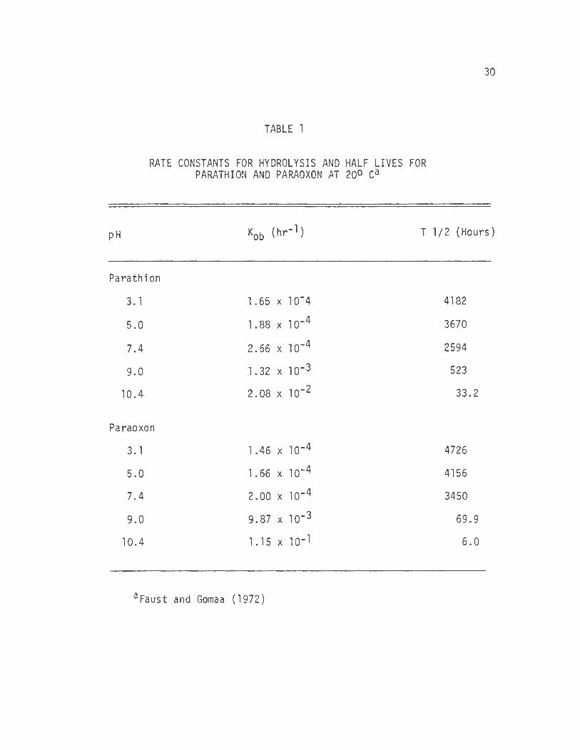
1. Rate Constants for Hydrolysis and Half Lives for Parathion and Paraoxon ........... .
2. Susceptibility of Orconectes Rusti cus to Parathion Exposure in Water . . . . . . . . . . . . . . .
3. Susceptibility of Viviparus Mall eatus to Parathion Exposure in Water . . . . . . . . . . . . . . . .
4. Susceptibility of Orconectes Rusticus to Paraoxon Exposure in Water . . . . . . . . . . . . . . . .
5. Susceptibility of Viviparus Malleatus to Paraoxon Exposure in Water . . . . . . . . . . . .
6. In Vitro Metabolism of Parathion with the Production ~of .e_-Nitrophenol Measured Spectrophotometrically
7. Efficiency of Thin Layer Chromatography Procedures
8. Efficiency of the Ether Extraction of Parathion from Water Samples ........ .
9. Parathion Metabolism and Excretion into the Water Environment by Orconectes Rusticus .
10. Parathion Metabolism and Excretion into the Water Environment by Viviparus Malleatus .
11. Accumulation of Parathion and Metabolites in Orconectes Rusticus Tissues Following Exposure
12. Accumulation of Parathion and Metabolites in Viviparus Malleatus Tissues Following Exposure .
13. Metabolism of 14c Labeled Parathion into .e_-Nitrophenol, Paraoxon, and Diethyl Phosphorothionate by Viviparus Malleatus . . .
.
.
.
.
.
.
14. Metabolism of 14c Labe l ed Parathi on to .e_-Nitrophenol, Paraoxon, and Diethyl Phosphorothionate by
. .
. .
. .
. .
. .
. .
. .
.
.
.
.
.
.
30
50
50
52
52
54
57
58
60
61
62 '
64
65
Orconectes Rusticus . . . . . . . . . . . . . 66
vii
15. In Vitro Metabolism of Paraoxon to p-Nitrophenol ~by Orconectes and Viviparus Determined
Spectrophotometrically ........... .
viii
68
LIST OF ILLUSTRATIONS
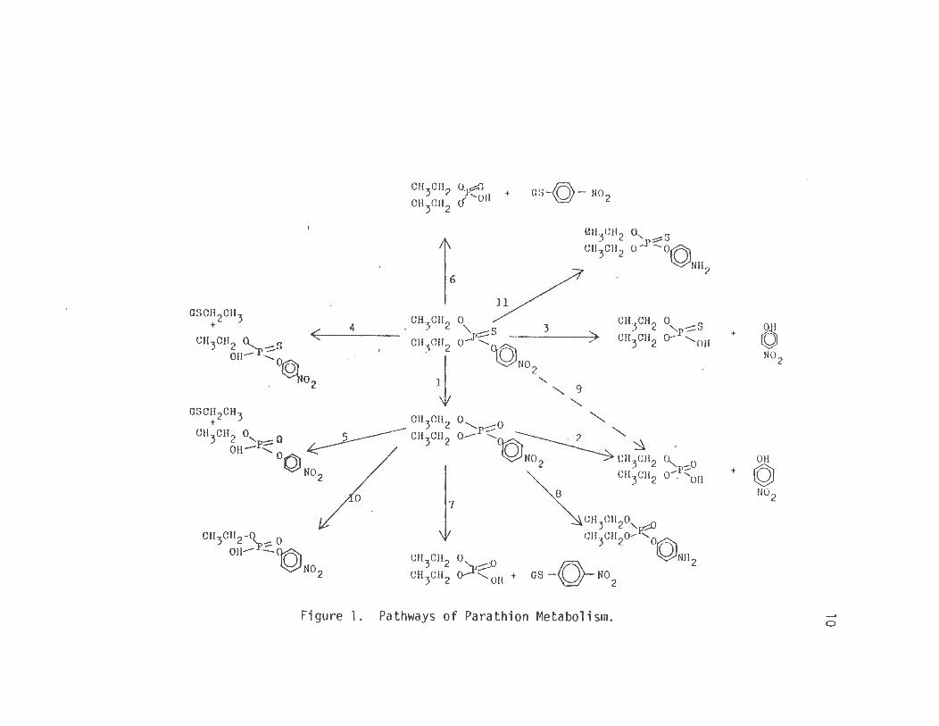
1. Pathways of Parathion Metabolism .... 10
ix
I. Introduction
The organophosphates are a very large class of compounds as
there are over 50,000 compounds in that category today. Originally
developed in Germany as nerve gases, the most important ones being
tabun, sarin, and soman, they have since been found to be of great
or unknown above the level of the solvent bath present in the chroma
tography tank. The solvent system employed consisted of hexane,
chloroform, and methanol 7:2:1, all pesticide grade. One hundred ml of
solvent mixture was used with this amount forming a 1 cm deep pool of
solvent in the · tank. A large square of filter paper was placed in the
tank to assure saturation.
The solvent front was allowed to develop to 10 cm before the
plate was removed from the tank to dry. Identification of the separated
spots was aided by the use of Rhodamine B 0.1 mg/ml in ethanol which
was sprayed on the plate until a light pink color covered the plate.
The plate was then viewed under ul traviolent light where the parathion
44
and metabolite spots were plainly visible against a light background.
When radioactive compounds were to be recovered, the sprayed
plates were marked to isolate the desired area of the plate to be
scraped by tracing around the area with a dissecting needle. That area
of the coating was scraped from the plate with a small spatula. The
silia gell coating was then retrieved with the use of a glass wool
plugged eye dropped attached to a vacuum hose. Care was taken to be
sure no significant amount of coating was lost in this process and
controls were run to determine the effectiveness and reliability of
this process. The glass wool containing the Silica Gel G and compounds
was then deposited into a scintillation vial containing Hydromix
Packard Liquid Scintillation Counter. Blanks and controls were
included to determine the effect of the glass wool, Silica Gel G and
Rhodamine B spray. Raw counts were used for all subsequent data
calculations.
5. Elution Procedure
Parathion and metabolites were recovered from the TLC plates
in liquid form for subsequent gas liquid chromatograph characterization
or as a comparison to the above procedure by scraping the area of the
plate and then using the eye dropper technique to transfer the coating
along with glass wool to a vial containing 5 ml of anhydrous ethyl
ether. This mixture was shaken and poured into a funnel with filter
paper. The filtering funnel was rinsed with 5 ml more of ether, and
the liquid containing the parathion or metabolites was evaporated with
45
suction down to less than 4 ml. The ether was then placed into a
scintillation counting vial containing 10 ml of Hydromix and counted or
analyzed with the gas chromatograph.
6. Extraction of Parathion and Metabolites from Water Samples
The extraction of parathion, paraoxon, .P_-nitrophenol, diethyl
phosphate and diethyl phosphorothionate from 500 ml samples of water
was done using anhydrous ethyl ether as the sole extracting solvent.
The water sample was placed in a large (1000 ml) separatory
funnel and extracted with 40 ml of ether. The sample was shaken for 2
minutes. The ether layers from three consecutive extractions were
pooled and passed through a glass column containing 3 inches of
anhydrous sodium sulfate to remove any water. Ether layers that were
in a semigel state had additional 20 ml ether portions added to
re-establish the more liquid state before passage through the column.
The dried sample was then placed in rotary evaporator flasks and the
volume reduced under suction but without heat. The sample was then
either spotted on a thin layer chromatography plate or injected into
the gas chromatograph.
7. Extraction of Parathion and Metabolites Excreted into Water Samples by Crayfish
Four crayfish were individually placed into 1 liter beakers
contain i ng 500 ml of glass distilled water. The water in two of the
beakers then had 14c parathion labeled in the ethyl group added while
the other two beakers had 14c parathion ring labeled added. The
amounts of parathion added to all of the beakers were such that a
concentration of 100 ppb was attained. One and one-half hours after
exposure, the crayfish were removed from the water and frozen for
46
subsequent analysis of their tissues for parathion and metabolites.
The water in which they were exposed was immediately extracted employ
ing the water extraction procedure to determine if any metabolites
could be detected.
8. Extraction of Parathion and Metabolites from Crayfish and Snail Tissue
The recovery of parathion and metabolites from the tissues
of crayfish following exposure to parathion in water was accomplished
using the crayfish that had been exposed to 100 ppb parathion and then
frozen. The same procedure for extraction from tissue was performed on
the whole snail tissue from the snails that were exposed to radio-
activity labeled parathion at a concentration of 320 ppm for 48 hours.
The day following the exposure, the hepatopancreas and tail
muscles of the 14c-ethyl parathion exposed crayfish tissue were pooled
as were the tissues from the crayfish exposed to the ring labeled
compound. The tissues were homogenized with a blade homogenizer in
9 ml of ether for 30 seconds. The homogenate was then scraped into
centrifuge tubes containing 9 ml of ether. The tissue attached to the
homogenizer blades was rinsed into the tube with additional ether. The
centrifuge tube containing the tissue and ether was shaken for 2 minutes
and then centrifuged at low speed to separate the tissues from ether
layer.
Following the centrifugation the ether layer was removed with
47
a Pasteur pipet and transferred to a rotary evaporator flask. The
sample was then evaporated without heat to a volume small enough to be
spotted onto TLC plates. The plates were developed and sprayed as
described along with standards for identification of the products. The
spots were removed and counted in the liquid scintillation counter to
quantify the amount of product recovered.
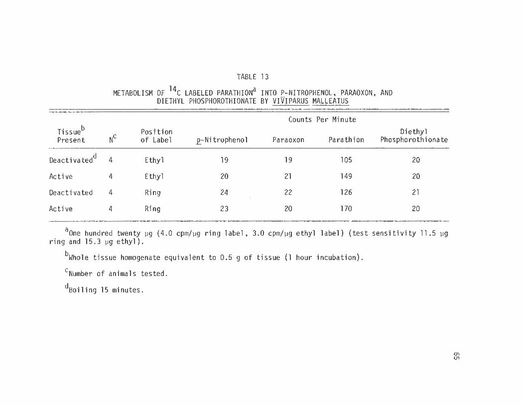
9. Production of 2_-Nitrophenol, Paraoxon, Diethyl Phosphate or Diethyl Phosphorothionate from Parathion via in vitro Metabolism by Orconectes or Viviparus Tissues
The procedure for this assay was almost identical to that for
the 2_-nitrophenol spectrophotometric assay.
Tissues from snail or crayfish were removed and homogenized
in NaCl, MgS04, nicotinamide in the same way. The tissue was incubated
in the same way with the parathion added to initiated the reaction
containing a known amount of radioactive parathion. Ring labeled 14c
parathion was used to follow the fate of parathion, paraoxon and
2_-nitrophenol while 14c ethyl label was employed to investigate the
possible production of diethyl phosphate and diethyl phosphorothionate.
The reaction was stopped by the addition of 5 ml of cold ether
and the mixture was stored in glass stoppered test tubes. The mixture
48
was extracted with two 5 ml portions of ether by shaking the sample
with ether for 2 minutes and removing the ether layer and depositing it
into flash evaporator flask. The extraction solvent was then evapora
ted under suction without heat to a small volume (300 µl) which was
subsequently spotted on thin layer chromatography plates. The plates
were developed and sprayed as previously described and the spots
corresponding to metabolites were scraped and placed in scintillation
vials. The amount of parathion that was metabolized to the -other
products was determined by the relative amounts of radioactivity found
in parathion spot versus the .E_-nitrophenol, paraoxon, diethyl phosphate
and diethyl phosphorothionate spots.
IV. Results
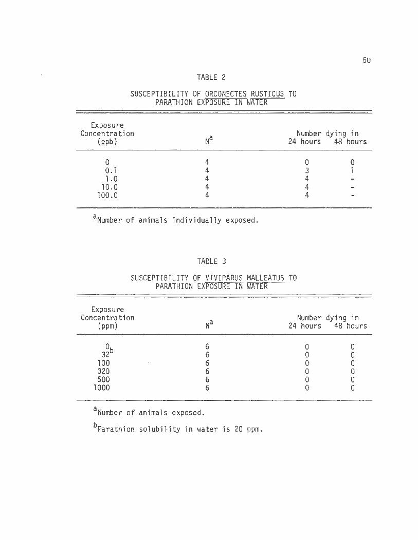
A. Parathion Toxicity
The fresh water snail Viviparus malleatus was not sensitive to
parathion in the dose range to which the animal was exposed in these
experiments. A level of 1000 ppm failed to exert any observable toxic
effects on the snail (table 3). The solubility of parathion in water
is only 20 ppm so the attempt to create a 1000 ppm concentration was to
be certain that saturation was reached.
The crayfish Orconectes rusticus demonstrated extreme sensitiv
ity to parathion exposure in its water environment. Concentrations as
low as 1 .0 parts per billion (ppb) produced death in 100% of the cray
fish exposed in less than 24 hours (table 2). The toxicity was evi
denced by twitching movements and exaggerated muscle contractions in
response to provocation immediately before death.
The concentration of 0.1 ppb was the only concentration that
did not produce 100% lethality in 24 hours. Three of the four cray
fish tested at that concentration did die in 24 hours. The remaining
animal dies in less than 48 hours.
It should be noted that it was necessary to employ new tygon
tubing, new air stones and new beakers for this study as it was shown
that beakers that had contained parathion and were subsequently cleaned
could still leach enough parathion to cause death to crayfish.
49
50
TABLE 2
SUSCEPTIBILITY OF ORCONECTES RUSTICUS TO PARATHION EXPOSURE IN WATER
Exposure Concentration
(ppb)
0 0. 1 1.0
10.0 100.0
4 4 4 4 4
aNumber of animals individually exposed.
TABLE 3
Number dying in 24 hours 48 hours
0 3 4 4 4
0 1
SUSCEPTIBILITY OF VIVIPARUS MALLEATUS TO PARATHION EXPOSURE IN ~JATER
Exposure Concentration
Na (ppm)
0 6 32b 6
100 6 320 6 500 6
1000 6
aNumber of animals exposed .
bParathion solubility in water is 20 ppm.
Number dying in 24 hours 48 hours
0 0 0 0 0 0
0 0 0 0 0 0
51
The calculation of Tlm50 values for these data was not possible
because of the lack of snail susceptibility and extreme crayfish
sensitivity.
The resistance of the snails to parathion was further studied
by injecting the snail directly with parathion. The two snails that
were each injected with 5.0 mg (approximately 250 mg/kg) of parathion
showed no toxic sign in 96 hours.
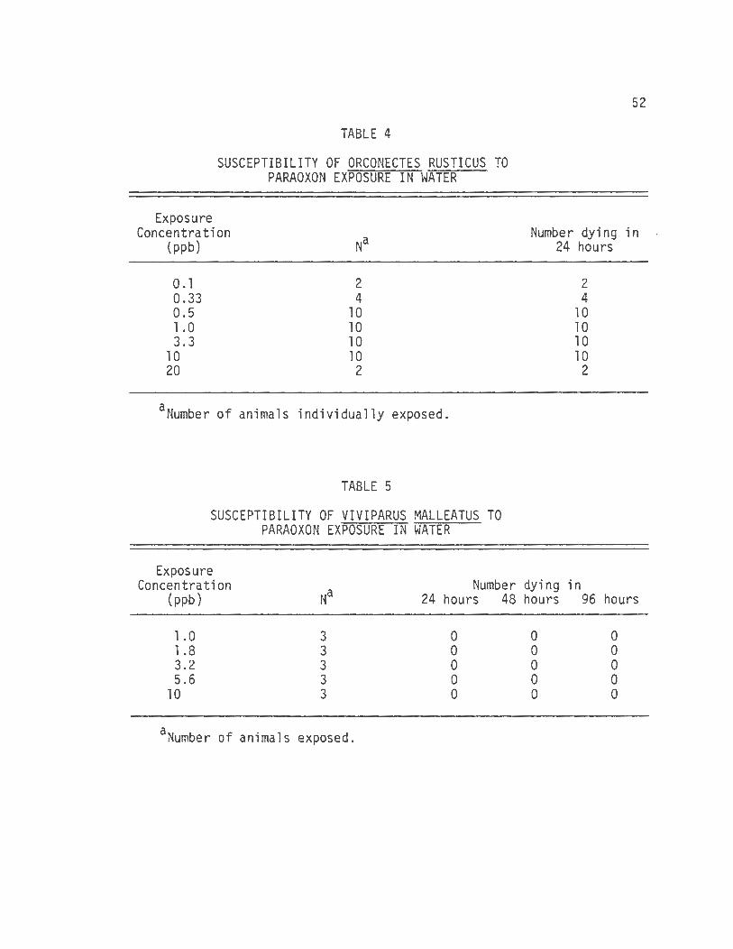
B. Paraoxon Toxicity
The susceptibility of Viviparus malleatus or Orconectes
rusticus was determined for the oxygen analog of parathion, paraoxon.
These data show no snail mortability due to paraoxon exposure at 10
ppm exposure in 96 hours (table 5).
The crayfish was extremely senstitive to paraoxon (table 4).
All of the animals exposed to concentrations ranging from 0.1 to 20 ppb
died in less than 24 hours and exhibited the same toxic signs (e.g.,
twitching movements and exaggerated muscle contractions in response to
provocation) as those that dies in the parathion exposure experiment.
The resistance of the snails to paraoxon was studied by
injecting paraoxon directly into two snails. The snails that were each
injected with 1.0 mg (approximately 50 mg/kg) of paraoxon showed no
toxic signs in 96 hours.
C. Q_-Nitrophenol Spectrophotometric Assay
The spectrophotometric _p_-nitrophenol assay measured the
production of .P_-nitrophenol from parathion by crayfish hepatopancreas,
TABLE 4
SUSCEPTIBILITY OF ORCONECTES RUSTICUS TO PARAOXON EXPOSURE IN WATER
Exposure Concentration
(ppb)
0 .1 0.33 0.5 1.0 3.3
10 20
2 4
10 10 10 10
2
aNumber of animals individua11y exposed.
TABLE 5
SUSCEPTIBILITY OF VIVIPARUS MALLEATUS TO PARAOXON EXPOSURE IN WATER
Exposure
52
Number dying in 24 hours
2 4
10 10 10 10
2
Concentration Na
Number dying in {ppb) 24 hours 48 hours 96 hours
1.0 3 0 0 0 1.8 3 0 0 0 3.2 3 0 0 0
. 5 .6 3 0 0 0 10 3 0 0 0
aNumber of animals exposed.
53
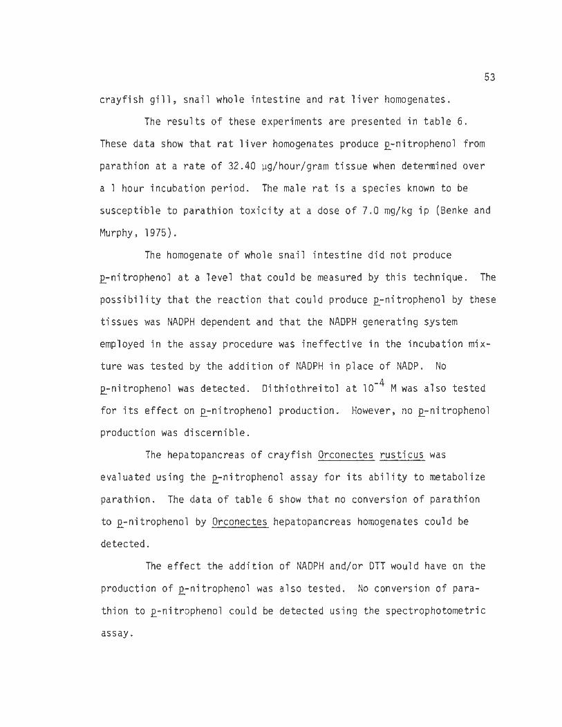
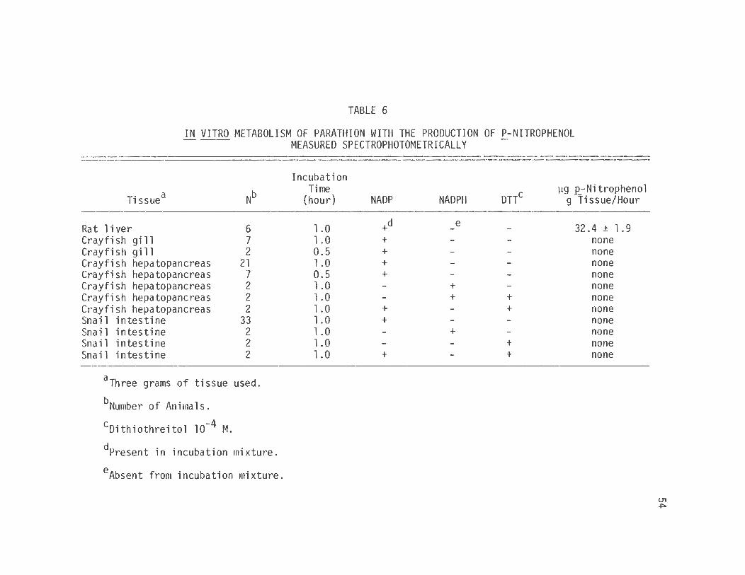
crayfish gill, snail whole intestine and rat liver homogenates .
The results of these experiments are presented in table 6.
These data show that rat liver homogenates produce .E_-nitrophenol from
parathion at a rate of 32.40 µg/hour/gram tissue when determined over
a 1 hour incubation period. The male rat is a species known to be
susceptible to parathion toxicity at a dose of 7.0 mg/kg ip (Benke and
Murphy, 1975) .
The homogenate of whole snail intestine did not produce
.E_-nitrophenol at a level that could be measured by this technique. The
possibility that the reaction that could produce .E_-nitrophenol by these
tissues was NADPH dependent and that the NADPH generating system
employed in the assay procedure was ineffective in the incubation mix
ture was tested by the addition of NADPH in place of NADP. No
.E_-nitrophenol was detected. Dithiothreitol at 10-4 M was also tested
for its effect on .E_-nitrophenol production. However, no .E_-nitrophenol
production was discernible.
The hepatopancreas of crayfish Orconectes rusticus was
evaluated using the .E_-nitrophenol assay for its ability to metabolize
parathion. The data of table 6 show that no conversion of parathion
to .E_-nitrophenol by Orconectes hepatopancreas homogenates could be
detected.
The effect the addition of NADPH and/or OTT would have on the
production of .E_- nitrophenol was also tested. No convers i on of para
thion to .E_-nitrophenol could be detected using the spectrophotometric
assay.
TABLE 6
IN VITRO METABOLISM OF PARATHION WITH THE PRODUCTION OF P-NITROPHENOL ~~ MEASURED SPECTROPHOTOMETRICALLY
Tissuea
Rat 1 i ver Crayfish gi 11 Crayfish gi 11 Crayfish hepatopancreas Crayfish hepatopancreas Crayfish hepatopancreas Crayfish hepatopancreas Crayfish hepatopancreas Snail intestine Snail intestine Snail intestine Snail intestine
dEther used to elute sample (10 ml added and then evaporated to less than 4 ml for counting.
57
346 388 375 414
+ 412
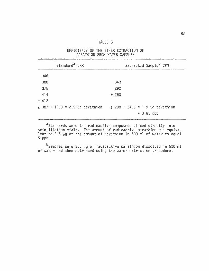
TABLE 8
EFFICIENCY OF THE ETHER EXTRACTION OF PARATHION FROM WATER SAMPLES
Standarda CPM
343 292
+ 260
Extracted Sampleb CPM
x 387 ± 12.0 = 2.5 µg parathion x 298 ± 24.0 = 1 .9 µg parathion = 3.85 ppb
aStandards were the radioactive compounds placed directly into scintillation vials. The amount of radioactive parathion was equivalent to 2.5 µg or the amount of parathion in 500 ml of water to equal 5 ppb.
bsamples were 2.5 µg of radioactive parathion dissolved in 500 ml of water and then extracted using the water extraction procedure.
58
59
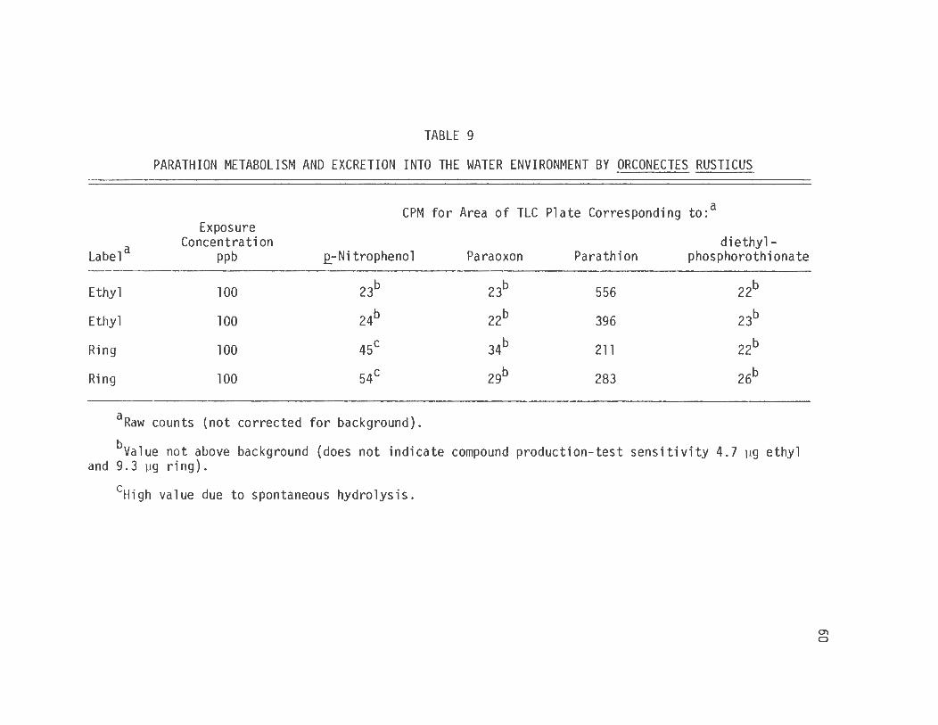
extraction, thin layer chromatographic separation and scintillation
counter quantification technique was used for that determination. The
data in table 9 show that unquestionably the vast majority of the
total amount of compound recovered from the exposure was in the form of
parathion.
The snails were exposed to 320 ppm of ring labeled parathion
for 48 hours with subsequent extraction of the aquarium water as with
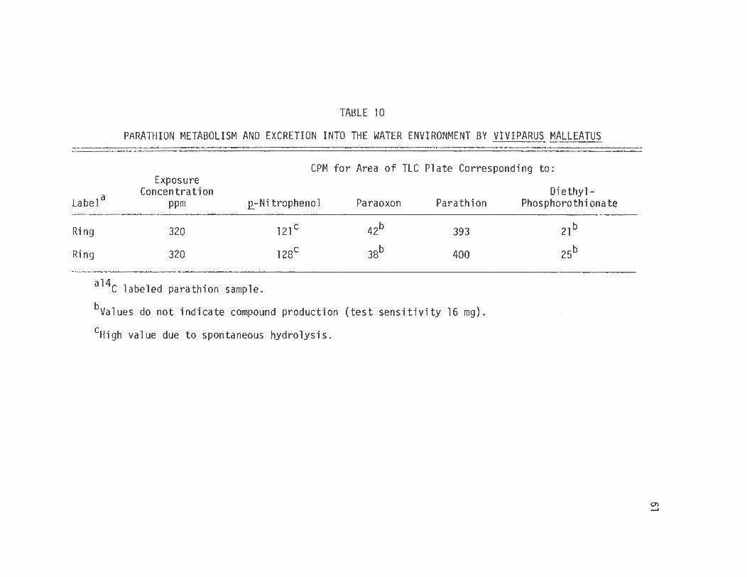
the crayfish (table 10). The results presented in tables 9 and 10
show that for both the crayfish and the snail there was no excretion
of parathion metabolite into the water environment. There was no
production of 2_-nitrophenol with either species. However high values
for 2_-nitrophenol were obtained which was shown to be due to spontane
ous hydrolysis.
F. Evaluation of Parathion and Metabolites Accumulation in Crayfish and Snail Tissues Following Parathion
Exposure in a Water Environment
To study the accumulation of parathion by tissues of the
species animals were exposed to 100 ppb 14c labeled parathion for
one-half hour and were then sacrificed for subsequent analysis. The
extraction of labeled 14c parathion and metabolites from crayfish
tissues showed that there was no indication of any accumulation of
these compounds in hepatopancreas or muscle tissues. The data (table
11) show that when hepatopancreas or muscle samples from animals
exposed to ring or ethyl labeled 14c parathion were pooled, no accumu-
lation could be detected.
TABLE 9
PARATHION METABOLISM AND EXCRETION INTO THE WATER ENVIRONMENT BY ORCONECTES RUSTICUS
CPM for Area of TLC Plate Corresponding to:a Exposure
aO = tissue (ViviEarus intestine equivalent to 0.5 g, Orconectes hepatopancreas equivalent to 0.1 g) deactivated by boiling 15 minutes.
+ = tissue active.
bNumber of samples tested.
eyes = 10 µl of 10 µg/µl paraoxon in ethanol added to incubation mixture.
no = paraoxon not added to incubation mixture.
dAbsorbance measured at 410 nm ± standard error.
V. Discussion
The investigation of the metabolism of parathion by fresh
water invertebrates is more than an academic exercise in pesticide
metabolism and detection. The problems that followed the use of the
chlorinated hydrocarbon pesticides were in part due to the lack in
understanding of the environmental consequences of their use and
ignorance as to the effects the compounds would have on organisms other
than the target species and man.
The increase in use of the organophosphates along with a
limited understanding of their toxicity and metabolism in nonmammals is
a situation analogous to the one that led to problems with chlori:;rated
hydrocarbon pesticides. Nicholson et al. (1962) did investigate the
environmental exposure consequences of parathion and showed that para
thion would accumulate in a farm pond, and that it would persist in the
environment for at least nine months. These times are similar to those
that were reported by Faust (1964).
Nicholson et al. (1962) showed that following the normal
agricultural spraying of an orchard, concentrations of l .22 ppb of
parathion could be detected in a farm pond that was subject to rain
runoff from that orchard. The concentration of parathion recovered
from the bottom mud samples from that pond were even greater than the
concentrations in the water. Nicholson's evaluation of the fauna
69
70
present in the farm pond showed no crayfish present.
The environmental implications of parathion use were considered
an integral part of the stimulus for this project, as the possible
toxocity and/or metabolism of parathion by Orconectes rusticus and
Viviparus malleatus would be a result of environmental exposure. The
determination of parathion-induceci toxicity to Orconectes or Viviparus
was investigated with an awareness of the environmental exposures
possible and an interest as to the possible accumulation of the toxin
and/or metabolites in the species.
The results obtained by the exposure and injection toxicity
experiments have shown that the snail Viviparus malleatus is not
susceptible to acute parathion induced toxicity, and no accumulation of
parathion or metabolites were detected in the snail. This correlates
with the work of Yu and Sanborn (1975) who could not detect any
accumulation of parathion in snail tissues following exposure to the
pesticide. The toxicity data derived concerning the crayfish
Orconectes rusticus demonstrates the opposite, that Orconectes is
susceptible to acute parathion induced toxicity at the extremely low
concentration of 1.0 ppb. Realizing that parathion concentrations of
greater than l .0 prb can be attained in natural waters as a result of
the normal agricultural use of parathion and that no crayfish were
reported in the pond investigated by Nicholson et al. (1962), the
possibility that the use of parathion may have deleterious effects on
the crayfish populations gains credence.
The reasons why parathion exposure was toxic to Orconectes
71
and not to Viviparus were evaluated by this project. The fact that
acute toxicity due to parathion exposure is due to its inhibition of
AChE had been shown by many investigators and that evidence has been
presented in the literature review section. It is important to
remember that parathion itself is not the compound that exerts the
inhibition of the AChE enzyme but rather the desulfurated metabolite of
parathion, paraoxon. Previous research has shown that not only must
parathion be metabolized to paraoxon to produce toxic effects but
parathion can be metabolized directly to relatively nontoxic meta
bolites diethyl phosphorothionate plus .P_-nitrophenol or diethyl phos
phate plus .P_-nitrophenol, through the same intermediate which gives
paraoxon. The toxic compound paraoxon can be detoxified prior to its
aging to diethyl phosphate and.P_-nitrophenol. Many routes of metabolism
of parathion and paraoxon other than these two important pathways have
also been elucidated, such as the glutathion dependent alkyl and aryl
transformations. These pathways are all presented in Figure 1. The
important point is that these reactions occur and that the rates at
which they compete for parathion and paraoxon influences the toxicity
of parathion exhibited in that particular species (Benke and Murphy,
1975).
The parathion exposure experiments showed that Viviparus was
not sensitive to parathion in its water environment even when the
solubility of parathion in the water was exceeded while Orconectes was
sensitive to parathion in its water at 1 ppb. The question that pre
sented itself was why there was such a species difference in
72
susceptibility. Refering to Figure 1 and with an understanding of the
data from other investigators the possible mechanisms for these results
were envisioned. The snail could be resistant to parathion exposure
due to the following reasons:
1. Parathion was not entering the shell of the snail.
2. Parathion was entering the shell but not being metabolized to
paraoxon, which if produced would cause toxicity.
3. The parathion was being metabolized extremely rapidly and
efficiently via pathways 3, 4, 6, 9 or 11 (Fig. 1) to the nontoxic
metabolites or being bound to tissues where no metabolism could
occur.
4. The parathion was being metabolized to paraoxon (pathway 1, Fig. 1)
very slowly while pathways 2, 5, 7, 8 or 10 were operating very
quickly.
5. The snail may be converting parathion to paraoxon but be
insensitive to paraoxon.
The goal of the investigation of the resistance of Viviparus
malleatus to parathion was then to design experiments to determine
which of these mechanisms was responsible for the lack of parathion
toxicity in the snail.
The possibility that the parathion was not entering the shell
of the snail was evaluated by injecting snails with 5.0 mg/kg. The
results show that there were no toxic signs demonstrated by the snails.
Paraoxon was also injected directly into snails. Two snails were
exposed to 50 mg/kg of paraoxon by direct injection with no toxic
73
signs demonstrated in 96 hours. The results of these two experiments
establish that the ability of the compound to enter the shell of the
snail was not a limiting factor in its lack of toxicity. The results
also show that the snail is resistant to paraoxon as well as parathion
so that the absence of pathway 1 of Figure 1 would not be the mechanism
responsible for the animals' resistance to the organophosphate.
The 2_-nitrophenol spectrophotometric assay was used with whole
snail homogenates to determine if the snail was capable of metabolizing
parathion via pathways 2, 3 or 9 of Figure 1. Since these pathways
are the most important detoxification pathways for parathion resistance,
the demonstration of measurable 2_-nitrophenol would show that the
parathion or paraoxon was being detoxified and could possibly be the
cause for the animals' resistance to parathion and paraoxon exposure.
The 2_-nitrophenol spectrophotometric assay with whole snail intestine
did not produce Q_-nitrophenol at a level that could be measured by
this technique. The reason for the lack of production was not due to
deficiency of NADPH for dependent enzyme reactions or the destruction
of essential sulfhydryl groups of the membranes or enzymes, as NADPH
and dithiothreitol were added. The same spectrophotometric assay
incorporating paraoxon in the place of parathion was also done to
determine if pathway 2 of Figure 1 was possible but not detected with
parathion in the incubation due to the absence of the conversion of
parathion to paraoxon (pathway 1, Fig. 1). Again, no 2_-nitrophenol was
produced indicating the lack of pathway 2 route of metabolism.
The Q_-nitrophenol assay is designed to demonstrate t he presence
74
of metabolism occurring but because the measured compound can be
produced by numerous pathways if the compound is detected the exact
pathway followed cannot be determined. A method to determine if
specific pathways were used in the metabolism of parathion would be to
determine the presence of the specific compounds paraoxon and diethyl
phosphorthionate, along with E_-nitrophenol and parathion.
The extraction of parathion and metabolites from water samples
measured the excretion of metabolites of parathion into the water
environments of snails. The results of that experiment (table 10) show
again that there was no metabolism followed by excretion of parathion
by the snail. To be certain that there was no metabolism of parathion
occurring by Viviparus malleatus an experiment was designed where
homogenates of snail tissue were incubated with radioactive parathion,
that mixture subsequently being extracted and the metabolites separated
by thin layer chromatography and quantified by liquid scintillation
counting. The results (table 13) further substantiate the conclusion
that parathion is not metabolized by Viviparus malleatus. The snail is
demonstrating a biologic resistance to parathion, as the inhibition of
AChE if present has no effect on the organism.
The crayfish demonstrated susceptibility to acute parathion
toxicity, and the muscle twitching signs were consistent with AChE
inhibition. The mechanism by which this toxicity was produced was
assumed to follow the desulfuration of parathion to paraoxon via
pathway 1 of Figure 1. This conversion would have to be at a rate
high enough to account for a paraoxon concentration that would cause
toxicity. The reaction rates for the toxic conversion reaction and
detoxification reactions has been shown to be an important factor in
the toxicity exhibited by species to parathion exposure does not
establish the existence or absence of detoxifying reactions by
Orconectes, only the existence of a toxic conversion reaction
75
The goal, then, for the investigation of the Orconectes
susceptibility to parathion was to demonstrate the presence of the
reaction coverting parathion to paraoxon and any other metabolic path
ways present as depicted in Figure 1. This is closely related to the
question of the accumulation ~f parathion and/or its metabolites in
crayfish tissues. The results presented in table 11 show that there
was no accumulation of these compounds in the gill, muscle or hepa
topancreas of crayfish following exposure to parathion.
The possibility that the crayfish toxicity was due to concen
trations of paraoxon too low to detect could not be overlooked in this
investigation considering the research of Carlson (1973) and Elmanlouk
and Gessner (1976). They reported that the hepatopancreas of the
lobster is the organ responsible for drug metabolism, but the
hepatopancreas of the lobster had very little, if any, observable
ability to convert parathion to paraoxon.
Establishing if paraoxon was capable of producing the toxicity
in crayfish was done by exposing the crayfish to paraoxon in the
aquarium water as had been done with parathion. The experimental
results showed that the crayfish was sensitive to paraoxon and that the
toxic signs exhibited by the crayfish following paraoxon exposure were
the same as those following parathion exposure. The concentration
range that produced the toxicity in the crayfish due to paraoxon
(table 4) was consistent with the theory that the parathion was
exhibiting its toxicity through conversion to paraoxon and subsequent
AChE inhibition.
The toxicity determination experiments did little to increase
the understanding of the metabolism of parathion by Orconectes.
Experimental procedures similar to the ones used to determine the
metabolism of parathion in snails needed to be done to determine the
pathways of metabolism of parathion and paraoxon in crayfish.
76
The .e_-nitrophenol spectrophotometric assay was used to
ascertain if any metabolism of parathion or paraoxon could be deter
mined with that technique. Rats are susceptible to parathion toxicity
at 7.0 mg/kg i.p. (Benke and Murphy, 1975) so they were used as
controls in these experiments to compare their metabolism of parathion
to that in the crayfish. The .e_-nitrophenol assay was perfonned on
rat liver homogenates as well as homogenates of crayfish hepatopancreas
and gill (t able 6). The results show t hat the rat liver homogenate
caused the production of 32.4 µg/hour/gram of tissue when determined
over a l hour incubation period, but the crayfish hepatopancreas and
gill homogenates which would be expected to be the organs capable of
metaboliz i ng parathion produced no .e_-nitrophenol that could be measured
spectrophotometrically. These results were not due to lack of NADPH
for dependent enzyme processes or the destruction of vital enzymes
since the addition both NADPH and dithiothreitol (l0-4 M) added to
some incubation samples as had been done in the snail tissue experi
ments, were not effective.
77
Since the E_-nitrophenol assay was done using both parathion and
paraoxon in the incubation flasks, the assay had the capability of
monitoring pathways 2, 3 and 9 of Figure 1 all of which cause the
production of E_-nitrophenol. No E_-nitrophenol could be detected by
this technique. However, because the crayfish were sensitive to the
parathion and paraoxon exposures other experimental designs were used
in an attempt to monitor some metabolism of parathion or paraoxon by
the crayfish or its tissues.
The possibility that the crayfish was able to absorb parathion
from its water environment and then to excrete metabolites of parathion
back into the water was evaluated. 14c Parathion labeled either in
the ethyl or ring positions was used for these exposure-excretion
experiments. Following the exposure of crayfish to labeled parathion
at a concentration of 100 ppb for 1-1/2 hours, the water in which the
crayfish were kept was extracted with ether, the samples separated by
thin layer chromatography and the appropriate spots scraped and counted
by liquid scintillation counting. The results showed that after the
spontaneous hydrolysis of stock parathion is considered there was no
excretion of parathion metabolites into the water that would be
indicative of parathion metabolism.
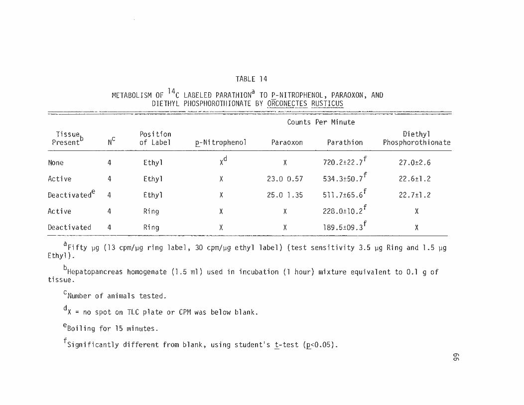
The ability to produce even minute amounts of parathion
metabolites by crayfish tissues was tested by the use of an incubation
technique similar to the E_-nitrophenol assay. Homogenates of crayfish
78
hepatopancreas were incubated with 14c parathion in either the ring or
ethyl positions. When the incubation was completed, the mixture was
extracted, separated and quantified by thin layer chromatography and
scintillation counting. The results (table 14) show that there was no
production of .e_-nitrophenol or paraoxon both of which would be detected
through the use of the ring label and no production of diethyl phos
phate or diethyl phosphorothionate detectable through the use of the
ethyl label.
Retrospectively, it is easy to propose a number of mechanisms
by which the metabolism of parathion and paraoxon could be responsible
for the crayfish exhibited toxicity without the metabolism being
detected by sensitive mechanisms. The crayfish could be so exquisitely
sensitive to paraoxon that a very small amount of metabolite below
detectability could be responsible. The paraoxon that was produced
could bind with the enzyme to cause AChE inhibition and remain attached
to the enzyme. The .1IJ.. vivo experiments demonstrated that the crayfish
are sensitive to very small quantities of paraoxon. Unfortunately the
.1IJ.. vitro techniques for the detection of .e_-nitrophenol are not quali
tative and quantitative enough to separate the nonspontaneous
hydrolytic production of .e_-nitrophenol from the small amounts of
2_-nitrophenol that would accompany any paraoxon inhibition of AChE.
The question that rises from these results is how is the
parathion producing its toxicity if no metabolism can be detected.
VI. Conclusions
The goal of determining the toxicity and metabolism of para
thion in the fresh water invertebrates Orconectes rusticus and
Viviparus malleatus made the extraction, detection and quantification
of minute quantities of organophosphate and its metabolites necessary.
Groups of the crayfish Orconectes rusticus when exposed to
1.0 ppb or greater of parathion all died. The snail Viviparus
malleatus was resistant to parathion exposure at the level of parathion
solubility and to direct injection of parathion with a dose of
approximately 50 mg/kg. The crayfish demonstrated extreme sensitivity
to paraoxon exposure and showed the same signs of cholinergic stimu
lation that were exhibited with parathion exposure, while the snail was
not effected.
The ether extraction of water samples for parathion and
metabolites was both efficient and reproducible. That technique
demonstrated that neither parathion or metabolites accumulated in the
tissues of Orconectes or Viviparus following exposure.
The thin layer chromatography technique used in the investi
gation was simple, reliable, specific, and sensitive. The combination
of Silica Gel G 250 µ plates developed in hexane, chloroform, and
methanol (7 :2:1) and sprayed with Rhodamine B (0.1 mg/ml in ethanol)
allowed for the ultravio let visualization of as little as 1 .0 µg of
79
organophosphate. Excellent separation of £_-nitrophenol, paraoxon,
parathion, and diethyl phosphorothionate was obtained.
80
The ability of the crayfish and the snail to metabolize
parathion was determined through the use of the £_-nitrophenol spectro
photometric assay, the investigation of excreted metabolites that were
extractable from water samples, the determination of the presence of
metabolites accumulating in tissue, detection of radioactive metabo
lites from tissue incubation experiments and the toxicity experiments.
All of the experimental data support the conclusion that there was no
metabolism of parathion by the snail and that this lack of metabolism
and the insensitivity of Viviparus to paraoxon form the basis for the
lack of toxicity exhibited by those compounds to the snail.
The data indicate no metabolism or accumulation of parathion
or its metabolites by the crayfish, but the conclusion that no
metabolism was taking place cannot be made as the toxicity experiments
do not support that contention.
The data from the £_-nitrophenol spectrophotometric assays with
parathion and paraoxon using either crayfish or snail tissue revealed
no production of £_-nitrophenol by either the snail or the crayfish
indicating that pathways 1, 2, 3 and 9 of Figure 1 could not be
demonstrated by that technique.
The extraction experiments show that there was no detectable
accumulation of parathion or metabolites in Orconectes or Viviparus
and no detectable excretion of parathion metabolites into the water
environment.
81
The experiments performed with radioactive parathion incubated
with either crayfish or snail tissues were unable to demonstrate any
metabolism of parathion, paraoxon, p_-nitrophenol, diethyl phosphoro
thionate and diethyl phosphate by crayfish or snails.
The resistance to parathion and paraoxon demonstrated by the
snail does not stem from enzyme systems capable of detoxifying the
compounds. The snail is biologically resistant to the inhibition of
AChE.
The mechanism by which parathion exhibits toxicity in
Orconectes cannot be determined from the results obtained by these
experiments. The extreme sensitivity to parathion and the clinical
signs demonstrated by the crayfish following parathion exposure are
consistent with the production of effective concentrations of paraoxon
that can inhibit the AChE at synapse and neuromuscular junctions.
However, until the production of some metabolism of parathion by some
crayfish tissue can be demonstrated more definitive conclusion cannot
be made.
REFERENCES
Alary, J., and Brodeur, J.: Studies on the mechanism of phenobarbitalinduced protection against parathion in adult female rats. J. Pharmacol. Exp. Ther. 169: 159-167, 1969.
Alary, J., and Brodeur, J.: Correlation between the activity of liver enzymes and the L050 of parathion in the rat. Can. J. Physiol. Pharmacol. 48: 829-831, 1970.
Albaugh, O. W.: Insecticide tolerances of two crayfish populations (Procambarus acutus) in south-central Texas. Bull. Environ. Contam. Toxicol. §_: 334-338, 1972.
Appleton, H. T., and Nakatsugawa, T.: Paraoxon deethylation in the metabolism of parathion. Pest. Biochem. Physiol. £: 286-294, 1972.
Bartels, E., and Nachmansohn, 0.: Organophosphate inhibitors of acetycholine-receptor and-esterase tested on electroplax. Arch. Biochem. Biophys. 133: 1-10, 1969.
Benke, G. M., Cheever, K. L., Mirer, F. E., and Murphy, S. 0.: Comparative toxicity, anticholinesterase action and metabolism of methyl parathion and parathion in sunfish and mice. Toxicol. Appl. Pharmacol. 28: 97-109, 1974.
Benke, G. M., and Murphy, S. 0.: Anticholinesterase action of methyl parathion, parathion and azinphosmethyl in mice and fish: onset and recovery of inhibition. Bull. Environ. Contam. Toxicol. 1£: 117-122, 1974.
Benke, G. M., and Murphy, S. 0.: The influence of age on the toxicity and metabolism of methyl parathion and parathion in male and female rats. Toxicol. Appl. Pharmacol. ll_: 253-269, 1975.
Black, W. 0., Wade, A. E., and Talbot, R. B.: A study of the effect of ovex on parathion toxicity in rats. Can. J. Physiol. Pharmacol. ~: 682-685, 1973.
Burchfield, H. P., and Storrs, E. E.: insecticides and metabolites. 1975.
82
Analysis for organophosphorus J. Chromatog. Sci. ll= 202-211,
Burkhard, N., and Voss, G.: Gas chromatographic residue method for iodofenphos and two of its metabolites in animal tissues, fat and milk. Pestic. Sci. l= 183-190, 1972.
83
Carlson, G. P.: Comparison of the metabolism of parathion by lobsters and rats. Bull. Environ. Contam. Toxicol. i= 296-300, 1973.
Chambers, J. E., and Yarbrough, J. D.: Organophosphate degradation by insecticide-resistant and susceptible populations of mosquitofish (Gambusia affinis). Pest. Biochem. Physiol. l= 312-316, 1973.
Chambers, J. E., and Yarbrough, J. D.: Parathion and methyl parathion toxicity to insecticide · resistant and susceptible mosquitofish (Gambusia affinis). Bull. Environ. Contam. Toxicol. 11: 315-320, 1974.
Coburn, J. A., and Chau, A. S. Y.: Confirmation of pesticide residue identity. VIII. Organophosphorus pesticides. J. Assoc. Off. Anal. Chem. 57: 1272-1278, 1974.
Coppage, D. L., and Mathews, E.: Short term effects of organophosphate pesticides on cholinesterase of esturine fishes and pink shrimp. Bull. Environ. Contam. Toxicol. ll= 483-488, 1974.
Cramer, P.A., and Hollingworth, R. M.: Unusual substrate specificity in the oxidative dearylation of paraoxon analogs by mouse hepatic microsomal enzymes. Biochem. Pharmacol. 25: 1799-1807, 1976. -
Dauterman, W. C.: Biological and nonbiological modifications of organophosphorus compounds. Bull. W.H.0. 44: 133-150, 1971.
DuBois, K. P., Kinoshita, F. K., and Frawley, J. P.: Quantitative measurement of inhibition of aliesterases, acylamidase, and cholinesterase by EPN and delnav. Toxicol. Appl. Peal. 12: 273-284, 1968. -
Elmamlouk, T. H., and Gessner, T.: Species difference in metabolism of parathion: apparent inability of hepatopancreas fractions to produce paraoxon. Comp. Biochem. Physiol. ~: 19-24, 1976.
Environmental Protection Agency: Pesticide Residue Analysis in Water (Training Manual) EPA-430/1-74-012, 1974 .
Faust, S. D.: Pollution of the water environment by organic pesticides. Clin. Pharmacol. Ther. i= 677-686, 1964.
Faust, S. D., and Gomaa, H. M.: Chemical hydrolysis of some organic.
Faust, S. 0., and Suffet, I. H.: Analysis for organic pesticides in aquatic environments. Micro organic matter in water. Arner. Soc. Test. Meter., Spec. Tech. Publ. 448: 24-64, 1969.
Faust, S. 0., and Suffet, I. H. 11 Analysis for pesticides and herbicides in the environment. 11 In Water and Water Pollution Handbook Vol. III, pp. 1249-1313, Edited by L. L. Ciaccio. New York: Marcell Dekker Publishers, 1972.
Gomaa, H. M., and Faust, S. 0.: Chemical hydrolysis and oxidation of parathion and paraoxon in aquatic environments. Advances in Chemistry III 11 Fate .of Organic Pesticides in the Aquatic Environment." N.J. Agricultural Expt. St. New Brunswick, N.J. 189-209' 1972.
Gunther, F. A., Ott, D. E., and Ittig, M.: The oxidation of parathion to paraoxon II, by use of ozone. Bull. Environ. Contam. Toxicol . .§_: 87-94, 1970.
Harbison, R. D.: Parathion-induced toxicity and phenobarbital-induced protection against parathion during pre.natal development. Toxicol. Appl. Pharmacol. ~: 482-493, 1975.
Hasselberg, R. J., and Jognson, J. L.: from fish, fish food and mud. ]_: 115-120, 1972.
Column extraction of pesticides Bull. Environ. Contam. Toxicol.
Hollingworth, R. M.: Dealkylation of organophosphorus esters by mouse liver enzymes in vitro and in vivo. J. Agr. Food Chem. 17: 987-996, 1969.- - --
Hollingworth, R. M., Alstott, R. L., and Litzenberg, R. D.: Glutathione S-Aryl transferase in the metabolism of parathion and its analogs. Life Sciences J.l: 191-199, 1973.
Joiner, R. L., and Baetcke, K. P.: chromatographic systems for of photoalteration products Chem. ~: 338-340, 1973.
Comparative studies of thin layer the separation and identification of parathion. J. Assoc. Off. Anal.
Kamataki, T., Belcher, D. H., and Neal, R. A.: Studies of the metabolism of diethyl Q_-nitrophenyl phosphorothionate (parathion) and benzphetamine using an apparently homogenous preparation of rat liver cytochrome P-450: effect of a cytochrome P-450 antibody preparation. Mol. Pharmacol. 12: 921-932, 1976.
Kamataki, T., Lee Lin, M. C. M., Belcher, D. H., and Neal, R. A.: Studies of the metabolism of parathion with an apparently homogenous preparation of rabbit liver cytochrome P-450 . J. Pharmacol. Expt. Ther . .1_: 180-189, 1976.
85
Kamataki, T., and Neal, R. A.: Metabolism of diethyl E_-nitrophenyl phosphorothionate (parathion) by a reconstituted mixed-function oxidase enzyme system: studies of the covalent binding of the sulfur atom. Mal. Pharmacol. ~: 933-944, 1976 .
Kliger, L., and Varon, B.: Parathion recovery from soils after a short contact period. Bull. Environ. Contam. Toxicol . .ll= 714-719, 1975.
Ku, Te-Yeh, and Dahm, P.A.: Effect of liver enzyme induction on paraoxon metabolism in the rat. Pest; Biochem. Physiol. 1_: 175-188, 1973.
Lenz, D. E., Deguehery, L. E., and Holton, J. S.: On the nature of the serum enzyme catalyzing paraoxon hydrolysis. Biophys. Acta. 321: 189-196, 1973.
Lichtenstein, E. P., Fuhremann, T. W., Hochberg, A. A., Zahlten 1 R. N., and Stratman, R. W.: Metabolism of 14c parathion and l~c paraoxon with fractions and subfractions of rat liver cells. J. Agr. Food. Chem. fl: 416-424, 1973.
MacNeil, J. 0., and Frei, R. W.: Quantitative thin layer chromatography of pesticides. J. of Chromatographic Sci . .ll= 279-285, 1975.
Matsumura, Furnia: Toxicology of insecticides, Plenum Press, New York, New York, 1975.
Metcalf, R. L., Sangha, G. K., and Kapoor, I. P.: Model ecosystem for the evaluation of pesticide biodegradability and ecological magnification. Environ. Sci. and Tech.§_: 709-713, 1971.
Miller, C. W., Zuckerman, B. M., and Charig, A~ J.: Water translocation of diazinon-14c and parathion-S 5 off a model cranberry bog and subsequent occurrence in fish and musse l s. Trans. Am. Fish. Soc. ~: 345-349, 1966.
Muncy, R. J., and Oliver, A. D.: Toxicity of ten insecticides to the red crawfish, Procambarus clarki (Girard). Trans. Am. Fish. Soc. 92: 428-431, 1963.
Neal, R. A.: Studi es on the metabolism of diethyl 4-nitrophenyl
phosphorothionate (parathion) in vitro. Biochem. J. 103: 183-191, 1967a. -
Neal, R. A.: Studies of the enzymatic mechanism of the metabolism of diethyl 4-nitrophenyl phosphorothionate (parathion) by rat liver microsomes. Biochem. J. 105: 289-297, 1967b.
86
Neal, R. A.: Enzymatic mechanism of metabolism of the phosphorothinate insecticides. Arch. Intern. Med. 128: 118-124, 1971.
Neal, R. A., and DuBois, K.: · Studies on the mechanism of detoxification and cholenergic phosphorothionates. J. Pharmacol. Exp. Th er. 148: 185-192, 1965.
Neskovic, N., Vitorovic, S., and Plesnicar, M.: The role of liver microsomal enzymes in the metabolism of parathion. Biochem. Pharmacol. £g_: 2943-2946, 1973.
Nicholson, H. P., Webb, H. J., Lauer, G. J., O' Brian, R. E., Grzenda, A. R., and Shankun, 0. W. : Insecticide contamination in a fanTI pond, Part I origin and duration. Trans. Am. Fish. Soc. 21_: 213-222, 1962.
Norman, B. J., Poore, R. E., and Neal, R. A.: Studies of the binding of sulfur released in the mixed-function oxidase-catalyzed metabolism of diethyl E_-nitrophenyl phosphate (paraoxon). Biochem. Pharmacol. £1: 1733-1744, 1974.
O'Brian, R. 0.: Insecticides Action and Metabolism. Academic Press, New York, 1967.
Potter, J. L., and O'Brian, R. 0.: Parathion activation by livers of aquatic and terrestrial vertebrates. Science 144: 55-56, 1964.
Ptashne, K. A., and Neal, R. A.: Reaction of parathion and malathion with peroxytrifluoroacetic acid, a model system for the mixed function oxidases. Biochem. }l: 3224-3228, 1972.
Ptashne, K. A., Wolcott, R. M., and Neal, R. A.: Oxygen-18 studies on the chemical mechanisms of the mixed function oxidase catalyzed desulfuration and dearylation reactions of parathion. J. Pharmacol. Exp. Ther. 179: 380-385, 1971.
Ripley, B. 0., Wilkinson, R. J., and Chau, A. S. Y.: Multiresidue analysis of fourteen organophosphorus pesticides in natural waters. J. Assoc . Off. Anal. Chem.~: 1033-1042, 1974.
Roth, J. A., and Neal, R. A.: Spectral studies of the binding of 0, 0-diethyl E_-nitrophenyl phosphorothionate (parathion) to
87
cytochrome P-450. Biochem. l!_: 955-96~, 1972.
Stahl, E. (ed.), Bolliger, H. R., Brenner, M., Ganshirt, H. M., H. K., Seiler, H., Stahl, E., and Waldi, D.: Thin Layer Chromatography, Academic Press, Inc., New York, 1965.
Stevens, J. T.: The role of binding in inhibition of hepatic microsomal metabolism by parathion and malathion. Life Sci. 14: 2215-2229, 1974.
Suffet, I. H., and Faust, S. D.: liquid-liquid extraction Organophosphates: choice Chem. 20: 52-56, 1972.
The p-value approach to quantitative of pesticides from water. l. of pH and solvent. J. Agric. Food
Suffet, I. H., and Faust, S. D.: Liquid-liquid extraction of organic pesticides from water: The p-value approach to quantitative extraction. Advances in Chemistry Jl.: 11-25, 1972.
Suffet, I. H., Faust, S. D., and Carey, W. F.: Gas-liquid chromatographic separation of some organophosphate pesticides, their hydrolysis products and oxons. Environ. Sci. and Tech. 1: 639-643' 1967.
Villeneuve, D. C., Phillips, W. E. J., and Syrotiuk, J.: Modification of microsomal enzyme activity and parathion toxicity in rats. Bull. Environ. Contam. Toxicol . .§_: 125-132, 1970.
White, E. R., Al-Adel, K. M., Winterlin, W. L., and Kilgore, W.W.: Extraction efficiency determinations of labeled systemic parathion residues. Bull. Environ. Contam. Toxicol . .lQ_: 140-144, 1973.
Whitehouse, L. W., and Ecobichon, D. J.: Paraoxon formation and hydrolysis by ma1TU11alian liver. Pest. Biochem. Physiol . .§_: 314-322' 1975.
Wolcott, R. M., Vaughn, K. W., and Neal, R. A.: Comparison of the mixed function oxidase-catalyzed metabolism of a series of dialkyl E_-nitrophenyl phosphorothionates. Toxicol. Appl. Phannacol. ~: 213-220, 1970.
Yu, Ching-Chieh, and Sanborn, J. R.: The fate of parathion in a model ecosystem. Bull. Environ. Contam. Toxicol. 13: 543-550, 1975.