15
THE ULTRASTRUCTURE OF DETRUSOR IN PATIENTS WITH BENIGN PROSTATIC HYPERPLASIA A. Lychkovsky, A. Zhuravchak, A. Shuliak, V. Kovalyshyn Lviv National Medical University by Danylo Galytsky
| Date post: | 13-Dec-2015 |
| Category: |
Documents |
| Upload: | clementine-ball |
| View: | 216 times |
| Download: | 0 times |

THE ULTRASTRUCTURE OF DETRUSOR IN PATIENTS WITH BENIGN PROSTATIC
HYPERPLASIA
A. Lychkovsky, A. Zhuravchak, A. Shuliak, V. Kovalyshyn
Lviv National Medical University by Danylo Galytsky

The benign prostatic hyperplasia (BPH) is widely spread disease in man over 50. According to the statistical data, hystological signs of BPH can be found in:• 50% males 50-59 years old
• 70% males 70-79 years old
• 90% males 90 years old and more
In the half of the patients BPH has negative influence on the quality of life

The results of current investigations show that clinical symptoms of BPH are also caused by detrusor hypoxia and insufficiency of energetic metabolism due to infravesical obstruction.

Materials and methods
• The ultrastructural investigations of detrusor were performed in 10 patients with BPH to whom open prostatectomy was done.
• We used the electronic microscope UEMB-100K with 1000 - 124000 magnifications

The electronmicroscopic investigation of detrusor in BPH patients reveals that it is composed mainly of smooth muscle cells which formed bundles and connective tissue. Smooth muscle cells are hypertrophic and have a few irregular shape nuclei each.
Fig. 1. Ultrastructure of the smooth muscle cells surrounded with connective tissue containing the collagen fibers in the detrrusor in the patient with BPH. х1200
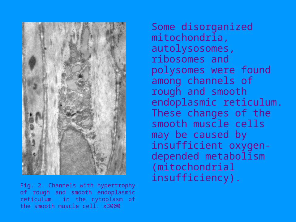
Some disorganized mitochondria, autolysosomes, ribosomes and polysomes were found among channels of rough and smooth endoplasmic reticulum. These changes of the smooth muscle cells may be caused by insufficient oxygen-depended metabolism (mitochondrial insufficiency).
Fig. 2. Channels with hypertrophy of rough and smooth endoplasmic reticulum in the cytoplasm of the smooth muscle cell. х3000

• A few disorganized mitochondria of smooth muscle cells are situated in the cortical layer adjacent to the cell membrane.
• In such cases the electronic density of cell membrane is increased.
Fig. 3. Disorganized mitochondria adjacent to cell membrane. х23000

There is the developed Golgi complex in the cytoplasm adjacent to the karyolemma containing a lot of lysosomes and microvesicles in the smooth muscle cell.
Fig. 4. The Golgi complex containing a lot of microvesicles and primary lysosomes in the cytoplasm of smooth muscle cell. х23000

Considerable changes of connective tissue, especially in parts adjacent to smooth muscle cells were observed in detrusor. Connective tissue is disorganized and contains fibroblasts with nuclei of irregular shape surrounded by collagen fibers.
Fig. 5. Ultrastructure of the disorganized fibroblast х4500

• There are fibroblasts with atrophia in the expanded intercellular space among the smooth muscle cells in the oedematous matrix of connective tissue .
• The nuclei of fibroblasts have irregular shape. Fragmented nucleus and hypetrophic nucleolus of fibroblasts and some smooth muscle cells is manifestation of preapoptosis.
Fig. 6. Expanded intercellular space with matrix, disorganized fibroblast and collagen fibers. х 1000

Smooth muscle cells situated in the disorganized connective tissue contain a lot of vacuoles. There are precipitates and coagulates in the matrix of this connective tissue
Fig. 7. Precipitates and coagulates in the matrix of the connective tissue arround of muscle cells х2000

Severe disturbances of microcirculation were observed in detrusor of patients with BPH. We noted disorganization of small and large capillaries. The wall of small capillary has partly swollen basal membrane and endothelial cells with oedema and low electronic density. Erythrocytes in such capillary were very close by situated to luminal surface of endothelium.
Fig 8. Ultrastructure of the disorganized connective tissue with small blood capillaries. х1000
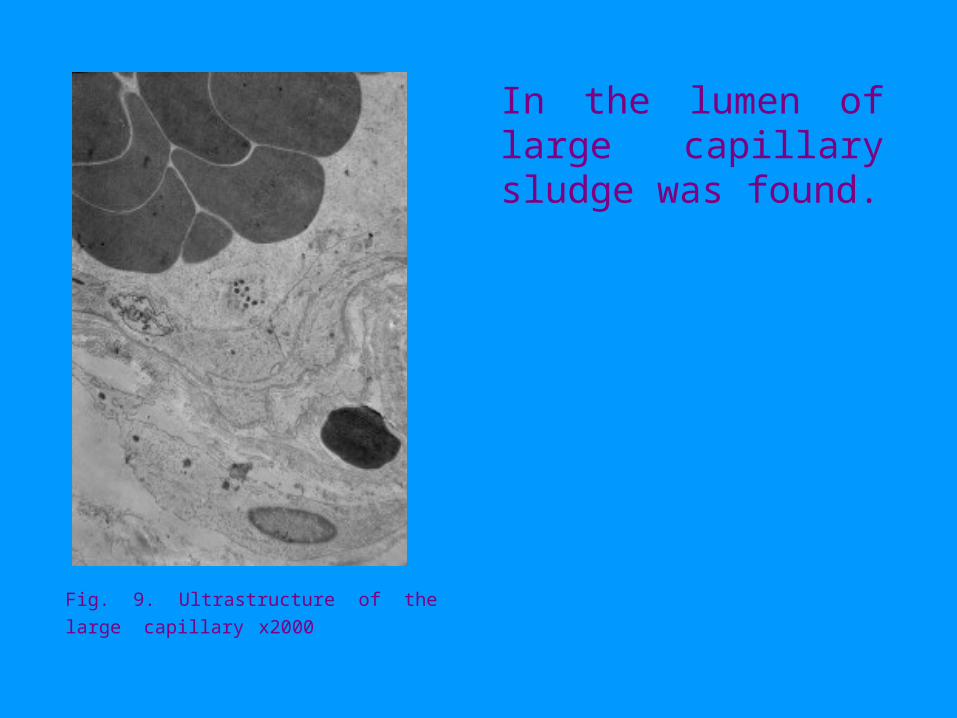
In the lumen of large capillary sludge was found.
Fig. 9. Ultrastructure of the large capillary х2000

The cluster of disorganized nonmielinated nerve fibers were found
Fig. 10. Bundle of the disorganized nerve fibers х2000

The ultrastructural investigation of detrusor testify that disturbance of microcirculation due to infravesical obstruction in patients with BPH cause preapoptosis of smooth muscle cells and fibroblasts.
Conclusion


