The vestibular system: multimodal integration and encoding of self-motion for motor control Kathleen E. Cullen Department of Physiology, McGill University, Montreal, Quebec H3G 1Y6, Canada Understanding how sensory pathways transmit infor- mation under natural conditions remains a major goal in neuroscience. The vestibular system plays a vital role in everyday life, contributing to a wide range of functions from reflexes to the highest levels of voluntary behavior. Recent experiments establishing that vestibular (self- motion) processing is inherently multimodal also pro- vide insight into a set of interrelated questions. What neural code is used to represent sensory information in vestibular pathways? How do the interactions between the organism and the environment shape encoding? How is self-motion information processing adjusted to meet the needs of specific tasks? This review highlights progress that has recently been made towards under- standing how the brain encodes and processes self- motion to ensure accurate motor control. Introduction The vestibular system encodes self-motion information by detecting the motion of the head in space. In turn, it provides us with our subjective sense of self-motion and orientation thereby playing a vital role in the stabilization of gaze, control of balance and posture. Neurophysiological and clinical studies have provided important insights into how, even at the earliest stages of processing, vestibular pathways integrate information from other modalities to generate appropriate and accurate behaviors (reviewed in [1]). The present review first considers the encoding of self- motion information at the earliest stages of vestibular processing, and next highlights the strategies of multimod- al integration that are used within vestibular pathways. It then considers the role of the vestibular system in ensuring the accuracy of three specific classes of behaviors: (i) the control of gaze to ensure clear vision during everyday activities, (ii) the production of the compensatory neck and limb movements required to ensure postural equilib- rium during both self generated and externally applied movements, and (iii) more complex voluntary motion tasks such as navigation and reaching. Taken together, the findings of recent behavioral, single-unit recording, and lesion studies emphasize the essential role of the multi- modal integration of vestibular with extra-vestibular sig- nals to ensure accurate motor control. Overview of the vestibular system The vestibular system is phylogenetically the oldest part of the inner ear, yet it was only recognized as an entity distinct from the cochlea in the middle of the 19th century. This is because, when the system is functioning normally, we are usually unaware of a distinct sensation arising from vestibular activity; it is integrated with visual, propriocep- tive and other extra-vestibular information such that com- bined experience leads to a sense of motion. For this reason, the significance of this sensory system is best appreciated by the study of patients for whom the daily activities that we take for granted become a significant challenge. For example, following complete vestibular loss, even the smallest head movements are accompanied by gaze instability and postural imbalance, which produce frequent and debilitating episodes of vertigo. Early vestibular processing and the sensory coding of self-motion: the sensory periphery To address the first major question – what neural code is used to represent vestibular sensory information? – recent studies have focused on the afferent fibers which innervate the vestibular sensory organs of the inner ear. The sensory organs comprise two types of sensors: the three semicircular canals, which sense angular acceleration in all three dimen- sions, and the two otolith organs (the saccule and utricle), which sense linear acceleration (i.e. gravity and translation- al movements) in all three dimensions. The afferent fibers of the vestibular component of the VIII nerve carry signals from the receptor cells of these sensory organs to the vestib- ular nuclei. In turn, the central neurons of the vestibular nuclei project to the neural structures that control eye move- ments, posture, and balance, as well as to upstream struc- tures involved in the computation of self-motion (Figure 1a). Individual afferent fibers innervating the sensory neu- roepithelium of either the canals or otoliths display diversity in discharge regularity in the absence of stimulation (Figure 1b). This regularity is typically quantified using a normalized coefficient of variation (CV*) and corresponds to distinct morphological as well as physiological properties [2]. The larger-diameter irregular afferent fibers can carry information from either the type I hair cells located at the center of neuroepithelium (C-irregulars) or both type I hair cells and type II hair cells (dimorphic or D-irregulars). By contrast, more regular afferent fibers preferentially carry information from type II hair cells in the peripheral Review Corresponding author: Cullen, K. ([email protected]) 0166-2236/$ – see front matter ß 2012 Elsevier Ltd. All rights reserved. doi:10.1016/j.tins.2011.12.001 Trends in Neurosciences, March 2012, Vol. 35, No. 3 185
Transcript
Review
The vestibular system: multimodalintegration and encoding ofself-motion for motor controlKathleen E. Cullen
Department of Physiology, McGill University, Montreal, Quebec H3G 1Y6, Canada
Understanding how sensory pathways transmit infor-mation under natural conditions remains a major goal inneuroscience. The vestibular system plays a vital role ineveryday life, contributing to a wide range of functionsfrom reflexes to the highest levels of voluntary behavior.Recent experiments establishing that vestibular (self-motion) processing is inherently multimodal also pro-vide insight into a set of interrelated questions. Whatneural code is used to represent sensory information investibular pathways? How do the interactions betweenthe organism and the environment shape encoding?How is self-motion information processing adjusted tomeet the needs of specific tasks? This review highlightsprogress that has recently been made towards under-standing how the brain encodes and processes self-motion to ensure accurate motor control.
IntroductionThe vestibular system encodes self-motion information bydetecting the motion of the head in space. In turn, itprovides us with our subjective sense of self-motion andorientation thereby playing a vital role in the stabilizationof gaze, control of balance and posture. Neurophysiologicaland clinical studies have provided important insights intohow, even at the earliest stages of processing, vestibularpathways integrate information from other modalities togenerate appropriate and accurate behaviors (reviewed in[1]). The present review first considers the encoding of self-motion information at the earliest stages of vestibularprocessing, and next highlights the strategies of multimod-al integration that are used within vestibular pathways. Itthen considers the role of the vestibular system in ensuringthe accuracy of three specific classes of behaviors: (i) thecontrol of gaze to ensure clear vision during everydayactivities, (ii) the production of the compensatory neckand limb movements required to ensure postural equilib-rium during both self generated and externally appliedmovements, and (iii) more complex voluntary motion taskssuch as navigation and reaching. Taken together, thefindings of recent behavioral, single-unit recording, andlesion studies emphasize the essential role of the multi-modal integration of vestibular with extra-vestibular sig-nals to ensure accurate motor control.
0166-2236/$ – see front matter � 2012 Elsevier Ltd. All rights reserved. doi:10.1016/j.tins.2011.
Overview of the vestibular systemThe vestibular system is phylogenetically the oldest part ofthe inner ear, yet it was only recognized as an entitydistinct from the cochlea in the middle of the 19th century.This is because, when the system is functioning normally,we are usually unaware of a distinct sensation arising fromvestibular activity; it is integrated with visual, propriocep-tive and other extra-vestibular information such that com-bined experience leads to a sense of motion. For thisreason, the significance of this sensory system is bestappreciated by the study of patients for whom the dailyactivities that we take for granted become a significantchallenge. For example, following complete vestibular loss,even the smallest head movements are accompanied bygaze instability and postural imbalance, which producefrequent and debilitating episodes of vertigo.
Early vestibular processing and the sensory coding of
self-motion: the sensory periphery
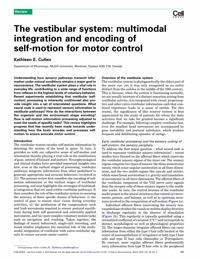
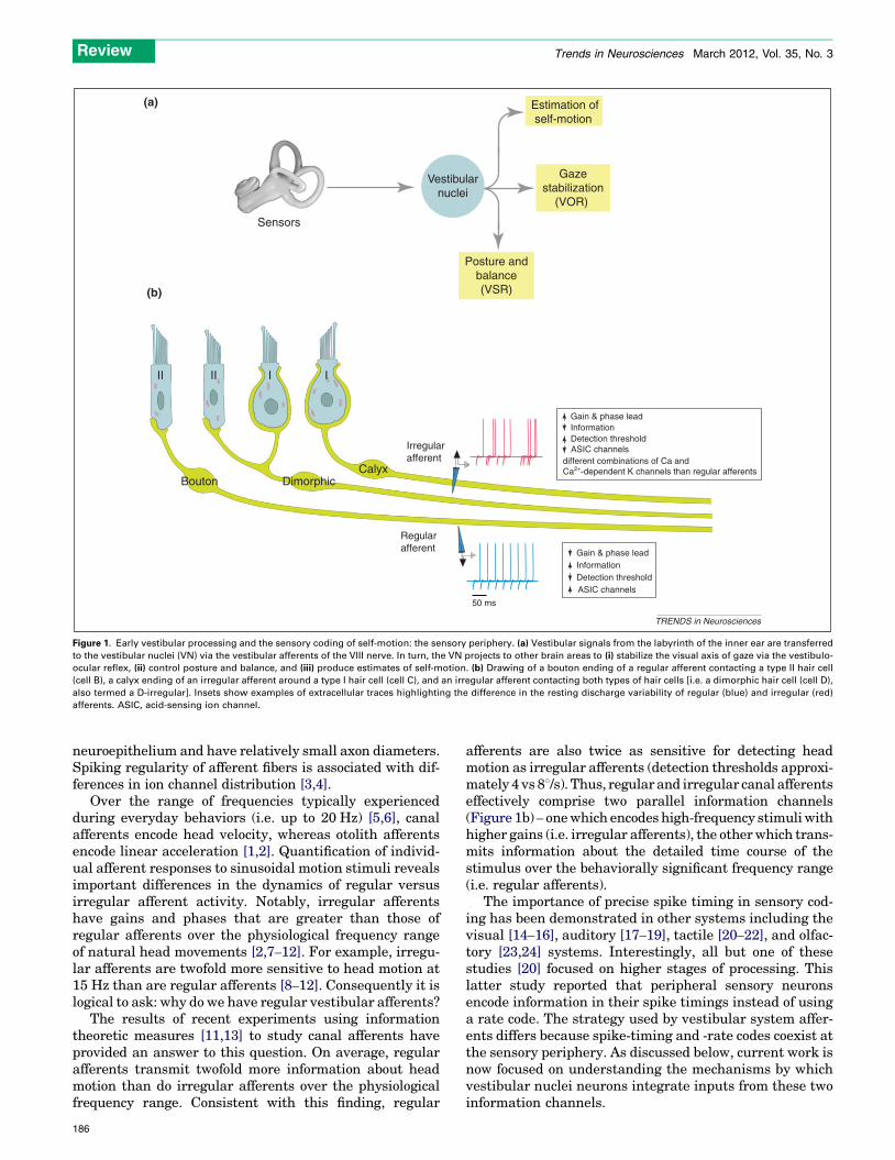
To address the first major question – what neural code isused to represent vestibular sensory information? – recentstudies have focused on the afferent fibers which innervatethe vestibular sensory organs of the inner ear. The sensoryorgans comprise two types of sensors: the three semicircularcanals, which sense angular acceleration in all three dimen-sions, and the two otolith organs (the saccule and utricle),which sense linear acceleration (i.e. gravity and translation-almovements) in all three dimensions. The afferent fibers ofthe vestibular component of the VIII nerve carry signalsfrom the receptor cells of these sensory organs to the vestib-ular nuclei. In turn, the central neurons of the vestibularnuclei project to the neural structures that control eyemove-ments, posture, and balance, as well as to upstream struc-tures involved in the computation of self-motion (Figure 1a).
Individual afferent fibers innervating the sensory neu-roepitheliumofeither the canals orotolithsdisplaydiversityin discharge regularity in the absence of stimulation(Figure 1b). This regularity is typically quantified using anormalized coefficient of variation (CV*) and corresponds todistinct morphological as well as physiological properties[2]. The larger-diameter irregular afferent fibers can carryinformation from either the type I hair cells located at thecenter of neuroepithelium (C-irregulars) or both type I haircells and type II hair cells (dimorphic or D-irregulars).By contrast, more regular afferent fibers preferentiallycarry information from type II hair cells in the peripheral
12.001 Trends in Neurosciences, March 2012, Vol. 35, No. 3 185
different combinations of Ca and Ca2+-dependent K channels than regular afferents
ASIC channels
ASIC channels
Sensors
Estimation ofself-motion
Posture andbalance(VSR)
Gazestabilization
(VOR)
Bouton DimorphicCalyx
Vestibularnuclei
TRENDS in Neurosciences
Figure 1. Early vestibular processing and the sensory coding of self-motion: the sensory periphery. (a) Vestibular signals from the labyrinth of the inner ear are transferred
to the vestibular nuclei (VN) via the vestibular afferents of the VIII nerve. In turn, the VN projects to other brain areas to (i) stabilize the visual axis of gaze via the vestibulo-
ocular reflex, (ii) control posture and balance, and (iii) produce estimates of self-motion. (b) Drawing of a bouton ending of a regular afferent contacting a type II hair cell
(cell B), a calyx ending of an irregular afferent around a type I hair cell (cell C), and an irregular afferent contacting both types of hair cells [i.e. a dimorphic hair cell (cell D),
also termed a D-irregular]. Insets show examples of extracellular traces highlighting the difference in the resting discharge variability of regular (blue) and irregular (red)
afferents. ASIC, acid-sensing ion channel.
Review Trends in Neurosciences March 2012, Vol. 35, No. 3
neuroepithelium and have relatively small axon diameters.Spiking regularity of afferent fibers is associated with dif-ferences in ion channel distribution [3,4].
Over the range of frequencies typically experiencedduring everyday behaviors (i.e. up to 20 Hz) [5,6], canalafferents encode head velocity, whereas otolith afferentsencode linear acceleration [1,2]. Quantification of individ-ual afferent responses to sinusoidal motion stimuli revealsimportant differences in the dynamics of regular versusirregular afferent activity. Notably, irregular afferentshave gains and phases that are greater than those ofregular afferents over the physiological frequency rangeof natural head movements [2,7–12]. For example, irregu-lar afferents are twofold more sensitive to head motion at15 Hz than are regular afferents [8–12]. Consequently it islogical to ask: why do we have regular vestibular afferents?
The results of recent experiments using informationtheoretic measures [11,13] to study canal afferents haveprovided an answer to this question. On average, regularafferents transmit twofold more information about headmotion than do irregular afferents over the physiologicalfrequency range. Consistent with this finding, regular
186
afferents are also twice as sensitive for detecting headmotion as irregular afferents (detection thresholds approxi-mately 4 vs 88/s). Thus, regular and irregular canal afferentseffectively comprise two parallel information channels(Figure 1b) – onewhich encodes high-frequency stimuliwithhigher gains (i.e. irregular afferents), the otherwhich trans-mits information about the detailed time course of thestimulus over the behaviorally significant frequency range(i.e. regular afferents).
The importance of precise spike timing in sensory cod-ing has been demonstrated in other systems including thevisual [14–16], auditory [17–19], tactile [20–22], and olfac-tory [23,24] systems. Interestingly, all but one of thesestudies [20] focused on higher stages of processing. Thislatter study reported that peripheral sensory neuronsencode information in their spike timings instead of usinga rate code. The strategy used by vestibular system affer-ents differs because spike-timing and -rate codes coexist atthe sensory periphery. As discussed below, current work isnow focused on understanding the mechanisms by whichvestibular nuclei neurons integrate inputs from these twoinformation channels.
Review Trends in Neurosciences March 2012, Vol. 35, No. 3
Early central vestibular processing and the sensory
coding of self-motion: central neurons
The responses of the vestibular nucleus neurons, to whichthe afferent fibers directly project, have been well charac-terized in head-restrained alert monkeys (reviewed in[25]). Traditionally, these neurons have been groupedaccording to differences in their sensitivity to eye motionand passive head motion, as well as to differences in theirconnectivity. Here, for the purpose of simplicity, two maincategories are considered: (i) vestibulo-ocular reflex (VOR)neurons, and (ii) posture/self-motion neurons (Figure 2a).
The most direct pathway that mediates the VOR com-prises a three neuron arc: vestibular nerve afferents proj-ect to central neurons in the vestibular nuclei (i.e. VOR
[(Figure_2)TD$FIG]
1
2
3
4
5
Gai
n (s
p/s)
/(°/
s)
0
40
80
120
Pha
se (
°)
Frequency (Hz)Frequency (Hz)
(b)
100 101 100 101
VO
PVP
FTN
0.5Hz 15Hz
50º/s
1s 100ms
50sp/s Firing rate
Head velocity
Estimate
(a)
VO
PVP
FTN
Ca
Vestibularnuclei
Vestibularsensors
Irregular
Regular PVP
FTN
VO20 ms0.
1mv
Figure 2. Early vestibular processing and the sensory coding of self-motion: central neur
afferents can be categorized into two main categories: (i) neurons that control and m
(i.e. PVPs and FTNs), and (ii) neurons that control posture and balance, and also pro
neurons). (b) Firing-rate response of an example VO neuron in the vestibular nucleus
below show response gains averaged for populations of VO, PVP, and FTN neurons rec
relatively higher gains which increase more dramatically at higher frequencies. Side ba
from [33] (c) Average detection threshold values for regular (blue) and irregular (red) affe
alert monkeys. Estimates of the information transmitted by a pooled population of 12 V
(green) for comparison. Side bands show +/– SEM. Data are replotted from [13].
neurons), which in turn project to extraocular motoneur-ons. Themajority of VORneurons are the so-called position-vestibular-pause (PVP) neurons; a distinct group of neuronswhich derive their name from the signals they carry duringpassive head rotations and eye movements. In addition, asecond class of neuron, termed floccular target neurons(FTN), also contribute to the direct VOR pathway. Notably,FTNs receive input from the flocculus of the cerebellumas well as from the vestibular nerve. The responses ofFTNs complement those of PVP neurons during our dailyactivities, and play a vital role in calibrating the VORto maintain excellent performance in response to theeffects of aging aswell as changes in environmental require-ments, such as those brought about by the corrective
Frequency (Hz)
Det
ectio
n th
resh
old
(deg
/s)
Regular Irregular
Human perception
Best regular
VO(single)
VO(population)
(c) Key:
1
12
libration/modulation
Direct
16
12
8
4
00 1 4 8 12 16
Gaze stabilization
(VOR)
Posture and balance(VSR)
Estimation of self-motion
TRENDS in Neurosciences
ons. (a) Neurons in the vestibular nuclei that receive direct input from the vestibular
odulate the vestibulo-ocular reflex to ensure gaze stability during everyday life
ject to higher-order structures involved in the estimation of self-motion (i.e. VO
recorded in alert monkeys during sinusoidal head rotation at 0.5 and 15 Hz. Plots
orded over a wide range of frequencies of head rotation. Note, PVP neurons have
nds show +/– SEM. VO data are replotted from [12]. FTN and VO data are replotted
rents, and VO neurons (grey) at different frequencies of sinusoidal head rotation in
O neurons (black) as well as human behavioral thresholds [37] are superimposed
187
Review Trends in Neurosciences March 2012, Vol. 35, No. 3
lens worn to correct myopia or during the motor learningrequired during prism adaptation ([26] for review).
The second category of vestibular nuclei neurons are thevestibular-only (VO, alternatively termed non-eye move-ment) neurons. VO neurons, as do VOR neurons, receivedirect inputs from the vestibular nerve. However, theseneurons do not project to oculomotor structures, and thusdo not contribute to the VOR. Instead, many of theseneurons project to the spinal cord and are thought tomediate, at least in part, the vestibular spinal reflexes([27] for review). In addition, VO neurons are reciprocallyinterconnected with the nodulus/uvula of the cerebellum[28] and appear to be the source of vestibular input tovestibular-sensitive neurons in thalamus and cortex[29,30]. Thus, whereas PVPs mediate the (VOR), stabilizegaze and ensure clear vision during daily activities, VOneurons are the substrate by which the vestibular systemplays a crucial role in ensuring postural equilibrium aswell as the higher-order vestibular processing required forstable spatial orientation.
Computational analyses of vestibular processing: linear
control system approach
The common view that early vestibular processing is fun-damentally linear has long made it an attractive model forthe study of sensorimotor integration. Over the past de-cade, investigations using a linear systems approach havebeen directed towards understanding early vestibular pro-cessing over the physiologically relevant frequency rangeof motion [13,31,32]. Interestingly, the response dynamicsof both VO neurons [13] and PVP neurons [33,34] arenearly comparable to those of irregular and regular affer-ents, respectively. By contrast, FTN neurons appear to be anotable exception; they show remarkably flat gain (andphase) tuning [33]. The functional implications of thesedifferences, summarized in Figure 2b for the gain responseof each of these three central neuron classes, are not yetfully understood.
Computational analyses of the vestibular system:
information transmission, detection thresholds, and
spike timing
As reviewed above, neural variability plays an importantrole determining the strategy used by vestibular afferentfibers to encodebehaviorally relevant stimuli (i.e.Figure1b).Although regular afferents transmit information aboutsensory input in a spike-timing code, irregular afferentsuse a rate code. However, there is no evidence that differentafferent classes preferentially contribute to different vestib-ular pathways (e.g. oculomotor versus vestibulo-spinal)[35,36].
How then is the information encoded by these twostreams of afferent input combined at the next stage ofprocessing? Recent experiments on VO neurons provideinsight into this question [13]. First, although VO neuronresponse gains are generally greater than those of individ-ual afferents, VO neurons transmit less information andhave significantly greater velocity detection thresholdsthan even the relatively ‘noisy’ irregular afferents(Figure 2c). Second, combining the responses of manyVO neurons (i.e. >20) lowers velocity detection thresholds,
188
but values remain higher than those measured duringbehavioral experiments (�2.5 vs 0.5–18/s) [37]. Thus, thereis an apparent discrepancy between the precision of codingat sequential stages of vestibular processing and the abilityof the brain to estimate self-motion.
The higher variability displayed by vestibular centralneurons could potentially serve to prevent phase-locking orentrainment [38,39]. For example, in the visual systemthalamic relay cells transmit detailed information in theirspike trains [14,15], whereas cortical neurons display largevariability in their responses [40]. However, in response tomore naturalistic stimuli, network interactions amongvisual cortical neurons can sharpen timing reliability[16]. A crucial assumption of prior analyses of vestibularprocessing (Figure 2c) is that the ability of a neuron toreconstruct the stimulus (i.e. coding fraction) can be mea-sured by computing the coding fraction [41,42]. On theother hand, given that (i) coding fractions compute thequality of the linear reconstruction of the stimulus, and (ii)coding fractions are low for central vestibular neurons (i.e.VO cells), it is important to consider whether this assump-tion is valid. Experiments directed towards understandingthe implication of non-linear behaviors such as phase-lock-ing are likely to provide new insights into how self-motioninformation is encoded for the subsequent computation ofself-motion as well as gaze and posture control.
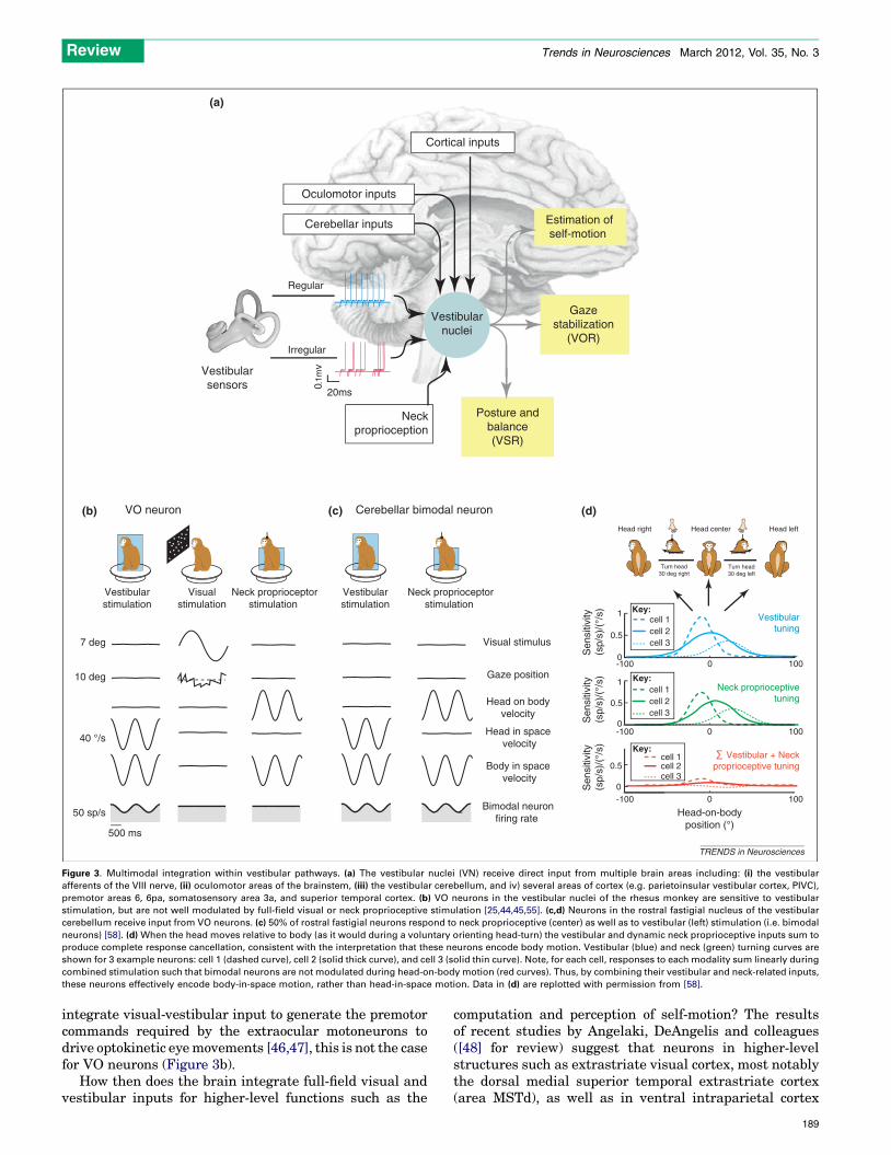
Multimodal integration within vestibular pathwaysVestibular inputs are not the only source of self-motioninformation. Somatosensory, proprioceptive, and visualinputs as well as motor-related signals also provide self-motion cues as an organism interacts with its environment.A distinguishing aspect of early vestibular processing isthat it combines multimodal sensory information at thefirst stage of central processing. Thus a second majorquestion is: how does the interaction of an organism withits environment shape and alter vestibular encoding?Figure 3a illustrates the sources of the extra-vestibularsensory inputs as well as premotor signals related to thegeneration of eye and head movements that are relayed tothe vestibular nuclei.
Integration of vestibular and visual inputs
Optic flow information provides an important sensory cuefor self-motion, capable of generating powerful sensationseven when a subject is stationary. Optic flow informationalso induces the generation of optokinetic eye movementsthat complement the VOR to ensure stable gaze duringself-motion at lower frequencies. It was initially thoughtthat all neurons in the vestibular nuclei are driven by large-field visual as well as vestibular stimulation [28,43]. Thisidea was theoretically very attractive because it provided aphysiological substrate by which the brain could combinevisual and vestibular signals to estimate self-motion. How-ever, visual-vestibular convergence is not as prevalent asinitially believed. Notably, although neurons sensitive toeye-movement show clear eye-movement related modula-tion during large-field visual (i.e. optokinetic) as well asduring vestibular stimulation, VO neurons do not showrobust modulation in response to optokinetic stimulation[44,45]. Thus, whereas VOR neurons (PVPs and FTNs)
[(Figure_3)TD$FIG]
(c)
(a)
Vestibularsensors
Estimation ofself-motion
Oculomotor inputs
Cerebellar inputs
Cortical inputs
20ms
Irregular
Posture andbalance(VSR)
Gazestabilization
(VOR)
Neckproprioception
Regular
0.1m
v
(b)
50 sp/s
40 °/s
7 deg
10 deg
Vestibularstimulation
Visualstimulation
500 ms
Neck proprioceptorstimulation
Head in space velocity
Body in space velocity
Bimodal neuronfiring rate
Gaze position
Head on bodyvelocity
Vestibularstimulation
Visual stimulus
VO neuron Cerebellar bimodal neuron (d)Head right Head center Head left
-100
Vestibulartuning
0
1
0.5
0
Neck proprioceptivetuning
100
-100 0 1000
Head-on-bodyposition (°)
Sen
sitiv
ity(s
p/s)
/(°/
s) 1
0.5
cell 1cell 2cell 3
cell 1Key:
cell 2cell 3
Vestibular + Neck proprioceptive tuning
Neck proprioceptorstimulation
-100 0 100
Sen
sitiv
ity(s
p/s)
/(°/
s)
0.5
0
cell 1cell 2cell 3
Sen
sitiv
ity(s
p/s)
/(°/
s)
Vestibularnuclei
Key:
Key:
Turn head30 deg left
Turn head30 deg right
TRENDS in Neurosciences
Figure 3. Multimodal integration within vestibular pathways. (a) The vestibular nuclei (VN) receive direct input from multiple brain areas including: (i) the vestibular
afferents of the VIII nerve, (ii) oculomotor areas of the brainstem, (iii) the vestibular cerebellum, and iv) several areas of cortex (e.g. parietoinsular vestibular cortex, PIVC),
premotor areas 6, 6pa, somatosensory area 3a, and superior temporal cortex. (b) VO neurons in the vestibular nuclei of the rhesus monkey are sensitive to vestibular
stimulation, but are not well modulated by full-field visual or neck proprioceptive stimulation [25,44,45,55]. (c,d) Neurons in the rostral fastigial nucleus of the vestibular
cerebellum receive input from VO neurons. (c) 50% of rostral fastigial neurons respond to neck proprioceptive (center) as well as to vestibular (left) stimulation (i.e. bimodal
neurons) [58]. (d) When the head moves relative to body (as it would during a voluntary orienting head-turn) the vestibular and dynamic neck proprioceptive inputs sum to
produce complete response cancellation, consistent with the interpretation that these neurons encode body motion. Vestibular (blue) and neck (green) turning curves are
shown for 3 example neurons: cell 1 (dashed curve), cell 2 (solid thick curve), and cell 3 (solid thin curve). Note, for each cell, responses to each modality sum linearly during
combined stimulation such that bimodal neurons are not modulated during head-on-body motion (red curves). Thus, by combining their vestibular and neck-related inputs,
these neurons effectively encode body-in-space motion, rather than head-in-space motion. Data in (d) are replotted with permission from [58].
Review Trends in Neurosciences March 2012, Vol. 35, No. 3
integrate visual-vestibular input to generate the premotorcommands required by the extraocular motoneurons todrive optokinetic eyemovements [46,47], this is not the casefor VO neurons (Figure 3b).
How then does the brain integrate full-field visual andvestibular inputs for higher-level functions such as the
computation and perception of self-motion? The resultsof recent studies by Angelaki, DeAngelis and colleagues([48] for review) suggest that neurons in higher-levelstructures such as extrastriate visual cortex, most notablythe dorsal medial superior temporal extrastriate cortex(area MSTd), as well as in ventral intraparietal cortex
189
Review Trends in Neurosciences March 2012, Vol. 35, No. 3
(area VIP), respond both tomotion in darkness as well as tooptic flow stimuli. Responses to motion in the dark areeliminated following bilateral labyrinthectomy [49,50],consistent with the proposal that neurons integrate ves-tibular and visual signals to compute self-motion.
Integration of vestibular and somatosensory/
proprioceptive inputs
Somatosensory/proprioceptive inputs reach the vestibularnuclei by means of dorsal-root axons as well as second-order neurons (reviewed in [25]). In addition, cerebellarand cortical areas sensitive to such inputs send directprojections to the vestibular nuclei (reviewed in [51–53])making this area a likely candidate for encoding bodymotion. In decerebrate or anesthetized preparations, pas-sive neck proprioceptive stimulation influences the activityof vestibular nuclei neurons ([27,54] for review). However,passive activation of proprioceptors does not directly affectneuronal responses in alert rhesus monkey (Figure 3c)[55]. By contrast, the same stimulation can affect theresponses of both VO neurons and VOR neurons in otherspecies of primate (i.e. squirrel monkey [56] and cynomol-gus monkey [57]). One possible explanation for this speciesdifference is that neck-related inputs to vestibular path-ways are particularly crucial for postural stabilization inthose primates that make their home in a challenging 3Darboreal environment.
Proprioceptive–vestibular integration is typically an-tagonistic in species where both inputs drive vestibularnuclei neurons. As a result, when the head moves relativeto body (for example, during a voluntary orienting head-turn), neurons fire less robustly than for comparable headmotion produced by whole-body motion (i.e. a condition inwhich only the vestibular system is stimulated). Striking-ly, recent studies have reported complete cancellation ofvestibular modulation by proprioceptive inputs within therostral fastigial nucleus of the cerebellum (Figure 3c,d)[58], a nucleus which is reciprocally connected to thevestibular nucleus [59,60]). Approximately half of the neu-rons in this region are sensitive to proprioceptive as well asvestibular inputs (Figure 3c; bimodal neurons), whereasthe other half are only sensitive to vestibular input (unim-odal neurons). When delivered in isolation, the vestibular-and proprioceptive-related responses of bimodal neuronshave comparable tuning (e.g. strength and location ofmaximal response) that varies as a function of head-on-body position (Figure 3d). Accordingly, although theirprocessing of each sensory modality is intrinsically nonlin-ear, responses sum linearly during combined stimulationsuch that bimodal neurons robustly encode body-in-spacemotion. Unimodal neurons, by contrast, encode head-in-space motion much in the same way as the VO neurons ofthe vestibular nuclei.
The integration of vestibular and proprioceptive infor-mation in the rostral fastigial nucleus of the vestibularcerebellum is vital for the accurate control of posture andbalance as well as higher-order functions such as self-motion perception. For example, the corrective movementsproduced by vestibulospinal reflexes must account forchanges in the position of the head relative to the body[61–63]. However, patients with lesions to this cerebellar
190
region do not exhibit the required changes in body swaythat normally occur when head-on-body position is alteredduring galvanic stimulation [64]. In addition, the conver-gence of vestibular and neck proprioceptive inputs is re-quired to perceive body motion independently of headmotion [65]. A prediction would be that body motion per-ception is also impaired in these patients.
Vestibular pathways and the control of motor behaviorThe final question to be addressed in this review is: how isthe processing of self-motion information adjusted to meetthe needs of specific tasks? Below I consider how, bycombining vestibular with extra-vestibular signals, thebrain effectively shapes behaviors. I first consider thevestibulo-ocular reflex (VOR), whose relative simplicityhas made it an excellent model system for bridging thegap between neuronal circuits and behavior. I then consid-er more complex voluntary behaviors including voluntaryorienting movements, reaching, and navigation.
The VOR: complementary response dynamics ensure
stable gaze
In our daily lives we move through the world and, at thesame time, maintain stable gaze. This is because the VORproduces compensatory eye movements of equal and oppo-site magnitude to head rotations to stabilize the visual axis(i.e. gaze) relative to space. TheVOR is arguably our fastestbehavior; in response to head movement, eye movementsare generated with a latency of only 5–6 ms [6]. This shortlatency is consistent with the minimal synaptic and axonaldelays of the three-neuron pathway (e.g. Figure 2a). Thus,the VOR reflex stabilizes gaze considerably faster (by anorder of magnitude) than would be possible via the mostrapid visually evoked eye movements (reviewed in [66]).
During natural behaviors such as active head-turns,walking, and running, the motion of the head in spacecan have frequency content approaching 20 Hz [6,67]. TheVOR shows remarkably compensatory gain (i.e. eye veloci-ty/head velocity = 1) as well as minimal phase lag overthe physiological relevant range of head movements(Figure 4a) [5,6]. The latter observation is particularlyimpressive considering that the VOR has a 5–6 ms latency,and thus the evoked eye movements would lag head move-ments by >308 at 15 Hz if not appropriately compensated.However, as reviewed above, linear-control system analy-sis has shown that both vestibular afferents and the PVPneurons (Figure 2b) to which they project are characterizedby the requisite phase leads.
The results of single-unit recordings have provided in-sight into how the VOR effectively stabilizes gaze across awide range of head velocities. The VOR is compensatory forhead velocities as large as 300–5008/s [6,10]. Even so, theresponses of a typical PVP neuron or FTNs (i) are silencedfor off-direction rotations at velocities of 100–2008/s and also(ii) demonstrate substantial non-linearities (i.e. firing-ratesaturation) for on-direction rotations at velocities >2008/s[12]. The apparent discrepancy between neuronal and be-havioral VOR responses can be reconciled by consideringthe next stage of neural processing. Specifically, recordingsfrom extraocular motoneurons have shown that the oculo-motor plant itself has complementary dynamics [68].
[(Figure_4)TD$FIG]
(a) (b)Gaze stabilization
15Hz40
º/s
1s 100ms
Inverted head velocityEye velocity
Key:
Vestibularafferent
PVPneuron
200°
/s Head velocity
Gaze velocity
VOR attenuation
Firing rate
Maximumattenuation
100ms
PVP MN
Gate
premotorsaccadic
drive
(c)
0.5Hz
Gaze redirection
TRENDS in Neurosciences
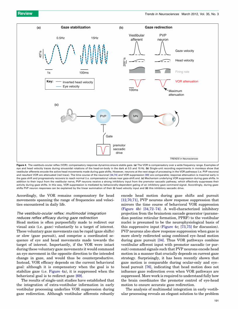
Figure 4. The vestibulo-ocular reflex (VOR): compensatory response dynamics ensure stable gaze. (a) The VOR is compensatory over a wide frequency range. Examples of
eye and head velocity traces during sinusoidal rotations of the head-on-body in the dark at 0.5 and 15 Hz. (b) Single-unit recording experiments in monkeys show that
vestibular afferents encode the active head movements made during gaze shifts. However, neurons at the next stage of processing in the VOR pathways (i.e. PVP neurons)
and resultant VOR are attenuated (red trace). The time course of the neuronal [34,74] and VOR suppression [69] are comparable; response attenuation is maximal early in
the gaze shift and progressively recovers to reach normal (i.e. compensatory) values near gaze-shift end. (c) Mechanism underlying VOR suppression during gaze shifts. In
addition to their input from the vestibular nerve, PVP neurons receive a strong inhibitory input from the premotor saccadic pathway, which effectively suppresses their
activity during gaze shifts. In this way, VOR suppression is mediated by behaviorally-dependent gating of an inhibitory gaze command signal. Accordingly, during gaze-
shifts PVP neuron responses can be explained by the linear summation of their (i) head velocity input and (ii) this inhibitory saccadic drive.
Review Trends in Neurosciences March 2012, Vol. 35, No. 3
Accordingly, the VOR remains compensatory for headmovements spanning the range of frequencies and veloci-ties encountered in daily life.
The vestibulo-ocular reflex: multimodal integration
reduces reflex efficacy during gaze redirection
Head motion is often purposefully made to redirect ourvisual axis (i.e. gaze) voluntarily to a target of interest.These voluntary gaze movements can be rapid (gaze shifts)or slow (gaze pursuit), and comprise a coordinated se-quence of eye and head movements made towards thetarget of interest. Importantly, if the VOR were intactduring these voluntary gazemovements it would commandan eye movement in the opposite direction to the intendedchange in gaze, and would thus be counterproductive.Instead, VOR efficacy depends on the current behavioralgoal: although it is compensatory when the goal is tostabilize gaze (i.e. Figure 4a), it is suppressed when thebehavioral goal is to redirect gaze [69].
The results of single-unit studies have established thatthe integration of extra-vestibular information in earlyvestibular processing underlies VOR suppression duringgaze redirection. Although vestibular afferents robustly
encode head motion during gaze shifts and pursuit[12,70,71], PVP neurons show response suppression thatmirrors the time course of behavioral VOR suppression(Figure 4b) [34,72–74]. A well-characterized inhibitoryprojection from the brainstem saccade generator (parame-dian pontine reticular formation, PPRF) to the vestibularnuclei is presumed to be the neurophysiological basis ofthis suppressive input (Figure 4c; [73,75] for discussion).PVP neurons also show response suppression when gaze ismore slowly redirected using combined eye–head motionduring gaze pursuit [34]. Thus VOR pathways combinevestibular afferent input with premotor saccadic (or pur-suit) command signals such that PVP neurons encode headmotion in a manner that crucially depends on current gazestrategy. Surprisingly, it has been recently shown thatgaze motion is comparable during ocular-only and eye–
head pursuit [76], indicating that head motion does notinfluence gaze redirection even when VOR pathways aresuppressed. More work is required to understand fully howthe brain coordinates the premotor control of eye-headmotion to ensure accurate gaze redirection.
The analysis of multimodal integration in early vestib-ular processing reveals an elegant solution to the problem
191
Review Trends in Neurosciences March 2012, Vol. 35, No. 3
of adjusting VOR reflex efficacy as a function of behavioralgoals. The VOR is robust and compensatory over a widerange of velocities and frequencies when the goal is tostabilize gaze during head motion. However, when thegoal is to redirect gaze, reflex efficacy is suppressed byan efferent copy of the command to redirect gaze voluntar-ily. Similarly, other features of gaze strategy (e.g. fixationdistance and gaze eccentricity) have been shown to modu-late VOR pathway responses [77–79] and in turn modulateVOR gain [80]. Understanding the distributed nature ofthe premotor circuitry responsible for these computationsremains a challenge for ongoing and future investigations.
Balance and the computation of self-motion:
mechanisms for the differential processing of actively
generated versus passive head movement
The vestibular system is often described as the balancesystem because it plays a vital role in ensuring stable body
[(Figure_5)TD$FIG]
Mossy fibers
Pc
Electroreceptorafferents
Output
Parallelfibers
P
-Proprioception,-Electric organ motor efference copy
-Sensory in(vestibular,propriocep-Movemenefference c
M
Vestibularafferent
Reafference
TotalExafference
Proprioception
World
(a)Muscle
(b) (c)Mormyrid electrosensorylateral line lobe
Headvelocity
Headvelocity
Firingrate
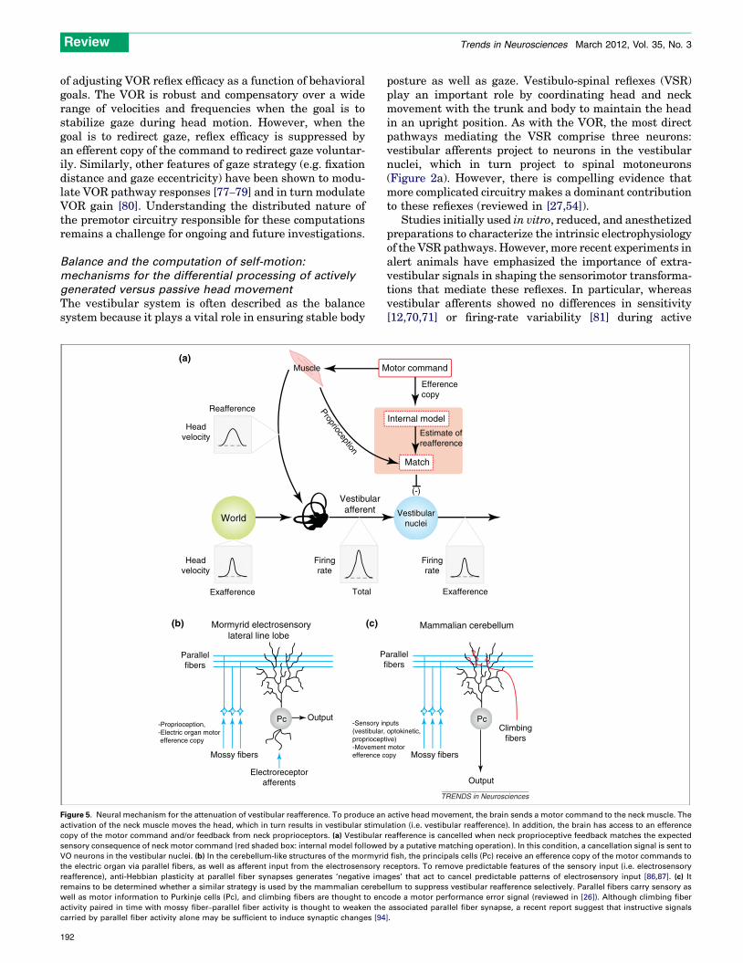
Figure 5. Neural mechanism for the attenuation of vestibular reafference. To produce an
activation of the neck muscle moves the head, which in turn results in vestibular stimu
copy of the motor command and/or feedback from neck proprioceptors. (a) Vestibular
sensory consequence of neck motor command (red shaded box: internal model followed
VO neurons in the vestibular nuclei. (b) In the cerebellum-like structures of the mormyri
the electric organ via parallel fibers, as well as afferent input from the electrosensory r
reafference), anti-Hebbian plasticity at parallel fiber synapses generates ‘negative ima
remains to be determined whether a similar strategy is used by the mammalian cerebe
well as motor information to Purkinje cells (Pc), and climbing fibers are thought to enc
activity paired in time with mossy fiber–parallel fiber activity is thought to weaken the
carried by parallel fiber activity alone may be sufficient to induce synaptic changes [94
192
posture as well as gaze. Vestibulo-spinal reflexes (VSR)play an important role by coordinating head and neckmovement with the trunk and body to maintain the headin an upright position. As with the VOR, the most directpathways mediating the VSR comprise three neurons:vestibular afferents project to neurons in the vestibularnuclei, which in turn project to spinal motoneurons(Figure 2a). However, there is compelling evidence thatmore complicated circuitry makes a dominant contributionto these reflexes (reviewed in [27,54]).
Studies initially used in vitro, reduced, and anesthetizedpreparations to characterize the intrinsic electrophysiologyof the VSR pathways. However, more recent experiments inalert animals have emphasized the importance of extra-vestibular signals in shaping the sensorimotor transforma-tions that mediate these reflexes. In particular, whereasvestibular afferents showed no differences in sensitivity[12,70,71] or firing-rate variability [81] during active
Mossy fibers
Climbingfibers
Output
arallelfibers
puts optokinetic,tive)t motoropy
otor command
Efferencecopy
Vestibularnuclei
Estimate ofreafference
Internal model
Match
Exafference
Mammalian cerebellum
Pc
(-)
Firingrate
TRENDS in Neurosciences
active head movement, the brain sends a motor command to the neck muscle. The
lation (i.e. vestibular reafference). In addition, the brain has access to an efference
reafference is cancelled when neck proprioceptive feedback matches the expected
by a putative matching operation). In this condition, a cancellation signal is sent to
d fish, the principals cells (Pc) receive an efference copy of the motor commands to
eceptors. To remove predictable features of the sensory input (i.e. electrosensory
ges’ that act to cancel predictable patterns of electrosensory input [86,87]. (c) It
llum to suppress vestibular reafference selectively. Parallel fibers carry sensory as
ode a motor performance error signal (reviewed in [26]). Although climbing fiber
associated parallel fiber synapse, a recent report suggest that instructive signals
].
Review Trends in Neurosciences March 2012, Vol. 35, No. 3
movements, vestibular-only (VO) neurons show strikingdifferences in the two conditions [55,82,83]. Specifically,VO neurons robustly respond to passive head movementsbut during active head movements their responses aremarkedly (70%) attenuated. Because these neurons projectinto VSR pathways (Figure 2a), this finding has led to theproposal that the VCR is turned off during voluntary headmovements [25,83].
Progress has been made towards understanding themechanism responsible for the selective cancellation ofneuronal responses to active head motion. Notably, byexperimentally controlling the correspondence betweenintended and actual head movement [83,84], it has beenshown that a cancellation signal is exclusively generated inconditions where the activation of neck proprioceptorsmatches themotor-generated expectation (Figure 5a). Thisresult provides support for the idea that an internal modelof the sensory consequences of active head motion is usedto suppress reafference (i.e. the vestibular stimulation thatresults from our own actions) selectively at the level of thevestibular nuclei. This general mechanism has notablesimilarity to that used by mormyrid fish to cancel electro-sensory reafference. The cerebellum-like structures ofthese fish act as adaptive filters, removing predictablefeatures of the sensory input ([85] for review). Recently,a combination of in vitro, in vivo, and computational stud-ies have provided direct insight into how anti-Hebbiansynaptic plasticity underlies cancellation of the electrosen-sory consequences of behavior of the fish [86,87]. It remainsto be determined whether a similar strategy is used in themammalian cerebellum to suppress vestibular reafferenceselectively (Figure 5b vs c).
The differential processing of active versus passive headmovements has important implications for voluntarymotorcontrol versus balance. Although it is helpful to stabilize thehead/body to compensate for unexpected movements (suchas those experienced while riding on the subway), thestabilizing commands produced by an intact VSR wouldbe counterproductive during active movements. According-ly, turning off vestibulospinal reflexes is functionally advan-tageous. Moreover, because VO neurons can continue toencode information reliably about passive self-motion dur-ing the execution of voluntary head-turns [55,82], vestibulo-spinal pathways continue to selectively adjust postural tonein response to head movement that the brain does notexpect. Such selectivity is fundamental to ensuring accuratemotor control. For example, the ability to recover fromtripping over an obstacle while walking or running requiresa selective but robust postural response to the unexpectedcomponent of vestibular stimulation. Finally, the differen-tial processing of active versus passive head movements isalso likely to have important implications for the computa-tion of self-motion because VO neurons have reciprocalinterconnections with regions of the vestibular cerebellum(see above) and vestibular thalamus [88].
Voluntary behavior: steering, reaching and navigation
As reviewed above, the processing of self-motion informa-tion is inherently multimodal; the integration of vestibularand extra-vestibular inputs has important implications forthe control of the vestibulo-ocular and vestibulospinal
reflexes which function to ensure stable gaze and posture,as well as for the processing of self-motion information forhigher-order functions. Recent studies of more complexbehaviors including voluntary orientingmovements, steer-ing, navigation, and even reaching have furthered ourunderstanding of the pervasive role of the vestibular sys-tem in voluntary motor control.
During self-motion, the ability to distinguish betweenactively-generated and passively-applied head movementsis not only important for shaping motor commands, but isalso crucial for ensuring perceptual stability ([89] for re-view). The active movements produced by orienting headand body movements are differentially encoded at the firstcentral stage of vestibular processing. How is self-motionencodedwhen it is voluntarily controlled in less direct ways– for example by driving a car? Single-unit experiments inmonkeys reveal that all vestibular nuclei neurons respondto vestibular input during ‘self-generated’ driving as if ithad been externally applied [34,55]. However, corticalneurons in MSTd show enhanced responses to virtual(i.e. visual) self-movement when monkeys steer astraight-ahead course, using optic flow cues [90]. Thus,at this higher level stage of processing, the brain appears tocombine steering-related (i.e. motor/motor preparation)signals with self-motion (i.e. vestibular, proprioceptiveand visual) information. It remains to be determinedwhether further training in a task such as steering wouldlead to the construction of an accurate internalmodel of thevehicle being driven (in this case themotion platform of themonkey) and, in turn, suppression of sensory responsesearlier in vestibular processing.
Finally, a current emerging area of interest is the role ofself-motion (i.e. vestibular) information in ensuring behav-ioral accuracy during complex voluntary behaviors, suchas, navigation and reaching. The discovery that vestibularreafference is suppressed early in processing has impor-tant implications for understanding how self-motion infor-mation is encoded during these everyday activities. Forexample, head-direction cells in the hippocampal forma-tion combine extra-vestibular information with vestibularinput to compute distinct estimates of heading directionduring active versus passive navigation [91,92]. In addi-tion, there is accumulating evidence that the brain usesvestibular signals to generate the appropriate reachingmotor command required to maintain accuracy duringself-motion ([93] for review). More work is needed to un-derstand how the brain integrates vestibular versus extra-vestibular cues during the voluntary self-motion producedduring these everyday activities.
Concluding remarksDuring everyday life, the brain combines vestibular andextra-vestibular cues – for example visual and/or proprio-ceptive information– to constructanestimateof self-motion.Significant progress has recently been made towardsanswering three interrelated questions: what neural codeis used to represent vestibular sensory information? Howdoes the interaction of the organism with the environmentshape and alter encoding? How is the processing of self-motion information adjusted to meet the needs of specifictasks?
193
Box 1. Outstanding questions
� What neural code is used to represent vestibular sensory
information?
One assumption of prior analyses is that neurons encode
information in a linear manner. However, recent analyses reveal
that irregular afferents and PVP neurons are characterized by
marked phase-locking in response to motion �20 Hz [9,33],
suggesting a role for non-linear coding in the sensorimotor
transformations that mediate the VOR at higher frequencies.
Similarly, a preliminary report suggests that phase-locking in VO
neurons is regulated by variability (e.g. synaptic noise) [39]. More
work is required to understand the strategy used to encode
behaviorally relevant vestibular stimuli.
� What is the functional role of the information encoded by
vestibular cerebellum during self-motion?
The head and body motion signals encoded by vestibular
cerebellum are known to play an important role in the production
of accurate postural control. However, the vestibular cerebellum
also sends ascending projections to the posterolateral ventral
nucleus of the thalamus. Patients with midline cerebellar lesions
exhibit reduced vestibular perception [95], and a prediction would
be that body motion perception would be also impaired in these
patients.
� What information is encoded by cortical areas that contribute to
the perception of self-motion? Do these areas distinguish actively
generated from passive self-motion?
Neurons at the first central stage of vestibular processing (VN) can
distinguish between self-generated and passive movements. Further
studies of the cellular mechanisms which underlie this computation,
as well as the functional significance of the information that is
ultimately sent upstream for subsequent computation, will be key to
understanding how the brain perceives self-motion.
� How is self-motion information encoded by the hippocampal
formation during navigation?
Vestibular input is required for the generation of the directional
signal encoded by head direction cells during navigation [91,92],
and directional tuning is thought to be created by means of online
integration of the angular head velocity of the animal (reviewed in
[96]). To date, however, most studies report a relative response
increase during active motion ([97–99]; [100] for an exception),
which is unexpected given that passive, not active, motion is more
robustly encoded in early vestibular processing (Figure 5a). Inter-
estingly, hippocampal place cells are characterized by the similar
discrepancy (i.e. a relative response increase during active motion;
compare [101,102]). Thus, how the hippocampal formation com-
bines extra-vestibular information with vestibular input to encode
self-motion during navigation remains an open question.
� How is vestibular information processed to predict the conse-
quence of the rotation dynamics during reaching?
Changes in vestibular input can affect ongoing reaching move-
ments [103–105]. In addition, vestibular signals that could poten-
tially influence reach planning and execution have been described
in somatosensory cortex, as well as in parietal cortex [106–108].
Although the relative influences of vestibular versus extra-vestib-
ular (i.e. motor efference copy and proprioceptive information)
remain to be determined precisely, current evidence suggest that
arm movements during reaching are altered in a manner consistent
with the hypothesis that vestibular signals are used to predict
Coriolis forces [109,110].
Review Trends in Neurosciences March 2012, Vol. 35, No. 3
First, the central vestibular system receives inputfrom two parallel information channels: regular afferentstransmit detailed information about head rotationsthrough precise spike-timing, whereas irregular afferentsrespond to high-frequency features exclusively throughchanges in firing rate. Second, the brain combines infor-mation from the vestibular sensors with extra-vestibularcues, such as proprioception andmotor efference signals, atthe earliest stages of central vestibular processing to com-pute estimates of self-motion. As an organism interactswith its environment, the resulting multimodal inflow isused to provide (i) robust estimates of self-motion (forexample, when visual as well as vestibular cues are avail-able), and (ii) estimates of the motion of neighboring partsof the body (e.g. body versus head motion) to ensure stableposture and perception. Third and finally, vestibular pro-cessing is shaped as a function of context during reflexbehavior, as well as during more complex voluntary beha-viors such as orienting, steering, navigation and reaching.Taken together, recent results provide new evidence thataction alters the sensory encoding of self-motion by thebrain at the earliest stages to ensure the accurate control ofbehavior in everyday life. Future studies need to considernot only how the neural code is used to represent self-motion by central pathways when multiple inputs arecombined, but also how differences in the behavioral con-text govern the nature of what defines the optimal compu-tation (Box 1). A better understanding of how the brainencodes and processes self-motion will provide vital insightinto the fundamental question of how we anticipate theconsequences of current or potential actions, and in turnstimulate a re-evaluation of the traditional separationbetween action and perception.
194
AcknowledgmentsThe author acknowledges support from the Canadian Institutes of HealthResearch (CIHR), the National Institutes of Health (DC002390), and theFonds Quebecois de la Recherche sur la Nature et les Technologies(FQNRT), and thanks Jerome Carriot, Diana Mitchell, Jessica Brooks,Mohsen Jamali, Alexis Dale, Carla Kalkhoven, Adam Schneider for helpwith figures and comments on the manuscript.
References1 Angelaki, D.E. and Cullen, K.E. (2008) Vestibular system: the many
facets of a multimodal sense. Ann. Rev. Neurosci. 31, 125–1502 Goldberg, J.M. (2000) Afferent diversity and the organization of
central vestibular pathways. Exp. Brain Res. 130, 277–2973 Eatock, R.A. andSonger, J.E. (2011) Vestibular hair cells and afferents:
two channels for head motion signals. Ann. Rev. Neurosci. 34, 501–5344 Kalluri, R. et al. (2010) Ion channels set spike timing regularity
of mammalian vestibular afferent neurons. J. Neurophysiol. 104,2034–2051
5 Armand, M. and Minor, L.B. (2001) Relationship between time- andfrequency-domain analyses of angular head movements in thesquirrel monkey. J. Comput. Neurosci. 11, 217–239
6 Huterer, M. and Cullen, K.E. (2002) Vestibuloocular reflex dynamicsduring high-frequency and high-acceleration rotations of the head onbody in rhesus monkey. J. Neurophysiol. 88, 13–28
7 Haque, A. et al. (2004) Spatial tuning and dynamics of vestibularsemicircular canal afferents in rhesus monkeys. Exp. Brain Res. 155,81–90
8 Hullar, T.E. et al. (2005) Responses of irregularly dischargingchinchilla semicircular canal vestibular-nerve afferents duringhigh-frequency head rotations. J. Neurophysiol. 93, 2777–2786
9 Ramachandran, R. and Lisberger, S.G. (2006) Transformation ofvestibular signals into motor commands in the vestibuloocularreflex pathways of monkeys. J. Neurophysiol. 96, 1061–1074
10 Sadeghi, S.G. et al. (2006) Dynamics of the horizontal vestibuloocularreflex after unilateral labyrinthectomy: response to high frequency,high acceleration, and high velocity rotations. Exp. Brain Res. 175,471–484
11 Sadeghi, S.G. et al. (2007) Neural variability, detection thresholds,and information transmission in the vestibular system. J. Neurosci.27, 771–781
Review Trends in Neurosciences March 2012, Vol. 35, No. 3
12 Sadeghi, S.G. et al. (2007) Response of vestibular-nerve afferents toactive and passive rotations under normal conditions and afterunilateral labyrinthectomy. J. Neurophysiol. 97, 1503–1514
13 Massot, C. et al. (2011) Information transmission and detectionthresholds in the vestibular nuclei: single neurons vs populationencoding. J. Neurophysiol. 105, 1798–1814
14 Reinagel, P. and Reid, R.C. (2000) Temporal coding of visualinformation in the thalamus. J. Neurosci. 20, 5392–5400
15 Desbordes, G. et al. (2008) Timing precision in population coding ofnatural scenes in the early visual system. PLoS Biol. 6, e324
16 Haider, B. et al. (2010) Synaptic and network mechanisms of sparseand reliable visual cortical activity during nonclassical receptive fieldstimulation. Neuron 65, 107–121
17 Kayser, C. et al. (2010) Millisecond encoding precision of auditorycortex neurons. Proc. Natl. Acad. Sci. U.S.A. 107, 16976–16981
18 Bizley, J.K. et al. (2010) Neural ensemble codes for stimulusperiodicity in auditory cortex. J. Neurosci. 30, 5078–5091
19 Sharpee, T.O. et al. (2011) Hierarchical representations in theauditory cortex. Curr. Opin. Neurobiol. 21, 761–767
20 Johansson, R.S. and Birznieks, I. (2004) First spikes in ensembles ofhuman tactile afferents code complex spatial fingertip events. Nat.Neurosci. 7, 170–177
21 Jones, L.M. et al. (2004) Robust temporal coding in the trigeminalsystem. Science 304, 1986–1989
22 Scaglione, A. et al. (2011) Trial-to-trial variability in the responses ofneurons carries information about stimulus location in the ratwhisker thalamus. Proc. Natl. Acad. Sci. U.S.A. 108, 14956–14961
23 Ito, I. et al. (2008) Sparse odor representation and olfactory learning.Nat. Neurosci. 11, 1177–1184
24 Giridhar, S. et al. (2011) Timescale-dependent shaping of correlationby olfactory bulb lateral inhibition. Proc. Natl. Acad. Sci. U.S.A. 108,5843–5848
25 Cullen, K.E. and Roy, J.E. (2004) Signal processing in the vestibularsystem during active versus passive headmovements. J. Neurophysiol.91, 1919–1933
26 Cullen, K.E. (2008) Procedural learning: VOR. In Learning andMemory: A Comprehensive Reference (Byrne, J.H., ed.), pp. 383–
402, Academic Press/Elsevier27 Goldberg, J.M. and Cullen, K.E. (2011) Vestibular control of the head:
possible functions of the vestibulocollic reflex. Exp. Brain Res. 210,331–345
28 Reisine, H. and Raphan, T. (1992) Unit activity in the vestibularnuclei of monkeys during off-vertical axis rotation. Ann. N. Y. Acad.Sci. 656, 954–956
29 Grusser, O.J. et al. (1990) Vestibular neurones in the parieto-insularcortex of monkeys (Macaca fascicularis): visual and neck receptorresponses. J. Physiol. 430, 559–583
30 Lang, W. et al. (1979) Vestibular projections to the monkey thalamus:an autoradiographic study. Brain Res. 177, 3–17
31 Dickman, J.D. and Angelaki, D.E. (2004) Dynamics of vestibularneurons during rotational motion in alert rhesus monkeys. Exp.Brain Res. 155, 91–101
32 Sadeghi, S.G. et al. (2010) Neural correlates of motor learning in thevestibulo-ocular reflex: dynamic regulation of multimodal integrationin the macaque vestibular system. J. Neurosci. 30, 10158–10168
33 Ramachandran, R. and Lisberger, S.G. (2008) Neural substrate ofmodified and unmodified pathways for learning in monkeyvestibuloocular reflex. J. Neurophysiol. 100, 1868–1878
34 Roy, J.E. and Cullen, K.E. (2002) Vestibuloocular reflex signalmodulation during voluntary and passive head movements.J. Neurophysiol. 87, 2337–2357
35 Boyle, R. et al. (1992) Inputs from regularly and irregularly dischargingvestibular nerve afferents to secondary neurons in squirrel monkeyvestibular nuclei. III. Correlation with vestibulospinal andvestibuloocular output pathways. J. Neurophysiol. 68, 471–484
36 Highstein, S.M. et al. (1987) Inputs from regularly and irregularlydischarging vestibular nerve afferents to secondary neurons in thevestibular nuclei of the squirrel monkey. II. Correlation withoutput pathways of secondary neurons. J. Neurophysiol. 58,719–738
37 Grabherr, L. et al. (2008) Vestibular thresholds for yaw rotation aboutan earth-vertical axis as a function of frequency. Exp. Brain Res. 186,677–681
38 Stein, R.B. et al. (2005) Neuronal variability: noise or part of thesignal? Nat. Rev. Neurosci. 6, 389–397
39 Schneider, A.D. et al. (2011) In vivo conditions induce faithfulencoding of stimuli by recording nonlinear synchronization investibular sensory neurons. PLoS Comput. Biol. 7, e1002120
40 London, M. et al. (2010) Sensitivity to perturbations in vivo implieshigh noise and suggests rate coding in cortex. Nature 466, 123–127
41 Gabbiani, F. et al. (1996) From stimulus encoding to feature extractionin weakly electric fish. Nature 384, 564–567
42 Rieke, F. et al. (1996) Spikes: Exploring The Neural Code. MIT Press43 Waespe, W. and Henn, V. (1977) Neuronal activity in the vestibular
nuclei of the alert monkey during vestibular and optokineticstimulation. Exp. Brain Res. 27, 523–538
44 Beraneck, M. and Cullen, K.E. (2007) Activity of vestibular nucleineurons during vestibular and optokinetic stimulation in the alertmouse. J. Neurophysiol. 98, 1549–1565
45 Bryan, A.S. and Angelaki, D.E. (2009) Optokinetic and vestibularresponsiveness in the macaque rostral vestibular and fastigial nuclei.J. Neurophysiol. 101, 714–720
46 Cullen, K.E. et al. (1993) Firing behavior of brain stem neurons duringvoluntary cancellation of the horizontal vestibuloocular reflex. II. Eyemovement related neurons. J. Neurophysiol. 70, 844–856
47 Scudder, C.A. and Fuchs, A.F. (1992) Physiological and behavioralidentification of vestibular nucleus neurons mediating the horizontalvestibuloocular reflex in trained rhesus monkeys. J. Neurophysiol. 68,244–264
48 Angelaki, D.E. et al. (2011) Visual and vestibular cue integrationfor heading perception in extrastriate visual cortex. J. Physiol. 589,825–833
49 Gu, Y. et al. (2007) A functional link between area MSTd andheading perception based on vestibular signals. Nat. Neurosci. 10,1038–1047
50 Takahashi, K. et al. (2007) Multimodal coding of three-dimensionalrotation and translation in area MSTd: comparison of visual andvestibular selectivity. J. Neurosci. 27, 9742–9756
51 Manzoni, D. (2007) The cerebellum and sensorimotor coupling:looking at the problem from the perspective of vestibular reflexes.Cerebellum 6, 24–37
52 Guldin, W.O. and Grusser, O.J. (1998) Is there a vestibular cortex?Trends Neurosci. 21, 254–259
53 Wilson, V.J. et al. (1999) Cortical influences on the vestibular nuclei ofthe cat. Exp. Brain Res. 125, 1–13
54 Wilson, V.J. and Schor, R.H. (1999) The neural substrate of thevestibulocollic reflex. What needs to be learned. Exp. Brain Res.129, 483–493
55 Roy, J.E. and Cullen, K.E. (2001) Selective processing of vestibularreafference during self-generated head motion. J. Neurosci. 21,2131–2142
56 Gdowski, G.T. and McCrea, R.A. (2000) Neck proprioceptiveinputs to primate vestibular nucleus neurons. Exp. Brain Res.135, 511–526
57 Sadeghi, S.G. et al. (2009) Different neural strategies for multimodalintegration: comparison of two macaque monkey species. Exp. BrainRes. 195, 45–57
58 Brooks, J.X. and Cullen, K.E. (2009) Multimodal integration in rostralfastigial nucleus provides an estimate of body movement. J. Neurosci.29, 10499–10511
59 Furuya, N. et al. (1975) Functional organization of vestibulofastigialprojection in the horizontal semicircular canal system in the cat. Exp.Brain Res. 24, 75–87
60 Shimazu, H. and Smith, C.M. (1971) Cerebellar and labyrinthineinfluences on single vestibular neurons identified by naturalstimuli. J. Neurophysiol. 34, 493–508
61 Kennedy, P.M. and Inglis, J.T. (2002) Interaction effects of galvanicvestibular stimulation and head position on the soleus H reflex inhumans. Clin. Neurophysiol. 113, 1709–1714
62 Tokita, T. et al. (1989) Modulation by head and trunk positions of thevestibulo-spinal reflexes evoked by galvanic stimulation of thelabyrinth. Observations by labyrinthine evoked EMG. ActaOtolaryngol. 107, 327–332
63 Tokita, T. et al. (1991) Studies on vestibulo-spinal reflexes byexamination of labyrinthine-evoked EMGs of lower limbs. ActaOtolaryngol. Suppl. 481, 328–332
195
Review Trends in Neurosciences March 2012, Vol. 35, No. 3
64 Kammermeier, S. et al. (2009) Vestibular-neck interaction incerebellar patients. Ann. N. Y. Acad. Sci. 1164, 394–399
65 Mergner, T. et al. (1991) Human perception of horizontal trunk andhead rotation in space during vestibular and neck stimulation. Exp.Brain Res. 85, 389–404
66 Buttner, U. and Kremmyda, O. (2007) Smooth pursuit eyemovementsand optokinetic nystagmus. Dev. Ophthalmol. 40, 76–89
67 Grossman, G.E. et al. (1988) Frequency and velocity of rotational headperturbations during locomotion. Exp. Brain Res. 70, 470–476
68 Sylvestre, P.A. and Cullen, K.E. (1999) Quantitative analysis ofabducens neuron discharge dynamics during saccadic and slow eyemovements. J. Neurophysiol. 82, 2612–2632
69 Cullen, K.E. et al. (2004) Time course of vestibuloocular reflexsuppression during gaze shifts. J. Neurophysiol. 92, 3408–3422
70 Cullen, K.E. and Minor, L.B. (2002) Semicircular canal afferentssimilarly encode active and passive head-on-body rotations:implications for the role of vestibular efference. J. Neurosci. 22, RC226
71 Jamali, M. et al. (2009) Response of vestibular nerve afferentsinnervating utricle and saccule during passive and activetranslations. J. Neurophysiol. 101, 141–149
72 McCrea, R.A. and Gdowski, G.T. (2003) Firing behaviour of squirrelmonkey eye movement-related vestibular nucleus neurons duringgaze saccades. J. Physiol. 546, 207–224
73 Roy, J.E. and Cullen, K.E. (1998) A neural correlate for vestibulo-ocular reflex suppression during voluntary eye-head gaze shifts. Nat.Neurosci. 1, 404–410
74 Fuchs, A.F. et al. (2005) Behavior of the position vestibular pause(PVP) interneurons of the vestibuloocular reflex during head-free gazeshifts in the monkey. J. Neurophysiol. 94, 4481–4490
75 Gandhi, N.J. et al. (2008) Coordination of eye and head components ofmovements evoked by stimulation of the paramedian pontinereticular formation. Exp. Brain Res. 189, 35–47
76 Ackerley, R. and Barnes, G.R. (2011) The interaction of visual,vestibular and extra-retinal mechanisms in the control of head andgaze during head-free pursuit. J. Physiol. 589, 1627–1642
77 Chen-Huang, C. and McCrea, R.A. (1999) Effects of viewing distanceon the responses of vestibular neurons to combined angular and linearvestibular stimulation. J. Neurophysiol. 81, 2538–2557
78 Meng, H. et al. (2005) Pursuit–vestibular interactions in brainstem neurons during rotation and translation. J. Neurophysiol. 93,3418–3433
79 Meng, H. and Angelaki, D.E. (2006) Neural correlates of thedependence of compensatory eye movements during translation ontarget distance and eccentricity. J. Neurophysiol. 95, 2530–2540
80 Zhou, W. et al. (2007) Multiplicative computation in the vestibulo-ocular reflex (VOR). J. Neurophysiol. 97, 2780–2789
81 Cullen, K.E. et al. (2011) Internal models of self-motion: computationsthat suppress vestibular reafference in early vestibular processing.Exp. Brain Res. 210, 377–388
82 McCrea, R.A. et al. (1999) Firing behavior of vestibular neurons duringactive and passive head movements: vestibulo-spinal and other non-eye-movement related neurons. J. Neurophysiol. 82, 416–428
83 Roy, J.E. and Cullen, K.E. (2004) Dissociating self-generated frompassively applied head motion: neural mechanisms in the vestibularnuclei. J. Neurosci. 24, 2102–2111
84 Cullen, K.E. et al. (2009) How actions alter sensory processing:reafference in the vestibular system. Ann. N. Y. Acad. Sci. 1164, 29–36
85 Bastian, J. and Zakon, H.H. (2005) Plasticity of sense organs andbrain. In Electroreception (Bullock, T.H. et al., eds), pp. 195–228,Springer
86 Bell, C.C. et al. (2008) Cerebellum-like structures and theirimplications for cerebellar function. Annu. Rev. Neurosci. 31, 1–24
87 Requarth, T. and Sawtell, N.B. (2011) Neuralmechanisms for filteringself-generated sensory signals in cerebellum-like circuits. Curr. Opin.Neurobiol. 21, 602–608
196
88 Marlinski, V. and McCrea, R.A. (2009) Self-motion signals investibular nuclei neurons projecting to the thalamus in the alertsquirrel monkey. J. Neurophysiol. 101, 1730–1741
89 Cullen, K.E. (2004) Sensory signals during active versus passivemovement. Curr. Opin. Neurobiol. 14, 698–706
90 Page, W.K. and Duffy, C.J. (2008) Cortical neuronal responses to opticflow are shaped by visual strategies for steering. Cereb. Cortex 18,727–739
91 Muir, G.M. et al. (2009) Disruption of the head direction cell signalafter occlusion of the semicircular canals in the freely movingchinchilla. J. Neurosci. 29, 14521–14533
92 Stackman, R.W. and Taube, J.S. (1997) Firing properties of headdirection cells in the rat anterior thalamic nucleus: dependence onvestibular input. J. Neurosci. 17, 4349–4358
93 Lackner, J.R. and DiZio, P. (2005) Motor control and learningin altered dynamic environments. Curr. Opin. Neurobiol. 15,653–659
94 Ke, M.C. et al. (2009) Elimination of climbing fiber instructive signalsduring motor learning. Nat. Neurosci. 12, 1171–1179
95 Bronstein, A.M. et al. (2008) Reduced self-motion perception inpatients with midline cerebellar lesions. Neuroreport 19, 691–
69396 Taube, J.S. (2007) The head direction signal: origins and sensory-
motor integration. Ann. Rev. Neurosci. 30, 181–20797 Zugaro, M.B. et al. (2002) Peak firing rates of rat anterodorsal
thalamic head direction cells are higher during faster passiverotations. Hippocampus 12, 481–486
98 Stackman, R.W. et al. (2003) Passive transport disrupts directionalpath integration by rat head direction cells. J. Neurophysiol. 90,2862–2874
99 Bassett, J.P. et al. (2005) Passive movements of the head do notabolish anticipatory firing properties of head direction cells.J. Neurophysiol. 93, 1304–1316
100 Shinder, M.E. and Taube, J.S. (2011) Active and passive movementare encoded equally by head direction cells in the anterodorsalthalamus. J. Neurophysiol. 106, 788–800
101 Song, E.Y. et al. (2005) Role of active movement in place-specific firingof hippocampal neurons. Hippocampus 15, 8–17
102 Terrazas, A. et al. (2005) Self-motion and the hippocampal spatialmetric. J. Neurosci. 25, 8085–8096
103 Karnath, H.O. (1994) Subjective body orientation in neglect and theinteractive contribution of neck muscle proprioception and vestibularstimulation. Brain 117, 1001–1012
104 Mars, F. et al. (2003) Vestibular contribution to combined arm andtrunk motion. Exp. Brain Res. 150, 515–519
105 Bresciani, J.P. et al. (2002) Galvanic vestibular stimulation inhumans produces online arm movement deviations when reachingtowards memorized visual targets. Neurosci. Lett. 318, 34–38
106 Kawano, K. and Sasaki, M. (1984) Response properties of neuronsin posterior parietal cortex of monkey during visual-vestibularstimulation. II. Optokinetic neurons. J. Neurophysiol. 51, 352–
360107 Bottini, G. et al. (1994) Identification of the central vestibular
projections in man: a positron emission tomography activationstudy. Exp. Brain Res. 99, 164–169
108 Andersen, R.A. et al. (1999) The contributions of vestibular signals tothe representations of space in the posterior parietal cortex. Ann.N. Y. Acad. Sci. 871, 282–292
109 Bockisch, C.J. and Haslwanter, T. (2007) Vestibular contributionto the planning of reach trajectories. Exp. Brain Res. 182, 387–
397110 Guillaud, E. et al. (2011) Prediction of the body rotation-induced
torques on the arm during reaching movements: evidence froma proprioceptively deafferented subject. Neuropsychologia 49,2055–2059