1 One-Pot Ultrasonic Synthesis of Multifunctional Microbubbles and Microcapsules Using Synthetic Thiolated Macromolecules Francesca Cavalieri, a,b,c Meifang Zhou, b Frank Caruso, c * and Muthupandian Ashokkumar b * Materials Materials. Poly(methacrylic acid, sodium salt) (PMA), Mw 15 kDa, was purchased from Polysciences. Cystamine hydrochloride, dithiothreitol (DTT), N-hydroxysuccinimide (NHS), and N-(3-dimethylaminopropyl)-N′-ethylcarbodiimide hydrochloride (EDC), tris(hydroxymethyl)amino methane (Tris buffer), perfluorohexane, doxorubicin hydrochloride were purchased from Sigma- Aldrich and used as received. High-purity water with a resistivity greater than 18 MΩ cm was obtained from an in-line Millipore RiOs/Origin system. Polymer Synthesis (PMA SH ). PMA samples with varying thiol content (mol %) were synthesized by the modification of PMA with cystamine. Briefly, the PMA solution (475 mg of 30 wt % solution) was diluted with 5 mL of potassium phosphate buffer (0.1 M, pH 7.2). The resulting solution was charged with EDC (100 mg) and NHS (65 mg), and the mixture stirred for 15 min. After this time, cystamine hydrochloride (14–72 mg) was added to the mixture and the reaction was allowed to proceed overnight. The resulting mixture was dialyzed extensively against distilled water and the polymer was isolated via freeze-drying. The degree of functionalization was estimated from 1 H NMR, and corresponds to 5, 10, and 30 mol % modification. 1 H NMR (D 2 O) δ (ppm): 0.8 -CH 3 from PMAA; 1.6 -CH 2 from PMAA; 1.9; 2.5-3.4 -CH 2 from cystamine. Microbubbles and Microsphere Preparation. An aqueous solution of thiolated polymethacryclic acid (PMA SH ) (1 mL at 4% w/v) was treated in 50 mM of trishydroxymethylaminomethane-Tris- HCl buffer (pH 8) containing 60 mg DTT for 2 min. The pH of the solution was adjusted to ~3 and then 50 μL of perfluorohexane (PFH) was added to the solution. A 20 kHz ultrasound generator Supplementary Material (ESI) for Chemical Communications This journal is (c) The Royal Society of Chemistry 2011
Transcript
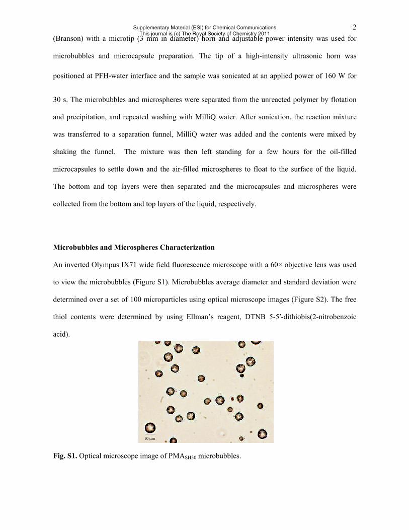
1
One-Pot Ultrasonic Synthesis of Multifunctional Microbubbles and Microcapsules Using Synthetic Thiolated Macromolecules Francesca Cavalieri,a,b,c Meifang Zhou,b Frank Caruso,c* and Muthupandian Ashokkumarb* Materials Materials. Poly(methacrylic acid, sodium salt) (PMA), Mw 15 kDa, was purchased from
Polysciences. Cystamine hydrochloride, dithiothreitol (DTT), N-hydroxysuccinimide (NHS), and