58
THYROID GLAND DR SYED UBAID Associate professor of surgery
| Date post: | 11-Apr-2017 |
| Category: |
Health & Medicine |
| Upload: | syed-ubaid |
| View: | 39 times |
| Download: | 3 times |

THYROID GLAND
DR SYED UBAIDAssociate professor of surgery

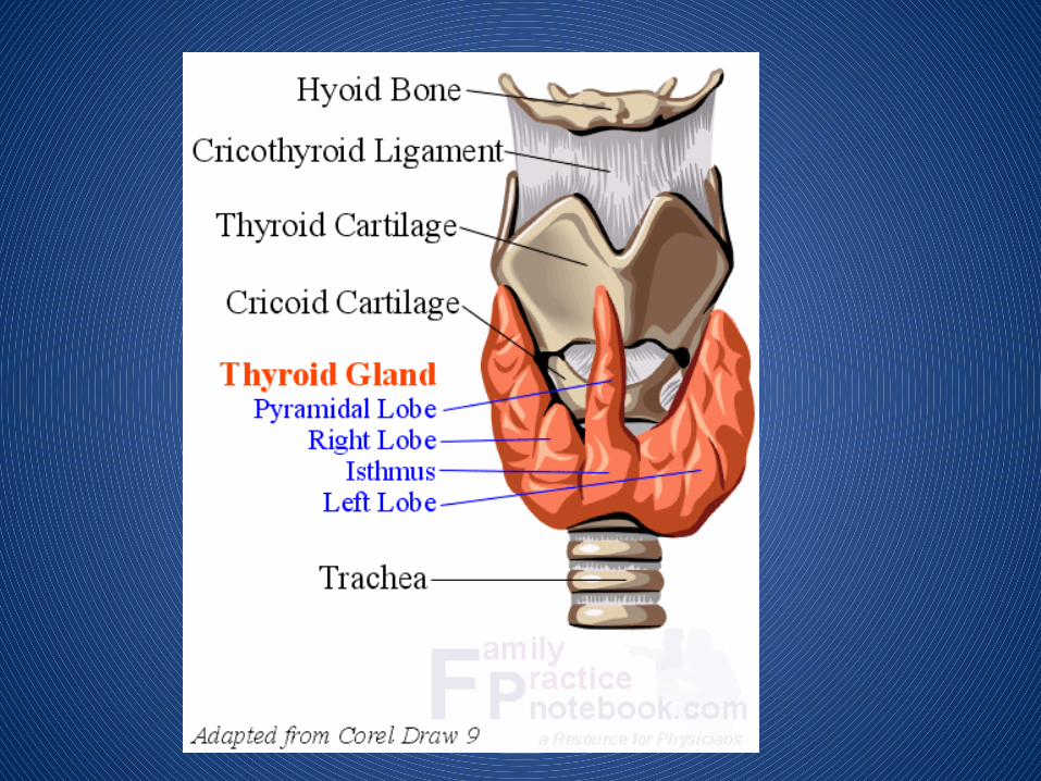
SURGICAL ANATOMY FROM GREEK thyreoeides= SHIELD SHAPE
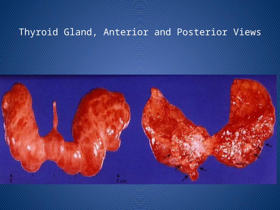
Thyroid Gland, Anterior and Posterior Views

SURGICAL ANATOMY
• TWO LOBES JOINED BY ISTHMUS• PYRAMIDAL LOBE (80%)PROJECT UPWARDS
FROM ISTHMUS OR EITHER OF THE LOBES• A FIBROMUSCULAR BAND levator glandulae
thyroideae DESCEND FROM THE BODY OF THE HYOID BONE TO ISTHMUS OR TO PYRAMIDAL LOBE

SURGICAL ANATOMY

SURGICAL ANATOMY
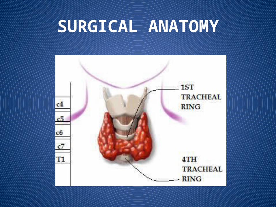
• GLAND LIES AGAINST C5,6,7 &T1 VERTEBRAE• EACH LOBE EXTENDS FROM MIDDLE OF
THYROID CARTILAGE TO 4TH OR 5TH TRACHEAL RING.
• ISTHMUS EXTENDS FROM 2ND TO THE 3RD TRACHEAL
SURGICAL ANATOMY
SURGICAL ANATOMY
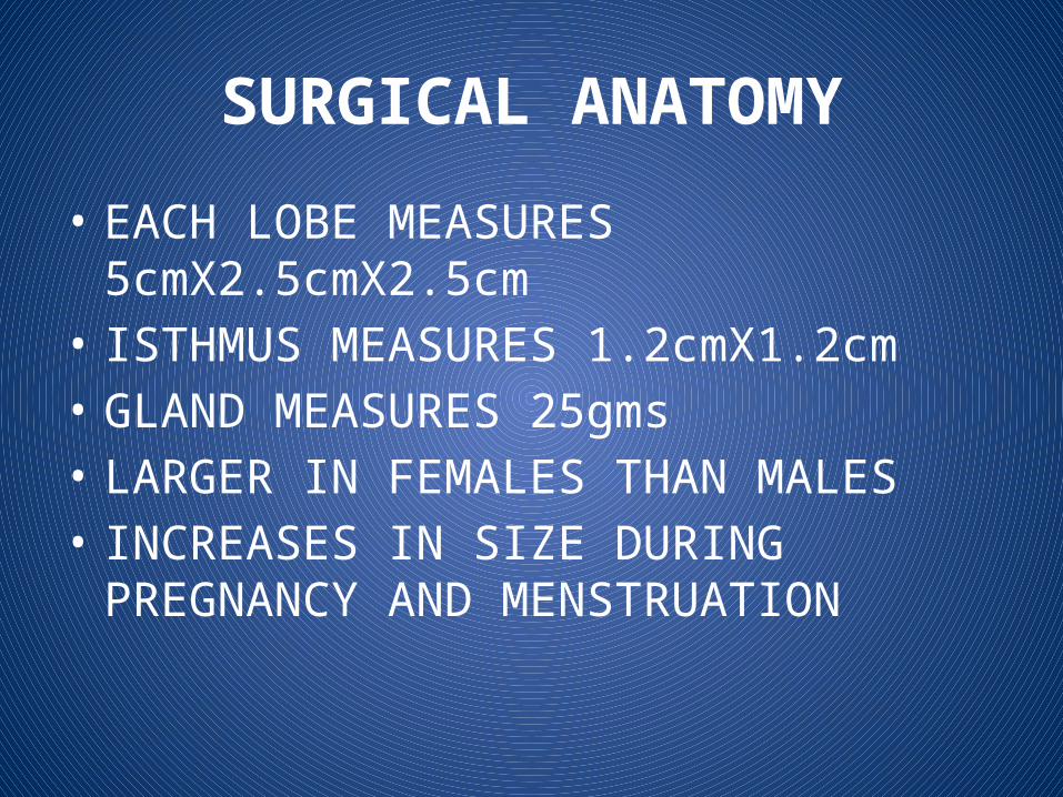
• EACH LOBE MEASURES 5cmX2.5cmX2.5cm• ISTHMUS MEASURES 1.2cmX1.2cm• GLAND MEASURES 25gms• LARGER IN FEMALES THAN MALES• INCREASES IN SIZE DURING PREGNANCY AND
MENSTRUATION

SURGICAL ANATOMY

SURGICAL ANATOMY
CAPSULES• TRUE PERIPHERAL CONDENSATION OF ‐
CONNECTIVE TISSUE OF GLAND
• FALSE/SURGICAL FROM PRETRACHEAL LAYER ‐OF DEEP CERVICAL FASCIA

SURGICAL ANATOMY
• CAPSULES
SURGICAL ANATOMY
SUSPENSORY LIGAMENT OF BERRY : THE PRETRACHEAL LAYER IS THIN ALONG THE
POSTERIOR BORDER OF THE LOBES, BUT THICK ON THE INNER SURFACE OF THE GLAND WHERE IT FORMS A SUSPENSORY LIGAMENT OF BERRY WHICH CONNECTS THE GLAND TO THE CRICOID CARTILAGE
SURGICAL ANATOMY
WHY THYROID MOVES WITH DEGLUTITION• DURING 1ST STAGE OF DEGLUTITION• HYOID BONE MOVES UP• PULLS PRETRACHEAL FASCIA UP• THIS PULLS LIGAMENT OF BERRY UP• THIS PULLS THYROID UP
SURGICAL ANATOMY
• ALL STRUCTURES ENCLOSED IN THE PRETRACHEAL FASCIA MOVES UP WITH DEGLUTITION
• THYROGLOSSAL CYST-SUBHYOID BURSITIS-PRE TRACHEAL LYMPH NODES-PRE LARYNGEAL LYMPH NODES
SURGICAL ANATOMY
VASCULAR ANATOMY
• HIGHLY VASCULAR• TWO ARTERIES• SUPERIOR THYROID ARTERY• INFERIOR THYROID ARTERY
SURGICAL ANATOMY
• 3 VEINS• SUP THYROID VEIN DRAINS INTO IJV OR COMMON FACIAL V.• MIDDLE THYROID VEIN DRAINS TO IJV• INFERIOR THYROID VEIN
INTO LEFT BRACHICEPHALIC V.• A 4TH VEIN OF KOCHER’S EMERGE B/W MIDDLE
AND INFERIOR VEINAND DRAIN INTO IJV

SURGICAL ANATOMY

SURGICAL ANATOMY
LYMPHATICS• PRIMARILY TO INTERNAL JUGULAR NODES• SUPERIOR POLE & MEDIAL ISTHMUS TO
SUPERIOR GROUP• LOWER POLE OF THYROID TO INFERIOR
GROUP• EMPTY INTO PRETRACHEAL & PARATRACHEAL
NODES

SURGICAL ANATOMY

SURGICAL ANATOMY
Innervation• Principally from ANS• Parasympatheticfibers –from vagus • Sympatheticfibers –from superior, middle, and
inferior ganglia of the sympathetic trunk• Enter the gland along with the blood vessels.
SURGICAL ANATOMY
• RECURRENT LARYNGEAL NERVE SUPPLIES THE INTRINSIC MUSCLE OF LARYNX EXCEPT CRICOTHYROID WHICH IS SUPPLIED BY EXTERNAL LARYNGEAL NERVE
• ACIDENTAL DAMAGE TO THIS NERVE DURING SURGERY CAUSES IPSILATERAL VOCAL CORD PARALYSIS & DIFFICULTY IN PHONATION
SURGICAL ANATOMY
• RT SIDE IT ORIGINATES FROM VAGUS CROSSES FIRST PART OF SUBCLAVIAN.A LOOPS UNDER IT RUNS OBLIQUE TO ENTER LARYNX AT LEVEL OF CRICOID

SURGICAL ANATOMY
• LEFT SIDE AFTER ORIGIN FROM VAGUS CROSSES AORTIC ARCH LOOPS POSTERIORLY AROUND LIGAMENTUM ARTERIOSUS ASCENDS MEDIALLY IN TRACHEO ESOPHAGEAL GROOVE

SURGICAL ANATOMY

SURGICAL ANATOMY
• SUPERIOR LARYNGEAL NERVE HAS INTERNAL BRANCH(SENSORY) & EXTERNAL BRANCH(MOTOR)HELPS IN VOCAL CORD TENSION AND PITCH OF VOICE
SURGICAL ANATOMY
• SUPERIOR LARYNGEAL NERVE RUNS IN CLLOSE PROXIMITY TO SUPERIOR POLE VESSELS, TO AVOID INJURY SUPERIOR POLE VESSELS SHOULD BE INDIVIDUALLY LIGATED & DIVIDED LOW ON THYROID GLAND AND DISSECTED LATERALLY TO CRICOTHYROID MUSCLE

SURGICAL ANATOMY
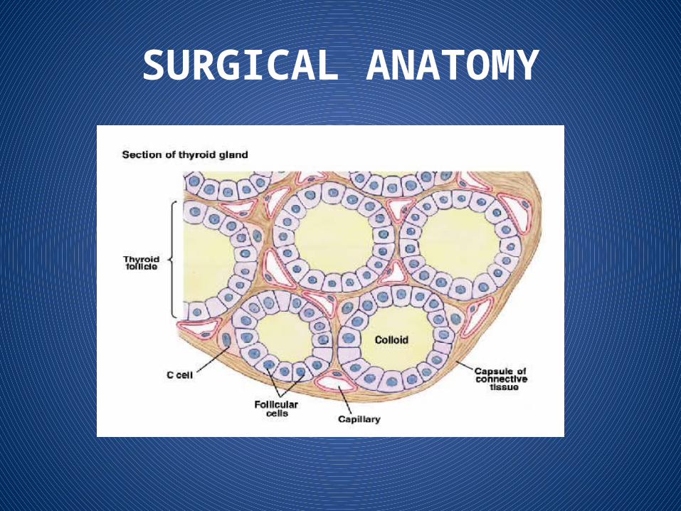
HISTOLOGY The functioning unit is the lobule supplied by a
single arteriole and consists of 24–40 follicles lined with cuboidal epithelium. The
follicle contains colloid in which thyroglobulin is stored.
SURGICAL ANATOMY
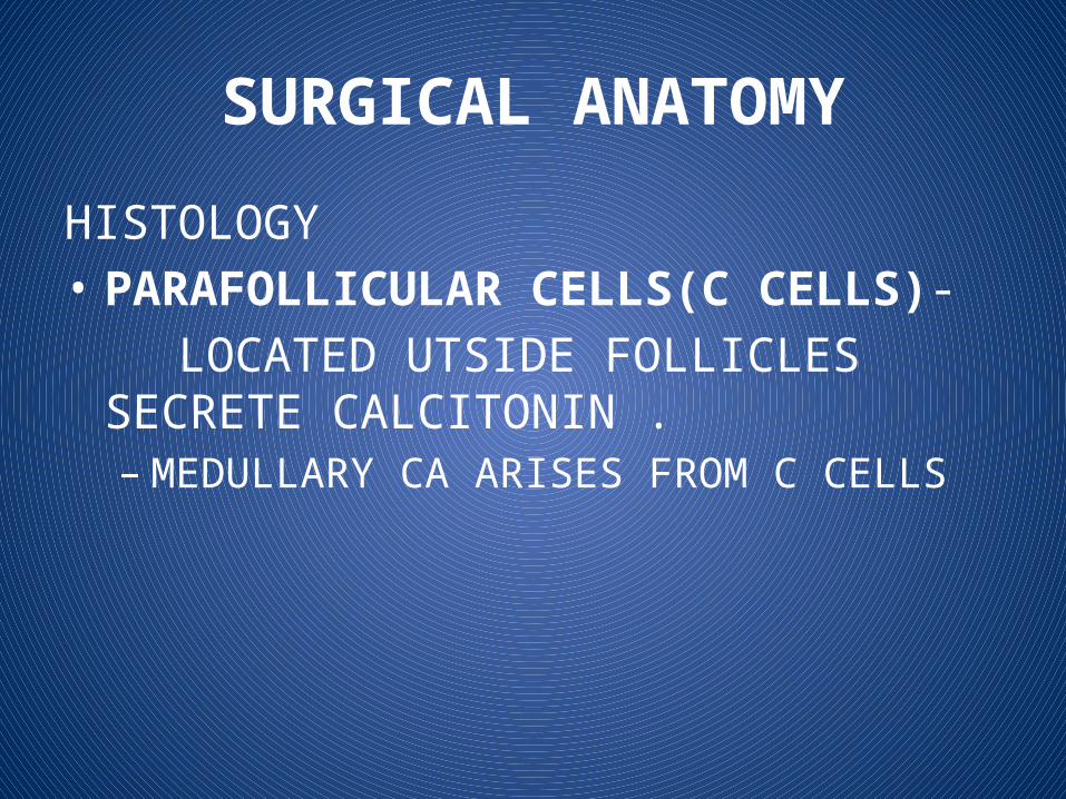
HISTOLOGY • PARAFOLLICULAR CELLS(C CELLS)- LOCATED UTSIDE FOLLICLES SECRETE
CALCITONIN . – MEDULLARY CA ARISES FROM C CELLS
SURGICAL ANATOMY

EMBRYOLOGY
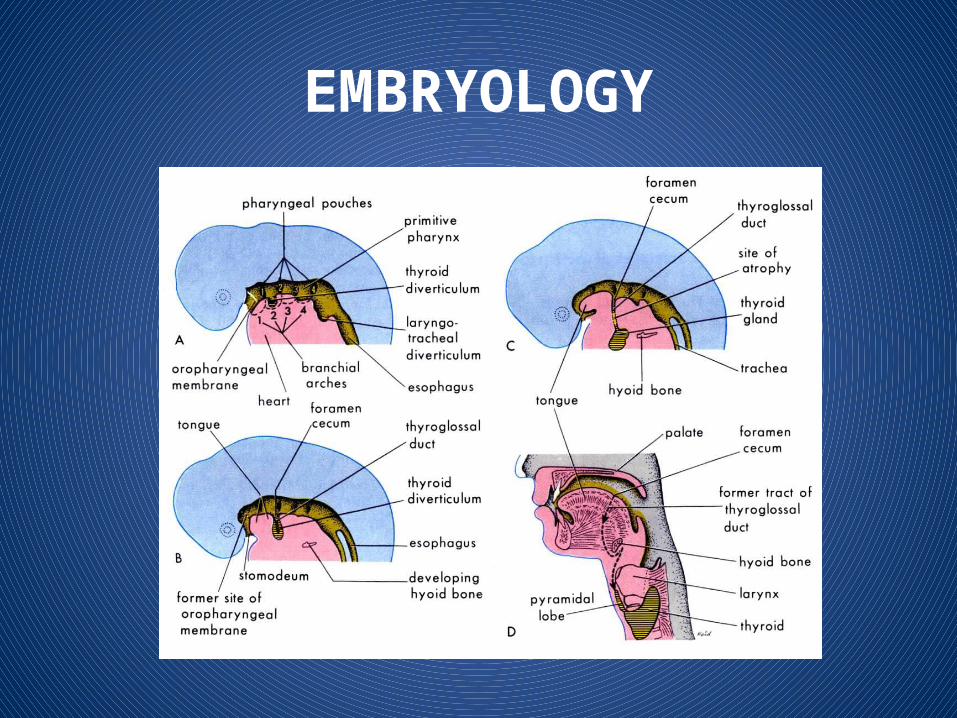
• The thyroglossal duct develops from the median bud of the pharynx. The foramen caecum at the base of the tongue is the vestigial remnant of the duct. This initially hollow structure migrates caudally and passes in close continuity with, and sometimes through, the developing hyoid cartilage.
EMBRYOLOGY
• The parathyroid glands develop from the third and fourth pharyngeal pouches
• The thymus also develops from the third pouch.• As it descends it takes the associated
parathyroid gland with it which explains why the inferior parathyroid which arises from the third pharyngeal pouch normally lies inferior to the superior gland.

EMBRYOLOGY
• However, the inferior parathyroid may be found anywhere along this line of descent.
• The developing thyroid lobes amalgamate• with the structures that arise in the fourth
pharyngeal pouch, i.e. the superior parathyroid gland and the ultimobranchial body.
• Parafollicular cells (C cells) from the neural crest reach the thyroid via the ultimobranchial body.
EMBRYOLOGY
Physiology The thyroid follicles secretes tri-iodothyronine(T3)and
thyroxin(T4) synthesis involves combination of iodine with tyrosine group
to form mono and di-iodotyrosine which are coupled to form T3 andT4.
The hormones are stored in follicles bound to thyrogobulin When hormones released in the blood they are bound to
plasma proteins and small amount remain free in the plasma . The metabolic effect of thyroid hormones are due to free
(unbound)T3 and T4. 90%of secreted hormones is T4 but T3is the active hormone
so, T4is converted to T3 peripherally.

Physiological control of secretion
Synthesis and libration of T3 and T4 is controlled by thyroid stimulating hormone(TSH)secreted by anterior pituitary gland.
TSH release is in turn controlled by thyrotropin releasing hormone (TRH)from hypothalamus .
Circulating T3and T4 exert –ve feedback mechanism on hypothalamus and anterior pituitary gland .
So, in hyperthyroidism where hormone level in blood is high ,TSH production is suppressed and vice versa.
PHYSIOLOGY
• IODINE –RAW MATERIAL FOR THYROID HORMONE SYNTHESIS
• INGESTED IODINE CONVERTED TO IODIDE BEFORE ABSORPTION
• 15O μg OF IODINE MINIMUM REQD FOR NORMAL THYROID FUNCTION OF WHICH 120μg ENTER THYROID AT NORMAL RATES OF HORMONE SYNTHESIS AND SECRETION

PHYSIOLOGY
• THYROGLOBULIN IS A GLYCOPROTEIN SYNTHESIZED IN THYROID CELLS AND SECRETED INTO THE COLLOID BY EXOCYTOSIS
• THYROGLOBULIN IS BOUND TO THYROID HORMONES TILL IT IS SECTRETED INTO BLOOD, AFTER WHICH IT IS INGESTED BACK INTO THE COLLOID
PHYSIOLOGY
• STEPS OF THYROID HORMONE SYNTHESIS 1. IODINE TRAPPING
2. OXIDATION3. IODINATION4. COUPLING5. STORAGE 6. RELEASE

PHYSIOLOGY
IODINE TRAPPING• IODINEAVAILABLE THROUGH CERTAIN FOODS (EG,
SEAFOOD, BREAD, DAIRY PRODUCTS), IODIZED SALT, OR DIETARY SUPPLEMENTS ETC
• THYROID CELL MEMBRANES FACING THE CAPILLARIES CONTAIN A SYMPORTER OR IODINE PUMP THAT TRANSPORTS Na+/I AGAINST ‐ELECTROCHEMICAL GRADIENT

Biosynthesis of T4 and T3
The process includes• Dietary iodine (I) ingestion• Active transport and uptake of iodide (I-) by thyroid gland• Oxidation of I- and iodination of thyroglobulin (Tg)
tyrosine residues • Coupling of iodotyrosine residues (MIT and DIT) to form
T4 and T3
• Proteolysis of Tg with release of T4 and T3 into the circulation
41

Regulation of TH synthesis/secretion

Normal circulatory concentrations
– T4 4.5-11 g/dL
– T3 60-180 ng/dL (~100-fold less than T4)
43

Carriers for Circulating Thyroid Hormones
• More than 99% of circulating T4 and T3 is bound to plasma carrier proteins– Thyroxine-binding globulin (TBG), binds about 75%– Transthyretin (TTR), also called thyroxine-binding
prealbumin (TBPA), binds about 10%-15%– Albumin binds about 7%– High-density lipoproteins (HDL), binds about 3%
• Carrier proteins can be affected by physiologic changes, drugs, and disease
44
PHYSIOLOGY
• PHYSIOLOGICAL ACTIONS• HEART INCREASE CARDIAC OUTPUT,HEART RATE• ADIPOSE TISSUE STIMULATE LIPOLYSIS

PHYSIOLOGY
• MUSCLE INCREASES PROTEIN BREAKDOWN• BONE PROMOTES NORMAL GROWTH AND SKELETAL DEVELOPMENT
PHYSIOLOGY
• NERVOUS SYSTEM PROMOTE NORMAL BRAIN DEVELOPMENT• GUT INCREASESRATE OF CHO ABSORPTION

Thyroid Hormone Plays a Major Role in Growth and Development
• Thyroid hormone initiates or sustains differentiation and growth– Stimulates formation of proteins– Is essential for normal brain development
• Essential for childhood growth– Untreated congenital hypothyroidism or chronic
hypothyroidism during childhood can result in incomplete development and mental retardation
48

Thyroid Hormones and the Central Nervous System (CNS)
• Thyroid hormones are essential for neural development and maturation and function of the CNS
• Decreased thyroid hormone concentrations may lead to alterations in cognitive function– Patients with hypothyroidism may develop
impairment of attention, slowed motor function, and poor memory
– Thyroid-replacement therapy may improve cognitive function when hypothyroidism is present
49

Thyroid Hormone Influences the Female Reproductive System
• Normal thyroid hormone function is important for reproductive function– Hypothyroidism may be associated with menstrual
disorders, infertility, risk of miscarriage, and other complications of pregnancy
50

Thyroid Hormone is Critical for Normal Bone Growth
– T3 also may participate in osteoblast differentiation and proliferation, and chondrocyte maturation leading to bone ossification
51

Thyroid Hormone Regulates Mitochondrial Activity
• T3 is considered the major regulator of mitochondrial activity
– A potent T3-dependent transcription factor of the
mitochondrial genome induces early stimulation of transcription and increases transcription factor (TFA) expression
– T3 stimulates oxygen consumption by the
mitochondria
52
Thyroid Hormones Stimulate Metabolic Activities in Most Tissues
• Thyroid hormones (specifically T3) regulate rate of
overall body metabolism– T3 increases basal metabolic rate
• Calorigenic effects– T3 increases oxygen consumption by most
peripheral tissues– Increases body heat production
53

Investigation
Laboratory investigation: -serum T3, T4.-serum TSH. -thyroid antibodies: in hashimoto’s disease.

Investigation
-chest and neck x-ray:Show descend of thyroid gland to thorax and
mediastanal shifting in retrosternal goitre. -iodine isotopesBy i.v injection of I131. Then, use gama rays to show
hot and cold nodules. -CT scanShow thyroid size and if there is compression to
trachea

Investigation
-Endoscopic investigation:-bronchoscopy: show compression and
infiltration of trachea by tumerBiopsy: -fine needle aspiration biopsy.-true-cut biopsy.

MIDLINE SWELLINGS
• Thyroid enlargement• Thyroglossal cyst• Dermoid cyst

THANK YOU


