78
Thyroid gland 1 & 2 Dr Heyam Awad FRCPath
Thyroid gland 1 & 2
Dr Heyam AwadFRCPath

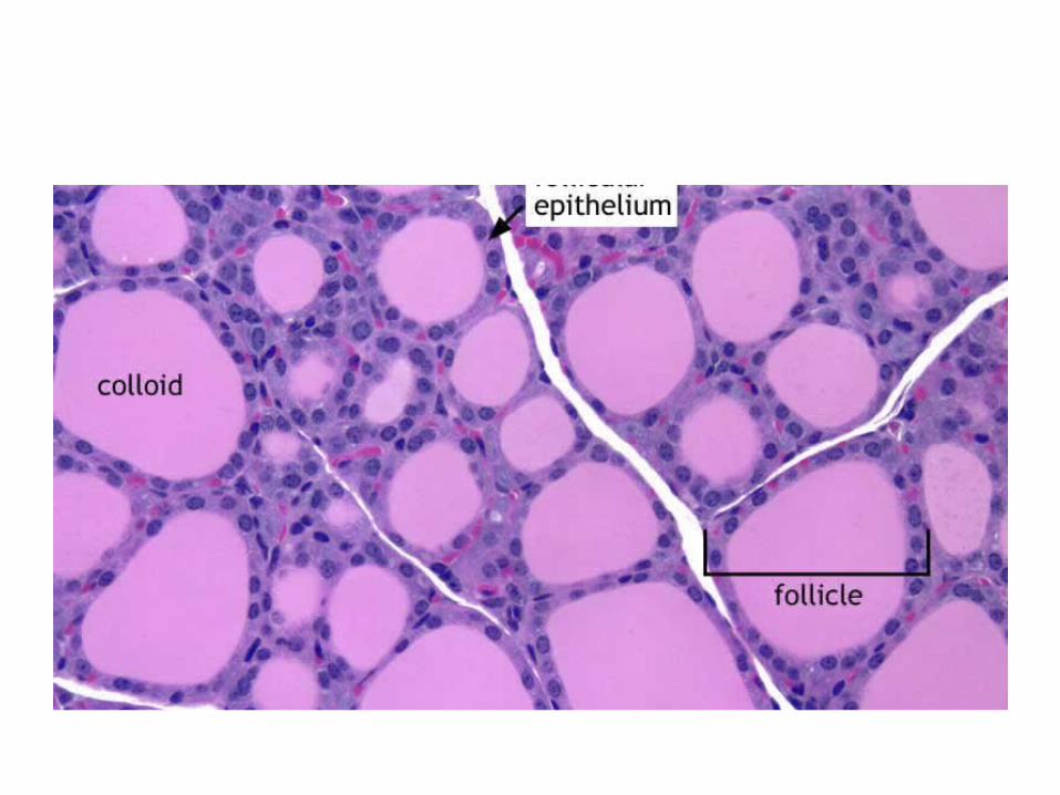
Thyroid gland

Thyroid hormones

Parafollicular cells
Thyroid diseases
• Hyerthyroidism• Hypothyroidism• Thyroiditis• Graves disease• Diffuse nontoxic goiter and multinodular
goiter• neoplasms
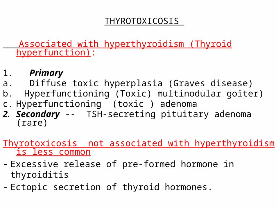
THYROTOXICOSIS
Associated with hyperthyroidism (Thyroid hyperfunction):
1. Primarya. Diffuse toxic hyperplasia (Graves disease)b. Hyperfunctioning (Toxic) multinodular goiter)c. Hyperfunctioning (toxic ) adenoma2. Secondary -- TSH-secreting pituitary adenoma (rare)
Thyrotoxicosis not associated with hyperthyroidism is less common
- Excessive release of pre-formed hormone in thyroiditis- Ectopic secretion of thyroid hormones.
Clinical manifestations of thyrotoxicosisa. Constitutional symptoms : warm flushed skin, heat
intolerance and excessive sweating - Weight loss despite increased appetite.b. Malabsorption, and diarrhea. c. Tachycardia and elderly patients may develop heart
failure due to aggravation of pre-existing heart disease
d. Nervousness, tremor, and irritability

e. A wide, staring gaze and lid lag because of sympathetic overstimulation of the levator palpebrae superioris
Note: True thyroid ophthalmopathy associated with proptosis is a feature seen only in Graves disease.
f. 50% develop proximal muscle weakness (thyroid myopathy).
g. Thyroid storm : Designates the abrupt onset of severe hyperthyroidism, and this condition occurs most commonly in individuals with Graves disease and it is a medical emergency because significant numbers of untreated patients die of cardiac arrhythmias

hyperthyroidism

Lab tests
• The measurement of serum TSH is the most useful single screening test for hyperthyroidism, because TSH levels are decreased even at the earliest stages, when the disease may still be subclinical

- Once the diagnosis of thyrotoxicosis has been confirmed measurement of radioactive iodine uptake by the thyroid gland often is valuable in determining the etiology
For example, such scans may show :a. Diffusely increased (whole-gland) uptake in Graves
disease,b. Increased uptake in a solitary nodule in toxic
adenomac. Or decreased uptake in thyroiditis.

Iodine scans

Cold nodule
HYPOTHYROIDISM : Primary causesa. - Worldwide, the most common cause of hypothyroidism
is dietary deficiency of iodine. b. In most developed countries, autoimmune diseases
predominate such as Hashimoto thyroiditis c. Genetic defects such as Thyroid dysgenesis or Congenital biosynthetic defect (dyshormogentic goiter). Secondary causes: Pituitary or hypothalamic disorder


hypothyroidism
• Cretinism• myxedema
Cretinism :Refers to hypothyroidism developing in infancy or early childhood
1. Endemic cretinism: in dietary iodine deficiency is endemic, including mountainous areas ( the Himalayas )
2. Sporadic cretinism. Caused by enzyme defects that interfere with thyroid hormone synthesis
Clinical features of cretinism include:
- Impaired development of skeletal system- short stature, - Coarse facial features, protruding tongue, umbilical
hernia. - Central nervous system, with mental retardation
Myxedema. or Gull syndrome : - Hypothyroidism in older children and adults and
characterized by: a. Patients are cold intolerant, and often obese. b. Generalized apathy and mental sluggishness that
in the early stages of disease may mimic depression
c. Broadening and coarsening of facial features
d. Enlargement of the tongue, and deepening of the voice.e. Bowel motility is decreased, resulting in constipation. f. Pericardial effusions are common; in later stages, the
heart is enlarged, and heart failure may supervene.g. Mucopolysaccharide-rich edematous fluid accumulates
in skin, subcutaneous tissue, and number of visceral sites

Lab tests
Serum TSH is the most sensitive screening test .a. The serum TSH is increased in primary
hypothyroidismb. The TSH is not increased in persons with
hypothyroidism caused by primary hypothalamic or pituitary disease.
c. Serum T4 is decreased hypothyroidism of any origin.
Thyroiditis Chronic Lymphocytic (Hashimoto) Thyroiditis- Is the most common cause of hypothyroidism in areas of
the world where iodine levels are sufficient. - It is characterized by gradual thyroid failure secondary
to autoimmune destruction of the thyroid gland- It is most prevalent between the ages of 45 and 65 years
and is more common in women than in men- It can occur in children and is a major cause of non-
endemic goiter in children
PATHOGENESIS :- Caused by breakdown in self-tolerance to thyroid antigens - Circulating autoantibodies against thyroid antigens are- present
in the vast majority of patients - Multiple immunologic mechanisms may contribute to thyroid
damage , I. Cytokine-mediated cell death: Excessive T cell activation
leads to the production of inflammatory cytokines such as IFN-γ in the thyroid with resultant recruitment and activation of macrophages and damage to follicles .
II. Binding of anti-thyroid antibodies (antithyroglobulin, and antithyroid peroxidase antibodies), followed by antibody- dependent cell-mediated cytotoxicity
III. T cell mediated cytotoxicity.

HASHIMOTO
. HASHIMOTO
- A significant genetic component is supported by thea. Concordance of disease in 40% of monozygotic twins, b. the presence of circulating antithyroid antibodies in 50%
of asymptomatic siblings of affected patients .

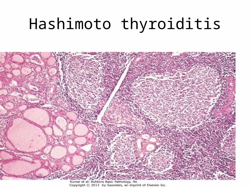
• Gross :- Diffuse and symmetric enlargement of the thyroid but
localized enlargement may be seen in some cases to raise suspicion for neoplasm
Microscopic examination reveals1. Infiltration by small lymphocytes, plasma cells, and well-
developed germinal centers 2. The thyroid follicles are atrophic 2. Some follicles are lined by epithelial cells with abundant
eosinophilic, cytoplasm, termed Hürthle cells and these Hurthle cells have numerous mitochondria

- Less commonly, the thyroid is small due to extensive fibrosis (fibrosing variant) but unlike Reidel thyroiditis, the fibrosis does not extend beyond the capsule of the gland.
Clinically , 1. Painless thyroid enlargement associated with some
degree of hypothyroidism,2. - In the usual clinical course, hypothyroidism develops
gradually.; however, it may be preceded by transient thyrotoxicosis due to disruption of thyroid follicles ,and secondary release of thyroid hormones (hashitoxicosis).
Hashimoto thyroiditis

- Patients with Hashimoto thyroiditis often :1. Have other autoimmune diseases2. .Are at increased risk for the development of B cell
non-Hodgkin lymphomas within the thyroid gland. Note: - The relationship between Hashimoto disease and
thyroid epithelial cancers remains controversial, with some morphologic and molecular studies suggesting a predisposition to papillary carcinomas

Subacute Granulomatous (de Quervain) Thyroiditis - Is much less common than Hashimoto disease- Is most common between the ages of 30 and 50 and,- More frequently in women than in men. - Is believed to be caused by a viral infection and a majority
of patients have a history of an upper respiratory infection just before the onset of thyroiditis.
Gross- The gland has intact capsule, and may be unilaterally or bilaterally enlarged.

Histologic examination reveals 1. Disruption of thyroid follicles, with extravasation of colloid leading to a neutrophilic infiltrate, which is replaced by
lymphocytes, plasma cells, and macrophages. 2. The extravasated colloid provokes a granulomatous reaction
with giant cells that contain fragments of colloid. 3. Healing occurs by resolution of inflammation and fibrosis. Clinical Features :-Acute onset characterized by neck pain ( with swallowing) - Fever, malaise, and variable enlargement of the thyroid. - Transient hyperthyroidism may occur as a result of disruption of
follicles and release of excessive hormones. - The leukocyte count is increased.

- With progression of disease and gland destruction, a transient hypothyroid phase may ensue.
- The condition typically is self-limited, with most patients returning to a euthyroid state within 6 to 8 weeks

Subacute Lymphocytic Thyroiditis :- Also is known as silent or painless thyroiditis;- And in a subset of patients the onset of disease follows -
pregnancy (postpartum thyroiditis). - Most likely to be autoimmune because circulating
antithyroid antibodies are found in a majority of patients - It mostly affects middle-aged women, who present with a-
painless neck mass or features of thyrotoxicosis
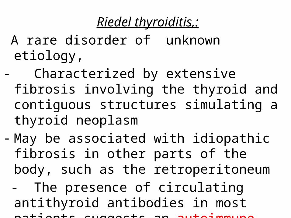
Riedel thyroiditis,: A rare disorder of unknown etiology, - Characterized by extensive fibrosis involving the thyroid
and contiguous structures simulating a thyroid neoplasm- May be associated with idiopathic fibrosis in other parts of
the body, such as the retroperitoneum - The presence of circulating antithyroid antibodies in most
patients suggests an autoimmune etiology

GRAVES DISEASE The most common cause of endogenous hyperthyroidism
with a peak incidence in women between the ages of 20 and 40.
Triad of manifestations: A. Thyrotoxicosis,. All patientsB. Localized, infiltrative dermopathy ( pretibial myxedema),
minority of cases and involves the skin overlying the shins, and manifests as scaly thickening
C. Infiltrative ophthalmopathy with resultant exophthalmos in 40% of patients
Exophthalmos is the result of increased volume of the retro-orbital connective tissues by
1. Marked infiltration of T cells with inflammatory edema2. Accumulation of glycosaminoglycans3. Increased numbers of adipocytes (fatty infiltration).
- These changes displace the eyeball forward, potentially interfering with the function of the extraocular muscles
- Exophthalmos may persist after successful treatment of the thyrotoxicosis, and may result in corneal injury.


PATHOGENESIS :- Genetic factors are important in the causation of Graves disease, the incidence is increased in relatives of affected patients, and the concordance rate in
monozygotic twins is 60%. - A genetic susceptibility is associated with the presence of HLA-DR3, - it is characterized by a breakdown in self-tolerance to
thyroid autoantigens, and is the production of multiple autoantibodies
Autoantibodies in GRAVES :
1. Thyroid-stimulating immunoglobulin:- An IgG antibody binds to the TSH receptor and mimics the action of TSH,
with resultant increased hormones 2. Thyroid growth-stimulating immunoglobulins:- Directed against the TSH receptor, and have been implicated in the
proliferation of follicular epithelium 3. TSH-binding inhibitor immunoglobulins: - Prevent TSH from binding to its receptor on thyroid cells and in so doing
may actually inhibit thyroid cell function, a finding explains why some patients with Graves spontaneously develop episodes of hypothyroidism.

Note: The coexistence of stimulating and inhibiting immunoglobulins in the serum of the same patient may explain why some patients with Graves disease spontaneously develop episodes of hypothyroidism
.Gross: Symmetrical enlargement of the thyroid gland with intact capsule,

On microscopic examination, a. The follicular cells in untreated cases are tall, and more
crowded and may result in formation of small papillaeb. Lymphoid infiltrates, consisting predominantly of T cells,
with few B cells and plasma cells are present throughout the interstitium; with formation of germinal centers
Laboratory findings and radiologic findings- Elevated serum free T4 and T3 and depressed serum TSH
- Because of ongoing stimulation of the thyroid follicles radioactive iodine uptake is increased, and radioiodine scans show a diffuse uptake of iodine.

DIFFUSE AND MULTINODULAR GOITER - Enlargement of the thyroid, or goiter, is the most
common manifestation of thyroid disease Mechanism : - The goiters reflect impaired synthesis of thyroid hormone
often caused by dietary iodine deficiency and this leads to to a compensatory rise in the serum TSH, which in turn causes hyperplasia of the follicular cells and, ultimately, gross enlargement of the thyroid gland .,

Goiters can be endemic or sporadic. I. Endemic goiter :Occurs in geographic areas where the
soil, water, and food supply contain little iodine.- The term endemic is used when goiters are present in
more than 10% of the population in a given region. - Such conditions are common in mountainous areas of the
world, including the Himalayas and the Andes but with increasing availability of iodine supplementation, the frequency and severity of endemic goiter have declined

II. Sporadic goiter : Less common than endemic goiter.- The condition is more common in females than in males,
with a peak incidence in puberty or young adulthood, when there is an increased physiologic demand for T4.
- It may be caused by several conditions, including the:a. Ingestion of substances that interfere with thyroid
hormone synthesis , such as excessive calcium and vegetables such as cabbage, cauliflower, sprouts, .
b. Hereditary enzymatic defects that interfere with thyroid hormone synthesis (dyshormonogenetic goiter).
-In most cases, the cause of sporadic goiter is not apparent.

MORPHOLOGY :- Initially, the gland is diffusely and symmetrically enlarged
(diffuse goiter) but later on it becomes multinodular goiter. On microscopic examination,a. The follicular epithelium may be hyperplastic in the early
stages of disease or flattened and cuboidal during periods of involution.
b. Colloid is abundant in the latter periods (colloid goiter). c. With time, recurrent episodes of hyperplasia and
involution produce a more irregular enlargement of thee thyroid, termed multinodular goiter and virtually all long-standing diffuse goiters convert into multinodular goiters.

Multinodular Goiter
- Multinodular goiters cause multilobulated, asymmetrically enlarged glands which attain massive size and old lesions often show fibrosis, hemorrhage, calcification
- Multinodular goiters typically are hormonally silent,- 10% of patients can manifest with thyrotoxicosis due to
the development of autonomous nodules producing hormone independent of TSH stimulation and this condition, called toxic multinodular goiter or Plummer syndrome

Clinical Features :a. The dominant features are mass effects of the goiter b. may cause airway obstruction, dysphagia, and
compression of large vessels in the neck and upper thorax
c. The incidence of malignancy in long-standing multinodular goiters is low (less than 5%) but not zero and concern for malignancy arises with goiters that demonstrate sudden changes in size or associated symptoms ( hoarseness)

Thyroid tumors : -present as solitary nodules.
- the majority of solitary nodules of the thyroid prove to be benign :
a. Follicular adenomas b. A dominant nodule in multinodular goiterc. Simple cysts or foci of thyroiditis
- Carcinomas of the thyroid, are uncommon, accounting for much less than 10% of solitary thyroid nodules.

- Several clinical criteria provide a clue to the nature of a given thyroid nodule: a. Solitary nodules, in general, are more likely to be
neoplastic than are multiple nodules.b. Nodules in younger patients are more likely to be
neoplastic than are those in older patients. c. Nodules in males are more likely to be neoplastic than
are those in females. d. Nodules that take up radioactive iodine in imaging
studies (hot nodules) are more likely to be benign than malignant.

Follicular adenomas- Are benign neoplasms derived from follicular epithelium. - solitary. - The tumor is demarcated and compressed the adjacent
thyroid parenchyma by a well-defined, intact capsule- - cold nodules on scanning but might be functional.

. Microscopic examination of follicular adenoma,- The cells are arranged in follicles and its variantsa. Hurthle cell adenoma: - The neoplastic cells show oxyphil or Hürthle cell change)
and its behavior is not different from those of a conventional adenoma.
b. Atypical adenoma: - The neoplastic cells exhibit focal nuclear atypia,
(endocrine atypia);and these features do not constitute evidence of malignancy

Follicular adenoma

- Thyroid adenomas :a. Carry an excellent prognosis b. and do not recur or metastasize c. and are not forerunners to carcinomas
- About 10% of cold nodules prove to be malignant and by contrast, malignancy is rare in hot nodules

Carcinomas :- Accounting for about 1.5% of all cancers - A female predominance has been noted among patients who
develop thyroid carcinoma in the early and middle adult years -***** cases manifesting in childhood and late adult life are
distributed equally between men and women- The major subtypes of thyroid carcinoma are are 1. Papillary carcinoma ( for more than 85% of cases)2. Follicular carcinoma (5% to 15% of cases)3. Anaplastic carcinoma (less than 5% of cases)4. Medullary carcinoma (5% of cases)

PATHOGENESISA. Genetic factorsA. Papillary thyroid carcinomas: 1. rearrangements of RET - The RET gene is not normally expressed in follicular cells but
in papillary cancers, chromosomal rearrangements place the tyrosine kinase domain of RET under the transcriptional control of genes that are constitutively expressed in the thyroid epithelium and the novel fusion proteins so formed are known as RET/PTC and are present in 20% to 40% of papillary thyroid cancers.
-
Genetics of papillary carcinoma/ continued
The frequency of RET/PTC rearrangements is significantly higher in papillary cancers arising after radiation exposure.
2. The second mechanism involves activating point mutations in BRAF, whose product is an intermediate
signaling component in the MAP kinase pathway
Note: RET/PTC rearrangements and BRAF point mutations are not observed in follicular adenomas or carcinomas.

GENETIC FACTORS
B. Follicular thyroid carcinomas: a. Gain-of-function point mutations of RAS and PIK3CA,b. Loss-of-function mutations of PTEN, a suppressor genec. A unique (2;3) translocation presents in one third to one
half of follicular carcinomas which creates a fusion gene composed of portions of PAX8, a gene that is important in thyroid development, and the peroxisome proliferator-activated receptor gene (PPARG), whose product is a nuclear receptor implicated in cell differentiation
,

GENETIC FACTORS
C. Anaplastic carcinomas:
Inactivation of TP53, restricted to anaplastic carcinomas and may also relate to their aggressive behavior
GENETIC FACTORS
D. Medullary thyroid carcinomas: - Arise from the C cells,. a. Familial medullary thyroid carcinomas occur in multiple
endocrine neoplasia type 2 (MEN-2) and are associated with germline RET proto-oncogene mutations .
b. RET mutations are also seen in approximately one half of nonfamilial (sporadic) medullary thyroid cancers.
B. Environmental Factors. a. The major risk factor to papillary thyroid cancer is
exposure to ionizing radiation, during the first 2 decades of life.
b. Deficiency of dietary iodine: and by extension, an association with goiter is linked with a higher frequency of follicular carcinomas.

Papillary Carcinoma :
Is the most common form- accounts for the majority of thyroid carcinomas associated
with previous exposure to ionizing radiation. - May occur at any age,
Gross: Either solitary or multifocal lesions - Some are well circumscribed and even encapsulated; others
infiltrate the adjacent parenchyma and the definitive diagnosis is made by microscopic examination

Microscopically: The diagnosis of papillary carcinoma is based on nuclear features even in the absence of a papillary architecture.
1. The nuclei of papillary carcinoma cells show:
a. optically clear nuclei, or "Orphan Annie eye" nuclei , seen on histological but not cytological preparations ( formalin artefact)
b. Have invaginations of the cytoplasm to the nucleus ( pseudoinclusions)
2. papillary architecture is common3. Concentrically calcified structures(psammoma bodies)4. Foci of lymphatic permeation by tumor cells are present, but
invasion of blood vessels is relatively uncommon5. Metastases to cervical lymph nodes in half of cases.

Papillary carcinoma

Psammoma bodies

Clinical Features of papillary carcinomasa. Are nonfunctional tumors manifest as painless masses in
the neck, either within the thyroid or as metastasis in a cervical lymph node
b. Are indolent lesions, with 10-year survival rates of 95%. c. The presence of isolated cervical nodal metastases does
not have influence on good prognosis of these lesions. d. In a minority of patients, hematogenous metastases are
present at the time of diagnosis, most commonly to lung.

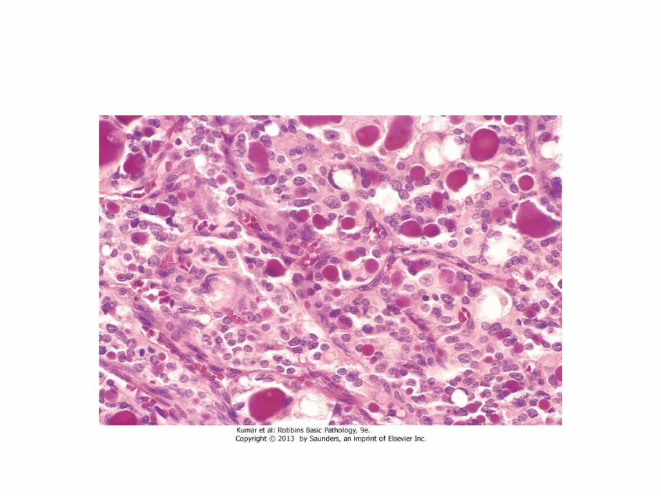
Follicular Carcinoma :
-- More common in women and in areas with dietary iodine deficiency .- The peak incidence between the ages of 40 and 60 years
On microscopic examination,- Are composed of fairly uniform cells forming small follicles, - In other cases, follicular differentiation is less apparent- It may bea. widely invasive, infiltrating the thyroid parenchyma and extrathyroidal
soft tissues, or b. Minimally invasive that may be impossible to distinguish from follicular
adenomas on gross examination and the .
- requires extensive histologic sampling to exclude capsular and/or vascular invasion
Clinical Features - Manifest most frequently as solitary cold thyroid nodules.- Tend to metastasize through the bloodstream
(hematogenous dissemination) to lungs, bone, and liver. - Regional nodal metastases are uncommon . - As many as half of patients with widely invasive
carcinomas succumb to their disease within 10 years, while less than 10% of patients with minimally invasive follicular carcinomas die within the same time span.

Follicular carcinoma
3. Anaplastic Carcinoma - Are undifferentiated tumors of the thyroid epithelium,- The mean age of 65 years.- They are aggressive, with a mortality rate of 100%. - Approximately a quarter of patients have a past history a
well-differentiated carcinoma, and a 1/4th harbor a well-differentiated tumor in the resected specimen.
- Metastases to distant sites are common, but death occurs in less than 1 year as a result of aggressive local growth which compromise of vital structures in the neck.
4. Medullary Carcinoma - neuroendocrine neoplasms.- Secrete calcitonin, the measurement of which plays
an important role in the diagnosis and postoperative follow-up evaluation of patients.
- In some cases, the tumor cells elaborate somatostatin, serotonin, and vasoactive intestinal peptide (VIP)

- Are sporadic in about 70% of cases and the remaining 30% are familial cases
a. Occurring in the setting of MEN syndrome 2A or 2B,b. or familial medullary thyroid carcinoma without an
associated MEN syndrome
Note: Both familial and sporadic forms demonstrate activating RET mutations.
Cases associated with MEN-2A or MEN-2B show
multicentric C cell hyperplasia in the surrounding thyroid parenchyma, a feature usually absent in sporadic lesions.
These foci are believed to represent the precursor lesions from which medullary carcinomas arise.

Clinical Features - The sporadic cases manifests most often as a
mass in the neck, sometimes associated with compression effects such as dysphagia or hoarseness.
- In some instances, the initial manifestations are caused by the secretion of a peptide hormone (e.g., diarrhea caused by the secretion of VIP).

- Screening of the patient's relatives for elevated calcitonin levels or RET mutations permits early detection of tumors in familial cases. ,
- All members of MEN-2 carrying RET mutations are offered prophylactic thyroidectomies to prevent the
development of medullary carcinomas- Often, the only finding in the resected thyroid of these
asymptomatic carriers is the presence of C cell hyperplasia or small (<1 cm) micromedullary carcinomas.- Recent studies have shown that specific RET mutations
correlate with an aggressive behavior in medullary carcinomas.

Medullary carcinoma


