Thyroid Hormone Replacement Therapy: Three ‘Simple’ Questions, Complex Answers
Antonio C. Bianco Sabina Casula
Division of Endocrinology, Diabetes and Metabolism, University of Miami Miller School of Medicine, Miami, Fla. , USA
Hypothyroidism affects about 3.7% of the general population in the United States [1] , reaching levels of up to 8% in areas with high prevalence of iodine deficiency [2] . At first sight, treatment for hypothyroidism, regard-less of its etiology, seems quite straightforward. Accord-ing to current guidelines the standard of care is treatment based on hormonal replacement therapy with daily ad-ministration of levothyroxine, the pro-hormone pro-duced exclusively by the thyroid gland [3] . The rationale is that the deiodinases, thioredoxin-fold containing se-lenoenzymes that metabolize thyroid hormone and are present in multiple extrathyroidal tissues, activate thy-roxine (T 4 ) and produce physiological amounts of the bi-ologically active thyroid hormone, triiodothyronine (T 3 ) [4] . The observation that circulating levels of T 3 and TSH can be normalized in levothyroxine-treated hypothy-roid patients reassures physicians that euthyroidism is achieved and probably contributed for the replacement of porcine thyroid preparations by the synthetic form of le-vothyroxine currently used [5, 6] . In fact, monitoring se-rum levels of TSH (and free T 4 (FT 4 )) became an integral part of the routine to follow the therapeutic efficacy of thyroid hormone replacement. However, despite normal-ization of these biochemical parameters, about 15% of those treated with levothyroxine replacement therapy alone do not achieve clinical euthyroidism and experi-ence some level of psychological impairment [7] .
Received: March 30, 2012 Accepted after revision: May 3, 2012 Published online: June 27, 2012
Dr. Antonio C. Bianco University of Miami Miller School of Medicine 1400 N.W. 10th Avenue, Suite 601 Miami, FL 33136 (USA) Tel. +1 305 243 5631, E-Mail abianco @ med.miami.edu
Treatment of Hypothyroidism Eur Thyroid J 2012;1:88–98 89
The persistency of a relatively small number of clini-cally symptomatic patients has led to an explosion of al-ternative treatment strategies, including the reawakening of desiccated porcine thyroid and development of new ‘thyroid supplements’ that take advantage of regulatory loopholes to avoid governmental oversight. This has cre-ated greater awareness in the medical community, with concerns gravitating mostly around two areas, namely (i) defining what is missing in our understanding of thyroid hormone signaling (transport across cell membranes and metabolism) that prevents us from developing a treat-ment strategy that is effective for 100% of the patients, and (ii) preventing the widespread usage of ‘thyroid for-mulas’ that in many cases leads to long-term subclinical or clinical thyrotoxicosis and their well-known conse-quences.
Basic Principles of Thyroid Hormone Transport,
Metabolism and Action
T 3 enters the target cells through a few specific thy-roid hormone transporters, including monocarboxylate transporter (MCT)8, MCT10, and organic anion-trans-porting polypeptide 1C1 (OATP) [8] . Once inside the cells, T 3 gains access to the cell nucleus where it interacts with two forms of nuclear receptors (TR � and TR � ); both TRs are unevenly distributed throughout the body, with virtually every cell expressing either one or both recep-tors. This modulates the expression of specific sets of T 3 -responsive genes, thus producing T 3 -dependent biolog-ical effects, e.g. positive cardiac chronotropism, bone resorption, acceleration of energy expenditure [9–11] ( fig. 1 ).
In healthy adult individuals, about 80–90% of the ex-trathyroidal T 3 is produced by deiodination of T 4 via the type I (D 1 ) and type II (D 2 ) deiodinases [12, 13] , which are widely expressed throughout extrathyroidal organs and tissues: D 1 , in liver and kidney, and D 2 , in the central ner-vous system, bone, skin, pituitary gland, brown adipose tissue and in minute amounts in skeletal muscle and heart [14, 15] . Thus, there are two sources of T 3 bound to tissue TR at any given time, i.e. (i) direct thyroid secretion or (ii) extrathyroidal deiodination of T 4 [14, 16] . There is also a third deiodinase, D 3 , which can inactivate both T 4 and T 3 and is expressed mostly during embryonic life [17] ; in healthy adults, D 3 expression remains only in a handful of tissues, including brain, skin, heart and pan-creatic � -cells [14, 18] . However, during disease process-es, D 3 expression can be enhanced severalfold or ectopi-
cally activated in most tissues, including liver, skeletal muscle and heart via signals such as ischemia and/or hypoxia [19, 20] .
The First Question: Can Plasma and Tissue T 3
Concentrations Be Normalized in Levothyroxine-
Treated Hypothyroid Individuals?
Serum T 3 concentrations are expected to be normal in levothyroxine-treated hypothyroid individuals [21–23] . This would indicate that the deiodinase pathways are suf-ficient to normalize T 3 levels in the plasma, provided that enough T 4 is available. However, a recent large-scale cross-sectional study involving about 3,900 euthyroid volunteers and about 1,800 athyreotic patients kept on replacement therapy with levothyroxine indicates that se-rum T 3 is consistently lower in the hypothyroid patients, although within the normal range [24] . Furthermore, in approximately 15% of these hypothyroid patients, serum T 3 is not normalized despite normal serum TSH [24] . In addition, it seems that further increases in the dose of le-vothyroxine would not result in normalization of serum T 3 without bringing serum TSH below the normal range. Of note, when all individuals are stratified as a function of their serum TSH, it is clear that for any given serum TSH, the levothyroxine-treated hypothyroid patients ex-hibit significantly lower serum T 3 [24] .
One additional point to keep in mind is that, as op-posed to T 4 , T 3 is mostly an intracellular hormone [14, 25] . Yes, it is true that plasma and tissue T 3 are at equilib-rium at all times, but the sizes of both pools are not the same [14] and both T 3 production and T 3 degradation are intracellular events [26, 27] . Deiodinase-mediated T 3 production takes place inside the T 3 -target cells and thus in any given cell there is a chance that the TR-bound T 3 was produced locally (within that very same cell) and found its way to the cell nucleus before exiting the cell or reaching the plasma ( fig. 2 ). The odds of this happening vary from tissue to tissue and depend among other things on the local activity of the deiodinases. At the same time, D 3 -mediated T 2 inactivation also takes place inside the cells and thus some of the T 3 produced intracellularly might be degraded before reaching the plasma [27] .
Thus, plasma T 3 level is a poor predictor of tissue T 3 concentration because it does not account for the intra-cellular production/inactivation of T 3 via the deiodinase pathways. As a consequence, a normal serum T 3 does not mean that the T 3 content in all tissues is normal. In fact, a mouse with targeted inactivation of D 2 (D 2 KO) has nor-
Bianco/Casula
Eur Thyroid J 2012;1:88–9890
mal serum T 3 levels, but its brain has only half as much T 3 when compared to a normal mouse [28, 29] . Even the mouse with combined D 1 /D 2 inactivation exhibits nor-mal serum T 3 , revealing a remarkable ability of the mu-rine thyroid to upregulate T 3 secretion when extrathyroi-dal T 3 production is abolished [30, 31] . A similar compen-satory mechanism is expected to exist in humans, even though the human thyroid contributes much less to the daily T 3 production. Thus, based on the mouse studies, it is very unlikely that patients with a ‘defect’ in the activat-ing deiodinase pathway would be identified by a low se-rum T 3 . Remarkably, each of these animals has a normal serum T 3 concentration and an increased serum T 4 con-centration. The elevations in serum T 4 concentration may
result from increased thyroidal secretion and/or de-creased clearance, but in either case it is fascinating that the hypothalamic-pituitary-thyroid axis could be wired such that adjustments in serum T 4 concentrations are made in order to maintain serum T 3 concentrations [32] . Thus, it is tempting to speculate that serum T 3 plays a critical role for some cells/tissues, perhaps the ones that do not exhibit significant deiodinase expression.
Only direct measurements of tissue T 3 can answer the first question. Human tissues have been processed for T 3 content largely in the context of embryonic development or non-thyroidal illnesses [33–35] , and not to address the ‘first’ question. Only studies in rats have addressed this point and the answer is a resonant ‘no’, i.e. extrathyroidal
T490 μg/day
T330 μg/day
25 μg/day
5 μg/day
D1
D2 20 μg/day
5 μg/day
TSH
T4
T3
D2
Development Growth Metabolism Neurocognition
T3 target cell
T3 TR
Fetal growth from 8 to 40 weeks
Fig. 1. Major aspects of thyroid hormone economy in healthy hu-man subjects. The human thyroid produces approximately 90 � g of T 4 and 5 � g of T 3 daily; deiodinases (D 1 and D 2 ) in extrathyroi-dal tissues are responsible for the production of approximately 25 � g daily. Plasma T 3 enters thyroid hormone target cells and binds
to TRs, changing the expression of T 3 -responsive genes. The sub-sequent changes in specific mRNA levels underlie the biological effects of thyroid hormone in various tissues during development, growth, metabolism and neurocognition.
Treatment of Hypothyroidism Eur Thyroid J 2012;1:88–98 91
metabolism of T 4 does not normalize T 3 content in most tissues [36] . However, prudence should be exercised while extrapolating rodent data to humans given that in rats the thyroidal contribution to T 3 production is much larger than in humans, about 40% [14] .
Nonetheless, when thyroidectomized rats were given a range of T 4 doses (0.2–8.0 � g/100 g b.w./day), no single dose of T 4 was able to restore normal serum TSH, T 4 and T 3 , as well as T 4 and T 3 in all tissues, or at least to restore T 3 simultaneously in plasma and all tissues, except for the brain [36] . Indeed, central to our discussion is the fact that T 3 content in the cerebral cortex and cerebellum was indeed normalized over a wide range of T 4 doses, even by doses that were not sufficient to normalize serum TSH [36] ( fig. 3 ). Thus, the normal rat brain (and probably the human brain as well) contains a highly efficient D 2 -me-
diated mechanism that maintains its T 3 concentration based on circulating T 4 . This agrees with the undeniable observation that about 85–90% of all patients with hypo-thyroidism on levothyroxine therapy alone are clinically and biochemically euthyroid, living normal healthy lives.
The Second Question: Can a Variability/Defect in
Thyroid Hormone Metabolism and/or Transport
Affect Tissue T 3 and Be Clinically Relevant?
The fascinating aspect of the thyroid hormone trans-port across cell membranes combined with the deiodin-ase-mediated control of thyroid hormone action is that thyroid hormone signaling can be customized in a cell- and time-specific fashion, independently of serum T 3 lev-
T4T3
D1
D2 20 μg/day
5 μg/day
TSH
T4
T3
D2
L-ThyroxineT3T3
T4T3
T3 TR
Neurocognition
T2
Tissue (¾) Plasma (¼)
90 μg/day30 μg/day
25 μg/day
D2
D3
Fig. 2. Serum T 3 does not reflect brain T 3 levels. Three-quarters of the extrathyroidal T 3 are located inside the cells. Despite the existing equilibrium between the plasma and tissue compart-ments, serum T 3 does not faithfully reflect tissue T 3 levels because
plasma T 3 is not the only source of tissue T 3 . In fact, substantial amounts of T 3 are constantly produced and degraded inside spe-cific cells and tissues, e.g. brain, pituitary gland or brown adipose tissue.
Bianco/Casula
Eur Thyroid J 2012;1:88–9892
els [8, 11, 16] . In fact, in healthy adult individuals, serum levels of T 4 and T 3 are remarkably constant throughout life [37] , unlike the T 3 tissue content that can change rap-idly in response to a number of developmental, metabol-ic and environmental cues [38] . Thus, it is logical to sup-pose that those patients who still experience neurocogni-tive impairment despite normalization of serum TSH, T 4 and T 3 concentrations lack sufficient T 3 in discrete brain areas due to a variability/defect in D 2 and/or D 3 pathways or thyroid hormone transport in the brain.
Deiodinase Pathways Loss-of-function mutations have not been reported in
any of the deiodinase genes. However, there is a report of two families in which 3 affected individuals exhibited transient growth retardation as a result of defective deio-dinase expression due to a broad deficiency in selenopro-tein synthesis [39] . This is an extremely rare syndrome that affects the synthesis of the three deiodinases. No data are available on whether such individuals exhibit altera-tions in tissue T 3 content. However, because it is so rare, it is unlikely to impact significantly the present discus-sion.
A D 1 -deficient mouse (D 1 KO) exhibits elevated serum levels of T 4 and rT 3 , whereas serum TSH and T 3 as well as several indices of peripheral thyroid status are unaffected [40, 41] . However, D 1 deficiency results in increased fecal excretion of endogenous iodothyronines suggesting that D 1 may play a major role in limiting the impact of iodine deficiency [41] . At the same time, in humans a single nu-cleotide polymorphism rs2235544 of DIO1 gene has been identified [42] in association with an increase in free T 3 and a decrease in FT 4 and rT 3 with no effect on serum
TSH levels. Similarly, carriers of the D 1b -G/T (rs12095080) allele in elderly individuals had higher serum T 3 and T 3 /rT 3 [43] . On the other hand, D 1a -C/T (rs11206244) carri-ers had higher serum FT 4 and rT 3 , lower T 3 , and lower T 3 /rT 3 [43] . Despite biochemical differences in thyroid hor-mone serum levels, no data are available regarding tissue T 3 levels and, more importantly, no clinical syndrome has been identified in carriers of these polymorphisms.
The D 2 KO mouse exhibits a rich phenotype based on alterations of tissue T 3 . The BAT [44–46] , brain and pitu-itary gland [31, 47] , skeleton [48] , skeletal muscle [49, 50] and lungs [51] have all been extensively studied with ma-jor phenotypes attributed to deficient local generation of T 3 via the D 2 pathway. This of course opens the door for the existence of individuals with clinical syndromes caused by potential variability/defects in the D 2 pathway. This is particularly true for the brain, where T 3 content is dramatically affected by the D 2 pathway [31, 52] . D 2 is re-sponsible for more than half of the T 3 present in the mu-rine brain [52] . Accordingly, D 2 KO animals have half as much brain T 3 content as their wild-type siblings [31] , greatly supporting the idea that any interference in the D 2 pathway could affect brain function and/or result in in-tellectual or cognitive symptoms.
A potentially relevant polymorphism in the DIO2 gene (Thr92AlaD 2 ) has been described in about 15% of normal individuals [53] ( fig. 4 a, b). This was originally associated with insulin resistance and increased BMI [53] , and subsequently with type 2 diabetes mellitus [54] . A recent case-control study with 1,057 type 2 diabetes patients and 516 non-diabetic subjects indicated that the frequencies of D 2 Ala92Ala homozygosity were 16.4% (n = 173) versus 12.0% (n = 62) in diabetic versus controls,
00
100
200
300
400
500
0.5 1.0 1.5 2.0
T3
T3 dose (μg/100 g BW • day)T3 infusion
00
100
200
300
400
500
1 2 3 4 5 6 7 8
T3
T4 dose (μg/100 g BW • day)T4 infusion
T4
T4
T 4 o
r T3
leve
ls re
lati
veto
eut
hyro
id c
ontr
ols
(%)
Fig. 3. T 3 and T 4 levels in the cerebral cor-tex of hypothyroid rats infused with either T 4 or T 3 . Note that in T 4 -infused rats, brain T 3 normalizes at doses of T 4 that do not normalize brain T 4 levels. In addition, even at much higher T 4 doses, brain T 3 re-mains normal despite almost 4-fold higher brain T 4 levels. These observations high-light the critical role played by D 2 and D 3 in brain T 3 homeostasis. Brain T 3 content can also be normalized in T 3 -infused rats but only when the doses of infused T 3 are approximately 3-fold higher than the physiological replacement dose of T 3 (about 0.3 � g/100 g b.w.). Modified from Escobar-Morreale et al. [36] .
Treatment of Hypothyroidism Eur Thyroid J 2012;1:88–98 93
respectively, resulting in an adjusted odds ratio of 1.41 (CI 95% 1.03–1.94, p = 0.03) [55] . These data indicate that the homozygosity for D 2 Thr92Ala polymorphism is associ-ated with increased risk for type 2 diabetes, a conclusion that was supported by a meta-analysis including 11,033 individuals [55] . Today there is a much broader spectrum of diseases and conditions that have been associated with the Thr92AlaD 2 polymorphism, including mental retar-
dation [56] , hypertension [57] , osteoarthritis [58] , bipolar disorder [59] , clinical course and myocardial remodeling [60] , accelerated bone turnover [61] , response to lung in-jury [51, 62] , indicating that indeed this locus (and the Thr92AlaD 2 polymorphism) is clinically relevant ( ta-ble 1 ).
Within the context of this discussion, the logical as-sumption is that the Thr92AlaD 2 polymorphism results in decreased D 2 activity and thus localized tissue T 3 de-ficiency and hypothyroidism. However, different groups have failed to detect differences in enzyme kinetics (K m (T 4 ) and V max ) of the Thr92AlaD 2 protein when tran-siently expressed in cultured cells [54, 63] . A single study in tissue samples of individuals with the Thr92AlaD 2 polymorphism revealed decreased V max in biopsies of skeletal muscle and thyroid gland [54] . However, the data in skeletal muscle have since lost relevance given the sub-sequent discovery that special technical considerations are needed to correctly assay skeletal muscle D 2 activi-ty [15, 64] , which were not considered in the said study [54] . Nevertheless, the reported decrease in thyroidal Thr92AlaD 2 V max remains unchallenged, albeit not yet reproduced by other groups. Two studies in patients in-directly support the view that Thr92AlaD 2 is a catalyti-cally less active enzyme: (i) higher doses of levothyroxine were needed to achieve target TSH levels in 191 thyroid-
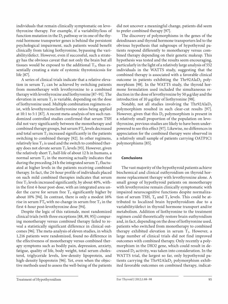
Table 1. Clinical features associated with the Thr92AlaD2 poly-morphism
Dora [55]Mental retardation Guo [56]Hypertension Gumieniak [57]Osteoarthritis Meulenbelt [58]Bipolar disorder He [59]Clinical manifestations of thyrotoxic
cardiomyopathy Grineva [60]Accelerated bone turnover Heemstra [61]Response to lung injury Barca-Mayo [51]
Ma [62]
Thr92Ala DIO2
a b
Fig. 4. 3D model of D 2 . a 3D model of the enzyme’s active center is shown including the critical selenocysteine (Sec) at position 133. b Most of the enzyme’s structure is shown including the 18-amino-acid loop that controls D 2 half-life [101] and contains the Thr92Ala polymorphism. Modified from Callebaut et al. [102] .
Bianco/Casula
Eur Thyroid J 2012;1:88–9894
ectomized individuals carrying Thr92AlaD 2 polymor-phism [65] and (ii) the finding that the Thr92AlaD 2 poly-morphism is associated with a delayed T 3 secretion in re-sponse to TRH stimulation [66] . Furthermore, all studies agree that patients with the Thr92AlaD 2 polymorphism have no alterations in all other thyroid function tests [63] .
At the same time, it is important to highlight that the literature about the Thr92AlaD 2 polymorphism is con-troversial, with poor reproducibility amongst different studies [67–72] . This suggests that additional unidenti-fied linkage factors such as ethnic background could play a significant role in the physiological and clinical rele-vance of the Thr92AlaD 2 polymorphism [71, 73] . If future studies by other groups identify and isolate such factors and confirm the observation that Thr92AlaD 2 polymor-phism limits D 2 ’s ability to produce T 3 , then ‘yes’ a vari-ability/defect in the deiodination pathway that controls tissue T 3 could be clinically relevant based on the multi-ple phenotypes associated with said polymorphism. At the time of this writing, other DIO2 polymorphisms have been reported, but their clinical relevance is even less well established [74] .
The D 3 KO mouse exhibits the richest phenotype of all deiodinase KO animals, which stems from elevated tissue T 3 during developmental and post-natal life [18, 75–77] . The D 3 KO mouse has central hypothyroidism (as a result of enhanced T 3 signaling in the hypothalamus), growth delay and a major central nervous system phenotype in-cluding problems with survival and maturation of cone photoreceptors [77] and in cochlear development and au-ditory function [78] , as well as aggressiveness and infer-tility [75, 76, 79] . However, despite the potential for mul-tiple clinical symptoms in humans, no relevant DIO3 mutations or polymorphisms or mutations have been de-scribed in humans that are relevant for the ‘second’ ques-tion.
Thyroid Hormone Transport Pathways It is clear that localized thyroid hormone deficiency in
the brain could result from a variability/defect in thyroid transport across cell membranes as well. In order for T 3 to establish a transcriptional footprint in the brain, both T 3 and T 4 need to move across the blood-brain barrier, in and out of cells. T 4 is taken up by D 2 -containing astro-cytes and tanycytes and the resulting locally generated T 3 must then exit these D 2 -containing cells and enter TR-containing neighboring neurons to finally trigger its transcriptional effects [80, 81] . A variability/defect in any of these steps could affect intracellular T 3 content and lead to localized brain hypothyroidism. This is illustrated
in patients with the Allan-Herndon-Dudley syndrome, carriers of an inactivating mutation in the X-linked MCT8 gene, a member of the MCT family that preferen-tially transports T 3 across cell membranes and is highly expressed in several organs including the brain. Patients with this syndrome exhibit psychomotor retardation and neurological impairment, indicating brain-specific hy-pothyroidism during development [82, 83] .
Other thyroid hormone transporters have been iden-tified, including the blood-brain barrier-specific anion transporter OATP1C1, mainly expressed in capillaries throughout the brain for which T 4 has a high affinity and specificity [8, 11] . In rats, OATP1C1 mRNA and protein are up- or downregulated depending on the T 4 serum lev-els, suggesting that this transporter plays a role in pre-serving physiologic concentrations of T 4 (and thus T 3 ) in the brain [84] . Interestingly, a clinical analysis of 141 hypothyroid participants of a randomized clinical trial revealed that OATP1C1 polymorphisms are associated with fatigue and depression but were not linked to neu-rocognitive dysfunction [85] .
The Third Question: Can a Deficiency in Brain
T 3 Be Restored by Treatment with Combined
Levothyroxine and Liothyronine Therapy?
There is circumstantial evidence supporting the para-digm that a variability/defect in thyroid hormone trans-port and/or metabolism could lead to insufficient T 3 in discrete brain areas of levothyroxine-treated patients ex-plaining their residual neurocognitive impairment de-spite normalization of serum TSH, T 4 and T 3 concen-trations. However, what makes this issue particularly challenging is that such a variability/defect(s) would be silenced by a functional thyroid gland, only to become clinically relevant after the onset of hypothyroidism and treatment with levothyroxine. This could be an indica-tion that the small amounts of T 3 contained in thyroid secretion would be sufficient to compensate for such a variability/defect, making it unlikely that levothyroxine alone would restore brain T 3 in such individuals. In fact, even when the transport/metabolism pathways in the rat brain are fully functional, brain T 3 did not increase (and remained fairly stable) in hypothyroid rats receiving a wide range (20-fold) of T 4 doses [36] . In contrast, brain T 3 increased progressively in hypothyroid rats treated with a much narrower range (8-fold) of T 3 doses [86] .
Thus, it is conceivable that administration of liothy-ronine could restore brain euthyroidism in hypothyroid
Treatment of Hypothyroidism Eur Thyroid J 2012;1:88–98 95
individuals that remain clinically symptomatic on levo-thyroxine therapy. For example, if a variability/loss of function mutation in the D 2 pathway or in one of the thy-roid hormone transporter genes is behind the persistent psychological impairment, such patients would benefit clinically from taking liothyronine, bypassing the vari-ability/defect. However, even if successful, such a strate-gy has the obvious caveat that not only the brain but all tissues would be exposed to the additional T 3 , thus es-sentially creating a state of systemic thyrotoxicosis for life [87] .
A series of clinical trials indicate that a relative eleva-tion in serum T 3 can be achieved by switching patients from monotherapy with levothyroxine to a combined therapy with levothyroxine and liothyronine [87–91] . The elevation in serum T 3 is variable, depending on the dose of liothyronine used. Multiple combination regimens ex-ist, with levothyroxine:liothyronine ratios being applied at 10: 1 to 5: 1 [87] . A recent meta-analysis of ten such ran-domized controlled studies confirmed that serum TSH did not vary significantly between the monotherapy and combined therapy groups, but serum FT 4 levels decreased and total serum T 3 increased significantly in the patients switching to combined therapy [92] . In other regimens, relatively less T 3 is used and the switch to combined ther-apy does not elevate serum T 3 levels [93] . However, given the relatively short T 3 half-life of about 12 h in humans, a normal serum T 3 in the morning actually indicates that during the preceding 24 h the integrated serum T 3 fluctu-ated at higher levels in the patients receiving combined therapy. In fact, the 24-hour profile of individuals placed on such mild combined therapies indicates that serum free T 3 levels increased significantly, by about 40%, with-in the first 4-hour post-dose, with an integrated area un-der the curve for serum free T 3 significantly higher by about 10% [94] . In contrast, there is only a modest 16% rise in serum FT 4 with no change in serum free T 3 in the first 4-hour post-levothyroxine dose [94] .
Despite the logic of this rationale, most randomized clinical trials (with three exceptions [88, 89, 95] ) compar-ing monotherapy versus combined therapy failed to re-veal a statistically significant difference in clinical out-comes [96] . The meta-analysis of eleven studies, in which 1,216 patients were randomized, found no difference in the effectiveness of monotherapy versus combined ther-apy symptoms such as bodily pain, depression, anxiety, fatigue, quality of life, body weight, total serum choles-terol, triglyceride levels, low-density lipoprotein, and high-density lipoprotein [96] . Yet, even when the objec-tive methods used to assess the well-being of the patients
did not uncover a meaningful change, patients did seem to prefer combined therapy [97] .
The discovery of polymorphisms in the genes of the deiodinases and thyroid hormone transporters led to the obvious hypothesis that subgroups of hypothyroid pa-tients respond differently to monotherapy versus com-bined therapy depending on their genetic makeup. This hypothesis was tested and the results seem encouraging, particularly in the light of a relatively large analysis of 552 individuals in the WATTS study, suggesting that the combined therapy is associated with a favorable clinical outcome in patients exhibiting the Thr92AlaD 2 poly-morphism [98] . In the WATTS study, the thyroid hor-mone formulation used included the simultaneous re-duction in the dose of levothyroxine by 50 � g/day and the introduction of 10 � g/day of liothyronine [98] .
Notably, not all studies involving the Thr92AlaD 2 polymorphism resulted in such clear-cut results [97] . However, given that this D 2 polymorphism is present in a relatively small proportion of the population on levo-thyroxine, previous studies are likely to have been under-powered to see this effect [97] . Likewise, no differences in appreciation for the combined therapy were observed in a relatively small sample of patients carrying OATP1C1 polymorphisms [85] .
Conclusions
The vast majority of the hypothyroid patients achieve biochemical and clinical euthyroidism on thyroid hor-mone replacement therapy with levothyroxine alone. A small group of hypothyroid patients on monotherapy with levothyroxine remain clinically symptomatic with impaired neurocognitive functions despite normaliza-tion of serum TSH, T 4 and T 3 levels. This could be at-tributed to localized brain hypothyroidism due to a variability/defect in thyroid hormone transport and/or metabolism. Addition of liothyronine to the treatment regimen could theoretically restore brain euthyroidism and, in fact, depending on the dose of liothyronine used, patients who switched from monotherapy to combined therapy exhibited elevation in serum T 3 . However, a large number of clinical trials did not find improved outcomes with combined therapy. Only recently a poly-morphism in the DIO2 gene, which could result in de-creased D 2 activity, was taken into consideration. In the WATTS trial, the largest so far, only hypothyroid pa-tients carrying the Thr92AlaD 2 polymorphism exhib-ited favorable outcomes on combined therapy, indicat-
Bianco/Casula
Eur Thyroid J 2012;1:88–9896
ing that personalized medicine is catching up with thy-roid disease. More studies are needed to further evaluate this question, but in the meantime the report of a slow-release liothyronine preparation seems to offer a more stable T 3 level over time [99, 100] , opening the doors to an appealing new approach to mildly and steadily ele-vate serum T 3 in some hypothyroid patients to offset variability/defects in thyroid hormone signaling in the brain.
Acknowledgement
The authors are grateful to Dr. Brian Kim for critically reading the manuscript and to the NIH support, DK58538, DK65055, DK77148.
Disclosure Statement
The authors have no conflicts of interest to disclose.
References
1 Aoki Y, et al: Serum TSH and total T 4 in the United States population and their associa-tion with participant characteristics: Na-tional Health and Nutrition Examination Survey (NHANES 1999–2002). Thyroid 2007; 17: 1211–1223.
2 Madhukar Aryal, et al: A prevalence of thy-roid dysfunction in Kathmandu University Hospital, Nepal. Biomed Res 2010; 21: 4.
3 Singer PA, et al: Treatment guidelines for pa-tients with hyperthyroidism and hypothy-roidism. JAMA 1995; 273: 808–812.
4 Larsen PR, Silva JE, Kaplan MM: Relation-ships between circulating and intracellular thyroid hormones: physiological and clinical implications. Endocr Rev 1981; 2: 87–102.
5 Murray GR: The life-history of the first case of myxoedema treated by thyroid extract. Br Med J 1920;i:359–360.
6 Braverman LE, Ingbar SH, Sterling K: Con-version of thyroxine (T 4 ) to triiodothyronine (T 3 ) in athyreotic subjects. J Clin Invest 1970; 49: 855–864.
7 Saravanan P, et al: Psychological well-being in patients on ‘adequate’ doses of L -thyrox-ine: results of a large, controlled community-based questionnaire study. Clin Endocrinol (Oxf) 2002; 57: 577–585.
8 Visser WE, Friesema EC, Visser TJ: Thyroid hormone transporters: the knowns and the unknowns. Mol Endocrinol 2011; 25: 1–14.
9 Wu Y, Koenig RJ: Gene regulation by thyroid hormone. Trends Endocrinol Metab 2000; 11: 207–211.
10 Zhang J, Lazar MA: The mechanism of ac-tion of thyroid hormones. Annu Rev Physiol 2000; 62: 439–466.
11 Jansen J, et al: Thyroid hormone transport-ers in health and disease. Thyroid 2005; 15: 757–768.
12 Saberi M, Sterling FH, Utiger RD: Reduction in extrathyroidal triiodothyronine produc-tion by propylthiouracil in man. J Clin Invest 1975; 55: 218–223.
13 Geffner DL, Azukizawa M, Hershman JM: Propylthiouracil blocks extrathyroidal con-version of thyroxine to triiodothyronine and augments thyrotropin secretion in man. J Clin Invest 1975; 55: 224–229.
14 Bianco AC, et al: Biochemistry, cellular and molecular biology and physiological roles of the iodothyronine selenodeiodinases. En-docr Rev 2002; 23: 38–89.
15 Ramadan W, et al: Type-2 iodothyronine 5 � -deiodinase in skeletal muscle of C57BL/6 mice. I. Identity, subcellular localization, and characterization. Endocrinology 2011; 152: 3082–3092.
16 Gereben B, et al: Cellular and molecular ba-sis of deiodinase-regulated thyroid hormone signaling. Endocr Rev 2008; 29: 898–938.
17 Galton VA: The roles of the iodothyronine deiodinases in mammalian development. Thyroid 2005; 15: 823–834.
18 Medina MC, et al: The thyroid hormone-in-activating type III deiodinase is expressed in mouse and human � -cells and its targeted inactivation impairs insulin secretion. En-docrinology 2011; 152: 3717–3727.
19 Peeters RP, et al: Reduced activation and in-creased inactivation of thyroid hormone in tissues of critically ill patients. J Clin Endo-crinol Metab 2003; 88: 3202–3211.
20 Huang SA, Bianco AC: Reawakened interest in type III iodothyronine deiodinase in crit-ical illness and injury. Nat Clin Pract Endo-crinol Metab 2008; 4: 148–155.
21 Braverman LE, et al: Effects of replacement doses of sodium L -thyroxine on the periph-eral metabolism of thyroxine and triiodo-thyronine in man. J Clin Invest 1973; 52: 1010–1017.
22 Stock JM, Surks MI, Oppenheimer JH: Re-placement dosage of L -thyroxine in hypothy-roidism. A re-evaluation. N Engl J Med 1974; 290: 529–533.
23 Jonklaas J, et al: Triiodothyronine levels in athyreotic individuals during levothyroxine therapy. JAMA 2008; 299: 769–777.
24 Gullo D, et al: Levothyroxine monotherapy cannot guarantee euthyroidism in all athy-reotic patients. PLoS One 2011; 6:e22552.
25 Oppenheimer JH, Schwartz HL, Surks MI: Determination of common parameters of io-dothyronine metabolism and distribution in man by noncompartmental analysis. J Clin Endocrinol Metab 1975; 41: 319–324.
26 Baqui MM, et al: Distinct subcellular local-ization of transiently expressed types 1 and 2 iodothyronine deiodinases as determined by immunofluorescence confocal microscopy. Endocrinology 2000; 141: 4309–4312.
27 Friesema EC, et al: Thyroid hormone trans-port by the human monocarboxylate trans-porter 8 and its rate-limiting role in intracel-lular metabolism. Mol Endocrinol 2006; 20: 2761–2772.
28 Schneider MJ, et al: Targeted disruption of the type 2 selenodeiodinase gene (DIO2) re-sults in a phenotype of pituitary resistance to T 4 . Mol Endocrinol 2001; 15: 2137–2148.
29 Hall JA, Bianco AC: Triumphs of the thyroid despite lesser conversion. Endocrinology 2009; 150: 2502–2504.
30 Christoffolete MA, et al: Mice with impaired extrathyroidal thyroxine to 3,5,3 � -triio-dothyronine conversion maintain normal serum 3,5,3 � -triiodothyronine concentra-tions. Endocrinology 2007; 148: 954–960.
31 Galton VA, et al: Life without T 4 to T 3 conver-sion: studies in mice devoid of the 5 � -deiodinases. Endocrinology 2009; 150: 2957–2963.
32 Bianco AC, Kim BW: Deiodinases: implica-tions of the local control of thyroid hormone action. J Clin Investigation 2006; 116: 2571–2579.
33 Campos-Barros A, et al: Phenolic and tyrosyl ring iodothyronine deiodination and thy-roid hormone concentrations in the human central nervous system. J Clin Endocrinol Metab 1996; 81: 2179–2185.
34 Peeters RP, et al: Tissue thyroid hormone lev-els in critical illness. J Clin Endocrinol Metab 2005; 90: 6498–6507.
35 Kester MH, et al: Iodothyronine levels in the human developing brain: major regulatory roles of iodothyronine deiodinases in differ-ent areas. J Clin Endocrinol Metab 2004; 89: 3117–3128.
36 Escobar-Morreale HF, et al: Replacement therapy for hypothyroidism with thyroxine alone does not ensure euthyroidism in all tis-sues, as studied in thyroidectomized rats. J Clin Invest 1995; 96: 2828–2838.
Treatment of Hypothyroidism Eur Thyroid J 2012;1:88–98 97
37 Andersen S, et al: Biologic variation is im-portant for interpretation of thyroid func-tion tests. Thyroid 2003; 13: 1069–1078.
38 Bianco AC, Silva JE: Cold exposure rapidly induces virtual saturation of brown adipose tissue nuclear T 3 receptors. Am J Physiol 1988; 255:E496–E503.
39 Dumitrescu AM, et al: Mutations in SECISBP2 result in abnormal thyroid hor-mone metabolism. Nat Gen 2005; 37: 1247–1252.
40 Berry MJ, et al: Physiological and genetic analyses of inbred mouse strains with a type I iodothyronine 5 � -deiodinase deficiency. J Clin Invest 1993; 92: 1517–1528.
41 Schneider MJ, et al: Targeted disruption of the type 1 selenodeiodinase gene (Dio1) re-sults in marked changes in thyroid hormone economy in mice. Endocrinology 2006; 147: 580–589.
42 Dayan CM, Panicker V: Novel insights into thyroid hormones from the study of com-mon genetic variation. Nat Rev Endocrinol 2009; 5: 211–218.
43 De Jong FJ, et al: The association of polymor-phisms in the type 1 and 2 deiodinase genes with circulating thyroid hormone parame-ters and atrophy of the medial temporal lobe. J Clin Endocrinol Metab 2007; 92: 636–640.
44 De Jesus LA, et al: The type 2 iodothyronine deiodinase is essential for adaptive thermo-genesis in brown adipose tissue. J Clin Invest 2001; 108: 1379–1385.
45 Christoffolete MA, et al: Mice with targeted disruption of the DIO2 gene have cold-in-duced overexpression of the uncoupling pro-tein 1 gene but fail to increase brown adipose tissue lipogenesis and adaptive thermogen-esis. Diabetes 2004; 53: 577–584.
46 Hall JA, et al: Absence of thyroid hormone activation during development underlies a permanent defect in adaptive thermogene-sis. Endocrinology 2010; 151: 4573–4582.
47 Ng L, et al: Hearing loss and retarded cochle-ar development in mice lacking type 2 iodo-thyronine deiodinase. Proc Natl Acad Sci USA 2004; 101: 3474–3479.
48 Bassett JH, et al: Optimal bone strength and mineralization requires the type 2 iodothy-ronine deiodinase in osteoblasts. Proc Natl Acad Sci USA 2010; 107: 7604–7609.
49 Marsili A, et al: Type II iodothyronine deio-dinase provides intracellular 3,5,3 � -triio-dothyronine to normal and regenerating mouse skeletal muscle. Am J Physiol Endo-crinol Metab 2011; 301:E818–E824.
50 Dentice M, et al: The FoxO3/type 2 deiodin-ase pathway is required for normal mouse myogenesis and muscle regeneration. J Clin Invest 2011; 120: 4021–4030.
51 Barca-Mayo O, et al: Role of type 2 deiodin-ase in response to acute lung injury in mice. Proc Natl Acad Sci USA 2011; 108:E1321–E1329.
52 Crantz FR, Silva JE, Larsen PR: Analysis of the sources and quantity of 3,5,3 � -triio-dothyronine specifically bound to nuclear receptors in rat cerebral cortex and cerebel-lum. Endocrinology 1982; 110: 367–375.
53 Mentuccia D, et al: Association between a novel variant of the human type 2 deiodinase gene Thr92Ala and insulin resistance: evi-dence of interaction with the Trp64Arg vari-ant of the � 3 -adrenergic receptor. Diabetes 2002; 51: 880–883.
54 Canani LH, et al: The type 2 deiodinase A/G (Thr92Ala) polymorphism is associated with decreased enzyme velocity and in-creased insulin resistance in patients with type 2 diabetes mellitus. J Clin Endocrinol Metab 2005; 90: 3472–3478.
55 Dora JM, et al: Association of the type 2 de-iodinase Thr92Ala polymorphism with type 2 diabetes: case-control study and meta-analysis. Eur J Endocrinol 2010; 163: 427–434.
56 Guo TW, et al: Positive association of the DIO2 (deiodinase type 2) gene with mental retardation in the iodine-deficient areas of China. J Med Genet 2004; 41: 585–590.
57 Gumieniak O, et al: Ala92 type 2 deiodinase allele increases risk for the development of hypertension. Hypertension 2007; 49: 461–466.
58 Meulenbelt I, et al: Identification of DIO2 as new susceptibility locus for symptomatic Osteoarthritis. Hum Mol Genet 2008; 17: 1867–1875.
59 He B, et al: Association of genetic polymor-phisms in the type II deiodinase gene with bipolar disorder in a subset of Chinese popu-lation. Prog Neuropsychopharmacol Biol Psychiatry 2009; 33: 986–990.
60 Grineva E, et al: Type 2 deiodinase Thr92A-la polymorphism impact on clinical course and myocardial remodeling in patients with Graves’ disease. Cell Cycle 2009; 8: 2565–2569.
61 Heemstra KA, et al: The type 2 deiodinase Thr92Ala polymorphism is associated with increased bone turnover and decreased fem-oral neck bone mineral density. J Bone Min-er Res 2010; 25: 1385–1391.
62 Ma SF, et al: Type 2 deiodinase and host re-sponses of sepsis and acute lung injury. Am J Respir Cell Mol Biol 2011; 45: 1203–1211.
63 Peeters RP, et al: Polymorphisms in thyroid hormone pathway genes are associated with plasma TSH and iodothyronine levels in healthy subjects. J Clin Endocrinol Metab 2003; 88: 2880–2888.
64 Larsen PR: Type 2 iodothyronine deiodinase in human skeletal muscle: new insights into its physiological role and regulation. J Clin Endocrinol Metab 2009; 94: 1893–1895.
65 Torlontano M, et al: Type 2 deiodinase poly-morphism (Thr92Ala) predicts L -thyroxine dose to achieve target TSH levels in thyroid-ectomized patients. J Clin Endocrinol Metab 2008; 93: 910–913.
66 Butler PW, et al: The Thr92Ala 5 � -type 2 de-iodinase gene polymorphism is associated with a delayed triiodothyronine secretion in response to the thyrotropin-releasing hor-mone-stimulation test: a pharmacogenomic study. Thyroid 2011; 20: 1407–1412.
67 Canani LH, et al: Type 2 deiodinase Thr92A-la polymorphism is not associated with arte-rial hypertension in type 2 diabetes mellitus patients. Hypertension 2007; 49:e47–e48.
68 Grarup N, et al: Studies of the common DIO2 Thr92Ala polymorphism and metabolic phenotypes in 7342 Danish white subjects. J Clin Endocrinol Metab 2007; 92: 363–366.
69 Gumieniak O, Williams GH: Response to type 2 deiodinase Thr92Ala polymorphism is not associated with arterial hypertension in type 2 diabetes mellitus patients. Hyper-tension 2007; 49:e47–e48.
70 Heemstra KA, et al: Thr92Ala polymor-phism in the type 2 deiodinase is not associ-ated with T 4 dose in athyroid patients or pa-tients with Hashimoto thyroiditis. Clin En-docrinol (Oxf) 2009; 71: 279–283.
71 Nair S, et al: Association analyses of variants in the DIO2 gene with early-onset type 2 di-abetes mellitus in Pima Indians. Thyroid 2012; 22: 80–87.
72 Van der Deure WM, et al: Impact of thyroid function and polymorphisms in the type 2 deiodinase on blood pressure: the Rotterdam Study and the Rotterdam Scan Study. Clin Endocrinol (Oxf) 2009; 71: 137–144.
73 Mentuccia D, et al: The Thr92Ala deiodinase type 2 (DIO2) variant is not associated with type 2 diabetes or indices of insulin resis-tance in the old order of Amish. Thyroid 2005; 15: 1223–1227.
74 Peeters RP, van der Deure WM, Visser TJ: Genetic variation in thyroid hormone path-way genes; polymorphisms in the TSH re-ceptor and the iodothyronine deiodinases. Eur J Endocrinol 2006; 155: 655–662.
75 Hernandez A, et al: Type 3 deiodinase is crit-ical for the maturation and function of the thyroid axis. J Clin Invest 2006; 116: 476–484.
76 Hernandez A, et al: Type 3 deiodinase defi-ciency results in functional abnormalities at multiple levels of the thyroid axis. Endocri-nology 2007; 148: 5680–5687.
77 Ng L, et al: Type 3 deiodinase, a thyroid-hor-mone-inactivating enzyme, controls surviv-al and maturation of cone photoreceptors. J Neurosci 2010; 30: 3347–3357.
78 Ng L, et al: A protective role for type 3 deio-dinase, a thyroid hormone-inactivating en-zyme, in cochlear development and auditory function. Endocrinology 2009; 150: 1952–1960.
79 Hernandez A, et al: Type 3 deiodinase defi-ciency causes spatial and temporal altera-tions in brain T 3 signaling that are dissoci-ated from serum thyroid hormone levels. En-docrinology 2010; 151: 5550–5558.
Bianco/Casula
Eur Thyroid J 2012;1:88–9898
80 Freitas BC, et al: Paracrine signaling by glial cell-derived triiodothyronine activates neu-ronal gene expression in the rodent brain and human cells. J Clin Invest 2010; 120: 2206–2217.
81 Mohacsik P, et al: Thyroid hormone and the neuroglia: both source and target. J Thyroid Res 2011; 2011: 215718.
82 Dumitrescu AM, et al: A novel syndrome combining thyroid and neurological abnor-malities is associated with mutations in a monocarboxylate transporter gene. Am J Hum Genet 2004; 74: 168–175.
83 Friesema EC, et al: Association between mu-tations in a thyroid hormone transporter and severe X-linked psychomotor retardation. Lancet 2004; 364: 1435–1437.
84 Sugiyama D, et al: Functional characteriza-tion of rat brain-specific organic anion transporter (Oatp14) at the blood-brain bar-rier: high affinity transporter for thyroxine. J Biol Chem 2003; 278: 43489–43495.
85 Van der Deure W, Appelhof BC, Peeters RP, Wiersinga WM, Wekkig EM, Huyser J, Schene AH, Tijssen JGP, Hoogendijk WJG, Visser TJ, Fliers E: Polymorphism in the brain-specific thyroid hormone transporter OATP-C1 are associated with fatigue and de-pression in hypothyroid patients. Clin Endo-crinol 2008; 69: 8.
86 Escobar-Morreale HF, et al: Regulation of io-dothyronine deiodinase activity as studied in thyroidectomized rats infused with thy-roxine or triiodothyronine. Endocrinology 1997; 138: 2559–2568.
87 Appelhof BC, et al: Combined therapy with levothyroxine and liothyronine in two ra-tios, compared with levothyroxine mono-therapy in primary hypothyroidism: a dou-ble-blind, randomized, controlled clinical trial. J Clin Endocrinol Metab 2005; 90: 2666–2674.
88 Bunevicius R, et al: Thyroxine vs. thyroxine plus triiodothyronine in treatment of hypo-thyroidism after thyroidectomy for Graves’ disease. Endocrine 2002; 18: 129–133.
89 Bunevicius R, et al: Effects of thyroxine as compared with thyroxine plus triiodothyro-nine in patients with hypothyroidism. N Engl J Med 1999; 340: 424–429.
90 Valizadeh M, et al: Efficacy of combined le-vothyroxine and liothyronine as compared with levothyroxine monotherapy in primary hypothyroidism: a randomized controlled trial. Endocr Res 2009; 34: 80–89.
91 Rodriguez T, et al: Substitution of liothyro-nine at a 1: 5 ratio for a portion of levothyrox-ine: effect on fatigue, symptoms of depres-sion, and working memory versus treatment with levothyroxine alone. Endocr Pract 2005; 11: 223–233.
92 Ma C, et al: Thyroxine alone or thyroxine plus triiodothyronine replacement therapy for hypothyroidism. Nucl Med Commun 2009; 30: 586–593.
93 Saravanan P, et al: Partial substitution of thyroxine (T 4 ) with tri-iodothyronine in pa-tients on T 4 replacement therapy: results of a large community-based randomized con-trolled trial. J Clin Endocrinol Metab 2005; 90: 805–812.
94 Saravanan P, et al: Twenty-four hour hor-mone profiles of TSH, free T 3 and free T 4 in hypothyroid patients on combined T 3 /T 4 therapy. Exp Clin Endocrinol Diabetes 2007; 115: 261–267.
95 Nygaard B, et al: Effect of combination therapy with thyroxine (T 4 ) and 3,5,3 � -triio dothyronine versus T 4 monotherapy in patients with hypothyroidism, a double-blind, randomised cross-over study. Eur J Endocrinol 2009; 161: 895–902.
96 Grozinsky-Glasberg S, et al: Thyroxine-tri-iodothyronine combination therapy versus thyroxine monotherapy for clinical hypo-thyroidism: meta-analysis of randomized controlled trials. J Clin Endocrinol Metab 2006; 91: 2592–2599.
97 Appelhof BC, et al: Polymorphisms in type 2 deiodinase are not associated with well-being, neurocognitive functioning, and preference for combined thyroxine/3,5,3 � -triio dothyronine therapy. J Clin Endocri-nol Metab 2005; 90: 6296–6299.
98 Panicker V, et al: Common variation in the DIO2 gene predicts baseline psychological well-being and response to combination thyroxine plus triiodothyronine therapy in hypothyroid patients. J Clin Endocrinol Metab 2009; 94: 1623–1629.
99 Hennemann G, et al: Thyroxine plus low-dose, slow-release triiodothyronine re-placement in hypothyroidism: proof of principle. Thyroid 2004; 14: 271–275.
100 Frumess RD, Larsen PR: Correlation of se-rum triiodothyronine (T 3 ) and thyroxine (T 4 ) with biologic effects of thyroid hor-mone replacement in propylthiouracil-treated rats. Metabolism 1975; 24: 547–554.
101 Dentice M, et al: The Hedgehog-inducible ubiquitin ligase subunit WSB-1 modulates thyroid hormone activation and PTHrP se-cretion in the developing growth plate. Nat Cell Biol 2005; 7: 698–705.
102 Callebaut I, et al: The iodothyronine sele-nodeiodinases are thioredoxin-fold family proteins containing a glycoside hydrolase clan GH-A-like structure. J Biol Chem 2003; 278: 36887–36896.