Page 1
Title Glutathione peroxidase 2 in Saccharomyces cerevisiae isdistributed in mitochondria and involved in sporulation
Author(s) Ukai, Yuuta; Kishimoto, Tomoyuki; Ohdate, Takumi; Izawa,Singo; Inoue, Yoshiharu
Citation Biochemical and Biophysical Research Communications(2011), 411(3): 580-585
Issue Date 2011-08
URL http://hdl.handle.net/2433/145971
Right © 2011 Elsevier Inc.
Type Journal Article
Textversion author
Kyoto University
Page 2
1
Running title: Localization of Gpx2 in yeast mitochondria
Glutathione Peroxidase 2 in Saccharomyces cerevisiae Is Distributed in Mitochondria
and Involved in Sporulation
Yuuta Ukai, Tomoyuki Kishimoto, Takumi Ohdate1, Singo Izawa2, and Yoshiharu Inoue*
Laboratory of Molecular Microbiology, Division of Applied Life Sciences, Graduate School of
Agriculture, Kyoto University, Uji, Kyoto 611-0011, Japan
1 Present address: Department of Microbiology, Tohoku Pharmaceutical University, Sendai,
Japan.
2 Present address: Laboratory of Microbial Technology, Graduate School of Science and
Technology, Kyoto Institute of Technology, Kyoto, Japan.
*Corresponding author. Tel. +81 774-38-3773. Fax. +81 774-38-3789. E-mail address:
[email protected] (Y. Inoue).
Page 3
2
Abstract
Gpx2, one of three glutathione peroxidase homologues (Gpx1, Gpx2, and Gpx3) in
Saccharomyces cerevisiae, is an atypical 2-Cys peroxiredoxin that prefers to use thioredoxin as
a reducing agent in vitro. Despite Gpx2 being an antioxidant, no obvious phenotype of gpx2∆
mutant cells in terms of oxidative stress has yet been found. To gain a clue as to Gpx2’s
physiological function in vivo, here we identify its intracellular distribution. Gpx2 was found to
exist in the cytoplasm and mitochondria. In mitochondria, Gpx2 was associated with the outer
membrane of the cytoplasmic-side, as well as the inner membrane of the matrix-side. The redox
state of the mitochondrial Gpx2 was regulated by Trx1 and Trx2 (cytoplasmic thioredoxin), and
by Trx3 (mitochondrial matrix thioredoxin). In addition, we found that the disruption of GPX2
reduced the sporulation efficiency of diploid cells.
Key words: glutathione peroxidase, Saccharomyces cerevisiae, peroxiredoxin, mitochondria,
sporulation
1. Introduction
All organisms take up organic compounds from the environment, and derive electrons
from such compounds to regenerate reductive coenzymes for energy production. In particular,
aerobic organisms use molecular oxygen (O2) as the final acceptor of electrons at the final step
in the electron transfer system for ATP production. O2 accepts four electrons one by one, and
consequently, several intermediates are generated. O2 per se is a biradical, but it is not as
Page 4
3
reactive as the intermediates generated during the reduction of O2, which in turn are referred to
as reactive oxygen species. Hence, for all aerobic organisms, it is unavoidable that reactive
oxygen species are generated during energy production. Organisms of all types are armed with
several antioxidant systems. For example, one-electron-reduction of O2 yields superoxide anion
radicals (O2•—), which are disproportionated to O2 and hydrogen peroxide (H2O2) by superoxide
dismutase. The H2O2 thus formed is decomposed to O2 and H2O by catalase, or reduced to H2O
by peroxidases with different electron donors. Generally, peroxidases are named according to
the electron donors that they use, e. g. ascorbate peroxidase, glutathione peroxidase, and
thioredoxin peroxidase. We have previously reported that the budding yeast Saccharomyces
cerevisiae has three glutathione peroxidase (GPx) homologues (Gpx1, Gpx2, and Gpx3) [1].
Peroxiredoxins (Prxs) are ubiquitous antioxidants in all types of organisms [2], which are
characterized by certain common properties in that they do not contain a redox active cofactor
or prosthetic group, and contain conserved Cys residue(s). According to the number of
conserved Cys residues, Prxs are divided into two major groups, i.e. 1-Cys Prx and 2-Cys Prx.
In both Prxs, the N-terminal Cys, which is sometimes referred to as the peroxidatic Cys, is
conserved. In 2-Cys Prx, another Cys residue that is referred to as the resolving Cys is
conserved, and two intermolecular disulfide bonds are formed between the peroxidatic Cys and
resolving Cys as a consequence of the reduction of peroxides, and subsequently a Prx dimer is
formed. Meanwhile, in a family of atypical 2-Cys Prxs, an intramolecular disulfide bond is
formed within the same molecule. We have previously reported that Gpx1 and Gpx2 are
atypical 2-Cys peroxiredoxins using both glutathione and thioredoxin as regucing agents [3,4],
while Delaunay et al. [5] have reported that Gpx3 shows thioredoxin peroxidase activity in
Page 5
4
vitro.
Since the expression of GPX3 is constitutive, and besides being an antioxidant, Gpx3
functions as a redox sensor and transducer of the oxidative stress-responsive transcription factor
Yap1 [5], gpx3∆ mutant cells exhibit a clear phenotype in terms of the loss of the antioxidant
[1]. Meanwhile, even though Gpx1 and Gpx2 show peroxidase activity in vitro [3,4], neither
gpx1∆ nor gpx2∆ cells demonstrate any obvious phenotypes in terms of oxidative stress [1]. To
gain a clue as to Gpx2’s physiological function in vivo, we determined its intracellular
localization. Here we show that Gpx2 is distributed in both the cytoplasm and mitochondria. We
explored the roles of Gpx2 with respect to mitochondria, and found that Gpx2 is involved in the
process of sporulation, i. e. the loss of Gpx2 decreased the sporulation efficiency of diploid
cells.
2. Materials and methods
2.1. Strains and media
The S. cerevisiae strains used in this study are summarized in Supplemental Table1.
Cells were grown in YPD medium (2% glucose, 1% yeast extract, and 2% peptone), or
synthetic dextrose (SD) medium (2% glucose, 0.67% yeast nitrogen base without amino acid)
with appropriate amino acids and bases at 28ºC. For peroxide treatment, 0.4 mM H2O2 was
added to the culture, in which logarithmically growing cells are involved, and incubated for 60
min.
2.2. Gene disruption
Page 6
5
To construct a gpx2Δ::his5+ strain in BY4741 (MATa his3-∆1 leu2-∆0 met15-∆0
ura3-∆0), pUG27 [6] was digested with BglII and SacI, and the resultant fragment containing
his5+ was replaced with KanMX4 at the gpx2Δ::KanMX4 locus of BY4741 (Invitrogen). To
construct a gpx2Δ::his5+ strain in YPH252, the allele of gpx2Δ::his5+ in BY4741 was amplified
by PCR with the following primers : GPX2-1, 5’-TTACCGTTGTCGACCTTGCTCTAC-3’,
and GPX2RA, 5’-TCAGAAGCAGGACACCTGTAGAGCTAGCCA-3’. The PCR fragment
was introduced into YPH252. To construct gpx2∆::TRP1, pUGI4 [1] was digested with Tth111I
followed by Klenow treatment, and the TRP1 gene was ligated (pUC19-gpx2∆::TRP1). The
gpx2∆::TRP1 fragment was amplified by PCR using the primers GPX2-FT
(5’-TTAATGTTGCCTCCAAGTGC-3’) and GPX2-RT (5’-TTGGTCCAAGGACGATGG-3’)
with pUC19-gpx2∆::TRP1 as a template DNA. The PCR fragment was introduced into YPH250
to disrupt GPX2.
FMP45 was amplified by PCR with Fmp45-F,
5’-GAAAGAGGAATTCAGGTTTAACGCTT-3’, and Fmp45-R,
5’-TTGGAGGTAGAAGCTTAGTTATGGTAC-3’. Fmp45-F and Fmp45-R are designed with
EcoRI and HindIII sites, respectively (underlined). The resultant PCR fragment was cloned into
the EcoRI-HindIII sites of pUC19 to yield pUC-FMP45. To disrupt the FMP45 gene, the
following two plasmids were constructed: pUC-FMP45 was digested with Eco47III and HincII
to delete a part of the open reading frame of FMP45, and URA3 or LEU2 was inserted to yield
pUC19-fmp45∆::URA3 and pUC19-fmp45∆::URA3, respectively. To construct fmp45∆::URA3
and fmp45∆::LEU2 strains, PCR was performed with primers Fmp45-F and Fmp45-R using
pUC19-fmp45∆::URA3 or pUC19-fmp45∆::LEU2 as a template, and the amplified fragment
Page 7
6
was introduced into YPH250 and YPH252, respectively.
2.3. Western blotting
After SDS-polyacrylamide gel electrophoresis (SDS-PAGE), separated proteins were
transferred to a PVDF membrane (Immobilon, Millipore). To detect proteins of interest,
antibodies for each protein [anti-Gpx2 [4], anti-Por1 (Cell Signaling), anti-Dpm1 (Cell
Signaling), anti-Pgk1 (Cell Signaling), anti-Tom70 [7], anti-Tim50 [7], and anti-Hsp60 [7]
antibodies] were used. Anti-rabbit IgG conjugated with alkaline phosphatase (Cell Signaling)
was used as the secondary antibody, and the immunoreacted bands were visualized with a
BCIP-NBT kit (Nacalai tesque).
2.4. Subcellular fractionation
The subcellular fractionation experiment, and sucrose density gradient
ultracentrifugation was done essentially as described by Meisinger et al. [8]. Details as to the
experimental conditions are described in the Supplemental Materials and methods.
2.5. Determination of submitochondrial localization of Gpx2
Treatment of mitochondria with proteinase K, NaCl, sonication, and Na2CO3 to
determine the localization of proteins was done essentially as described by Yamamoto et al. [9].
Details as to the experimental conditions are described in the Supplemental Materials and
methods.
Page 8
7
2.6. Determination of the redox states of mitochondrial Gpx2
The modification of Cys residues of Gpx2 was performed basically as described by
Tanaka et al. [4]. The crude mitochondria (200 µg protein) in 200 µl of SEM [250 mM sucrose,
1 mM EDTA, and 10 mM MOPS (pH 7.2)] buffer were mixed with 50 µl of 100% TCA, the
mixture was centrifuged at 13000 x g for 10 min at 4ºC, and the precipitates were washed once
with acetone. After removal of the acetone, the dried materials were suspended with 40 µ l of
sample buffer [80 mM Tris-HCl (pH 6.8), 2% SDS, 6 M urea, 1 mM PMSF, and 0.05%
bromophenol blue] containing 20 mM 4’-acetamido-4’-maleimidylstilbene-2, 2’-disulfonic acid
(AMS) (Molecular Probes). A small portion (approximately 4µl) of 1 M Tris-HCl (pH 8.0) was
added to adjust the pH of the sample, and the mixture was boiled for 2 min. Twenty microliters
of the sample was subjected to non-reducing SDS-PAGE followed by Western blotting using
anti-Gpx2 antibody to determine the redox state of Gpx2 in mitochondria.
2.7. Determination of sporulation efficiency
Diploid cells grown for 1 day on YPD agar plates were suspended in a 0.85% NaCl
solution, and diluted with a 0.85% NaCl solution to bring the A610 to 0.1. The cell suspension
was spotted in quintuplicate (5 µl each) onto plates containing sporulation medium (1%
potassium acetate, 0.1% yeast extract, 0.05% glucose, 2% agar, and appropriate amino acids),
and incubated at 25ºC. On each day of incubation, all the cells of one of five spots were
recovered, and suspended in the 0.85% NaCl solution. The numbers of sporulated cells forming
spores were counted using a microscope.
Page 9
8
3. Results
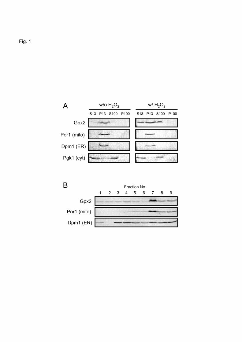
3.1. Gpx2 is distributed in both mitochondria and cytoplasm
Though Gpx2 is an atypical 2-Cys Prx that prefers to use thioredoxin as a reducing
agent in vitro [4], gpx2∆ cells do not exhibit any obvious phenotype regarding peroxide
sensitivity. To gain insights into its function in vivo, we tried to determine the subcellular
localization of Gpx2. We found that Gpx2 was present in not only soluble fractions but also
insoluble fractions, so it is feasible that Gpx2 exists in some organelles. To address this
possibility, we fractionated cell homogenates by a subsequent centrifugation, and found that
Gpx2 was predominantly located in the crude mitochondrial fractions (P13) and in the
cytoplasmic fractions (S13) (Fig. 1A). In H2O2-treated cells, the overall Gpx2 level was
increased as reported previously [1], and Gpx2 was detected in both the crude mitochondria and
cytoplasm (Fig. 1A).
In the preparation of mitochondria by centrifugation, the ER contaminates the crude
mitochondrial fractions. As shown in Fig. 1A, both Por1 (mitochondrial marker protein) and
Dpm1 (ER marker protein) were detected in the crude mitochondrial fractions (P13). Therefore,
to eliminate the ER, these samples were subjected to a three-step sucrose density gradient
ultracentrifugation. As shown in Fig. 1B, the pattern of fractionation of Gpx2 coincided with
that of Por1 but not Dpm1. Therefore, Gpx2 is likely to be located in both mitochondria and the
cytoplasm in yeast cells.
3.2. Submitochondrial localization of Gpx2
Next, we determined the submitochondrial localization of Gpx2. Mitochondria were
Page 10
9
treated with proteinase K (PK). The principle of this experiment is as follows: in whole
mitochondria, PK can digest only proteins associated with the surface of mitochondria; in
mitoplasts, PK can also digest proteins in the intermembrane space and those associated with
the surface of the inner membrane facing the intermembrane space; and in solubilized
mitoplasts, PK can also digest proteins in the matrix. As shown in Fig. 2A, when whole
mitochondria were treated with PK, Tom70, an outer membrane-integrated protein, was
digested completely, because a large part of Tom70 is exposed at the surface of the
mitochondrial outer membrane. However, approximately half of the mitochondrial Gpx2 was
not digested under the same conditions. These results imply that some Gpx2s seem to exist on
the surface of the outer membrane of mitochondria, but the rest are likely to be sequestered in
mitochondria where PK cannot reach. Next, when mitoplasts were treated with PK, Gpx2 still
remains within the mitoplasts, which implies that Gpx2 exists neither in the intermembrane
space nor on the surface of the inner membrane facing the intermembrane space. In the PK
treatment of solubilized mitoplasts, the rest of the Gpx2s were digested, a pattern which
resembles Hsp60, a matrix marker protein. Therefore, Gpx2 seems to exist in the matrix also.
3.3. Gpx2 is associated with the surface of mitochondrial membranes via ionic interaction
To further explore the mode of existence of Gpx2 in the submitochondrial
compartment, we treated mitochondria with NaCl, sonication, and Na2CO3. As shown in Fig. 2B,
some Gpx2 was released from whole mitochondria to the supernatant fractions after treatment
with 0.5 M NaCl for 60 min. However, Tom70 was not released from whole mitochondria
under the same conditions, because this protein is integrated in the outer mitochondrial
Page 11
10
membrane. This result indicates that Gpx2 is not a membrane-integrated protein, but associated
with the outer membrane through the ionic interaction.
Sonication disrupts the mitochondrial membrane. After sonication, the disrupted
mitochondria were centrifuged. As a result, Gpx2 was mainly detected in the precipitates (Fig.
2B). This behavior in terms of protein localization resembles that of Tom70 and Tim50,
integrated in the outer and inner membrane of mitochondria, respectively; but is different from
that of Hsp60, a soluble protein in the matrix. This result also indicates that Gpx2 is a
membrane-associated protein.
Na2CO3 also destroys mitochondria. After centrifugation of the NO2CO3-treated
mitochondria, Gpx2 was mainly detected in the supernatants. This behavior resembles that of
Tam41 (data not shown), a peripheral membrane protein [10], but is different from that of
Tom70 and Tim50, membrane-integrated proteins (Fig. 2B). This result indicates that Gpx2 is a
peripheral membrane protein not a membrane-integrated protein. Taken together, Gpx2 is a
peripheral protein positioned on the outer membrane of mitochondria facing the cytoplasm via
ionic interaction, as well as on the inner membrane facing the matrix.
3.4. Redox regulation of mitochondrial Gpx2
Previously, we have shown that the redox state of the bulk cellular Gpx2 is
predominantly regulated by Trx1 and Trx2 (cytoplasmic thioredoxin) [4]. Here, we focused on
the redox regulation of mitochondrial Gpx2. The redox state of mitochondrial Gpx2 was
determined by the AMS modification assay. As shown in Fig. 3, approximately half of the
mitochondrial Gpx2 was in the oxidized form when mitochondria were isolated from wild-type
Page 12
11
cells without treatment with H2O2. Previously, we have reported that the bulk cellular Gpx2 was
essentially in the reduced form in wild-type cells untreated with peroxides [4]. The occurrence
of oxidized Gpx2 in mitochondria prepared from H2O2-untreated cells is presumably due to
oxidation during the preparation. Nonetheless, the proportion of oxidized Gpx2 in mitochondria
prepared from trx1Δtrx2Δ cells was higher than that of wild-type cells, which indicates that
Trx1 and Trx2 have roles in the redox regulation of mitochondrial Gpx2 in cells untreated with
H2O2. Furthermore, the proportion of oxidized Gpx2 in mitochondria was greatly increased
when mitochondria were isolated from trx3Δ cells as well as trx1Δtrx2Δ cells treated with H2O2.
Considering the localization of thioredoxin, i. e. Trx1 and Trx2 in the cytoplasm and Trx3 in the
mitochondrial matrix, Trx1 and Trx2 may reduce Gpx2 associated with the outer membrane of
mitochondria, while Trx3 may reduce that associated with the surface of the inner membrane of
the matrix-side. It should be noted that the level of Gpx2 is high in trx1∆trx2∆ cells untreated
with H2O2, because thioredoxin (Trx1 and Trx2) is a negative regulator of Yap1 [11], a
transcriptional activator of GPX2 [1,12]; and therefore, Yap1 is constitutively activated in the
trx1∆trx2∆ mutant, thereby enhancing GPX2 expression.
3.5. Effects of GPX2’s disruption on sporulation
Comprehensive analyses of the physical interactions of yeast proteins have been
conducted using different analytical methods. It has been reported that Gpx2 physically interacts
with several proteins. Since we have demonstrated that Gpx2 is distributed in mitochondria, we
focused on Fmp45, a mitochondrial membrane-integral protein. Tarassov et al. [13] revealed the
physical interaction between Gpx2 and Fmp45 in vivo with a protein-fragment complementation
Page 13
12
assay. It has been reported that a deficiency in Fmp45 reduces sporulation efficiency [14]. We
confirmed that the sporulation efficiency of fmp45∆/fmp45∆ diploid cells was reduced (Fig. 4).
So, we determined the effect of the loss of Gpx2 on the sporulation efficiency of diploid cells.
As shown in Fig. 4, the sporulation efficiency of gpx2Δ/gpx2Δ cells was decreased compared
with that of GPX2/GPX2 cells, whose rate was comparable to that of fmp45∆/fmp45∆ cells.
Next, we determined whether any correlation between Gpx2 and Fmp45 exists in terms of the
reduction in sporulation efficiency. The sporulation efficiency of the
fmp45∆gpx2∆/fmp45∆gpx2∆ mutant did not decrease further compared with that of the
fmp45∆/fmp45∆ cells (Fig. 4).
4. Discussion
The cellular localization of a protein provides a powerful hint as to its function.
Previously, we have tried to determine the subcellular distrubution of Gpx2 using a GFP-tagged
Gpx2, and found that the fluorescence derived from Gpx2-GFP was observed as punctate foci
scattered within the cell (Tsuzi, D., Maeta, K., Izawa, S., and Inoue, Y., unpublished results);
however, direct evidence that Gpx2 exists in distinctive organelles has not been obtained.
Additionally, since the gpx2∆ mutant did not exhibit obvious phenotypes, it is difficult to verify
whether GFP-tagged Gpx2 is correctly distributed in the cell and functioning properly through
complementation of the mutant’s phenotype. Here, we adopted biochemical approaches to
determine the localization of Gpx2.
We have demonstrated that Gpx2 is present in both the cytoplasm and mitochondria.
The mitochondrial Gpx2 shows a characteristic distribution, i. e. some is associated with the
Page 14
13
surface of the outer membrane on the cytoplasmic-side, while the rest is associated with the
surface of the inner membrane on the matrix-side. Considering the results of biochemical
analyses, Gpx2 seems to be recruited to the mitochondrial outer membrane through ionic
interaction (Fig. 2B). The pI value of Gpx2 is calculated to be 8.8 based on its amino acid
sequence, therefore, Gpx2 is likely to be positively charged at a physiological pH, which may
enable this protein to associate with the mitochondrial membranes that are negatively charged
due to phosphorus groups of phospholipids. We found that Gpx2 carrying an HA-tag at the C
terminus, which has a lower pI (5.5) value, was not properly localized to mitochondria
(Supplemental Fig. S1). Since Gpx2-HA is supposed to be negatively charged at a physiological
pH, the ionic interaction with the mitochondrial membrane might have been inhibited. Hence,
the positive charge of Gpx2 might be one of the factors that enable this protein to attach to the
mitochondrial outer membrane. In addition, Gpx2 does not have the predicted signal sequence
at the N terminus for translocation into mitochondria. Indeed, the apparent molecular weight of
the mitochondrial Gpx2 was the same as that of the cytoplasmic protein. In addition, the Gpx2
protein carrying a FLAG-tag at the N terminus was properly translocated to mitochondria with
Western blotting using anti-FLAG antibody (Supplemental Fig. S1), and therefore, the
processing of the N terminus is not likely to occur during the translocation of Gpx2 into
mitochondria. Hence, the mechanism by which Gpx2 is translocated into the mitochondrial
matrix is of considerable interest.
Avery and Avery [15] have reported that Gpx2 is active toward phospholipid
hydroperoxide in vitro. Among mammalian GPxs, only PHGPx/GPx4 (phospholipid
hydroperoxide GPx) is able to attack glycerophosphate hydroperoxides, and Gpx2 is the
Page 15
14
orthologue of PHGPx/GPx4 [4]. Therefore, it is feasible that Gpx2 protects the integrity of the
mitochondrial membrane by preventing the peroxidation of phospholipids. On the other hand,
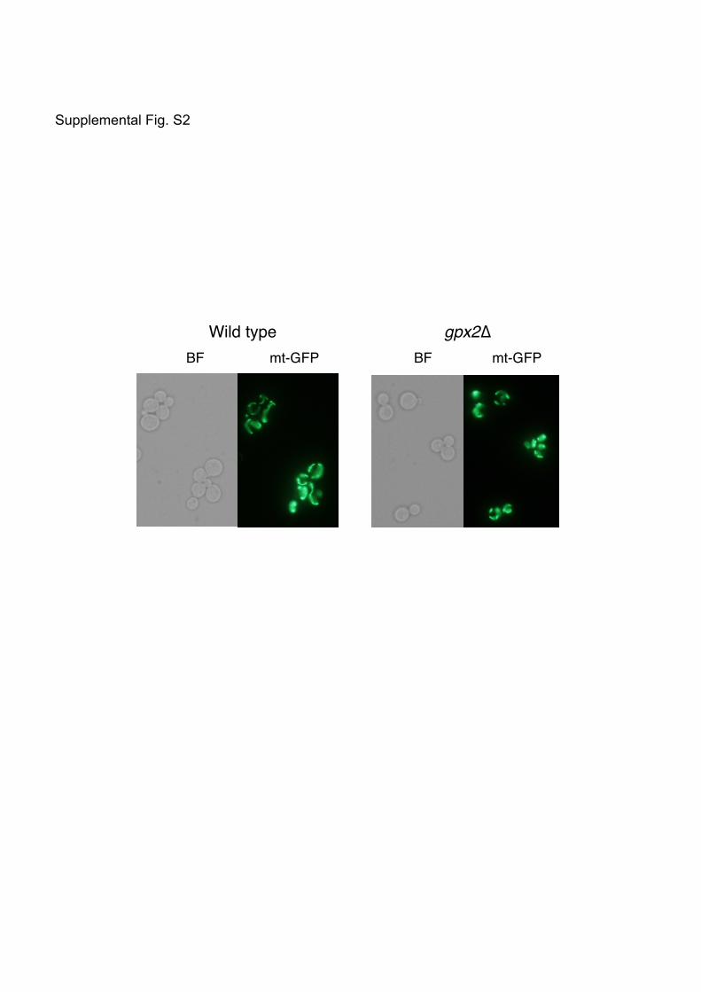
Entian et al. [16] have reported that gpx2∆ cells have round-shaped mitochondria as observed
under an electron microscope. These authors named the GPX2 gene AMI1 for “aberrant
mitochondria”. In logarithmically growing yeast cells, mitochondria show a tubular form with a
branched network along the cell cortex [for a review, see Ref. 17]. In our preliminary
experiments using the mitochondrial matrix-targeting GFP (mt-GFP) [18], the proportion of
cells possessing the mitochondria in the network structure seemed somehow lower among
gpx2Δ cells than wild-type cells (Supplemental Fig. S2). Considering the localization of Gpx2
(i.e. the surface of the outer membrane of mitochondria), the loss of Gpx2 might influence the
function of the factor(s) involved in the fusion of mitochondria, such as Fzo1-Mgm1-Ugo1
complex [19-21]. In addition, if Gpx2 functions as an antioxidant to reduce phospholipid
hydroperoxides in the mitochondrial membrane, its loss may reduce the integrity of the
mitochondrial membrane to decrease the efficiency of mitochondrial membrane docking.
However, since it is difficult to quantitate the change of mitochondrial morphology, we need to
define an index to evaluate the morphological change of mitochondria.
In addition to the cellular localization of proteins, the physical and/or genetic
interaction among gene products whose the functions are known would provide a clue as to the
related physiological role of a function-unknown partner. Fmp45 is a mitochondrial
membrane-integral protein, and involved in the process of sporulation. It has been reported that
Fmp45 physically interacts with Gpx2 [13]. Considering that Gpx2 is associated with the
mitochondrial membrane, it is feasible that these proteins share some functions in mitochondria.
Page 16
15
It has been well established that unimpaired mitochondrial function is necessary for sporulation
[22]. Here we have demonstrated that diploid cells defective in GPX2 show reduced efficiency
in sporulation. Additionally, the simultaneous disruption of FMP45 and GPX2 did not have an
additive effect in terms of the reduction of sporulation efficiency as compared with
fmp45∆/fmp45∆ diploid cells (Fig. 4). This result implies that Gpx2 functions in the same
regulatory pathway of sporulation as Fmp45, which seems to support the notion of physical
interaction between Fmp45 and Gpx2. Further study is now underway to figure out the function
of mitochondrial Gpx2 in the process of sporulation.
Acknowledgements
We thank Dr. T. Endo for antibodies for Tom70, Tim50, and Hsp60, and Dr. W.
Neupert for mt-GFP plasmids. We are also grateful to Dr. F. Ling for technical advice on the
preparation of mitochondria, and evaluation of the morphological change in mitochondria.
References
[1] Y. Inoue, T. Matsuda, S. Izawa, A. Kimura, Genetic analysis of glutathione peroxidase in
oxidative stress response of Saccharomyces cerevisiae. J. Biol. Chem. 274 (1999)
27002-27009.
[2] B. Hofmann, H.J. Hecht, L. Flohé, Peroxiredoxins, Biol. Chem. 383 (2002) 347–364.
[3] T. Ohdate, K. Kita, Y. Inoue, Kinetics and redox regulation of Gpx1, an atypical 2-Cys
peroxiredoxin, in Saccharomyces cerevisiae, FEMS Yeast Res. 10 (2010) 787-790.
[4] T. Tanaka, S. Izawa, Y. Inoue, GPX2, encoding a phospholipid hydroperoxide glutathione
peroxidase homologue, codes for an atypical 2-Cys peroxiredoxin in Saccharomyces
cerevisiae, J. Biol. Chem. 280 (2005) 42078-42087.
[5] A. Delaunay, D. Pflieger, M.B. Barrault, J. Vinh, M.B. Toledano, A thiol peroxidase is an
Page 17
16
H2O2 receptor and redox-transducer in gene activation, Cell 111 (2002) 471–481.
[6] U. Güldener, J. Heinisch, G.J. Köhler, D. Voss, J.H. Hegemann, A second set of loxP
marker cassettes for Cre-mediated multiple gene knockouts in budding yeast, Nucleic
Acids Res. 30 (2002) e23.
[7] M. Naoe, Y. Ohwa, D. Ishikawa, C. Ohshima, S. Nishikawa, H. Yamamoto, T. Endo,
Identification of Tim40 that mediates protein sorting to the mitochondrial intermembrane
space, J. Biol. Chem. 279 (2004) 47815-47821.
[8] C. Meisinger, T. Sommer, N. Pfanner, Purification of Saccharomcyes cerevisiae
mitochondria devoid of microsomal and cytosolic contaminations, Anal. Biochem. 287
(2000) 339–342.
[9] H. Yamamoto, M. Esaki, T. Kanamori, Y. Tamura, S. Nishikawa, T. Endo, Tim50 is a
subunit of the TIM23 complex that links protein translocation across the outer and inner
mitochondrial membranes, Cell 111 (2002) 519–528.
[10] Y. Tamura, Y. Harada, K. Yamano, K. Watanabe, D. Ishikawa, C. Ohshima, S. Nishikawa,
H. Yamamoto, T. Endo, Identification of Tam41 maintaining integrity of the TIM23
protein translocator complex in mitochondria, J. Cell Biol. 174 (2006) 631–637.
[11] S. Izawa, K. Maeda, K. Sugiyama, J. Mano, Y. Inoue, A. Kimura, Thioredoxin deficiency
causes the constitutive activation of Yap1, an AP-1-like transcription factor in
Saccharomyces cerevisiae, J. Biol. Chem. 274 (1999) 28459-28465.
[12] D. Tsuzi, K. Maeta, Y. Takatsume, S. Izawa, Y. Inoue, Regulation of the yeast
phospholipid hydroperoxide glutathione peroxidase GPX2 by oxidative stress is mediated
by Yap1 and Skn7, FEBS Lett. 565 (2004) 148-154.
[13] K. Tarassov, V. Messier, C.R. Landry, S. Radinovic, M.M. Serna Molina, I. Shames, Y.
Malitskaya, J. Vogel, H. Bussey, S.W. Michnick, An in vivo map of the yeast protein
interactome, Science 320 (2008) 1465-1470.
[14] M.E. Young, T.S. Karpova, B. Brugger, D.M. Moschenross, G.K. Wang, R. Schneiter, F.T.
Wieland, J.A. Cooper, The Sur7p family defines novel cortical domains in Saccharomyces
cerevisiae, affects sphingolipid metabolism, and is involved in sporulation, Mol. Cell. Biol.
22 (2002) 927-934.
[15] A.M. Avery, S.V. Avery, Saccharomyces cerevisiae expresses three phospholipids
hydroperoxide glutathione peroxidases, J. Biol. Chem. 276 (2010) 33730-33735.
[16] K.D. Entian, T. Schuster, J.H. Hegemann, D. Becher, H. Feldmann, U. Guldener, R. Gotz,
M. Hansen, C.P. Hollenberg, G. Jansen, W. Kramer, S. Klein, P. Kotter, J. Kricke, H.
Page 18
17
Launhardt, G. Mannhaupt, A. Maierl, P. Meyer, W. Mewes, T. Munder, R.K. Niedenthal,
R.M. Ramezani, A. Rohmer, A. Romer, M. Rose, B. Schafer, M.L. Siegler, J. Vetter, N.
Wilhelm, K. Wolf, F.K. Zimmermann, A. Zollner, A. Hinnen, Functional analysis of 150
deletion mutants in Saccharomyces cerevisiae by a systematic approach, Mol. Gen. Genet.
262 (1999) 683-702.
[17] K. Okamoto, J.M. Shaw, Mitochondrial morphology and dynamics in yeast and
multicellular eukaryote, Annu. Rev. Genet. 39 (2005) 503–536.
[18] B. Westermann, W. Neupert, Mitochondria-targeted green fluorescent proteins: convenient
tools for the study of organelle biogenesis in Saccharomyces cerevisiae, Yeast 16 (2000)
1421-1427.
[19] E.D. Wong, J.A. Wagner, S.V. Scott, V. Okreglak, T.J. Holewinske, A. Cassidy-Stone, J.
Nunnari, The intramitochondrial dynamin-related GTPase, Mgm1p, is a component of a
protein complex that mediates mitochondrial fusion, J. Cell Biol. 160 (2003) 303–311.
[20] H. Sesaki, R.E. Jensen, Ugo1p links the Fzo1p and Mgm1p GTPases for mitochondrial
fusion. J. Biol. Chem. 279 (2004) 28298–28303.
[21] H. Sesaki, S.M. Southard, M.P. Yaffe, R.E. Jensen, Mgm1p, a dynamin-related GTPase, is
essential for fusion of the mitochondrial outer membrane, Mol. Biol. Cell 14 (2003)
2342–2356.
[22] A.M. Neiman, Ascospore formation in the yeast Saccharomyces cerevisiae, Microbiol.
Mol. Biol. Rev. 69 (2005) 565-584.
Page 19
18
Figure legends
Fig. 1. Subcellular localization of Gpx2
(A) The distribution of proteins in each fraction was analyzed by Western blotting. S13
(supernatants) and P13 (crude mitochondria) were separated by the centrifugation of
homogenates at 13000 x g for 10 min. S100 (supernatants) and P100 (pellets) were separated by
the centrifugation of S13 fractions at 100000 x g for 20 min. Por1, Dpm1, and Pgk1 are the
marker proteins for mitochondria (mito), endoplasmic reticulum (ER), and cytoplasm (cyt),
respectively. (B) P13 fractions were subjected to the ultracentrifugation of a three-step sucrose
density gradient at 134000 x g for 1 h at 2ºC.
Fig. 2. Determination of mitochondrial localization of Gpx2
(A) Whole mitochondria, mitoplasts, and solubilized mitoplasts were treated with various
concentrations of PK as indicated in the figure for 30 min on ice. OM and IM represent the
outer membrane and inner membrane of mitochondria, respectively. (B) The mitochondria and
mitoplasts were treated as follows: mitochondria were treated with 0.5 M NaCl or 0.1 M
Na2CO3 for 60 min on ice, while mitoplasts were treated with sonication (50 W, 15 sec, 3 sets).
After centrifugation of each sample at 100000 x g for 30 min at 4ºC, pellets (Ppt) and
supernatants (Sup) were subjected to SDS-PAGE followed by Western blotting. OM and IM
represent the outer membrane and inner membrane of mitochondria, respectively.
Fig. 3. Redox regulation of mitochondrial Gpx2
Page 20
19
Cells (WT, trx1∆trx2∆, and trx3∆) were cultured in SD medium to a log phase of growth, and
treated with or without 0.4 mM H2O2 for 1 h. After the isolation of mitochondria, the
mitochondrial Gpx2 was modified with AMS. Proteins were separated by non-reducing
SDS-PAGE.
Fig. 4. Effects of the loss of Gpx2 on sporulation
Diploid cells (GPX2/GPX2, gpx2∆/gpx2∆, fmp45∆/fmp45∆, and gpx2∆fmp45∆/gpx2∆fmp45∆)
were incubated on sporulation medium agar plates at 25ºC. Approximately 1000 cells were
counted each day, and the proportion of sporulated cells (asci with four spores) versus total cells
counted is shown. Values represent the average for three independent experiments ± standard
deviation.
Page 21
Gpx2
Por1 (mito)
Dpm1 (ER)
Pgk1 (cyt)
w/o H2O2 w/ H2O2
S13 P13 S100 P100 S13 P13 S100 P100
Fig. 1
Gpx2
Por1 (mito)
Dpm1 (ER)
Fraction No 1 2 3 4 5 6 7 8 9
A
B
Page 22
Gpx2
Tom70 (OM)
Hsp60 (matrix)
Tim50 (IM)
(µg/ml)
Whole mitochondria Mitoplasts
Solubilized mitoplasts
PK 0 10 25 50 0 10 25 50 0 10 25 50
Fig. 2
Gpx2
Tom70 (OM)
Hsp60 (matrix)
Tim50 (IM)
Sup Ppt
NaCl Na2CO3 Sonic Sup Ppt Sup Ppt
Whole mitochondria Mitoplasts
A
B
Page 23
Fig. 3
Gpx2
WT trx3∆ Reduced Gpx2
Oxidized Gpx2
w/o H2O2 w/ H2O2
trx1/2∆ WT trx3∆ trx1/2∆
Page 24
Fig. 4
Spo
rula
tion
(%)
Time (day) 3.5 5.5
0
5
10
15
20
25 GPX2/GPX2 gpx2∆/gpx2∆ fmp45∆/fmp45∆ gpx2∆fmp45∆/gpx2∆fmp45∆
Page 25
SUPPLEMENTARY DATA Supplemental Materials and methods Subcellular fractionation The subcellular fractionation experiment was done essentially as described by Meisinger et al. [1] with slight modifications. Cells grown to a log phase of growth were treated with 4 mg/ml Zymolyase-100T (Seikagaku Kogyo Co.) in potassium phosphate buffer (pH 7.4) containing 1.2 M sorbitol at 30ºC for 60 min to obtain spheroplasts. After the homogenization of spheroplasts by 30 strokes in a Dounce glass homogenizer in 15 ml of ice-cold homogenization buffer [0.6 M sorbitol, 10 mM Tris-HCl (pH 7.4), 1 mM EDTA, and 1 mM phenylmethyl sulfonyl fluoride (PMSF)], cell debris and nuclei were removed by centrifugation at 1500 x g for 5 min. The supernatants were centrifuged at 1800 x g for 10 min twice, and the resultant supernatants were further centrifuged at 13000 x g for 10 min to precipitate crude mitochondria. The crude mitochondrial pellets were washed with SEM buffer [250 mM sucrose, 1 mM EDTA, and 10 mM MOPS (pH 7.2)], and again centrifuged at 13000 x g for 10 min. The resulting crude mitochondrial fractions (P13) were then resuspended in SEM buffer to give a final concentration of 1 mg of protein per ml. To prepare the cytoplasmic fraction, the supernatants after the preparation of crude mitochondria (S13), were centrifuged at 100000 x g for 20 min. The resultant supernatants were S100 fractions, and the resultant precipitates were P100 fractions. Each fraction was subjected to SDS-PAGE followed by Western blotting using respective antibodies to verify the localization of marker proteins. To separate ER from the crude mitochondrial fractions, the crude mitochondria (250 µg protein) in 250 µl of SEM buffer were loaded onto the top of a three-step sucrose density gradient [0.75 ml 60%, 2 ml 32%, 0.75 ml 23%, 0.75 ml 15% (w/v) sucrose in EM buffer (10 mM MOPS, pH 7.2, and 1 mM EDTA)]. The centrifugation was done at 134000 x g for 1 h at 2ºC with a RPS40-T rotor (himac CP75β, Hitachi Koki). Each fraction (500 µl) was collected from the top of the tube (9 fractions), and proteins were precipitated by adding 125 µl of 100% trichloroacetic acid (TCA). The mixture was centrifuged at 13000 x g for 10 min at 4ºC, and the precipitates were washed once with acetone. After removal of the acetone, the dried materials were suspended in 40 µl of 1 M Tris solution. The suspension was mixed with 10 µl of 5x sample buffer, and the mixture was boiled for 5 min. The sample was subjected to SDS-PAGE followed by Western blotting.
Proteinase K treatment Treatment of mitochondria with proteinase K to determine the localization of proteins was done essentially as described by Yamamoto et al. [2] with slight modifications. Briefly, the crude mitochondria (200 µg protein) were suspended in 200 µl of TS buffer [0.6 M sorbitol, and 10 mM Tris–HCl (pH 7.4)]. Mitoplasts were generated with osmotic swelling by suspending the crude mitochondria (200 µg protein) in a 10-fold volume of 10 mM Tris–HCl (pH 7.4) buffer for 20 min on ice. The mitoplasts were collected by centrifugation at 13000 x g
Page 26
for 10 min, and suspended in 200 µl of TS buffer. Solubilized mitoplasts were generated from the mitoplasts (200 µg protein) by treatment with 0.1% Triton-X100 in 200 µl of TS buffer for 20 min on ice. Each suspension of crude mitochondria, mitoplasts, and solubilized mitoplasts was treated with 10-50 µg/ml of proteinase K (Wako) for 30 min on ice. The reaction was stopped by addition of 1 mM PMSF for 5 min on ice, and 50 µl of 100% TCA was added. The precipitates were washed once with acetone. After removal of the acetone, the dried materials were suspended with 32 µl of 1 M Tris solution. The suspension was mixed with 8 µl of 5x sample buffer, and the mixture was boiled for 5 min. The sample was subjected to reducing SDS-PAGE followed by Western blotting. NaCl, sonication, and Na2CO3 treatment Treatment of mitochondria with NaCl, sonication, and Na2CO3 to determine the localization of proteins was done essentially as described by Yamamoto et al. [2] with slight modifications. Briefly, the crude mitochondria (200 µg protein) were treated in 200 µl of 0.5 M NaCl or 0.1 M Na2CO3 in TS buffer containing 1 mM PMSF for 30 min on ice. Mitoplasts (2 mg of protein) prepared by hypo-osmotic swelling as described previously were treated with sonication (50 W, 15 sec, 3 sets) in 2 ml of TS buffer. Each solution was centrifuged at 100000 x g for 30 min at 4ºC. The precipitates were suspended in 32 µl of 1 M Tris solution, and the suspension was mixed with 8 µl of 5x sample buffer. The supernatants were mixed in 50 µl of 100% TCA, the mixture was centrifuged at 13000 x g for 10 min at 4ºC, and the precipitates were washed once with acetone. After removal of the acetone, the dried materials were suspended with 32 µl of 1 M Tris solution. The suspension was mixed with 8 µl of 5x sample buffer, and the mixtures were boiled for 5 min. The sample was subjected to SDS-PAGE followed by Western blotting. References [1] C. Meisinger, T. Sommer, N. Pfanner, Purification of Saccharomcyes cerevisiae
mitochondria devoid of microsomal and cytosolic contaminations, Anal. Biochem. 287 (2000) 339–342.
[2] H. Yamamoto, M. Esaki, T. Kanamori, Y. Tamura, S. Nishikawa, T. Endo, Tim50 is a subunit of the TIM23 complex that links protein translocation across the outer and inner mitochondrial membranes, Cell 111 (2002) 519–528.
Page 27
Supplemental Table 1
Yeast strains used in this study
Strain Relavant genotype
YPH250 MATa trp1-Δ1 his3-Δ200 leu2-1 lys2-801 ade2-101 ura3-52
YPH252 MATα trp1-Δ1 his3-Δ200 leu2-1 lys2-801 ade2-101 ura3-52
YPH274 MATa/MATα YPH250 x YPH252
gpx2∆ YPH250, gpx2∆::TRP1
gpx2∆ YPH252, gpx2∆::his5+
trx1∆trx2∆ YPH250, trx1∆::URA3 trx2∆::HIS3
trx3∆ YPH250, trx3∆::TRP1
GPX2/GPX2 same as YPH274
gpx2∆/gpx2∆ YPH250 (gpx2∆::TRP1) x YPH252 (gpx2∆::his5+)
fmp45∆/fmp45∆ YPH250 (fmp45∆::URA3) x YPH252 (fmp45∆::LEU2)
gpx2∆fmp45∆/gpx2∆fmp45∆ YPH250 (gpx2∆::TRP1 fmp45∆::URA3) x YPH252 (gpx2∆::his5+
fmp45∆::LEU2)
Page 28
Legends for Supplemental Figures Supplemental Fig. S1. Localization of epitope-tagged Gpx2 Cells carrying plasmids for the HA-tagged GPX2 at the C terminus or FLAG-tagged GPX2 at the N terminus were cultured in SD medium until a log phase of growth, and mitochondria were prepared by sucrose density ultracentrifugation. Gpx2-HA and FLAG-Gpx2 were detected by anti-HA monoclonal antibody and anti-FLAG monoclonal antibody, respectively. Por1 and Dpm1 are the marker proteins for mitochondria (mito) and endoplasmic reticulum (ER), respectively. Supplemental Fig. S2. Effects of the loss of Gpx2 on mitochondrial morphology Cells (wild type and gpx2∆) carrying mitochondrial matrix-targeting GFP (mt-GFP) were cultured in SD medium to a log phase of growth, and the morphology of mitochondria was observed with fluorescence microscopy.
Page 29
Gpx2-HA
Por1 (mito)
Dpm1 (ER)
FLAG-Gpx2
Por1 (mito)
Dpm1 (ER)
Supplemental Fig. S1
1 2 3 4 5 6 7 8 9
1 2 3 4 5 6 7 8 9
Page 30
BF mt-GFP BF mt-GFP Wild type gpx2∆
Supplemental Fig. S2


![Helically Twisted Tetracene: Synthesis, Crystal …...Helically Twisted Tetracene: Synthesis, Crystal Structure, and Photophysical Properties of Hexabenzo[a,c,fg,j,l,op]tetracene Yuuta](https://static.documents.pub/doc/80x56/5e4ef0a5936ab37407486d54/helically-twisted-tetracene-synthesis-crystal-helically-twisted-tetracene.jpg)


