44
Topic Teaching Paediatric Respiratory Tract Disorders Albert M. Li / Dorothy Chan Department of Paediatrics Prince of Wales Hospital
| Date post: | 31-Dec-2015 |
| Category: |
Documents |
| Upload: | hayes-nguyen |
| View: | 37 times |
| Download: | 2 times |
Topic TeachingPaediatric Respiratory Tract Disorders
Albert M. Li / Dorothy Chan
Department of Paediatrics
Prince of Wales Hospital

Introduction
• Respiratory disorders are the most common clinical problems seen in primary care and hospital practice.
• Vast majority of symptomatic respiratory infections are due to respiratory viruses. Most commonly seen in young preschool children, as a result of the relative immaturity of the host defence mechanisms and anatomical development.
• Lung development is not complete until about 3 years of age and growth in size and physiological development continue into adulthood. Thus respiratory diseases in early childhood may cause significant morbidity in adult life.
Introduction
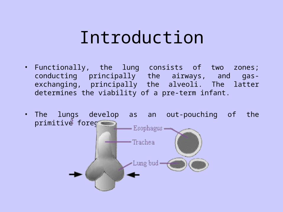
• Functionally, the lung consists of two zones; conducting principally the airways, and gas-exchanging, principally the alveoli. The latter determines the viability of a pre-term infant.
• The lungs develop as an out-pouching of the primitive foregut.
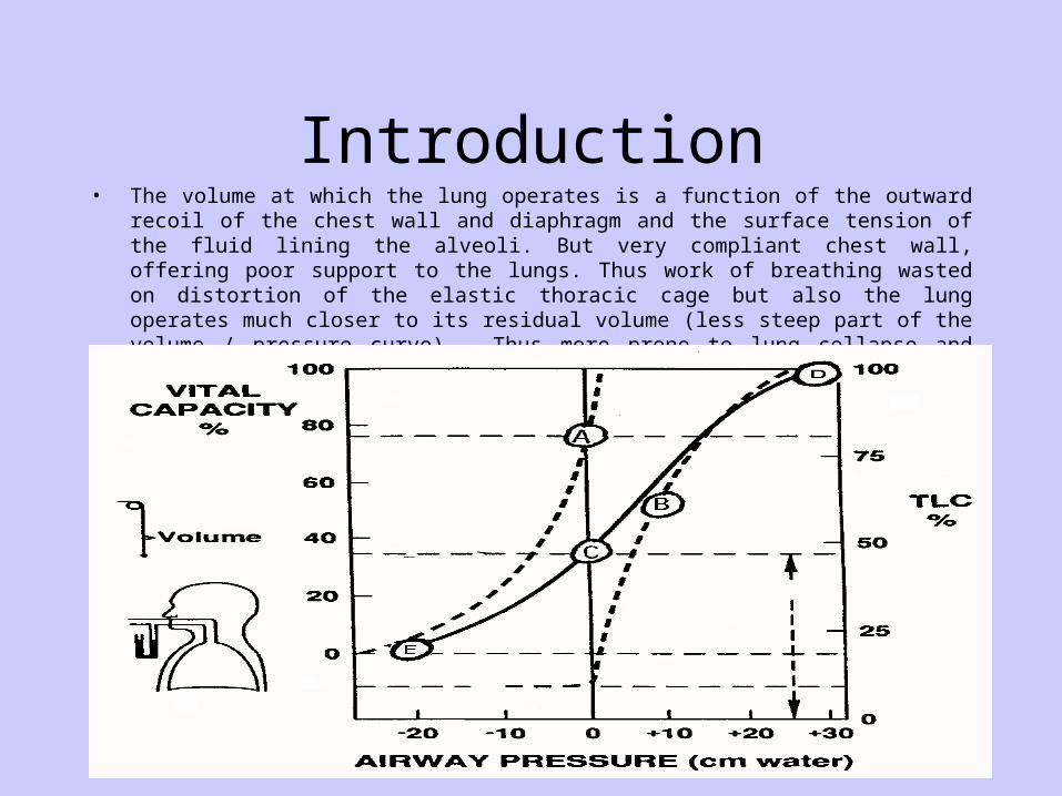
Introduction• The volume at which the lung operates is a function of the outward recoil of the chest wall and
diaphragm and the surface tension of the fluid lining the alveoli. But very compliant chest wall, offering poor support to the lungs. Thus work of breathing wasted on distortion of the elastic thoracic cage but also the lung operates much closer to its residual volume (less steep part of the volume / pressure curve). Thus more prone to lung collapse and this results in shunting of blood through poorly aerated parts causing hypoxaemia.
Introduction
• Large cranial vault but small facial skeleton, relative large size of the lymphoid tissue, thus over-crowding in the upper airway space. Any narrowing increases greatly the resistance to air flow especially during inspiration. A much greater force is needed to generate to overcome this increased resistance, which may further encourage the collapse of the airway.
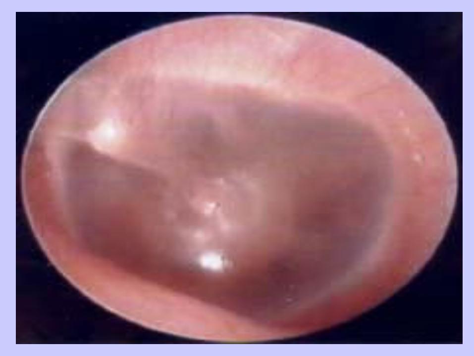
• Another problem with overcrowding of the pharyngeal space and hypertrophy of the lymphatic tissue in this region is impairment of eustachain tube drainage, predisposing to the development of otitis media.

Core problem / Presenting complaint Key diagnosis Related topics
Upper respiratory tract infection Acute pharyngitis Coryza(URTI) Tonsillitis Otitis media
Wheeze Asthma Bronchiolitis
Cough Post viral URTI Asthma
Stridor Viral croup Foreign body
Allergic reaction Epiglottitis
Breathlessness Pneumonia Cardiac disease
Chronic, productive cough Bronchiectasis Tuberculosis

Upper respiratory tract disorders
• Coryzal symptoms.
• Nasal discharge and obstruction can cause feeding difficulties.
• Very common, up to six symptomatic infections per year are the norm.
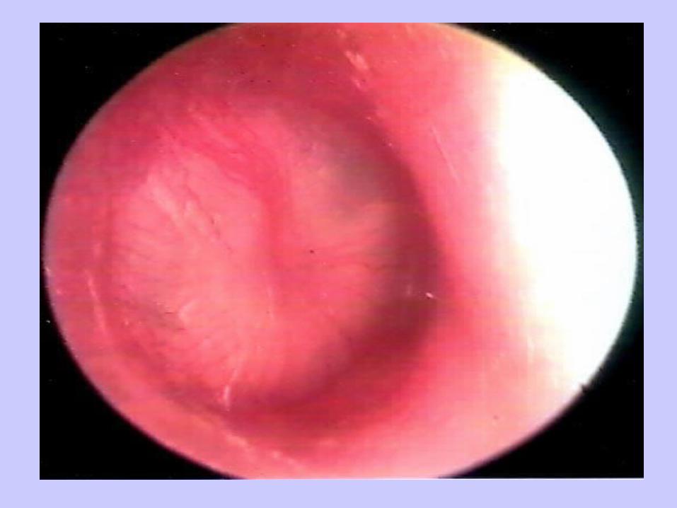
• Examination injected posterior pharynx, dull or pink ear drums, together with fever.
• Symptomatic relief. Antibiotics not necessary and can be harmful.

Upper respiratory tract disorders
• Noisy breathing
- stertor versus stridor versus wheeze
- stridor, acute versus chronic

Upper respiratory tract disorders
• Acute stridor
- viral croup
- foreign body inhalation
- allergic reaction
- epiglottitis
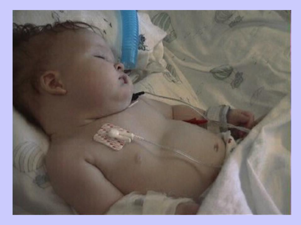
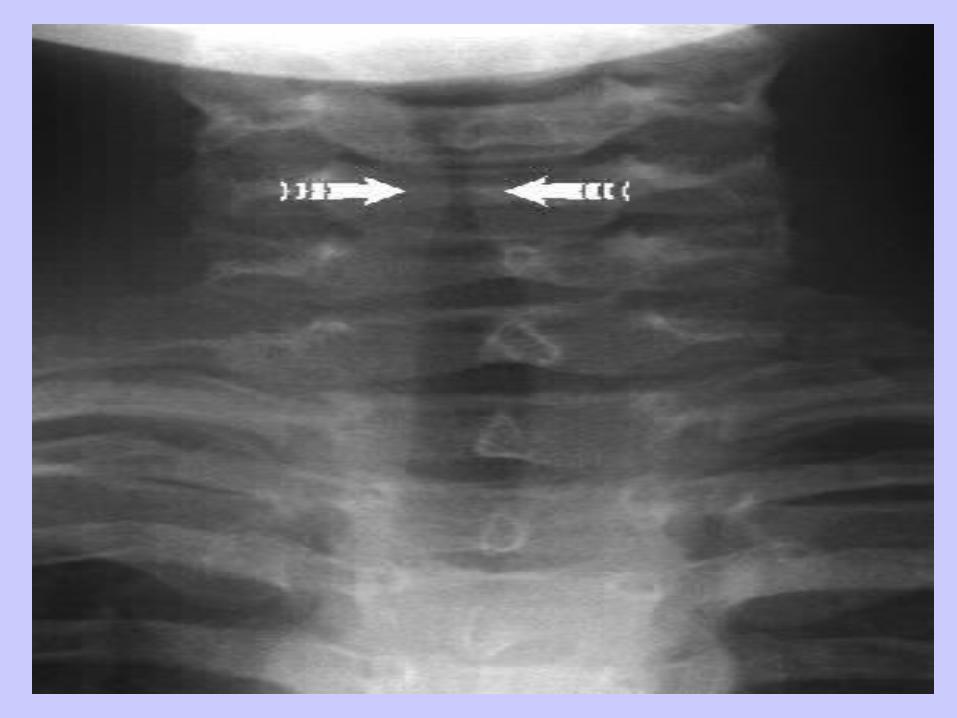
• Stridor is the audible breath sound heard due to turbulent airflow through the trachea and larger airways.

Viral croup
• Combination of stridor, barking cough, hoarseness of voice and respiratory distress due to upper airway obstruction. Clinical diagnosis.
• Usually preceded by coryzal symptoms.
• Parainfluenza viruses.
• Most important to exclude other possible causes of acute stridor.
• Supportive treatment + corticosteroid.
Lower respiratory tract disorders
• Wheeze - narrowing of small airways causing turbulence in air flow.
- common feature of respiratory disease in infants, reflect the smaller diameter of the airways.
- asthma is probably the most common cause of wheeze but other conditions have to be considered.
- nature of presentation and age are important distinguishing factors.

• Differential diagnosis of wheeze
Acute attacks
Initial Infection (bronchiolitis)
Foreign body inhalation
Recurrent Asthma
Recurrent infections (aspiration, immunodeficiency)
Ongoing symptoms Aspiration syndromes
Chronic lung conditions
External compression

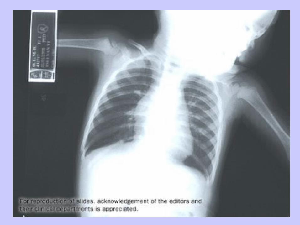
Bronchiolitis
• Classically coryzal symptoms followed by respiratory distress, wheeze and/or poor feeding.
• Age group.
• Aetiological agent.
• Seasonality.
• Pathophysiology; airway inflammation, epithelial shedding, airway obstruction, atelactasis, hypoxaemia.

Bronchiolitis
• Hydration and Oxygenation.
• Supportive measures.

• Bronchiolitis versus Asthma.
• Bronchiolitis leading to Asthma.
Asthma
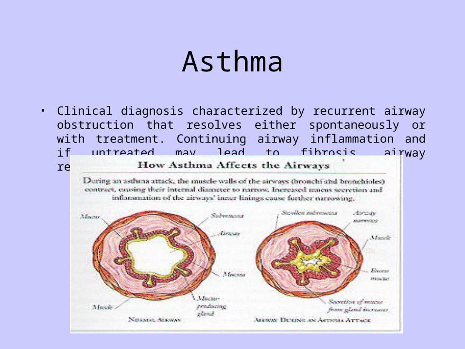
• Clinical diagnosis characterized by recurrent airway obstruction that resolves either spontaneously or with treatment. Continuing airway inflammation and if untreated may lead to fibrosis, airway remodeling.

Asthma
• Prevalence of the disease among the paediatric population may be as high as 15%.
• Increased prevalence recorded - people have become more aware
- diet
- more hygienic
- environmental factors
- diagnostic shift
• Strongly associated with atopy, skin prick test positive to inhaled allergen, in Hong Kong cockroach and house dust mite.

Asthma
• Questions to ask in history taking
- prenatal and birth history,
- family history,
- mode of infant feeding,
- other atopic diseases,
- presenting symptoms, recurrent attacks,
- time to recovery following viral infection,
- medications used and response,
- severity of disease,


Asthma
• Clinical diagnosis,
• Skin prick test to common allergens,
• Lung function test, obstructive diseases cause a greater fall in FEV1 compared to FVC. The ration of FEV1 to FVC is reduced.
• Response to bronchodilator challenge.
• Stimulation challenge.
• Chest radiograph.
• Blood tests.
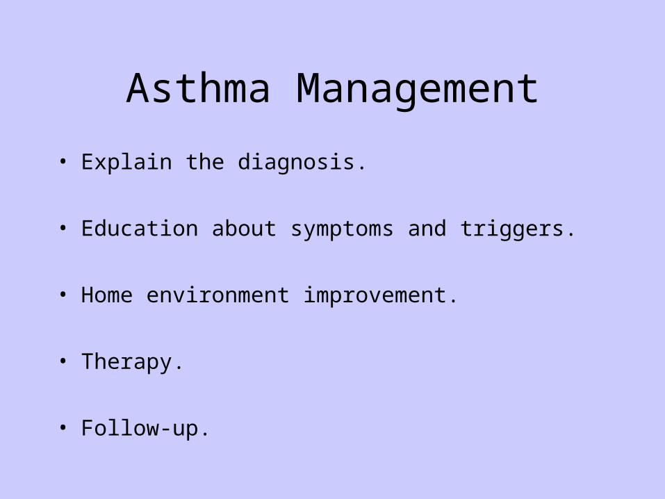
Asthma Management
• Explain the diagnosis.
• Education about symptoms and triggers.
• Home environment improvement.
• Therapy.
• Follow-up.


Cough• Cough does not equal to asthma.
• Can be the presentation of a wide range of respiratory problems.

Tuberculosis
• Symptoms are non-specific. Children are less likely to have open TB, thus acquired from an adult.
• Investigations.
• Contact tracing important.
• Vital to complete full course of therapy.
• DOTS.
• Use of multiple drugs, Isoniazid resistance.
• Follow-up, possible complications.


Breathlessness
• Breathlessness (dyspnoea) is symptom and tachypnoea is a sign.
• Lower respiratory tract problems, common causes are pneumonia and asthma.
• Pyrexia could give rise to tachypnoea.
children 0–2 months: less than 60/min children 2 months–1 year: less than 50/min children over 1 year: less than 40 min
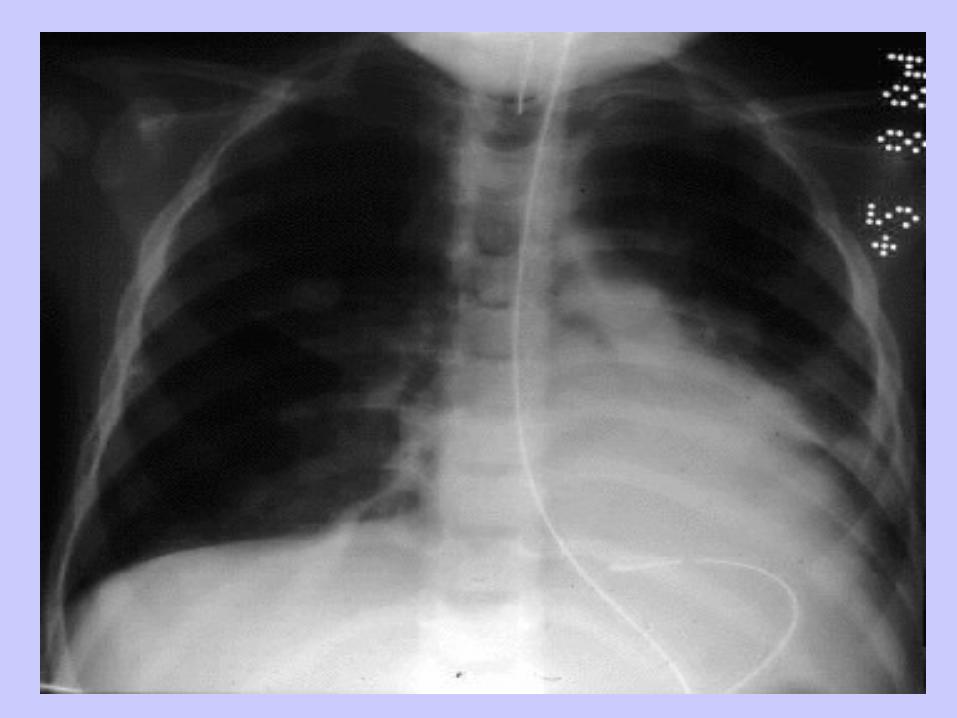
Pneumonia
• Upper respiratory tract infection versus pneumonia. Former,child may be miserable but not unwell, no chest signs. Latter, child, unwell, may appear toxic with respiratory distress.
• Rapid, high swinging fever in a toxic child is more in favour of a bacterial pneumonia.
• Likely causative agent depend on age of subject, neonates versus children up to around 4 years of age versus older children.
Pneumonia
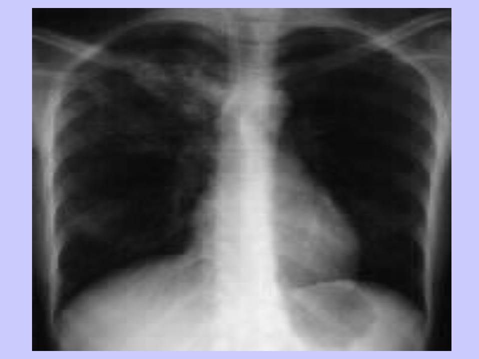
• Atypical organism in school aged children, flu-like symptoms, wheeze may be present in up to 50% of cases. CXR appearance often worse than the patient appears clinically.
• Tachypnoea is a reliable sign of bacterial pneumonia.
• Wheeze is unusual in genuine bacterial pneumonia.
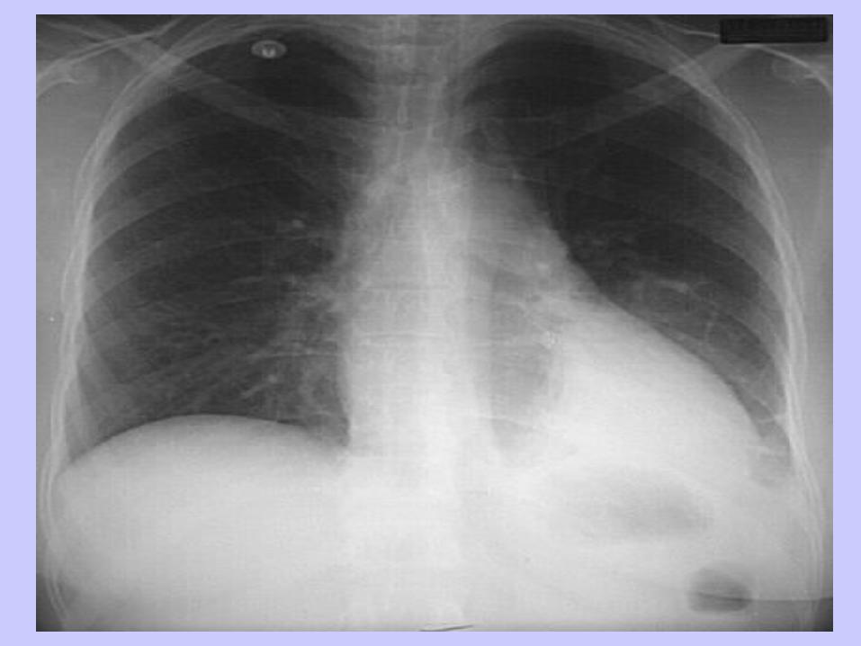
• CXR appearance not diagnostic of causative organism.
Pneumonia
• Blood tests
- complete blood picture, neutropilia versus lymphocytosis.
- CRP not that helpful, overlap in viral and bacterial cases.
- blood culture, low yield.
- viral titres, acute and convalescence.
- electrolytes, sodium.
• Other investigations
- naso-pharyngeal aspirate for virus screen.


Pneumonia
• Complications - para-pneumonic effusion.
- persistent fever, non-improving clinical course, think about empyema; a
collection of pus within the thoracic cavity.
What should be done??
- septic embolism causing cerebral abscess.
Recurrent episodes of pneumonia think about……………..


