2 ABSTRACT: The outcomes of treatment for gastric cancer in Japan have improved markedly as a result of early detection and extensive radical surgery. To date, the Japanese Gastric Cancer Association (JGCA) has recommended that non-early, potentially curable gastric cancers should be treated by D2 lymphadenectomy, and defined standard gastrectomy, which is the principal surgical procedure performed with curative intent, as resection of not less than two-thirds of the stomach with a D2 lymph node dissection. Laparoscopic surgery was launched in the early ‘90s. At that time, most laparoscopic surgeons applied laparoscopic surgery, using its minimally invasive nature, to less extended surgery. However, we assumed from the beginning that laparoscopic surgery should 1 K. Suda, 2 V. A. Kashchenko, 1 K. Ishikawa, 1 Y. Ishida, мI. Uyama 1 Fujita Health University 2 Sokolov’ Hospital № 122 of the Federal Medical and Biological Agency INSTITUTIONAL AFFILIATION: *Division of Upper GI, Department of Surgery, Fujita Health University, 1-98 Dengakugakubo, Kutsukake, Toyoake, Aichi, 470-1192 Japan. †Department of Surgery, L.G. Sokolov Memorial Hospital №122, 4 Kultury Pr. St. Petersburg 194291, Russia. ADDRESS CORRESPONDENCE AND REQUESTS FOR REPRINTS TO: Koichi Suda, MD, PhD Division of Upper GI, Department of Surgery, Fujita Health University, 1-98 Dengakugakubo, Kutsukake, Toyoake, Aichi, 470-1192 Japan TOTALLY LAPAROSCOPIC DISTAL GASTRECTOMY WITH D2 LYMPH NODE DISSECTION BASED ON JAPANESE GASTRIC CANCER TREATMENT GUIDELINES: PRINCIPLES AND METHODS 1. Introduction Gastric cancer remains a major public health problem in the world. Gastric cancer is the 4 th most common cancer and the 2 nd leading cause of cancer- related death 1,2) . The highest incidence of gastric cancer is found not only in East Asia including Japan but also in Russia 2) . In terms of prognosis of gastric cancer, 5-year relative survival was over 60% in Japan, whereas 25% in the Western countries 3) . There are following two major factors which may cause such a great difference in long-term outcomes: early detection of gastric cancer, and the extended D2 lymph node dissection 1) . We introduced laparoscopic assistance into moderate to advanced gastrointestinal surgery in 1995, and developed techniques for laparoscopic distal and total gastrectomy with D2 dissection for advanced gastric cancer, which were published for the first time in the world 1,4,5) . Since then, we have performed more than 1,000 laparosocpic gastrectomies. At present, the standard type of operation for curable gastric cancer at Fujita Health University is totally laparoscopic D2 gastrectomy 1) . We herein present the principles and methods of totally laparoscopic D2 distal gastrectomy with delta- shaped B-I anastomosis 6) . 2. Set up 2.1. List of instruments An operating surgeon basically uses Thunderbeat and “Mancina” with his/her right and left hands, respectively. An assistant surgeon does “Johann” and “Croce” with his/her cranial and caudal-sided hands, individually (Fig.1). All the details were shown in Table 1. 2.2. Patients The stage of the cancer is classified according to the 14th edition of the Japanese Classification of Gastric Carcinoma (JCGC) 7) . Cancer staging be suitable for meticulous dissection using the high quality of laparoscopic image with magnified visualization, and since mid- ‘90s, we have been doing totally laparoscopic D2 gastrectomy with intracorporeal anastomosis using linear staplers as the standard treatment for operable patients with resectable gastric cancer. In this article, we present technical aspects of totally laparoscopic D2 distal gastrectomy with delta-shaped B-I anastomosis based on our experience. KEY WORDS: gastric cancer, laparoscopic distal gastrectomy, D2 dissection, outermost layer-oriented medial approach, delta- shaped anastomosis.
ABSTRACT: The outcomes of treatment for gastric cancer in Japan have improved markedly as a result of early detection and extensive radical surgery. To date, the Japanese Gastric Cancer Association (JGCA) has recommended that non-early, potentially curable gastric cancers should be treated by D2 lymphadenectomy, and defined standard gastrectomy, which is the principal surgical procedure performed with curative intent, as resection of not less than two-thirds of the stomach with a D2 lymph node dissection. Laparoscopic surgery was launched in the early ‘90s. At that time, most laparoscopic surgeons applied laparoscopic surgery, using its minimally invasive nature, to less extended surgery. However, we assumed from the beginning that laparoscopic surgery should
1K. Suda, 2 V. A. Kashchenko, 1K. Ishikawa, 1Y. Ishida, мI. Uyama 1Fujita Health University
2Sokolov’ Hospital № 122 of the Federal Medical and Biological Agency
INSTITUTIONAL AFFILIATION:*Division of Upper GI, Department of Surgery, Fujita Health University, 1-98 Dengakugakubo, Kutsukake, Toyoake, Aichi, 470-1192 Japan.
†Department of Surgery, L.G. Sokolov Memorial Hospital №122, 4 Kultury Pr. St. Petersburg 194291, Russia.
ADDRESS CORRESPONDENCE AND REQUESTS FOR REPRINTS TO: Koichi Suda, MD, PhD
Division of Upper GI, Department of Surgery, Fujita Health University, 1-98 Dengakugakubo, Kutsukake, Toyoake, Aichi, 470-1192 Japan
TOTALLY LAPAROSCOPIC DISTAL GASTRECTOMY WITH D2 LYMPH NODE DISSECTION BASED
ON JAPANESE GASTRIC CANCER TREATMENT GUIDELINES: PRINCIPLES AND METHODS
1. IntroductionGastric cancer remains a major public health
problem in the world. Gastric cancer is the 4th most common cancer and the 2nd leading cause of cancer- related death1,2). The highest incidence of gastric cancer is found not only in East Asia including Japan but also in Russia2). In terms of prognosis of gastric cancer, 5-year relative survival was over 60% in Japan, whereas 25% in the Western countries3). There are following two major factors which may cause such a great difference in long-term outcomes: early detection of gastric cancer, and the extended D2 lymph node dissection1).
We introduced laparoscopic assistance into moderate to advanced gastrointestinal surgery in 1995, and developed techniques for laparoscopic distal and total gastrectomy with D2 dissection for advanced gastric cancer, which were published for the first time in the world1,4,5). Since then, we have performed more than 1,000 laparosocpic
gastrectomies. At present, the standard type of operation for curable gastric cancer at Fujita Health University is totally laparoscopic D2 gastrectomy1).
We herein present the principles and methods of totally laparoscopic D2 distal gastrectomy with delta-shaped B-I anastomosis6).
2. Set up
2.1. List of instrumentsAn operating surgeon basically uses Thunderbeat
and “Mancina” with his/her right and left hands, respectively. An assistant surgeon does “Johann” and “Croce” with his/her cranial and caudal-sided hands, individually (Fig.1). All the details were shown in Table 1.
2.2. PatientsThe stage of the cancer is classified according
to the 14th edition of the Japanese Classification of Gastric Carcinoma (JCGC) 7). Cancer staging
be suitable for meticulous dissection using the high quality of laparoscopic image with magnified visualization, and since mid-‘90s, we have been doing totally laparoscopic D2 gastrectomy with intracorporeal anastomosis using linear staplers as the standard treatment for operable patients with resectable gastric cancer. In this article, we present technical aspects of totally laparoscopic D2 distal gastrectomy with delta-shaped B-I anastomosis based on our experience.
is performed based on the findings of contrast-enhanced computed tomography, gastrography, endoscopic study, and endosonography before the beginning of any treatment and, when applicable, after the completion of chemotherapy. The patients with clinical T ≥ 2 cancer over 5 cm in size and/or a swollen locoregional lymph node over 1.5 cm in size undergo staging laparoscopy. Clinical Stage ≤ IIIC is determined to be resectable. Neoadjuvant chemotherapy (S-1 80
mg/m2 Day1-21 + CDDP 60 mg/m2 Day 8) is used for those with clinical T ≥ 2 as well as tumor ≥ 5.0cm in size and/or a swollen locoregional lymph node ≥ 1.5cm in size, unless the patients refuse it. Induction chemotherapy (S-1 80 mg/m2 Day 1-14 + CDDP 35 mg/m2 Day 8, or Docetaxel 30 mg/m2 Day 1, 15 + CDDP 30 mg/m2 Day 1, 15 + S-1 80 mg/m2 Day 1-14) is used for clinical Stage IV disease, and radical gastrectomy is conducted when downstaging is achieved.
Fig.1 Forceps and hemostats specialized for advanced laparoscopic surgery
Distal gastrectomy is used for the tumor localized to M and/or L area. D1+ lymphadenectomy is done for preoperative Stage IA disease, whereas D2 is done for preoperative Stage IB, II, and III diseases in accordance with the 3rd edition of the JGCA Guidelines8).
2.3. OR setupBasically, the operator stands on the patient’s
right side, except for #6 lymph node dissection (Fig.2). When the operator stands on the left side, the scrub nurse with the table should move from the caudal to the cranial side of the patient (Fig.2) just to avoid the cables connecting between the forceps and generators from getting tangled.
2.4. Patient’s positionThe patient is placed in a supine position with
legs apart, left arm extended, and 15-degree head-up tilt.
2.5. Trocar arrangement (Fig. 3) Camera: navel or mid-line below the navel RUP: one-finger caudally from the right
subcostal line, top of the right subphrenic “dome”, affecting the comfortableness in grasping adipose tissue including #11p
Note: The distance between Camera and RUP should be longer than eight fingers. RLP: Caudally on the median line between
Camera and RUP LUP: more than two-finger caudally from the
left subcostal line, affecting the comfortableness in #6 dissection
Note: The distance between Camera and LUP should also be longer than eight fingers. LLP: Caudally on the median line between
Camera and LUP Additional port: Cranially on the median line
between Camera and RUP, suitable for deeply dissecting suprapancreatic lymph nodes over the pancreas
Fig. 3 Trocar arrangement in laparoscopic gastrectomy
D2 dissection entails removal of the lymph nodes in the suprapancreatic area in distal gastrectomy1). Dissection of this area is technically demanding due to the serious risk of bleeding and/or pancreatic leakage derived from a major vessel or organ injury1,9,10). To improve the safety, efficacy, and reproducibility of suprapancreatic nodal dissection, we developed our original methodology called outermost layer-oriented medial approach1,11,12). In this approach, the layer between the autonomic nerve sheaths of the major arteries and the adipose tissue bearing lymphatic tissue is dissected1,11,12). We termed this layer as the outermost layer of the autonomic nerve (Fig.4) 1,12). To identify this layer throughout the dissection process, we developed an original surgical theory, “XYZ-axis”
Fig.5 XYZ-axis theory
CHA – common hepatic artery SPA – splenic artery, PHA – proper hepatic artery, LGA – left gastric artery, RGA – right gastric artery,
GDA – gastroduodenal artery, RGEA – right gastroepiploic artery, ASPDA – anterior superior pancreatoduodenal artery, RGEV – right gastroepiploic vein.
theory (Fig.5), consisting of the following three steps—(1) cut the serosal membrane on the suprapancreatic border; (2) dissect suprapancreatic adipose tissue caudocranially towards the junction of the three arteries (zero point) to find the outermost layer; (3) dissect the target adipose tissue mediolaterally along the layer spreading on the XZ and YZ axes.
3.2. Details of D2 dissection in distal gastrectomy
3.2.1. #4d dissectionThe operating surgeon stands to the right of the
patient. The assistant surgeon holds the greater curvature on the “watershed” dividing between the right and left gastroepiploic arteries (RGEA and LGEA) and raise it cranioventrally with his/her right hand. Subsequently, the assistant surgeon grasps the greater omentum near the transverse colon. Then, the operating surgeon gently holds the pedicle of the right gastroepiploic artery and vein (RGEA and RGEV) to create a triangle. The operating surgeon starts opening the bursa at a thin part of the greater omentum (Fig.6), and transects it along the border between adipose tissue belonging to the stomach and that belonging to the transverse colon referring to the “line” created by physiological adhesion (Fig.7a). Adhesion between the posterior aspect of the stomach and the pancreatic body should be detached as much as possible just to recover the original anatomy.
3.2.2. #4sb dissectionThe assistant surgeon holds the posterior aspect
of the upper area of the stomach and determine the pedicle including the LGEA/V originating from the pancreatic tail (Fig.7a). By dividing the bursa along the physiological adhesion line mentioned above, the root of the gastric branch of LGEA is easily exposed preserving the omental branch (Fig.7b). Then, adipose tissue including #4sb is removed out of the greater curvature from the “watershed” upto the avascular area between LGEA and short gastric arteries (SGAs) (Fig.7c).
3.2.3. #6 dissectionThe operating surgeon moves to the left of the
patient. Transverse colon is mobilized by dissecting fusion fascia and pancreatic head is widely exposed. The left aspect of the adipose tissue including #14v and 6 is dissected along the inferior border of the pancreas (Fig.8a). Subsequently by exposing the edge of the pancreatic head behind the duodenal bulb, RGEA and the autonomic nerve on the right of RGEA is exposed on the anterior and inferior aspect of the pancreatic head, respectively (Fig.8b). At this site, right gastroepiploic vein (RGEV) is running along the nerve, and the outermost layer of RGEA is widely exposed by dividing between the vein and nerve to facilitate #6v dissection (Fig.8b). Prepancreatic fascia is dissected along anterior
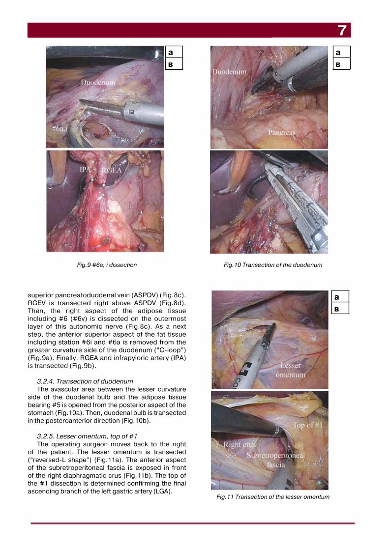
superior pancreatoduodenal vein (ASPDV) (Fig.8c). RGEV is transected right above ASPDV (Fig.8d). Then, the right aspect of the adipose tissue including #6 (#6v) is dissected on the outermost layer of this autonomic nerve (Fig.8c). As a next step, the anterior superior aspect of the fat tissue including station #6i and #6a is removed from the greater curvature side of the duodenum (“C-loop”) (Fig.9a). Finally, RGEA and infrapyloric artery (IPA) is transected (Fig.9b).
3.2.4. Transection of duodenumThe avascular area between the lesser curvature
side of the duodenal bulb and the adipose tissue bearing #5 is opened from the posterior aspect of the stomach (Fig.10a). Then, duodenal bulb is transected in the posteroanterior direction (Fig.10b).
3.2.5. Lesser omentum, top of #1The operating surgeon moves back to the right
of the patient. The lesser omentum is transected (“reversed-L shape”) (Fig.11a). The anterior aspect of the subretroperitoneal fascia is exposed in front of the right diaphragmatic crus (Fig.11b). The top of the #1 dissection is determined confirming the final ascending branch of the left gastric artery (LGA).
Fig.9 #6a, i dissection Fig.10 Transection of the duodenum
3.2.6. Rolling up the stomachTo facilitate suprapancreatic lymph node
dissection, the stomach is rolled up (Fig.12).
3.2.7. Probing the outermost layer of CHA and SPAThe assistant surgeon retracts the caudal
edge of the pancreatic body and stretches the gastropancreatic fold. The operating surgeon stretches the adipose tissue containing #8a and #11p carefully and dissected along the stably visualized outermost layer of the common hepatic artery (CHA) (Fig.13a) and the proximal part of the splenic artery (SPA) (Fig.13b). This dissection was continued along the outermost layer of the left lateral aspect of the proper hepatic artery (PHA) and the dorsal area of the right gastric artery (RGA).
3.2.8. #5 dissectionThe outermost layer of the nerve along PHA and
the cranial aspect of RGA is exposed (Fig.14a). The origin of RGA was divided by clips (Fig.14b).
3.2.9. Medial approach1,11)
The avascular space of the left gastric artery (LGA) is dissected bilaterally along the outermost layer (Fig.15a,b).
3.2.10. #12a dissectionThe fat tissue containing #8a, 9(R), and 12a is
lifted ventrally and laterally. To create a good surgical field, the operating surgeon stretches the thick nerve fibers along the PHA laterally, the assistant surgeon stretches the nerve fibers on the cranial side of the CHA caudally, and the assistant also retracts the target tissue medially (Fig.16a). Under this good surgical field, #12a lymph-nodes are dissected along the portal vein (PV) safely (Fig.16b).
3.2.11. #9(R) dissectionThe target fat tissue is completely dissected on
the outermost layer of the nerve plexus of the celiac artery, leading to complete mobilization of the target fat tissue containing #8a, 9(R), and 12a. Finally, the lymphatic connection between the target fat tissue and #16a2-inter is divided, and the fat tissue containing #8a, 9(R), and 12a is dissected along the right diaphragmatic crus (Fig.17). Left gastric vein (LGV) is transected on the way (Fig.18a).
3.2.12. #7 dissectionThe origin of LGA is exposed and divided by clips
(Fig.18b).
3.2.13. #11p dissectionThe massive area of the target fat tissue bearing
suprapancreatic lymph nodes is retracted laterally to the left by the assistant surgeon. #11p lymph nodes are freed from subretroperitoneal (Gerota’s) fascia, delineating the dorsal aspect of #11p (Fig.19a). The lateral aspect of the targeted fat tissue is dissected along the outermost layer of SPA (Fig.19b). To get a good surgical view around the dorsal area of SPA, the assistant surgeon caudally retracts the thick nerve fibers along the cranial edge of SPA (Fig.19b). Under this good surgical field, the lateral bottom aspect of the target fat tissue including #11p and 9(L) lymph-nodes is dissected along the splenic vein (SPV) safely (Fig.19c).
3.2.14. #9(L) dissectionThe fat tissue containing #11p and 9(L) is lifted,
and lymphatic connection between #9(L) and 16a2-lat is divided (Fig.20).
3.2.15. #1 and 3 dissectionThe adipose tissue bearing #1 and 3 is lifted by the
assistant surgeon’s right hand and the operating surgeon’s left hand (Fig.21a). The other hand of the assistant surgeon retracts the posterior aspect of the stomach ventrally (Fig.21a). Using this surgical field, #1 and 3 are dissected in the caudocranial direction (Fig.21b).
4.1. Intracorporeal anastomosis using linear staplers13) (Fig. 23)
Intracorporeal anastomosis is essential for totally laparoscopic gastrectomy, which is characterized by smaller wounds, less invasiveness, and better feasibility of a secure ablation in comparison with laparoscopy-assisted gastrectomy14). We have preferred
Fig.22 Transection of the stomach
Fig. 23 Intracorporeal anastomosis using linear staplers
Fig.24 Optimal size and shape of anastomosis created with linear staplers
intracorporeal anastomosis using linear staplers because of its handy, quick visible and reproducible natures. It could reduce anastomotic stenosis and wound infection without increasing anastomotic leakage in comparison with that using circular stapler15). Theoretically, intracorporeal anastomosis using linear staplers should create latero-lateral anastomosis in anti-peristaltic or normo-peristaltic manners. It could reduce Functional end-to-end anastomosis (FEEA)16) and Delta-shaped B-I anastomosis6) are categorized into the anti-peristaltic method. In this type of anastomosis, common stab incision is created at the afferent side, so that the common stab incision could be closed without concern of postoperative stricture using a linear stapler. In contrast, “overlap” method17) is categorized into the Normo-peristaltic method. In this type of anastomosism common stab incision is created at the efferent side, so that the common stab incision should be closed with hand-sewn technique just to prevent postoperative stricture.
4.2. Optimal size and shape of anastomosis created with linear staplers (Fig. 24)
There have been a couple of reports suggesting that the size of an anastomosis created using a 25 mm circular stapler is sufficient18). In the meantime, an isosceles triangular anastomosis is created by using two sets of linear staplers. The size of an isosceles triangle is at least as large as that of an anastomosis using a 25 mm circular stapler when the vertical angle ranges from 50 to 130 degree, maximized when the vertical angle comes to 90 degree. In other words, an isosceles right triangle, in which the sides are in the ratio 1:1: , must be the optimal shape of anastomosis created with linear staplers. Then, the 1st and the 2nd stapling should be created using 45 mm and 60 mm staplers, respectively.
4.3. Process flow diagram of selecting type of intracorporeal anastomosis (Fig. 25)
In practice, following distal gastrectomy, delta-shaped anastomosis is used when B-I anastomosis is technically possible. When B-I could not be used, then B-II anastomosis is applied for patients over 75 year of age or those with high surgical risk, whereas Roux-en Y anastomosis is used for patients under 75 year of age. In both B-II and Roux-en Y, FEEA is used as a standard type of anastomosis, but Overlap method is used for patients with relatively small remnant stomach. Following total gastrectomy, intra-abdominal Roux-en Y anastomosis is done using FEEA, whereas intra-thoracic Rouxe-en Y anastomosis is performed using Overlap method.
4.4. Keys for successful intracorporeal anastomosis Sufficient blood flow No twisting: 4.5.5., 4.5.6. Formation of isosceles right triangle: 4.5.4. Appropriate tension to the anastomosis:
4.5.2., 4.5.3.
4.5. Delta-shaped B-I anastomosisFollowing are the details of delta-shaped
anastomosis:
4.5.1. Transection of the duodenal bulb in the posteroanterior direction (Fig. 26)
4.5.2. Transection of the stomach (Fig. 27)The stomach is transected from the greater to
lesser curvature on the line between the prefinal branch of LGEA and final ascending branch of LGA irrespective of the location of the tumor.
Fig. 25 Process flow diagram of selecting type of intracorporeal anastomosis
Fig. 26 Transection of the duodenal bulb in the posteroanterior direction
Fig. 28 “Delta check”
Fig. 27 Transection of the stomach
Fig. 29 Creation of the entry holes
а
в
Fig. 30 1st stapling
4.5.3. “Delta check” (Fig. 28)It is confirmed whether the remnant stomach and
the duodenal stump could be anastomosed without too much tension.
4.5.4. Creation of the entry holes (Fig. 29)Small incisions are created on the greater curvature
side of the gastric stump and the posterior side of the duodenal stump. The size of the entry holes should be as small as 1 cm to create an isosceles right triangle after closure of the common stab incision.
4.5.5. Insertion of the cartridge fork into the remnant stomach, insertion of the anvil fork into the duodenal stump, 1st stapling (Fig. 30)
1st stapling is done putting the posterior walls of the stomach and duodenum together.
4.5.6. Temporary closure of the common stab incision, 2nd stapling, confirmation of complete full-thickness closure of the common stab incision (Fig. 31)
Fig. 32 Inversion of the greater curvature end of the 2nd stapling line
4.5.7. Inversion of the greater curvature end of the 2nd stapling line to avoid fistula formation between the anastomosis and GDA (Fig. 32)
5. ConclusionsIt has been clearly shown that laparoscopic
gastrectomy has considerable short-term benefits over open approach, even though further investigation would be required to demonstrate oncological safety of laparoscopic gastrectomy especially for advanced gastric cancer1,10). The principles and methods for totally laparoscopic gastrectomy based on our experience demonstrated in this article may help the other surgeons overcome technical difficulties in laparoscopic D2 gastrectomy and intracorporeal anastomosis.
Table 1 List of instruments used for totally laparascopic gastrectomy at Fujita Health University
Category Description Product name
Imaging
Monitor OEV-261H
NDS SC-WU26-A1511-1
Video System CV-190
Light Source CLV-190
Insufflations UHI-4
Scope LTF-S190-10
IMH20
Video Recorder
Energy
Ultrasonic(ThunderBeat) USG400
ESG400, WB50402W foot pedal
TC-E400
TD-TB400 (transducer)
TD-TB400 (transducer) - spare
TB-0545FC
TB-0535FC
MAJ-1871
MAJ-1872
MAJ-1873
MAJ-1876
MAJ-1870
WB50403W single foot pedal for bipolar)
MAJ-814 Pcode)
Electrosurgical FORCE TRIAD
HiQ
Dissector WA64300A ( with A60800A and A60201A ) Right hand forceps
WA64370A ( with A60800A and A60201A ) Fine Marryland
1) Uyama I, Suda K, Satoh S. Laparoscopic surgery for advanced gastric cancer: current status and future perspectives. J Gastric Cancer 2013;13:19-25.
2) Crew KD, Neugut AI. Epidemiology of gastric cancer. World J Gastroenterol 2006;12:354-362.
3) Dikken JL, van de Velde CJ, Coit DG, Shah MA, Verheij M, Cats A. Treatment of resectable gastric cancer. Therap Adv Gastroenterol 2012;5:49-69.
4) Uyama I, Sugioka A, Fujita J, Komori Y, Matsui H, Hasumi A. Laparoscopic total gastrectomy with distal pancreatosplenectomy and D2 lymphadenectomy for advanced gastric cancer. Gastric Cancer 1999;2:230-234.
5) Uyama I, Sugioka A, Matsui H, Fujita J, Komori Y, Hasumi A. Laparoscopic D2 lymph node dissection for advanced gastric cancer located in the middle or lower third portion of the stomach. Gastric Cancer 2000;3:50-55.
6) Kanaya S, Gomi T, Momoi H, Tamaki N, Isobe H, Katayama T, Wada Y, Ohtoshi M. Delta-shaped anastomosis in totally laparoscopic Billroth I gastrectomy: new technique of intraabdominal gastroduodenostomy. J Am Coll Surg 2002;195:284-287.
7) Japanese Gastric Cancer Association. Japanese Classification of Gastric Carcinoma: 3rd English edition. Gastric Cancer 2011;14:101-112.
8) Japanese Gastric Cancer Association. Japanese gastric cancer treatment guidelines 2010 (ver. 3). Gastric Cancer 2011;14:113-123.
9) Shinohara T, Kanaya S, Taniguchi K, Fujita T, Yanaga K, Uyama I. Laparoscopic total gastrectomy with D2 lymph node dissection for gastric cancer. Arch Surg 2009;144:1138-1142.
10) Shinohara T, Satoh S, Kanaya S, Ishida Y, Taniguchi K, Isogaki J, Inaba K, Yanaga K, Uyama I. Laparoscopic versus open D2 gastrectomy for advanced gastric cancer: a retrospective cohort study. Surg Endosc 2013;27:286-294.
11) Kanaya S, Haruta S, Kawamura Y, Yoshimura F, Inaba K, Hiramatsu Y, Ishida Y, Taniguchi K, Isogaki J, Uyama I. Video: laparoscopy distinctive technique for suprapancreatic lymph node dissection: medial approach for laparoscopic gastric cancer surgery. Surg Endosc 2011;25:3928-3929.
12) Uyama I, Kanaya S, Ishida Y, Inaba K, Suda K, Satoh S. Novel integrated robotic approach for suprapancreatic D2 nodal dissection for treating gastric cancer: technique and initial experience. World J Surg 2012;36:331-337.
13) Hosogi H, Kanaya S. Intracorporeal anastomosis in laparoscopic gastric cancer surgery. J Gastric Cancer 2012; 12:133-139.
14) Ikeda O, Sakaguchi Y, Aoki Y, Harimoto N, Taomoto J, Masuda T, Ohga T, Adachi E, Toh Y, Okamura T, Baba H. Advantages of totally laparoscopic distal gastrectomy over laparoscopically assisted distal gastrectomy for gastric cancer. Surg Endosc 2009; 23: 2374–2379.
15) Giordano S, Salminen P, Biancari F, Victorzon M. Linear stapler technique may be safer than circular in gastrojejunal anastomosis for laparoscopic Roux-en-Y gastric bypass: a meta-analysis of comparative studies. Obes Surg 2011;21:1958-1964.
16) Okabe H, Obama K, Tanaka E, Nomura A, Kawamura J, Nagayama S, Itami A, Watanabe G, Kanaya S, Sakai Y. Intracorporeal esophagojejunal anastomosis after laparoscopic total gastrectomy for patients with gastric cancer. Surg Endosc 2009; 23: 2167-2171.
17) Inaba K, Satoh S, Ishida Y, Taniguchi K, Isogaki J, Kanaya S, Uyama I. Overlap method: novel intracorporeal esophagojejunostomy after laparoscopic total gastrectomy. J Am Coll Surg 2010;211:e25-29.
18) Markar SR1, Penna M, Venkat-Ramen V, Karthikesalingam A, Hashemi M. Influence of circular stapler diameter on postoperative stenosis after laparoscopic gastrojejunal anastomosis in morbid obesity. Surg Obes Relat Dis 2012;8:230-235.