Page 1
Courtesy of Prof. Cecil Brownie
1
PESTICIDES
Definition: Agents (Physical, chemical or biological) designed to kill pests that
interfere with the comfort, health or economic well-being of man animal alike.
Recorded use of compounds in the control of pests goes back to 1000 BC, when
sulfur was used for such purpose.
Groups of Pesticides
Insecticides, Herbicides, Fungicides, Rodenticides, and Others (more effective and
safe)
Based on production: Herbicides > Insecticides > Fungicides > Rodenticides >
others.
RODENTICIDES: Pages 3 – 22
INSECTICIDES/ACARICIDES: Pages 23 – 49
HERBICIDES: Pages 50 – 59
MISCELLANEOUS: Pages 59 - 62
ToxicologyToxicology
All substances are All substances are
poisonous; there is none poisonous; there is none
that is not a poison. The that is not a poison. The
right dose differentiates right dose differentiates
a Poison and a Remedy a Poison and a Remedy
PhilipusPhilipus AureolusAureolus Theophrastus Theophrastus
BombastusBombastus von von HohenheimHohenheim
““ParacelcusParacelcus”” 1493 1493 –– 1541 1541
Page 2
Courtesy of Prof. Cecil Brownie
2
Rodenticides – Historical Development
Year Pesticides
1000 BC Sulfur used by Greeks
900 Arsenicals used by Chinese
1763 Nicotine as crude tobacco used as insecticide
Pre 1800 Arsenic/Lead
1800’s First use of Pyrethrins in Asia
First use of retinoids
Early 1900s Thallium sulfate/Phosphorus
Late 1940s Fluroacetacetate
(1080)/ANTU/Dicoumarol/Warfarin
Early 1950s Diphacinone/Norbromide
1850s (early post) Strychnine/Ricin
1939 Inscticidal property of DDT discovered – P Muller
1940-50 Development of Organochlorine insecticides
1944 Parathion synthesis
1950’s Development of carbamate insecticides
1963 First formamidine pesticide synthesis -
Chlordimeform
1970’s Modern Pyrethroids development.
1976 Pyriminil (vacor)/Brodifacoum
Pre 2000 Cholecalciferol/Bromethalin
Since then there have been continued development of more effective pesticides.
Currently in use today are > 600 pesticides, constituting 15,000 compounds in
3,500 formulations.
Page 3
Courtesy of Prof. Cecil Brownie
3
RODENTICIDES
Widely used by pest control operators and the public.
One of the most common causes of animal poisoning, with the majority
attributable to anticoagulant baits.
Relative incidence rates: Dogs > cats > livestock > horses
Generally non-selective.
Secondary poisoning or relay toxicity may occur with some rodenticides
(e.g.,fluoroacetate and strychnine).
SOME COMMON RODENTICIDES
Anticoaguants (Warfarin and second generation compounds)
Cholecalciferol
Bromethalin
Strychnine
1080
Thallium
ANTU
Zinc Phosphide
COMMON CLINICAL SIGNS (PRESENT [+] OR ABSENT [-]) ASSOCIATED WITH
RODENTICIDE POISONING IN COMPANION ANIMALS
Clinical Sign Anticoagulant Cholecalciferol Bromethalin Strychnine
Seizures rare rare + +
Hypercalcemia - + - -
Hemorrhage + + - -
Coagulopathy + - - -
Paralysis rare - + -
Onset Delayed Delayed Delayed Acute
Clinical Sign 1080 Thallium ANTU Zinc phosphide
Seizures + rare - +
Pulmonary edema - - + +
Skin lesions - + - -
Liver ± renal
involvement
- + - +
Onset Acute Acute to chronic Acute Acute
Page 4
Courtesy of Prof. Cecil Brownie
4
Anticoagulant Rodenticides
Source and Chemistry
Numerous types of anticoagulant rodenticides. All share a common mechanism of
action. Anticoagulants are also classified as either first- or second- generation by
their ability to kill warfarin-resistant rodents.
First-generation anticoagulants:
Warfarin (Final® and others). Warfarin was the first marketed anticoagulant and
therefore became the best known and most widely used. Relatively limited sales
today, due to the availability of more potent anticoagulants and the emergence of
warfarin-resistant rodents ( 3-(alpha-acetonylbenzyl)-4-hydroxycoumarin).
Pindone (Pival®, Pivalyn®, others). Pindone is also one of the early
anticoagulants which is still available for use in commensal rodent control.
(2-pivalyl-1, 3-indandione)
Chlorophacinone (Drat®, Mouse Out®, RoZol®, others) Diphacinone
(RoKill®, Ramik®, Ditrac®, others). Chlorophacinone and diphacinone are
similar in potency and are significantly more toxic than the anticoagulant
compounds developed earlier. Consequently, they are formulated at lower
concentrations.
Second-generation anticoagulants:
Brodifacoum (d-Con Mouse Prufe II®, Enforcer®, Talon®, Havoc®, others).
Brodifacoum is the most potent rodenticide currently available for commensal
rodents. It is available in 0.005% pellet formulations and in wax blocks. A
related rodenticide is Difenacoum which is registered by the US EPA but appears
to be used more widely in Europe.
3-[3-(4'bromo[1,1'-biphenyl]-4-yl)-1,2,3,4,-tetrahydro-1-naphalenyl]-4-hydroxy-
2H-1-benzophyran-2-one
Bromadiolone (Just One Bite®, Rat Arrest®, Maki®, Contrac®, others). It is
available in 0.005% pellet formulations and in wax blocks.
Difethialone (Generation®, D-cease®, Hombre®). This is a newer
aniticoagulant in the market. Limited information available on its toxicity. It
appears to be as toxic or may be slightly more toxic than brodifacoum.
Page 5
Courtesy of Prof. Cecil Brownie
5
Absorption, Distribution, Metabolism, and Excretion
1) GI absorption is generally high (> 90%)
2) Highly (> 95%) protein bound (albumin)
3) Liver metabolism
4) Renal excretion
5) Plasma T1/2 20-24h in dog (warfarin). Longer T1/2 with other second
generation anticoagulant rodenticides (e.g., mean T1/2 of 6 +/- 4h in dogs).
Toxicity:
Potential hazard to all species of mammals and birds.
Chronic low level exposures also result in the development of toxicity.
Animals that may be more susceptible to the development of toxicity include
Hypo-prothrombinemic juveniles, and patients with deficient clotting factor
production due to liver failure or gastrointestinal malabsorption syndromes.
Concurrent administration of highly protein bound drugs (e.g., phenylbutazone,
aspirin) or other disease states (e.g., chronic renal disease) may also predispose the
patient to the development of toxicity.
Risk of relay toxicity is considered moderate to high with second generation
anticoagulants.
Toxicity of Common Anticoagulant Rodenticides
Species Agent Single dose (mg/kg) Multiple doses
Cat Warfarin 5-50 1 mg/kg, 5 days
Diphacinone 15
Brodifacoum 25
Dog Warfarin 5-50 5 mg/kg, 5 - 15 days
Diphacinone 3
Brodifacoum 0.25-3.6
Pig Warfarin 3 0.05 mg/kg, 7 days
Diphacinone 150
Brodifacoum 0.5-2.0
Mechanism of Action:
Interfere with the vitamin K epoxide reductase enzyme. This enzyme is required
for the reconversion of inactive vitamin K1 to its active quinone form.
Ultimately results in decreased vitamin K- dependent clotting factor (factors II,
VII, IX, X) levels and vitamin K1 itself.
Page 6
Courtesy of Prof. Cecil Brownie
6
Pesticides – Anticoagulant Rodenticides
Clotting Factors T1/2
11 41h
V11 6.2h**
1X 13.9h
X 16.5h
** Diagnostic
Clinical Signs:
Hemorrhaging in K-dependent coagulation factors occur at rates that approximate
the coagulation factors half-lives (e.g., 6 - 41h; dog).
Slows the extrinsic/intrinsic, and common clotting pathways.
The severity and duration of the resultant coagulopathy is primarily dependent
upon the specific anticoagulant ingested. Warfarin anticoagulant rodenticides
will depress clotting factor amounts for 7-10 days while chlorophacinone,
diphacinone, and brodifacoum and other second generation products depress
activity for 3-4 weeks.
Animals remain asymptomatic until depletion of active clotting factors occurs,
therefore clinical signs generally do not develop until 1-2 days post ingestion.
Common clinical signs include depression, vomiting, anorexia, ataxia, diarrhea,
hemorrhage, melena, weakness and dyspnea. Many cases of anticoagulant
poisoning are subacute in nature, and animals may be presented with pale mucous
membranes, anemia, dyspnea, weakness, hematemesis, epistaxis, or bloody feces.
Other clinical signs may include hemorrhage into body cavities, hematuria, scleral
or subconjunctival hemorrhage, bruising or external bleeding, and shock. Sudden
deaths may occur as the result of hemorrhage into the pericardium, thorax,
mediastinum, abdomen or cranium.
Diagnosis:
Depends upon a history of exposure, development of compatible clinical signs, and
laboratory confirmation. Specimens obtained at postmortem for detection of
anticoagulants should include stomach contents, liver or unclotted blood (the
preferred specimen), and kidney.
A positive therapeutic response to vitamin K1 therapy may be supportive.
Clinical Pathology:
Page 7
Courtesy of Prof. Cecil Brownie
7
Decreased packed cell volume (PCV)
Prolonged bleeding from venipuncture or injection sites, delayed whole blood
clotting time (WBCT), increased activated clotting times (ACT, often elevated 2-
10-fold), as well as prolonged activated prothrombin time (PT, often elevated 2-6-
fold), one-stage prothrombin time (OSPT), or activated partial thromboplastin
(APTT, often elevated 2-4-fold) time. Since factor VII has the shortest half-life
(6.2 hours), the use of the PT test is the most sensitive tool in early diagnosis. The
measurement of PT times at 1 and 3 days after exposure is therefore recommended
to monitor an animal for the development of a coagulopathy.
Platelet count: normal or marginally low. Fibrin degradation products: normal.
Elevated levels of the carboxylated forms of the vitamin K dependent coagulation
factors (PIVKA)
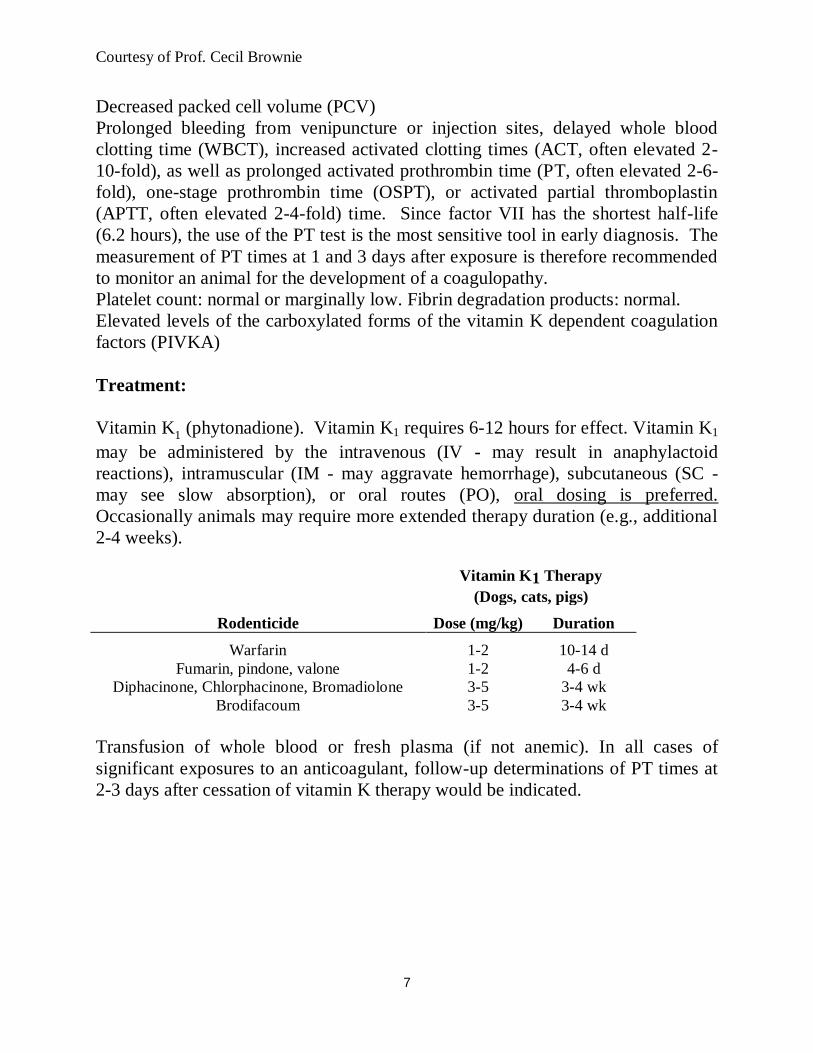
Treatment:
Vitamin K1 (phytonadione). Vitamin K1 requires 6-12 hours for effect. Vitamin K1
may be administered by the intravenous (IV - may result in anaphylactoid
reactions), intramuscular (IM - may aggravate hemorrhage), subcutaneous (SC -
may see slow absorption), or oral routes (PO), oral dosing is preferred.
Occasionally animals may require more extended therapy duration (e.g., additional
2-4 weeks).
Vitamin K1 Therapy
(Dogs, cats, pigs)
Rodenticide Dose (mg/kg) Duration
Warfarin 1-2 10-14 d
Fumarin, pindone, valone 1-2 4-6 d
Diphacinone, Chlorphacinone, Bromadiolone 3-5 3-4 wk
Brodifacoum 3-5 3-4 wk
Transfusion of whole blood or fresh plasma (if not anemic). In all cases of
significant exposures to an anticoagulant, follow-up determinations of PT times at
2-3 days after cessation of vitamin K therapy would be indicated.
Page 8
Courtesy of Prof. Cecil Brownie
8
Bromethalin
Source and Chemistry:
Bromethalin is a single-dose rodenticide that causes central nervous system
depression and paralysis, leading to death in 2 to 4 days. Trade names include
Vengeance®, Assault®, Trounce® , and others. Bromethalin-based rodenticides
are pelleted (often tan colored) grain-based products that contain 0.75 - 1.5 ounces
(21 - 42 gram) of bait in paper "place pack" envelopes.
Chemical name: N-methyl-2,4-dinitro-N-(2,4,6-tribromophenyl)-6-
(trifluoromethyl) benzenamine
Toxicity:
Reported LD50s for technical grade bromethalin include: 1.8 mg/kg in the cat, 4.7
mg/kg in the dog, 13 mg/kg in rabbits, and > 500 mg/kg in the guinea pigs.
Minimum toxic doses of bromethalin baits: 1.67 grams/kg (dog) and 0.30 grams/kg
(cat).
Minimal lethal doses of bromethalin baits are 2.5 grams/kg (dog) and 0.45
grams/kg (cat).
Bromethalin-based baits acute oral LD50s are 3.65 grams/kg (dog) and 1.1
grams/kg (cat).
Relay (secondary) toxicity may occur.
Mechanism of Action:
Page 9
Courtesy of Prof. Cecil Brownie
9
Bromethalin uncouples oxidative phosphorylation resulting in a lack of adequate
ATP concentrations, so that insufficient energy is available for Na+ - K+ ion
channel pumps resulting in the development of cerebral edema.
Absorption, Distribution, Metabolism, and Excretion:
Rapidly absorbed from the gastrointestinal tract, with peak plasma concentrations
occurring ~ 4-hr after ingestion.
Excretion however, is slow and a plasma half-life of 5.6 days has been reported.
Bromethalin is N-demethylated by the liver to a more toxic metabolite
(desmethylbromethalin).
Clinical Signs:
Animals that ingest oral doses of bromethalin at or above the LD50 generally
develop clinical signs within 24 hours. Bromethalin ingestion at these higher oral
doses often produces an acute syndrome that is characterized by severe muscle
tremors, hyperthermia, extreme hyper-excitability, and focal motor and generalized
seizures.
Lower oral dose of bromethalin (less than an LD50 of bait) produces a toxic
syndrome with a slower onset of clinical signs (e.g., 24 - 86 hours). This syndrome
is characterized by the development of hindlimb ataxia and/or paresis and/or
central nervous system depression, extensor rigidity, and opisthotonus, anisocoria,
seizures, tremors, coma, and mydriasis.
Diagnosis:
The diagnosis of bromethalin poisoning is dependent upon the presence of an
exposure history to a potentially toxic dose of a bromethalin-based rodenticide and
the subsequent development of appropriate clinical signs within an appropriate
time-frame.
Chemical confirmation of bromethalin residues is not widely available, and may
have limited clinical utility in cases in which a delay in presentation (i.e., the
chronic syndrome) occurs.
Page 10
Courtesy of Prof. Cecil Brownie
10
Treatment:
Prevention of absorption in recently poisoned animals with emetics, repeated
administrations of activated charcoal, and saline or osmotic cathartics. Dexamethasone and osmotic diuretics have been reported to be effective.
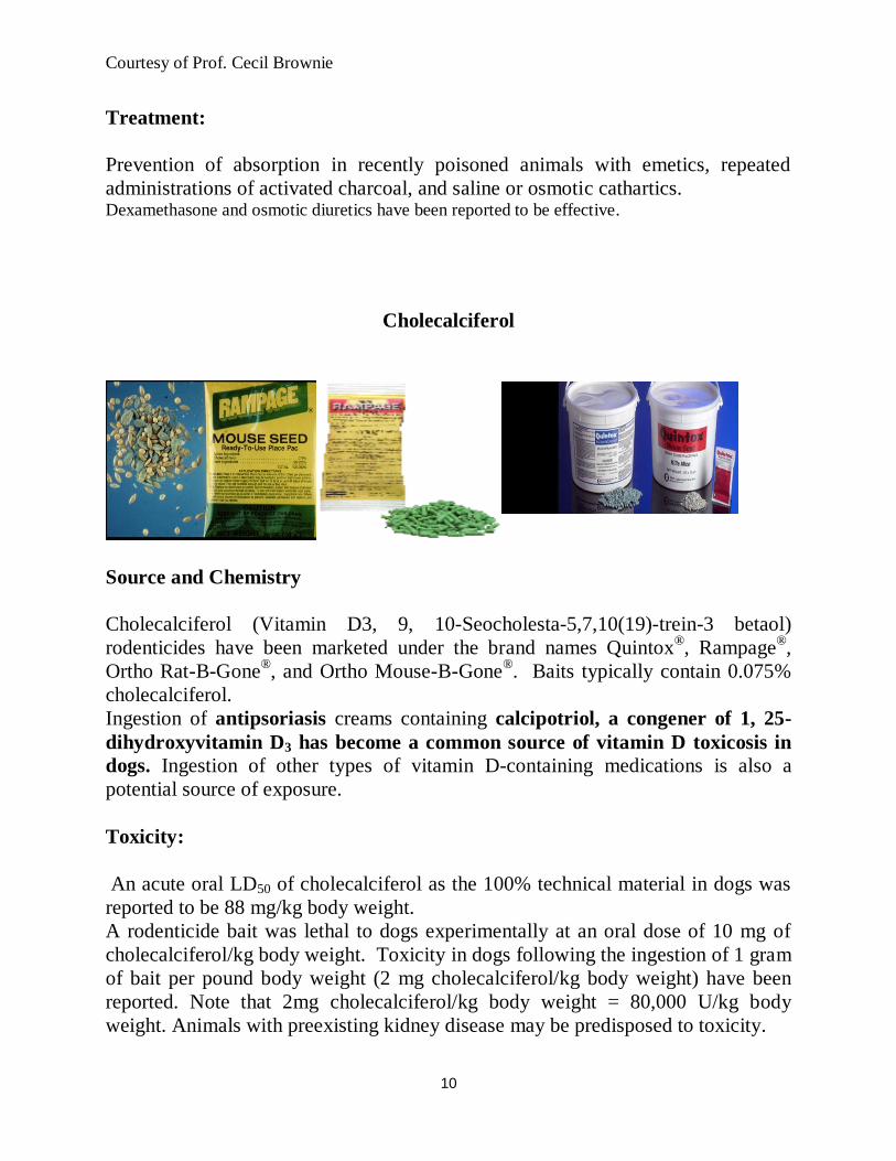
Cholecalciferol
Source and Chemistry
Cholecalciferol (Vitamin D3, 9, 10-Seocholesta-5,7,10(19)-trein-3 betaol)
rodenticides have been marketed under the brand names Quintox®, Rampage
®,
Ortho Rat-B-Gone®, and Ortho Mouse-B-Gone
®. Baits typically contain 0.075%
cholecalciferol.
Ingestion of antipsoriasis creams containing calcipotriol, a congener of 1, 25-
dihydroxyvitamin D3 has become a common source of vitamin D toxicosis in
dogs. Ingestion of other types of vitamin D-containing medications is also a
potential source of exposure.
Toxicity:
An acute oral LD50 of cholecalciferol as the 100% technical material in dogs was
reported to be 88 mg/kg body weight.
A rodenticide bait was lethal to dogs experimentally at an oral dose of 10 mg of
cholecalciferol/kg body weight. Toxicity in dogs following the ingestion of 1 gram
of bait per pound body weight (2 mg cholecalciferol/kg body weight) have been
reported. Note that 2mg cholecalciferol/kg body weight = 80,000 U/kg body
weight. Animals with preexisting kidney disease may be predisposed to toxicity.
Page 11
Courtesy of Prof. Cecil Brownie
11
Metabolism:
Cholecalciferol is fat soluble and is absorbed through chylomicrons into the
lymphatic system. Cholecalciferol is metabolized by the liver to 25-
hydroxycholecalciferol (25-OH-D3), which is the major circulating metabolite
during vitamin D excess. Further metabolism of 25-OH-D3 occurs in the kidney
where calcitriol (1,25-(OH)2-D3) is produced. Cholecalciferol and 25-
hydroxycholecalciferol have limited biological activity; calcitriol is the most potent
cholecalciferol metabolite in terms of enhancing bone resorption and intestinal
calcium transport.
Mechanism of Action:
Cholecalciferol and other vitamin D metabolites increase intestinal absorption of
calcium, stimulate bone resorption, and increase the renal tubular reabsorption of
calcium.
Clinical Signs:
The most common clinical signs include vomiting, depression, anorexia,
polydipsia, polyuria and diarrhea. Gastrointestinal and pulmonary hemorrhage
sometimes occur as an apparent result of dystrophic calcification, and should not
lead to a misdiagnosis of anticoagulant rodenticide toxicity.
CH2
HO 1
6
3
8
24
25
Vitamin D3: Cholecalciferol
CH2
HO 1
6
3
8
24
25
25-OHD 3: Calcifediol
OH
Hepatic microsomes
1,25-(OH)2D3: Calcitriol
CH2
HO 1
6
3
8
24
25 OH
OH
Renal
mitochondria
Page 12
Courtesy of Prof. Cecil Brownie
12
Clinical signs generally develop within 12-36 hours of ingestion of cholecalciferol.
Clinical Pathology:
Hypercalcemia (serum calcium > 12 mg/dl), and associated dystrophic
calcification.
Hyperproteinemia, hyperphosphatemia and azotemia can occur.
Urinalyses may reveal hyposthenuria (Uspg 1.001-1.007), proteinuria, and
glucosuria. Urine sediment examination occasionally reveals leukocytes,
erythrocytes, and casts in variable numbers; metabolic acidosis.
Pathology:
Gross lesions in dogs poisoned with cholecalciferol based rodenticides may
include diffuse hemorrhage in the gastric mucosa, duodenum, and jejunum.
Microscopic lesions may include necrosis and mineralization of the myocardium
and of the arterial intima.
Mineralization of glomerular capillary walls, renal cortical tubular basement
membranes, Bowman's capsules, and stomach has been described.
Diagnosis:
The diagnosis of a cholecalciferol rodenticide poisoning depends upon a history of
a potentially toxic level of exposure, appropriate clinical signs, and the
development of hypercalcemia.
Hyperphosphatemia and hypercalcemia tend to develop within 12 and 24 hours,
respectively, after the ingestion of a cholecalciferol based rodenticide.
Mineralization may be appreciated radiographically.
Elevated serum concentrations of cholecalciferol and its primary metabolites, 25-
hydroxycholecalciferol, 24,25-dihydroxy- cholecalciferol and/or 1,25-
dihydroxycholecalciferol, may also support a diagnosis.
Total kidney calcium concentrations may be elevated when compared to normal
animals.
Baseline serum calcium determinations are recommended for all cases of
potentially toxic ingestion. These serve as basis for comparison with subsequent
time points. The initial calcium values obtained are likely to be within the normal
range (even when potentially lethal doses are consumed) for up to several hours
after ingestion.
Page 13
Courtesy of Prof. Cecil Brownie
13
Treatment:
Treatment goals include –
1). Detoxification of the gastrointestinal tract to decrease cholecalciferol
absorption via administration of an emetic and activated charcoal with a
saline or osmotic cathartic
2). Correcting fluid and electrolyte imbalances.
3). Initiation of specific therapies that will prevent or reduce the
hypercalcemic state:
Calciuresis with IV 0.9 % sodium chloride
Furosemide at 2-5 mg/kg every 8-12 hours
Oral prednisone at 2 mg/kg every 12 hours
Salmon calcitonin sq at 4-6 IU/kg every 2 to 3 hours until serum
calcium levels stabilize
4). Rarely, seizure control, treatment of arrhythmias, and other symptomatic
therapies may be required.
Following significant exposures, serum calcium and BUN should be determined at
1, 2, and 3 day's post-exposure. If hypercalcemia (serum calcium > 12 mg/dl) is
present or the animal is symptomatic, then further diuresis and additional therapies
may be indicated.
Vitamin D-induced hypercalcemia often persists for several weeks requiring long
term management.
After the serum calcium is stabilized within the normal range, maintenance therapy
consisting of furosemide orally at 2-4.5 mg/kg every 8-12 hour and oral prednisone
at 2 mg/kg every 12 hour.
Calcitonin is the recommended drug for treatment of cholecalciferol-induced
hypercalcemia. Calcitonin by itself is ineffective for treatment of cholecalciferol-
induced hypercalcemia and is always accompanied by aggressive fluid, diuretic,
and corticosteroid administration. The half-life of calcitonin is short (3 to 4 hours);
therefore, multiple daily injections must be given for 2 to 3 weeks to maintain
blood calcium levels within the physiologic range. Prolonged calcitonin
administration is associated with undesirable adverse effects including vomiting
and anorexia. Decreased responsiveness has also been reported in humans.
Pamidronate is a potentially useful antidote against cholecalciferol toxicity in
dogs. Pamidronate (aminohydroxy-propylidene biphosphonate) and clodronate are
bisphonates - a new class of inhibitors of bone resorption. In a recent experimental
Page 14
Courtesy of Prof. Cecil Brownie
14
study (Rumbeiha et al., AJVR 61: 9-13, 2000), cholecalciferol-poisoned Beagle
dogs were treated at 1 and 4 days after cholecalciferol ingestion with intravenous
infusions of either 1.3 mg or 2.0 mg of pamidronate/kg in 0.9% saline (150 ml total
volume given during a 2-hr period). Treated dogs demonstrated decreased serum
calcium levels and improved renal function when compared to animals given saline
alone.
Strychnine
Source and Chemistry
Strychnine is a colorless crystal or white bitter tasting indole alkaloid powder
extracted from the seeds of the plants Strychnos nux-vomica and Strychnos
ignatii. Strychnine was first used in medicine (e.g., Nux vomica) and as an animal
poison since the sixteenth century.
Pesticidal applications: rat control.
Many commercially available baits are pelleted and are dyed either bright green or
red.
Preparations containing less than 0.5% strychnine are approved for subsoil use
against burrowing rodents (restricted use pesticide).
Continues to remain as a potential cause of toxicosis particularly in dogs.
Absorption, Distribution, Metabolism, and Excretion:
Oral exposure is common and vomiting often does not occur, stomach contents
may contain high levels of strychnine. Strychnine appears to be readily absorbed
from the intestinal tract
Excretion is accomplished in urine (5-20% unchanged) and through secretion into
the stomach.
Strychnine is ionized in acidic media (pK1 = 6.0, pK2 = 11.7). Therefore,
strychnine should be highly ionized in the stomach and not readily absorbed. Ion
trapping of strychnine occurs in the acid conditions of the stomach and urinary
excretion may be enhanced by acidification of the urine.
Highest concentrations of strychnine are found in blood, liver and kidney.
Hepatic microsomal enzymes metabolize strychnine.
Mechanism of Action:
Strychnine reversibly antagonizes the action of glycine in the spinal cord and
medulla. Net effect is reduced action of inhibitory postsynaptic neurons.
Page 15
Courtesy of Prof. Cecil Brownie
15
The physiologic effect of strychnine is uncontrolled reflex activity. All striated
muscle groups are affected, but the relatively more powerful extensors
predominate to produce symmetrical and generalized rigidity and tonic seizures.
Sublethal convulsant doses of strychnine may elevate blood pressure, heart rate
and right ventricular pressure in dogs.
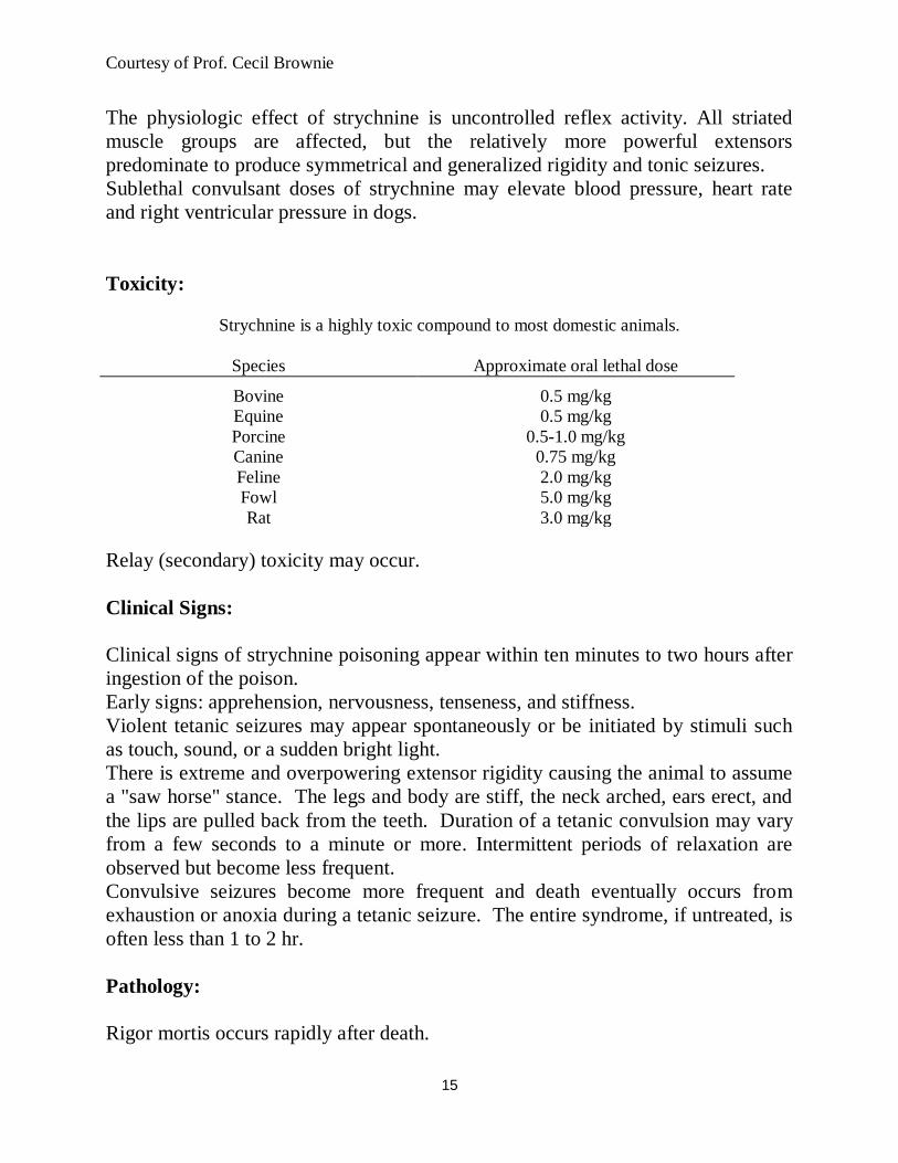
Toxicity:
Strychnine is a highly toxic compound to most domestic animals.
Species Approximate oral lethal dose
Bovine 0.5 mg/kg
Equine 0.5 mg/kg
Porcine 0.5-1.0 mg/kg
Canine 0.75 mg/kg
Feline 2.0 mg/kg
Fowl 5.0 mg/kg
Rat 3.0 mg/kg
Relay (secondary) toxicity may occur.
Clinical Signs:
Clinical signs of strychnine poisoning appear within ten minutes to two hours after
ingestion of the poison.
Early signs: apprehension, nervousness, tenseness, and stiffness.
Violent tetanic seizures may appear spontaneously or be initiated by stimuli such
as touch, sound, or a sudden bright light.
There is extreme and overpowering extensor rigidity causing the animal to assume
a "saw horse" stance. The legs and body are stiff, the neck arched, ears erect, and
the lips are pulled back from the teeth. Duration of a tetanic convulsion may vary
from a few seconds to a minute or more. Intermittent periods of relaxation are
observed but become less frequent.
Convulsive seizures become more frequent and death eventually occurs from
exhaustion or anoxia during a tetanic seizure. The entire syndrome, if untreated, is
often less than 1 to 2 hr.
Pathology:
Rigor mortis occurs rapidly after death.
Page 16
Courtesy of Prof. Cecil Brownie
16
No gross or microscopic lesions (e.g., neurons, axons, or myelin sheath)
characteristic of strychnine poisoning can be consistently detected.
Diagnosis:
Tentative diagnosis is usually based on history of ingestion, characteristic clinical
signs, and lack of lesions.
Characteristically, the stomach of strychnine-poisoned animals is filled with food
or bait that has not been completely digested.
Samples for analysis should include stomach contents, liver, kidney, urine, and
central nervous system. In addition, bait or vomitus should be kept for analysis.
Most chemical confirmations of strychnine poisoning are from stomach contents or
liver. Concentrations of strychnine in dogs dying of acute poisoning ranged from 0
to 52 ppm in liver and from 0 to 12,800 ppm in stomach contents.
Treatment:
Of prime concern in strychnine poisoning is prevention of asphyxia.
Morphine should be avoided, due to possibility of respiratory depression.
Ketamine should also be avoided, since it has motor stimulant effects in the brain.
Oral detoxification: Induce vomiting (e.g., apomorphine) if animals are not
hyperesthetic or convulsive.
Activated charcoal very effective (pretreatment with diazepam may be required).
In anesthetized animals, gastric lavage could be used.
Forced diuresis (5 % mannitol in 0.9 % sodium chloride administered at the rate of
6.6 ml/kg/hour) and acidification of the urine with ammonium chloride (132
mg/kg, PO) may enhance excretion of strychnine. Forced diuresis with
acidification has had limited benefit in many cases.
Facilities for positive pressure pulmonary ventilation, oxygen administration, and
warm quiet conditions should be readily available.
Control seizures e.g., diazepam (2-5 mg/kg, IV) is the drug of choice although
pentobarbital may be required. Resolution of signs generally occurs within 1-2 days
Page 17
Courtesy of Prof. Cecil Brownie
17
Zinc Phosphide
Source:
Zinc phosphide (Zn3P2) has been available commercially for rodent control since
1930.
Zinc phosphide, or occasionally aluminum phosphide, is employed in baits of
bread, bran mash, soaked wheat, damp rolled oats, or sugar at concentrations of
from 2 to 5 percent.
Zinc phosphide is a dull, grayish black powder, insoluble in water and having a
faint acetylene odor in atmospheric air.
Toxicity:
Most animals and poultry can be poisoned by 40 mg/kg depending upon the pH in
the stomach.
Oral LD50 of zinc phosphide in rats is 40.5 mg/kg body weight.
A dose of 40 mg/kg zinc phosphide administered to a dog by gelatin capsule
induced onset of convulsive seizures seven hours after administration, and the
animal expired within thirty minutes after the onset of convulsions. Dogs fed zinc
phosphide in amounts of 300 mg/kg can survive if the toxicant is given on an
empty stomach. Feeding dogs a normal ration stimulates gastric HCl secretion and
greatly increases their susceptibility to zinc phosphide.
Some commercial products contain tartar emetic to stimulate vomiting as a
protective measure in non-target species. Zinc phosphide itself has a strong
tendency to induce emesis in nonrodent animals, and it does not consistently cause
fatal poisoning when accidentally ingested.
Mechanism of Action:
Liberates phosphine gas upon acid hydrolysis in the stomach.
CNS effects are due to an unknown mechanism of action.
Page 18
Courtesy of Prof. Cecil Brownie
18
Clinical Signs:
Onset of poisoning is rapid, usually within fifteen minutes to four hours after
ingestion of a toxic amount of zinc phosphide. Death from large doses is usually
within three to five hours, and animals rarely survive longer than 24-48 hr. In
some instance's onset of clinical signs may be delayed as long as 12-18 hr after
ingestion.
Early signs: anorexia, lethargy, vomiting, tachypnea, and abdominal pain. Ataxia,
weakness, and recumbency follow. There may be terminal hypoxia, gasping for
breath, and struggling. Also, hyperesthesia, "running fits," and seizures can be
seen. Metabolic acidosis can occur.
Pulmonary edema. Liver and/or kidney failure may occur.
Pathology:
There is marked congestion of the lungs and interlobular edema in some cases.
Pleural effusion and sub-pleural hemorrhages may be seen. Myocardial
degeneration may occur.
The liver and kidney are extremely congested in acute cases. Renal tubular
degeneration, hyaline change, and necrosis may occur, gastritis. The freshly
opened stomach has a characteristic odor of acetylene.
Diagnosis:
History of exposure to zinc phosphide, accompanied by rapid death characterized
by dyspnea, vomiting, pulmonary edema, and visceral congestion suggests zinc
phosphide poisoning.
Chemical detection of zinc phosphide in stomach contents or gastric lavage is
possible.
Zinc levels in blood, liver, and kidney may be elevated.
Treatment:
Correct acidosis and hypocalcemia
If detected early, emesis should be induced. Activated charcoal.
Breakdown of zinc phosphide to phosphine may be retarded by gastric lavage with
aluminum or magnesium hydroxide gel.
Page 19
Courtesy of Prof. Cecil Brownie
19
MINOR RODENTICIDES
Alpha—Napthyl Thiourea (ANTU)
Alpha-naphthyl thiourea (ANTU) is used exclusively as a rodenticide.
Rodents are very susceptible to ANTU toxicity (rat oral LD50 6-7 mg/kg).
Dogs, cats, and swine are the most susceptible domestic animals. Mature and aged
dogs are more susceptible than young ones. The single oral lethal dose in the
mature dog is recorded to be between 10 and 50 mg/kg body weight. In young
dogs the single lethal dose is 85-100 mg/kg.
ANTU causes pulmonary edema.
Treatment
Symptomatic and supportive.
Phosphorus
Phosphorus is used little today because better, safer rodenticides are available. The
characteristic odor is described as garlic-like.
Lethal dose in most species 1-2 mg/kg.
Clinical Signs:
Phase l: Initial signs occur within hours of ingestion and characterized by
gastrointestinal, abdominal and circulatory signs: vomiting ± blood, cardiac
arrhythmias, no diarrhea, shock cyanosis, incoordination and coma may develop
with death occurring before the second and third phases appear.
Phase lI: an interim or latent phase (1-3 days after ingestion) in which apparent
recovery occurs.
Phase lII: recurrence of severe clinical signs characterized by vomiting and
occasionally hematemesis, icterus and hepatic failure and central nervous system
dysfunction. Hypoprothrombinemia and a tendency to bleeding especially from
gingiva, stomach, intestine or kidney. Hypoglycemia may be severe and liver
enzymes are elevated. Oliguria can develop attended by a rise in blood urea
nitrogen and other parameters associated with renal tubular damage. Urinalysis
Page 20
Courtesy of Prof. Cecil Brownie
20
reveals albuminuria, hematuria and increased concentrations of amino acids.
Phosphorus content of the blood is generally normal.
Treatment:
Symptomatic and supportive.
Sodium Monofluoracetate (compound 1080)
Sources:
The primary use of fluoroacetate is to control predators. In the United States,
fluoroacetate compounds are mixed with a black dye (nigrosine) and are available
only to licensed exterminators.
Dogs are very susceptible and as little as 0.05 mg/kg may be lethal.
Undergoes synthesis to fluorocitrate which then acts to inhibit the enzyme
aconitase in the Krebs or tricarboxylic acid cycle resulting in a buildup of citrate
and inhibition of cellular respiration.
Page 21
Courtesy of Prof. Cecil Brownie
21
Convulsions may be prominent or cardiac effects may be dominant. In all species
there is a characteristic latent period of from 0.5-2h after ingestion of 1080. When
signs commence, the onset is acute and the course rapid and usually violent.
Treatment:
Symptomatic and supportive (Decontaminate the GI tract and seizure control).
Thallium
Pesticide use prohibited since 1965.
The LD50 for most species is in the range of 10-15 mg/kg.
Thallium will accumulate in the body ---> chronic toxicity.
Rapidly absorbed from the GI tract.
Excreted both in the urine and in the feces but is only slowly removed from the
body.
Interferes with oxidative phosphorylation.
Clinical Signs:
Acute Form: 1-4 days of ingestion: severe gastric irritation, vomiting, severe
hemorrhagic diarrhea, abdominal pain and anorexia. Lingual ulcers have been
reported in cats. In the acute form the dog or cat may die from the severe gastritis,
depression and renal damage within three to six days. In the acute form motor
paralysis and trembling may occur.
Sub-acute Form: 2-7 days of ingestion: mild gastric distress with prominent skin
changes. There is a reddening of the skin and early pustular formation. This
usually begins on the ears and nose and then will proceed to involve the axillary
region, ventral abdomen and torso. Marked reddening of the oral membranes and
to a lesser extent the skin.
Chronic Form: 7-10 days of ingestion: loss of hair and the drying and scaling of
the skin.
Page 22
Courtesy of Prof. Cecil Brownie
22
Pathology:
Severe hemorrhagic gastroenteritis
Fatty degeneration and necrosis of the liver, and congestion and hemorrhage of the
spleen, heart and kidneys may be observed.
Hyperkeratosis, parakeratosis, hyperemia and some hyalinization of the skin.
Diagnosis:
Mixed GI and skin effects
Detection of thallium in the urine or tissues.
Treatment:
Symptomatic and supportive. Forced potassium diuresis.
Decontamination of GI tract (emetics, activated charcoal)
Prussian Blue
Page 23
Courtesy of Prof. Cecil Brownie
23
INSECTICIDES
Syndrome – Toxic response
Acute: Organophosphates/Carbamates
Pyrethrins/Pyrehroids
Arsenic
Organo-chlorine
Chronic: Delayed neuropahy with some Organophsphates and among some
animal and birds species
Residues:
Organo-chlorines (DDT, Mirex, and others)
Organophosphorus (OP) and Carbamate Insecticides
Source and Chemistry
The most prevalent and toxic chemical warfare agents are potent
organophosphorus compounds (soman, sarin, tabun). These compounds are not
used as insecticides. Carbamate insecticides, are derivatives of carbamic acid are
used for similar surface applications. The original carbamate, physostigmine, is a
plant alkaloid, derived from the "ordeal bean."
Animal poisonings from organophosphorus (e.g., Asuntol,
chlorpyrifos,dichlorvos)/carbamate (e.g. aldicarb, methomyl, propuxur, carbaryl,
carbofuran) insecticides commonly result from their deliberate dermal application
or accidental ingestion. Most of these insecticides are applied to surfaces of plants,
animals, soils, or household, floors, etc. Others are systemics (e.g., Spot-On®) that
are absorbed via any route by plants or animals and then distributed through the
organism on which the insect pest feeds.
An important source of toxicity in the cat results from the inappropriate use
of chlorpyrifos-based products used for the control of fleas and other ectoparasites.
Flea control formulations available for domestic animals that contain chlorpyrifos
include dips, sprays (polymer-based), and collars. However, except for flea
collars, chlorpyrifos is not approved for use on cats. The inappropriate use of
chlorpyrifos-based products on cats results in a significant number of poisoning
Page 24
Courtesy of Prof. Cecil Brownie
24
cases each year. Chlopyrifos and Diazinon based formulations have been
withdrawn from sale in the US in 2000 and 2004 respectively.
Environmental contamination: Concern varies with the specific
Compound and their route of exposure. Whereas the carbamates (except for
aldicarb in ground water and on vegetables) are less of a concern environmentally
the organophosphorus insecticides tend to be much less persistent than the
organochlorine insecticides.
Generic Name Trade Names
Acephate Orthene, Asataf, Pillarthene, Kitron, Aimthane, Ortran, Ortho 12420, Ortril,
Chrevron RE 12420, and Orthene 755
Chlorpyrifos Brodan, Detmol UA, Dowco 179, Dursban, Eradex, Lorsban, Piridane,
Stipend
Coumaphos Agridip, Asunthol, Meldane, Muscatox, Umbethion, Co-Ral, Asuntol, Bay
21, Baymix, Dilice, Resistox, Suntol, Negashunt
Diazinon Knox Out, Spectracide and Basudin
Dimethoate Cekuthoate, Chimigor 40, Cygon 400, Daphene, De-Fend, Demos NF,
Devigon, Dimate 267, Dimet, Dimethoat Tech 95%, Dimethopgen,
Ferkethion, Fostion MM, Perfekthion, Rogodan, Rogodial, Rogor, Roxion,
Sevigor, Trimetion
Absorption, Distribution, Metabolism, and Excretion:
Exposures – oral, ophthalmic, respiratory and dermal routes
Generally lipophilic compounds and are well absorbed, widely distributed,
accummulate in fat, liver metabolized, and excreted as metabolites in urine.
Organophosphates – Some OPs (especially those with a -P=S moiety, e.g.,
parathion) require metabolic activation, forming Oxons which irreversibly
phosphorylate cholinesterase, becoming more toxic over a longer duration due to
aging OP-Acetylcholinesterase complex rather than the new enzyme
(Acetylcholinesterase) synthesis.
Carbamates- Inhibit cholinesterase enzyme “carbamylation” ester. Complexation
is labile, reversible and of a shorter duration of action.
Toxicity:
Page 25
Courtesy of Prof. Cecil Brownie
25
Wide variation depending on dose and compound involved - Characterized in the
case of OPs as highly toxic (e.g parathion), intermediate toxicity (e.g. coumaphos)
or low toxicity (e.g. fenthion); and in the case of Carbamates as extremely toxic
(e.g. aldicarb, methomyl), highly toxic (e.g. propuxur, aminocarb) or moderate
toxicity (e.g. carbaryl).
LD50 Chlorpyrifos in the cat is between 10 and 40 mg/kg. Some cats may
develop severe clinical signs following exposures to even smaller doses especially
if chronically exposed.
Mechanism of Action:
Competitive inhibitors of cholinesterase enzymes (Acetylcholinesterase or true
Cholinesterase and Pseudo-cholinesterase or plasma cholinesterase) activity
Acetylcholinesterase – present in RBC membrane
Pseudo-cholinesterase – present in plasma, liver, pancreas and CNS.
Acetylcholine (ACH) is the mediator at junctions including those between
preganglionic and postganglionic neurons in both the parasympathetic and
sympathetic nervous system, smooth muscles or glands, motor nerves and skeletal
muscles, and some neuron to neuron junctions in the CNS.
Acetylcholinesterase (ACHE) is the enzyme that rapidly hydrolyzes ACH at these
locations. Actual physiological purposes of the enzymes located in blood are
unknown.
Clinical Signs:
Organophosphorus and carbamate insecticides are potent inhibitors of
acetylcholinesterase in mammals and produce three categories of effects namely:
muscarinic (salivation, lacrimation, bronchial secretion, vomiting, diarrhea),
nicotinic (tremors, respiratory paralysis), and CNS (seizures, miosis,
hyperactivity).
Death is usually due to one or more of the following effects: increased respiratory
tract secretions and bronchiolar constriction with hypoxia aggravated by
bradycardia; respiratory depression from nicotinic stimulation to the point of
paralysis; and respiratory paralysis from CNS depression due to central effects of
the insecticide (may be the primary cause of respiratory-failure in some species).
Page 26
Courtesy of Prof. Cecil Brownie
26
Horses--colic, abdominal pain, salivation, severe diarrhea (often watery),
dehydration.
Chlorpyrifos toxicosis in cats and bulls. The classical syndrome of OP
poisoning characterized by salivation, lacrimation, urination, and defecation
("SLUD") is rarely observed in cats or bulls except following acute oral exposure.
More typically, animals are presented following a delayed onset of 1 to 5 days after
dermal exposure. Clinical signs in cats (and bulls to some extent) may include
depression, ataxia, tremors (generally involving the head, neck, and back),
behavior changes (personality change, aggression), hyperactivity, and
hyperesthesia, as well as miosis or mydriasis. Other signs include anorexia,
salivation, vomiting, diarrhea, tachypnea, and dyspnea. Tachypnea, and dyspnea
result from excessive bronchoconstriction and hypersecretion and may be life-
threatening in overly stressed cats. Clinical signs commonly persist for several
weeks in topically dosed cats and bulls. This duration may be the result of
prolonged cholinesterase inhibition and debilitation that results in hypokalemia.
Although not specific for chlorpyrifos toxicosis, electromyographic (EMG)
abnormalities reported to occur in chlorpyrifos-poisoned cats include prolonged
insertion activity, fibrillation potentials, positive sharp waves, and high-frequency
discharges consistent with a neuropathy or neuromyopathy. Electromyographic
changes are most severe in the pelvic limbs.
Interestingly, in addition to the acute effects of organophosphorus insecticides,
some compounds (e.g. phosphoramidates, phosphonates) are associated with the
rare development of organophosphate induced delayed neuropathy (OPIDN),
characterized by central-peripheral distal axonopathy ("dying back
polyneuropathy"). For example, in addition to its acute effects, experimental
chlorpyrifos toxicosis has also associated with the development of OPIDN.
Although controversial, the postulated mechanism of action in the development of
OPIDN involves the phosphorylation and aging of a second class of esterase,
neuropathy target esterase (NTE). In the cat, this neuropathy is characterized by a
central-peripheral distal axonopathy ("dying back polyneuropathy"). The
appearance of clinical signs is delayed for up to 2 to 3 weeks after exposure. This
syndrome is rarely encountered clinically, but when it occurs it is characterized by
ataxia (waddling gait), hind-limb hypermetria, depressed conscious proprioception,
weakness, and an ascending paralysis.
Page 27
Courtesy of Prof. Cecil Brownie
27
Diagnosis:
A diagnosis is based upon a history of exposure, development of appropriate
clinical signs, and chemical analysis of stomach contents and tissues (available at
most diagnostic laboratories).
Test try dose of atropine: a pre-anesthetic dose of atropine and, if normal
atropinization occurs (normal extent and duration of rapid heart rate, dry mouth
and mydriasis), it tends to rule out poisoning from a cholinesterase inhibitor.
Pathology: Nonspecific. Salivation, Bronchial secretion or pulmonary edema.
Occasional hemorrhages in gastrointestinal tract serosa and mucosa.
Determination of reduced cholinesterase activity (< 50% normal activity) in blood,
brain, and retinal tissues is highly supportive of a diagnosis. Depending on the
diagnostic laboratory's preference, whole blood samples (collected in the
appropriate anticoagulant), serum, plasma, or brain tissue (one hemisphere) should
be frozen and shipped on ice.
Reduced whole blood cholinesterase activity may suggest exposure to an
organophosphate or carbamate and, although, not a conclusive test, should be
performed on animals presented alive. In most species, whole blood cholinesterase
is considerably less sensitive than pseudocholinesterase. Therefore, depression of
the activity of whole blood cholinesterase is more indicative of serious exposure or
toxicosis than plasma pseudocholinesterase. Conversely, plasma
pseudocholinesterase is a more sensitive test for detecting exposure than whole
blood cholinesterase. It is possible to have no plasma cholinesterase activity
detected in animals exposed to therapeutic (parasiticidal) amounts of OPs. In most
species, the red blood cells contribute the major fraction of the activity in total
blood cholinesterase. In most instances, the whole blood cholinesterase values of
poisoned animals fall below 25% of control values. Feline whole blood
cholinesterase activity is comprised primarily of pseudocholinesterases that are
extremely sensitive to inhibition by organophosphorus and carbamate insecticides.
Therefore, reduced cholinesterase activity may suggest exposure but may be
difficult to use prognostically. Blood cholinesterase activity may not be
representative of CNS cholinesterase activity in a symptomatic animal (false
negative).
Because the incubation time allows for decarbamylation, the MICHEL
METHOD of cholinesterase analysis may give false negative (false normal)
Page 28
Courtesy of Prof. Cecil Brownie
28
values for carbamate toxicoses. Effects of various insecticides on whole blood
acetylcholinesterase (AChEase) activity (incubation temperature and times).
Whole blood AChEase
Insecticides 37C; 5 min 37C; 60 min
Organophosphorus Decreased Decreased
Carbamates Decreased Normal
Pyrethrin and pyrethroids Normal Normal
Organochlorines Normal Normal
Rotenone Normal Normal
d-Limonene and linalool Normal Normal
Tissues of internal organs usually do not yield detectable residues of
organophosphate or carbamate compounds even in seriously poisoned animals.
Stomach or rumen contents are often important in confirming diagnoses after oral
exposure because the insecticides are most often detected in these samples (ship
frozen).
Differential Diagnoses:
Insecticides that are commonly used for the control of ectoparasites on companion
animals include organophosphorous, carbamates, pyrethrins, pyrethroids,
organochlorines, d-limonene, d-linolool, rotenone, pennyroyal oil, and amitraz. Of
these insecticides, organophosphorous, carbamates, pyrethrins, pyrethroids, and
citrus oil derivatives (d-limonene and d-linolool) may induce central nervous
system depression. Organophosphorous, carbamates, pyrethrins, pyrethroids,
rotenone, and organochlorine insecticide toxicoses commonly result in muscle
tremors and central nervous system stimulation.
Clinical Pathology:
Measurement of whole blood, brain, or retinal acetylcholinesterase activity is
diagnostically useful in cases in which an exposure to an organophosphorus or
carbamate insecticide may have occurred
Hypokalemia and elevation of serum creatine kinase activity has also been reported
in chlorpyrifos-poisoned cats.
Page 29
Courtesy of Prof. Cecil Brownie
29
Treatment:
Decontamination: Very early, emetics (only for very recent exposures; never
when contraindicated and constant monitoring will not be undertaken). Activated
charcoal (always use for any recent oral exposure) administered with appropriate
precautions to avoid aspiration. Thoroughly bathe with detergent all animals
exposed topically taking care to avoid exposure of human skin.
Atropine sulfate (0.1-0.2 mg/kg, intramuscular or intravenous, repeated as needed)
is antidotal, and can be used to control salivation and bronchial secretions
(muscarinic effects). Atropine will not control muscle tremors (nicotinic). The
initial dose of atropine should be divided, with one quarter given intravenously and
the remainder given either subcutaneously or intramuscularly. Since long-term
atropine therapy may be required, one should use the lowest dose that alleviates the
dyspnea and bradycardia. The reversal of excessive salivation may serve as a
useful clinical marker of effective atropinization. Do not attempt to monitor
atropinization using pupil size, since this is an unreliable indicator in cats and other
animals. The dose of atropine should be decreased or discontinued if tachycardia,
gastrointestinal stasis, severe behavioral changes (e.g., delirium), or hyperthermia
develop. Lower doses of atropine are required in horses to avoid a possibly lethal
atropine-induced colic. May want to avoid atropine sulfate altogether in horses
due to gut stasis, (except with life-threatening pulmonary or cardiac effects).
When used in horses, atropine is added to fluids and administered IV while
ausculting the abdomen. Administration is stopped prior to a reduction (to less
than normal) in gastrointestinal sounds. Some rabbits have atropinase and may
require higher dosing, begin at 1.0 mg/kg but may need to increase to 10 mg/kg. In
all cases, subsequent doses of atropine should depend on the reappearance and
severity of clinical signs.
Enzyme reactivators act on the organophosphorus insecticide-acetylcholinesterase
complex to free the enzyme and restore normal function. These agents are only
effective if covalent binding of the organophosphorus insecticide to
acetylcholinesterase (aging) has not yet occurred. Aging involves the
cholinesterase inhibitor-bound enzyme, and usually occurs within 24 hours of the
initial binding of the insecticide to the enzyme. Even with an extended
postexposure interval and possibility of aging, enzyme reactivators may still be
useful. Skin and subcutaneous fat may serve as a depository for some insecticides
(e.g., chlorpyrifos) following topical exposure, redistribution from this depot
results in continued enzyme exposure to the insecticide with subsequent aging.
Page 30
Courtesy of Prof. Cecil Brownie
30
The newly formed, but not aged enzyme-insecticide complex, is the site of action
for the enzyme reactivators.
Of the enzyme reactivators, pralidoxime chloride (Protopam chloride, Wyeth-
Ayerst Laboratories, Philadelphia, PA) has received the widest clinical use.
Pralidoxime chloride is given (20 mg/kg, IM, repeated every 12 hr), to relieve
tremors and other nicotinic signs and should be continued until these signs are
abolished or until additional prolonged benefit (lasting more than a day) is no
longer observed. Cattle and horses can be dosed at 10-20 mg/kg. Pralidoxime
chloride is generally of low toxicity; however, overdoses can cause tachycardia
and cardiac arrhythmias. Not effective for carbamate insecticide toxicosis.
Aggressive symptomatic and supportive care (hypokalemia is common in
chlorpyrifos-poisoned cats; oral potassium supplementation may be required) and
maintenance of caloric intake and hydration status. Artificial respiration may be
needed to counteract respiratory paralysis. It is essential to avoid hypoxia.
Seizures may be controlled with diazepam or barbiturate anticonvulsants.
Systemic acidosis may complicate OP poisoning. Sodium bicarbonate administered
at an initial dose of 5 mg/kg IV can be used to correct acidosis. Subsequent 1-3
mEq/kg IV doses may be required. Monitor acid base status. Animals should be
monitored for the development of chemical pneumonitis due to secondary
aspiration of hydrocarbon solvents if such formulations are ingested; and for
aspiration of gastric contents. Stress may cause secondary clinical problems, e.g.,
hemobartonella, reported in OP-poisoned cats.
It is critical that further exposures to organophosphorus and carbamate insecticides
be avoided until the animal is fully recovery. Exposure to another
acetylcholinesterase inhibitor should not be allowed until 4 to 6 weeks after the
initial exposure. It is unknown whether prior organophosphorus or carbamate
insecticide exposure increases the sensitivity of animals to other classes of
nonacetylcholinesterase-inhibiting insecticides.
Page 31
Courtesy of Prof. Cecil Brownie
31
Organochlorine Insecticides
Source and Chemistry
Animal exposures to organochlorine insecticides (e.g., lindane, endrin, DDT)
continue to result in serious neuro-toxicoses.
DDT type (diphenyl aliphatic)--includes DDT, methoxychlor, perthane, and
dicofol. Methoxychlor used a lot, including products for use around gardens,
animals.
Miscellaneous (lindane, mirex, kepone, paradichlorobenzene).
Lindane (hexachlorocyclohexane) is used in some veterinary insecticidal dips
and shampoos. Lindane toxicosis is most commonly the result of the overuse of
lindane-based insecticides on dogs or its inappropriate use on cats. Lindane
toxicosis is common in cats and dogs. Never approved for cats, used for dogs
(fleas, ticks, sarcoptic mange), also people (Qwell; for scabies). Some lindane
isomers are hepatocarcinogenic.
Cyclodiene type: aldrin, dieldrin, chlordane, endrin, heptachlor, "active
constituent" of toxaphene. Chlordane, heptachlor, aldrin used for termites. Aldrin
used to be used for corn root worm. Endrin has been used as an avicide, and
toxicosis in the cat can result from secondary poisoning from ingestion of endrin-
poisoned birds. Endrin at least in past used in "Rid-a-bird." Toxaphene used to be
made for animal dips, other uses.
Paradichlorobenzene (moth crystals, cakes, deodorant block used in diaper pales,
closets, rest rooms, and in other moth products occasionally including moth balls;
note: not the same as naphthalene; the most common compound in moth balls). Of
comparatively low toxicity, but does cause poisoning! Neurologic system toxicity
similar to other members of this group.
Absorption, Distribution, Metabolism, and Excretion:
Absorption variable. Typically very fat soluble.
Lindane like other organochlorine insecticides is very lipophilic, is
Rapidly absorbed, and tends to develop high fat and brain tissue
concentrations. As lindane and other organochlorine insecticides are very
lipophilic, they are rapidly absorbed, and tend to develop high fat and brain
tissue concentrations. Paradichlorobenzene metabolized to a phenolic and is
Page 32
Courtesy of Prof. Cecil Brownie
32
hepatotoxic. Aldrin is metabolized to dieldrin in the body and environment.
Heptachlor is metabolized to heptachlor epoxide. Dieldrin and heptachlor
epoxide are persistent in the body and the environment.
Toxicity:
LD50s in most species tend to be relatively high. Cyclodiene insecticides
cause more seizure activity and are more acutely toxic (lower LD50s) than
the DDT type organochlorines. LD50 endrin (cats) 3-6 mg/kg.
Lindane toxicosis is most commonly seen in cats (although dogs are sensitive) and
often follows the deliberate application of the product by the owner. The minimal
oral lethal dose for lindane in most species ranges from 5 to 25 mg/kg.
DDT: LD50 rat (oral) 113-2500 mg/kg, rat (iv) 47 mg/kg
DDT: unique aspects. Banned in 1972. DDT and its metabolites are potent
mixed-function oxidase inducers. Egg shell thinning--birds of prey
especially aquatic predators (e.g., bald eagles). O'P'DDD (synonymous with
TDE) and especially DDE are persistent metabolites in environment and
body. O'P'DDD--causes thinning of the adrenal cortex in the dog; therefore
used to treat Cushing's disease in the dog. Excessive use on dogs
can cause Addisonian crisis.
Mechanism of Action:
Methoxychlor, mirex, kepone, lindane, perthane, dicofol are similar to DDT
in structure and mechanism.Slows down the turning off of the Na+ influx
and inhibits the turning on of the K+ outflux; results in more of each cation
inside the nerve than normal. Therefore, the inside of nerve membrane is
more positive (partially depolarized) which decreases the threshold for
another action potential to occur. Sensory nerves are more readily affected
by DDT (and similar agents) than are motor nerves. EEG shows a diffuse
low amplitude, fast frequency pattern due to partial depolarization of
neurons.
Cyclodiene insecticides act by competitive inhibition of the binding of
GABA at its receptor. GABA is a more widely used inhibitory transmitter
in the CNS than glycine. Note the effects of GABA are facilitated by
benzodiazepines (such as Valium) and these drugs may therefore be of major
benefit in treatment. Lindane may also affect the GABA-receptor-ionophore
complex.
Page 33
Courtesy of Prof. Cecil Brownie
33
Clinical Signs:
All of these agents mainly cause tremors, salivation, ataxia, depression and
sometimes vomiting. Seizures may occur in cats at low doses or in other
species with very high doses.
Clinical signs of lindane toxicosis generally develop within 24 hours of
exposure.
Clinical signs may be progressive or explosive in nature, and commonly
include excitation, depression, tremors, clonic-tonic seizures, hyperactivity,
ataxia, circling, salivation, hyperthermia, and coma.
Electroencephalographic changes can include increased low amplitude-fast
frequency and spike activities.
Liver damage is occasionally observed several days after exposure.
Diagnosis:
History of exposure, appropriate clinical signs.
Pathology:
Liver damage is occasionally observed several days after exposure. There is
usually no histologic lesions in the brain or spinal cord.
Chemical analyses:
Brain analysis important for diagnosis of acute
toxicosis; submit ½ brain frozen (for organochlorine and other analyses and
virology). Other 1/2 should be fixed for histopathology to rule out infectious
(encephalitides), degenerative, or neoplastic diseases.
To determine sources, it may be appropriate to submit specimens for
analysis such as: feed; suspect insecticidal formulation (e.g., granules,
liquid, old containers, etc); gastrointestinal tract contents; hair (live animal)
or skin (dead animal) Liver, fat, and milk fat are the preferred samples to
assess residue contamination. It is essential to avoid any cross
contamination: e.g., from source material to animal or milk, from skin or
stomach contents to brain, etc.
Clinical Pathology:
Increased liver enzymes, azotemia occasionally observed
Page 34
Courtesy of Prof. Cecil Brownie
34
Treatment:
With dermal exposures bathe with detergent--avoid human exposure by the
use of heavy-gauge rubber gloves.
Recent oral exposure--emetic (small animals and possibly swine) (only if
presented very early), and not if any likelihood of seizures is apparent: must
avoid aspiration of stomach contents into lungs (as discussed with strychnine
previously), lavage. Usually use activated charcoal, saline cathartic.
Monitor liver function.
Seizure control is usually necessary for 24 hours or so, sometimes may need
to medicate for longer periods of time. Suggested drug for initial control is
diazepam (dogs) or, if it fails (or for other species), phenobarbital or
pentobarbital. For prolonged CNS stimulation, the drug of choice is
phenobarbital which may also stimulate mixed function oxidase activity to
shorten half-life.
Residues:
In addition to acute toxicoses residues in the fats of meat and milk are a
primary concern.
The tendency of organochlorine insecticides to store in fat is a function of
the rate of metabolism and excretion. Biological magnification is the
concentration of an agent in the lipids of successive predators. Can
sometimes result in chronic or acute (lethal) toxicoses. Most likely when
aquatic systems are involved because: 1) food chains are longer permitting a
greater number of bioconcentrations and 2) lipid soluble compounds are
readily acquired from the environment especially sediments and the surface
micro-layer of natural waters. These compounds are washed into streams
but are poorly soluble in the water column.
Methoxychlor is eliminated fairly rapidly but still had a 30-day withdrawal
at one time.
More rapid dechlorination and especially oxidation versus DDT.
DDT once had concern regarding cancer (e.g., human breast cancer) may
possibly be unfounded. Potent mixed function oxidase inducer. DDE, DDT
persistent in fat.
In general: For residue concerns monitor: Milk fat; values may
approximate body fat concentrations. Fat biopsy. In cattle, fat may be taken
from the tailhead, neck, and scrotum of steers. Prefer to avoid perirenal fat
even at necropsy unless other fat is unavailable. Can use experimentally
Page 35
Courtesy of Prof. Cecil Brownie
35
derived data for a particular organochlorine to calculate time needed for a
known concentration in body fat to fall below actionable level. Note: logical
to consult with a veterinary toxicologist when trying to address
recommendations for contaminated food animals.
Decontamination of Residue-Contaminated Livestock:
Essential to identify source and terminate exposure. Determine the value of
animals and the duration of pasturing/feeding to achieve decontamination.
Assess value of animals minus costs of decontamination vs. other options.
Consider all costs including feed, labor, biopsy, chemical analysis, killing
and burial of highly contaminated individuals and purchase of additional
animals. If animals must be destroyed, burial or other disposal should be
approved by regulatory officials before euthanasia.
Lactating animals tend to more rapidly eliminate insecticide residues due in
part to losses in milk fat. Young animals sometimes metabolize/excrete
significant amounts and, because of growth, dilute the residues and may
therefore (sometimes) not require specific detoxification procedures.
Placing fattened animals on pasture, so that they lean out, helps to hasten
removal of the organochlorine insecticide from body fat stores. If residues
are extremely high in fat, may need to monitor for neurologic effects as body
fat residues are mobilized.
The use of agents to promote metabolism or excretion such as phenobarbital,
mineral oil, or repeated activated charcoal for the purposes of lowering
residues in fat or milk fat are generally ineffective and not worthwhile.
Page 36
Courtesy of Prof. Cecil Brownie
36
Pyrethrin and Pyrethroid Insecticides
Source and Chemistry
Pyrethrin and pyrethroid insecticides are contained in a variety of insecticidal
formulations. Pyrethrins and pyrethroids are classified on the basis of their
neurophysiologic and toxicologic effects and chemical structure. Due to their
increasing use on dogs and cats, pyrethrin and pyrethroid toxicoses have become
more commonplace.
The pyrethrins are natural insecticidal esters of chrysanthemic acid and pyrethric
acid, and are obtained by extracting dried pyrethrum flowers (e.g., Chrysanthemum
cinerariifolium). Pyrethrins are natural insecticides and include pyrethrin I and II,
jasmolin I and II, cinerin I and II.
Pyrethroids (e.g. allethrin, tetramethrin, deltamethrin, fenvalerate) are synthetic
insecticides having greater chemical stability versus natural pyrethrins.
One product (Hartz Blockade®) contains a pyrethroid (fenvalerate) and the insect
repellent N,N-diethyl-m-toluamide (DEET).
Absorption, Distribution, Metabolism, and Excretion:
Rapid hydrolysis in the gastrointestinal tract and liver metabolism. Pyrethrins and
pyrethroids are highly lipophilic and undergo rapid absorption and distribution
following topical application. Orally administered pyrethrins and some
pyrethroids may undergo enterohepatic recirculation.
Toxicity:
Pyrethrin and pyrethroid insecticides are considered among the safest classes of
insecticides available. For example, rat oral LD50 values range from 128 mg/kg
for deltamethrin to 4640 mg/kg for tetramethrin. The low oral toxicity of
pyrethroids is in contrast to a much higher degree of toxicity (LD50's < 10 mg/kg)
following parenteral administration.
The introduction of the alpha cyano moiety found in some pyrethroids generally
results in an increase in toxicity to both insects and mammals.
The low acute oral toxicity of these insecticides is due in part to their rapid
hydrolysis in the digestive tract and metabolism by liver microsomal esterases.
Synergists (e.g., piperonyl butoxide, and N-octyl-bicycloheptene dicarboxiimide
[MGK 264]) are combined with pyrethrins and pyrethroids to enhance their
insecticidal activity and may enhance mammalian toxicity as well.
Page 37
Courtesy of Prof. Cecil Brownie
37
Mechanism of Action:
No one mechanism of action seems to account for all of the clinical signs observed
in mammals associated with the various types of pyrethrin and pyrethroid
insecticide toxicoses. Toxicologic mechanisms of action of these insecticides
include interference with sodium channels, enhanced sodium ion conductance, and
post-synaptic gamma aminobutyric acid (GABA) receptor-chloride ionophore
complex blockade.
Clinical Signs:
Most of our knowledge regarding the syndromes associated with pyrethrin and
pyrethroid insecticide toxicosis occurs from experimental studies that utilize
rodents. Type I pyrethroid poisoning in mice and rats produces a syndrome, refers
to as tremor or T syndrome, which is characterized by tremors, prostration, and
altered startle reflexes. Type II pyrethroid poisoning in mice and rats causes
ataxia, convulsions, hyperactivity, choreoathetosis, and profuse salivation, and is
referred to as the choreoathetosis/salivation or CS syndrome. All pyrethroids that
contain the alpha cyano phenoxybenzyl alcohol moiety (type II) produce the CS
syndrome, however, the CS syndrome is not exclusively produced by these alpha
cyano-containing pyrethroids, there are certain type I pyrethroids which also cause
the CS syndrome. Moreover, subsequent investigations have not found this
classification (type I = T syndrome; type II = CS syndrome) to be all inclusive;
some compounds produce a combination of the two syndromes leading to a tremor,
hypersalivation syndrome (TS syndrome). Furthermore, different stereoisomeric
forms (e.g. cis vs. trans isomers) of a given pyrethroid can produce different
syndromes.
The development and severity of clinical signs have been shown to be proportional
to the concentration of a given pyrethroid in nervous tissues.
Pyrethrin or pyrethroid toxicosis generally develops within hours of exposure, but
may be delayed if a result of prolonged exposure from dermal absorption or
grooming.
Clinical signs of pyrethrin and pyrethroid insecticide toxicosis in cats and dogs
include tremors, increased salivation, ataxia, vomiting, depression,
hyperexcitability or hyperactivity, seizures, dyspnea and death. In general, the
development of pyrethrin/pyrethroid toxicosis develops within hours of exposure,
but may be delayed as a result of prolonged exposure from dermal absorption or
grooming.
Page 38
Courtesy of Prof. Cecil Brownie
38
In sub-lethally exposed animals, the syndrome is considered reversible, with most
animals recovering within 72 hours.
Diagnosis:
A presumptive diagnosis of pyrethrin/pyrethroid poisoning is based upon a history
of a potentially toxic level of exposure to a pyrethrin or pyrethroid containing
insecticide, and the timely development of appropriate clinical signs.
No specific diagnostic test or histopathologic changes exists for the confirmation
of pyrethrin or pyrethroid toxicosis. The use of chemical analysis of pyrethrin or
pyrethroid residues on the skin or in the gastrointestinal tract of exposed animals
may be used to confirm topical or oral exposure to this class of insecticide. Often
tissue (especially brain) concentrations of pyrethroids may help to support a
tentative diagnosis of pyrethrin poisoning, however, direct correlations between
tissue residues and severity of clinical signs (including death) have not been
determined for domestic animals.
Differential Diagnoses:
A list of differential diagnoses for pyrethrin and pyrethroid toxicosis should
include other seizure or tremor producing neurotoxicants or disease states, and
other neurotoxic insecticide toxicoses (e.g., those from organophosphorus,
carbamate, and organochlorine insecticides). The determination of whole blood or
brain acetylcholinesterase activity can be used to rule out an exposure to a toxic
level of an organophosphorus or carbamate insecticide.
Treatment:
Treatment for pyrethrin and pyrethroid toxicosis primarily involves basic life
support, seizure control when needed, and prevention of further absorption of the
insecticide. The most severe clinical sign of toxicosis is seizure, and if untreated
may result in the death of the animal.
The following treatment recommendations for acute pyrethrin/pyrethroid
poisonings have been made: a) institution of life-saving symptomatic therapy as
needed, b) anticonvulsant therapy using diazepam or as a second choice
phenobarbital, c) bathing the animal with a mild liquid dish detergent or shampoo
to decrease further dermal absorption (when appropriate), d) oral administration of
activated charcoal and a saline or osmotic cathartic to interrupt possible
enterohepatic recirculation, and to decrease gastrointestinal absorption if oral
Page 39
Courtesy of Prof. Cecil Brownie
39
exposure has occurred (e.g. ingestion of dip, grooming), e) administration of
atropine to alleviate some clinical signs (if severe, e.g. diarrhea, salivation), and f)
other symptomatic and supportive therapy as needed.
The protective effect of diazepam is thought to be due to its interactions with
the GABA-benzodiazepine receptor. Due to the presumed extrapyramidal
stimulation from pyrethrins and pyrethroids, the use of phenothiazine tranquilizers
is considered to be contraindicated.
Citrus Oil Extracts (e.g., d-limonene)
Source and Chemistry
Crude citrus oil extracts have been formulated into preparations labelled for use on
pets, "to control itching due to" (in tiny letters), "fleas, ticks, and lice," (in giant
letters). This was an attempt to avoid EPA safety testing by avoidance of an
insecticidal claim on the label. These should not be confused with formulations
containing purified d-limonene another citrus derived agent.
d-Limonene has also been used as a wetting and dispersing agent, as a food flavor,
and as a fragrance in various detergents.
A third citrus derivative, linalool, is now on the market alone (in dip solutions) and
in a spray product in combination with d-limonene and the mixed function oxidase
inhibitor, piperonyl butoxide.
Absorption, Distribution, Metabolism, and Excretion:
When applied topically, at least some of the compound(s) is (are) absorbed through
the skin which probably accounts for some of the systemic effects. With regard to
the absorbed fraction of linalool, a major portion is conjugated in the liver to form
a glucuronide or sulfate conjugate with significant excretion in the urine (a total of
55% of administered radiolabel from linalool is excreted in the urine in one form
or another). Some linalool undergoes enterohepatic recycling. Orally
administered linalool is rapidly absorbed. Approximately 25% of orally
administered linalool is eliminated by metabolism to CO2.
Toxicity:
Rat oral LD50 s 2790 mg/kg.
Toxicosis resulting from citrus oil extracts is most likely in cats. Cats died at the
recommended concentrations of the preparation containing crude citrus oil.
Page 40
Courtesy of Prof. Cecil Brownie
40
Cats are, however, highly tolerant of d-limonene. For example, cats treated with
15 times the recommended concentration in the final dip solution survived with no
supportive or detoxification treatment. The cats treated at this level did, however,
display marked, although temporary signs of toxicosis.
Mechanism of Action:
The mechanism of action of these agents is not thoroughly understood. There is
evidence of both centrally and peripherally acting vasodilation. Prolonged vascular
effects of linalool appear to be related to nervous system mechanism of action.
Clinical Signs:
Cats treated with excessive amounts of citrus-based insecticides tend to display
ataxia, central nervous system depression (or generalized paralysis), and at least in
the case of d-limonene, profound hypothermia when exposed to high rates of
exposure. Cats exposed to crude citrus oil products may die after a period of
central nervous system depression. Cats given excessive exposure to the-spray
containing linalool, d-limonene and piperonyl butoxide were recumbent for up to 6
days after topical application.
With topical exposure to d-limonene alone, recovery in healthy cats should be
expected within 6-12 hours.
The only lesion likely to be observed in excessively treated cats is scrotal and
associated (self-trauma-induced) perineal dermatitis in male cats.
Diagnosis:
History of toxic exposure with development of appropriate signs. Do not ignore
the possibility of other more toxic agents being used on or around the cat and
causing the toxic syndrome (i.e., other insecticides or various other toxicants used
in the home or on the animal).
Treatment:
Bathing in a liquid dish detergent solution is recommended to remove any
significant residual insecticide. Keep the animal warm but well ventilated. Other
therapy is symptomatic and supportive. Atropine is NOT indicated.
Page 41
Courtesy of Prof. Cecil Brownie
41
Amitraz (Francodex, Mitaban, Mitac, Mitacur, Ovasyn, Preventic, Taktic,
Triatox, Zema)
A formamidine derivative insecticide (used as an acaracide and immiticide)
Available – various forms (powders, collars, sprays, dips, and topicals) and
concentrations
Exposures – collar ingestion, products misuse
Clinical signs – route of exposures and dose:
Topical – transient sedation lasting 48-72h.
Oral – more severe signs (depression, head pressing, ataxia, seizures, coma, ileus,
diarrhea, vomiting, hyper-salivation, polyuria, hypothermia, bradycardia, hyper- or
hypotension. and mydriasis.
Mechanism of action:
A diamide topical parasitticide – ( mechanism of action is poorly understood
10 a CNS apha2-adrenergic agonist and a weak monoamine oxidase inhibitor
And a mild serotonin and antiplatelet effect
Pharmacokinetics/Absorption, Distribution, Metabolism, and Excretion:
Pharmacologic action in animal is not well known but (follows closely to that of
human patients).
Rapid oral absorption (signs develop within 30 mins. - 2 hours)
Dermal absorption – minimal (some animals show clinical signs of toxicity when
dipped in amitraz)
Concentrations in tissues bile > liver > eye > intestines
Liver metabolized to active and inactive metabolites
Metabolites excretion - 10 urine with some fecal excretion
Toxicity:
LD50 PO dogs (100 mg/kg
4 mg/kg/day PO (90 days) Beagles – CNS depression, ataxia, hypothermia,
hyperglycemia, and increased pulse rates. No deaths reported.
Toxicity – amitraz-imprignated collars reported in all species studied
Page 42
Courtesy of Prof. Cecil Brownie
42
Systems affected:
CNS – depression, ataxia, coma, seizures
GI – bloat or ileus 20 to anticholinergic-like effects, hyper-salivation, vomiting,
diarrhea
Cardiovascular – hypertension or hypotension, bradycardia 20 to alpha2-
adrenergic receptor activity.
Endocrine/Metabolic – hyperglycemia, presumably by inhibiting insulin release;
hypothermia or hyperthermia
Respiratory – respiratory depression du to depression of the respiratory center of
the brain.
Ophthalmic – mydriasis, due to alpha2-adrenergic receptor activity
Renal/urologic – polyuria may result at higher overdoses due to ADH suppression
Hemic/lymphatic/immune – DIC 20 to severe hyperthermia from prolonged
seizures/tremors.
Signalment/history:
All breeds of dogs are affected. Toy breeds reported to be more susceptible to the
CNS effects
Cats (very sensitive) to toxicosis, developing signs at much lower doses
Horses are very sensitive to toxicoses.
Risk factors:
Contraindicated for canine patients <4 months of age
Geriatric and debilitated animals (toxicity even at normal doses
Not suitable for patients with seizure disorders (potentially lower the seizure
threshold)
Interactions with drugs, nutrients or environment:
Corticosteroids (other immune-suppressing drugs
MAOI (Selegiline) and those with MAOI-type activity
Tricyclic anti-depressants (amitriptyline, clomipramine)
SSRIs (fluoxetine and fluvoxamine)
Atypical antipsychotic agent
Anesthetic drugs with known adrenergic activity (Xylazine or medetomidine)
Cats ingesting amitraz should not have emesis induced with Xylazine due to risks
of severe CNS depression with 20 aspiration pneumonia.
Page 43
Courtesy of Prof. Cecil Brownie
43
Clinical signs:
Clinical signs (onset) within 30 mins. – 2h post ingestion (may be delayed for up to
10-12h)
Present – CNSdepression, tremors, ataxia, and GI signs (hyper-salivation and
bloat); hypothermia or hyperthermia may also be present.
Some seizuring or in patient with a history of having seizure. Often with mydriatic
Duration of clinical signs (quite long 3-7 days) without the use of reversal drugs.
Differential Diagnosis:
Toxicities: 5-HTP, Atypical antipsychotic agents, benzodiazepines, imidazolin,
methionine, Tremorgenic mycotoxins, nicotine, SSRIs, TCAs.
10 or 2
0 neurologic disease
10 metabolic diseases (renal, hepatic, hypoglycemia)
Diagnosis:
Baseline chemistry (CBC, renal values)
ECG and blood pressure monitoring (bradycardia and hypotension)
Monitor frequent blood glucose levels (diabetic patients in particular)
Pathological findings – nothing specific of significance
Treatment:
Detoxification
Emesis is not recommended for animals showing clinical sign
Asymptomatic - Early ingestion stage, not an aspiration risk; administer activated
one dose charcoal with cathartic
Patient stable on topical exposure, decontaminate with dish detergent and warm
water
Remove ingested pieces of collar (surgery, endoscopy)
Monitor TPR
Medications/antidotes – none specifically available. alpha2-adrenergic antagonists
(broad range of safety, multiple administrations due to short half-life) may help.
Antipamezole (Antisedan) 50ug/kg IM or Yohimbine 0.1mg/kg IV to reverse
severe sedation and bradycardia
Page 44
Courtesy of Prof. Cecil Brownie
44
Crystalloid IV fluid to maintain hydration and treat hypotension. Diuresis does
NOT speed elimination. Lingering hypotension, might include additional therapy
(colloid like Hetastarch and/or vasopressin therapy.
Tremor/seizures – treat with diazepam (valium) or barbiturates at ye lowest
effective dose (prevents severe sedation)
Diazepam 0.1-0.25 mg/kg, IV to effect PRN
Phenobarbital 2-4 mg/kg, IV to effect PRN
Protracted vomiting – antiemetic therapy
Ondasetron 0.1-o.2 mg/kg SQ, IV, q 8-12h.
Maropitant 1 mg/kg SQ q 24h.
Metoclopramide 0.2-0.5 mg/kg SQ, IMq 8-12h.
Precautions: Do not use Xylazine or medetomidine as emetic agents in amitraz
exposed patients.Do not treat atropine to treat bradycardia in these patients
(exacerbate hypertension and gastrointestinal stasis
Considerations: Endoscopic or surgical removal of amitraz-containing collar
Possible complications: Extended tremors/seizures duration – myoglobinuric
renal failure and DIC
Prognosis – generally good with early detection and treatment. Patients
showing CNS signs generally have poor prognosis
Poor prognosis in equid developing ileus secondary to amitraz toxicosis.
Boric Acid
Source and Chemistry:
Roach and ant poisons, which are often > ~5% boric acid (often called ortho-boric
acid), comprise the most common source causing toxicosis in small animals.
Sodium borate (borax) is used in cleaning clothing in washing machines. Sodium
perborate, which decomposes to form hydrogen peroxide and sodium borate, is
found in denture cleansers. Whether sodium borate breaks down to boric acid is
unknown.
Page 45
Courtesy of Prof. Cecil Brownie
45
Absorption, Distribution, Metabolism, and Excretion:
Soluble borates are absorbed from the gastrointestinal tract. Concentrated in the
kidney before excretion; probably related to the fact that the kidney is the organ
that is most damaged. Renal excretion is comparatively slow.
Toxicity:
Boric acid--oral LD50 in the rat is 2.68 g/kg to 4.08 g/kg. Borate oral LD50 in the
rat ranges from 4.5-4.98 g/kg. One gram of boric acid = 1.55 g Borax.
Sodium borate = 21.50% boron.
Mechanism of Action:
Exact mechanism is unknown; generally cytotoxic to all cells.
Clinical Signs:
Vomiting, diarrhea, anorexia. Muscle weakness and ataxia, possible
hyperpyrexia. Seizures possible, tremors may be seen prior to seizures if
they occur. Depression and lethargy. Oliguria or anuria, possibly hematuria,
and albuminuria. Erythematous skin eruptions develop in humans (and pet
monkeys) which may become generalized; may have a "boiled lobster"
appearance. Metabolic acidosis may occur.
Lesions may include gastroenteritis, nephrosis, fatty degeneration of
kidneys, hepatic damage, fatty degeneration of liver, possible cerebral
edema.
Diagnosis:
Appropriate clinical signs and/or lesions with a history of sufficient
exposure. A bench chemistry test is available for detecting boric acid in
urine. Oliguria, anuria, proteinuria, casts and red blood cells. Chronic
poisoning can occur.
Treatment:
For recent exposure, an emetic has been recommended or if contraindicated,
gastric lavage is suggested, either approach is followed by activated charcoal
administration. Symptomatic, plus fluid diuresis and peritoneal dialysis or
Page 46
Courtesy of Prof. Cecil Brownie
46
exchange transfusions. Peritoneal dialysis has been shown to be effective in
reducing blood levels of boric acid in human poisoning. Should seizures
occur in dogs, diazepam is recommended.
Rotenone
Source and Chemistry:
Rotenone-based insecticides are occasionally used on companion animals.
Rotenone is a natural insecticide derived from the root of the Derris plant.
Toxicity:
Rotenone is moderately toxic with acute oral LD50s in laboratory rodents
ranging from 13 to 130 mg/kg.
Mechanism of Action:
Rotenone is a highly potent mitochondrial poison, effectively blocking the
NADH dehydrogenase system.
Clinical Signs:
Clinical signs of rotenone toxicosis may include vomiting, central nervous
System depression, muscle tremors, seizures, dyspnea, and death.
Diagnosis:
History of exposure with development of appropriate signs.
Treatment:
Oral decontamination (emetics in asymptomatic animal, activated charcoal).
Symptomatic and supportive therapy.
Page 47
Courtesy of Prof. Cecil Brownie
47
Fipronil (Frontline®)
Source and Chemistry:
Fipronil is a phenylpyrazole insecticide which was introduced to the United States
for use in animal health, indoor pest control, and commercial turf in1996. Products
containing fibronil include: granular turf products, pet care flea and tick sprays, pet
care flea and tick topical solutions, roach and ant baits.
Frontline Spray Treatment contains 0.29 percent of the active ingredient, fipronil,
and is labeled for control of fleas and ticks on dogs, puppies, cats and kittens.
Frontline Top Spot for Cats and Frontline Top Spot for Dogs contain 9.7 percent
fipronil and are labeled for the same use as Frontline Spray Treatment but are
applied with a pre-measured pipette applicator to a single area of skin between the
pet's shoulder blades. Frontline Spray Treatment label directions state that
"Approximately 1 to 2 Pumps per pound of the animal's body weight will be
required." Each pump delivers ~1.5 milliliter (ml) of Frontline Spray Treatment.
Toxicity:
The technical product (96.5% fipronil) has a high order of toxicity with
respect to ingestion and inhalation in the rat, but appears to be less toxic via
skin absorption.
Absorption, Distribution, Metabolism, and Excretion:
The major route of fipronil excretion in rats is via feces. Excretion in the feces
ranges from 45-75% of the administered dose, while excretion in urine ranges from
5-25%.
Mechanism of Action:
Fipronil disrupts normal nerve function. The insecticidal action involves blocking
the lambda-aminobutyric acid (GABA)-gated chloride channel with much greater
sensitivity of this target in insects than in mammals.
Clinical Signs:
Fipronil may cause mild irritation to the eyes and slight skin irritation. It
does not sensitize the skin.
Page 48
Courtesy of Prof. Cecil Brownie
48
Signs of toxicity in rats include reduced feed consumption, anuria, increased
excitability, and seizures.
Technical fipronil caused a number of toxicological effects in chronic animal
studies at relatively low doses. Clinical signs of neurotoxicity were reported in rats
and dogs at doses as low as 0.07 and 1.0 milligrams per kilogram body weight per
day (mg/kg/day), respectively. Clinical signs of neurotoxicity were also reported
in a 21-day dermal exposure study in rabbits at a dose of 10 mg/kg/day, indicating
that neurotoxicity can result from dermal exposures.
Diagnosis and Treatment:
History of exposure with development of appropriate signs.
Oral decontamination (emetics in asymptomatic animal, activated charcoal).
Symptomatic and supportive therapy.
Insect Growth Regulators
A number of insect growth regulators have been developed for use on domestic
animals. Products include:
Advantage (imidacloprid 9.1%)
Program (lufenuron)
Sentinel (milbemysin + lufenuron)
Diflubenzuron (chitin inhibitor like lufenuron),
Pyripoxyfen
Fenoxycarb
Nylar
Methoprene
Insect growth regulators are juvenile hormone analogs (resemble the natural
growth factor found in the flea) that work by interfering with egg development and
molting from various life stages of the flea. The two most commonly available
IGRs are methoprene (Precor®) and fenoxycarb. IGRs are found in sprays,
foggers, and flea collars. They may be used on the pet or applied to the
environment. An advantage of the IGRs is their high margin of safety. These are
products that would be among the most safe for application in a household with
infants or other people intolerant to insecticides.
Insect development inhibitors work by interfering with a particular aspect of
development. Most of these products interfere with the synthesis of chitin, a
Page 49
Courtesy of Prof. Cecil Brownie
49
protein necessary for maturation and function of the flea exoskeleton. Chitin
inhibitors include lufenuron, pyriproxyfen, and cyromazine.
Imidacloprid
Imidacloprid works by binding to the insect's nicotinyl receptor sites on the
postsynaptic neuron, thus disrupting normal nerve transmission. Imidacloprid has
both multiple agonist and antagonist effects on the neuronal nicotinic acetylcholine
receptor-channels. Imidacloprid is selectively toxic to specific insect species. Even
though all insects and mammals have nicotinic receptors in the postsynaptic nerve
region, certain insect species have a larger proportion of nicotinic acetylcholine
receptors to which imidacloprid binds. Mammals and other organisms have larger
proportion of muscarinic receptors, or other types of nicotinic receptors, and these
receptors do not bind imidacloprid effectively. Imidacloprid is found in a variety of
commercial insecticides.Imidacloprid based insecticide formulations are available
as dustable powder, granular, seed dressing (flowable slurry concentrate), soluble
concentrate, suspension concentrate, and wettable powder. The products Admire,
Condifor, Gaucho, Premier, Premise, Provado, and Marathon all contain
imidacloprid as the active ingredient. Imidacloprid is moderately toxic. The oral
dose of technical grade imidacloprid that resulted in mortality to half of the test
animals (LD50) is 450 mg/kg body weight in rats, and 131 mg/kg in mice. The 24-
hour dermal LD50 in rats is >5,000 mg/kg. It is considered non-irritating to eyes
and skin (rabbits), and non-sensitizing to skin (guinea pigs).Clinical signs of
poisoning would be expected to be similar to nicotinic signs and symptoms,
including fatigue, twitching, cramps, and muscle weakness including the muscles
necessary for breathing. Some granular formulations may contain clays as inert
ingredients that may act as eye irritants.
Methoprene (Altosid, Apex, Diacon, Dianex, Kabat, Minex, Pharorid, Precor, ZR-
515).
Methoprene is classified by the U.S. Environmental Protection Agency (EPA) for
general use as both an insecticide and a growth regulator. Methoprene is referred to
as an insect growth regulator because it interferes with the maturation stages
through which an insect goes: from egg, larvae, and pupa, to adult. Methoprene is
relatively non-toxic when ingested or inhaled and slightly toxic by dermal
absorption. No overt signs of poisoning have been reported in incidents involving
Page 50
Courtesy of Prof. Cecil Brownie
50
accidental human exposure to methoprene. The oral LD50 for methoprene in rats is
greater than 34,600 milligrams per kilogram (mg/kg). The oral LD50 for
methoprene in dogs is between 5,000 and 10,000 mg/kg. The dermal LD50 for
methoprene in rabbits is between 3,038 - 10,250 mg/kg. In mammals, methoprene
is rapidly and completely broken down and excreted, mostly in the urine and feces.
Methoprene is excreted unchanged in cattle feces in amounts that are sufficient to
kill some fly larvae that breed in dung
Herbicides
A large group of diverse compounds (plants are target species affected):Arsenicals,
Atrazine – photosynthesis inhibitor; Bipyridyl derivatives (paraquat, diquat) –
disrupt cell membranes, Chlorophenoxy acids (2,4-D, 2,4,5-T) – plant growth
regulators, Glyphosate (roundup) – plant amino-acid synthesis inhibitor, etc.
In general, toxicoses are reported in dogs, cattle and horses.
Characterized by clinical signs which are:
Non-specific gastrointestinal (all)
Tremors, muscular twitching, myotonia (ex. 2,4-D)
Respiratory >> pulmonary fibrosis and death (paraquat).
In some cases their mechanism (s) of action have been ascertained -uncoupler of
oxidative phosphorylation (Chlorophenoxy acids) or
When metabolized (as is the case for paraquat, they form free radical >> damage
lung parenchyma (paraquat).
Chlorphenoxy Herbicides (2,4-D, MCPP, SILVEX, 2,4,5-T)
Sources
Phenoxyacetic acid (chlorinated) derivatives, such as 2,4-D (2,4-
dichlorophenoxyacetic acid), 2,4,5-T (2,4,5-trichlorophenoxyacetic acid), silvex,
MCPP, MCPA, and others may possibly be involved.
2,4,5-T was contaminated with very toxic chlorinated dibenzodioxins, especially
2,3,7,8 tetrachlorodibenzodioxin, but these are not encountered in 2,4-D.
Page 51
Courtesy of Prof. Cecil Brownie
51
Dibenzodioxins were the toxic contaminant found in the defoliant Agent Orange,
which was a 50/50 combination of 2,4-D and 2,4,5-T.
Toxicity:
In theory 2,4-D should not cause toxicoses in dogs which only have contact with it
via lawns treated with the herbicide, when it is used at recommended rates.
Whether there is a segment of the canine population that is exceedingly susceptible
is unknown.
2,4-D
LD50 dog 100 mg/kg (oral). Lethal to dogs at 25 mg/kg/day for 6 days.
Oral LD50 rat 375-1,200 mg/kg.
Toxicity observed in calves at 200 mg/kg, swine 100 mg/kg.
2,4,5-T
Oral LD50 dog approximately 100 mg/kg.
Oral LD50 rat 500 mg/kg, guinea pigs 381 mg/kg.
MCPA
Oral LD50 rat 612-1,200 mg/kg.
Dicamba (a benzoic acid herbicide with some toxic effects similar to the phenoxy
herbicides)
Oral LD50 rat 757-1414 mg/kg.
Mechanism of Action:
2,4-D has a direct effect on muscle membranes causing increased irritability and
rigidity followed by paralysis.
Absorption, Distribution, Metabolism, and Excretion:
2,4-D salts or esters are readily released in the gastric acid and rapidly absorbed
due to protonation (addition of hydrogen ion) of the free anion in the gastric acid.
Most of the herbicide is excreted in the urine as the unchanged acid. 2,4,5-T may
undergo enterohepatic circulation. Dogs are predisposed as compared to other
species possibly due to their poor ability to excrete organic acids in the urine. In
practice, also seem to eat inordinate amounts of phenoxy-herbicide treated grass.
Page 52
Courtesy of Prof. Cecil Brownie
52
Clinical Signs:
Dogs: Often the only signs seen in the field from moderately toxic doses of
phenoxy herbicides are related to gastrointestinal effects including vomiting,
diarrhea (occasionally bloody). With serious toxicosis as more often occurs after
exposure to concentrates or pools of sprays, or when dogs are present and heavily
exposed during spraying, muscular effects may predominate including a hesitancy
to move, rigidity of skeletal muscles, ataxia, and weakness especially in the rear
legs. Clearly neurologic signs including seizures, clonic spasms, opisthotonus, and
coma occur only with highly toxic doses. The most sensitive effect of 2,4-D
toxicosis in the dog is found by use of the electromyogram. Greatly increased
insertional activity (harmonic response upon insertion of the needle electrode)
occurs at lower doses. At much higher doses, nerve degeneration which is at least
partially reversible may also occur. This may be largely a result of damage to the
nerve at the neuromuscular junction. Additional effects, which may occasionally
occur in dogs, include oral ulcers, small intestine mucosal damage, renal tubular
degeneration, and focal hepatic necrosis.
Ruminants: Clinical signs include anorexia, depression, bloat, rumen atony,
weakness, and diarrhea. Oral mucosal ulceration may be noted. Congestion of
kidney, hyperemia of lymph nodes, and congested friable liver may be seen on
postmortem examination. Rumen stasis commonly observed.
Swine: Display diarrhea, ataxia, depression, vomiting, and weakness.
Phenoxyherbicides may cause an increased palatability of some types of poisonous
plants due to abnormal plant carbohydrate metabolism. Phenoxy herbicides can
also increase the concentration of nitrates and cyanogenic glycosides in some
plants.
Clinical Pathology:
Elevation of CPK, SGOT, LDH.
Treatment:
Usually there is an oral exposure from drinking or eating the herbicide or from
eating heavily treated grass. Grooming of 2,4-D from the body by the animal may
prolong exposure. The use of activated charcoal and a saline cathartic for very
Page 53
Courtesy of Prof. Cecil Brownie
53
recent exposures is indicated, along with a bath in a detergent. Ion-trapping of 2,4-
D may enhance the removal from the tissues and promote its excretion in the urine.
Fluids containing 1-2 mEq/kg bicarbonate are therefore indicated, if normal renal
function is present. A bland diet and good nursing care should be provided. Most
animals should be expected to make an uneventful recovery.
Dinitrophenol and Related Herbicides
Sources
Various dinitro derivatives of cresol and phenol are used as insecticides acaricides,
fungicides, and herbicides (nonselective biocides). They are applied in sufficient
amounts to wet the target organism for contact action. Included in these
compounds are: dinitrophenol (2,4-dinitrophenol, an insecticide and acaricide,
fungicide which is phytotoxic to green plants); dinoseb (2-sec-butyl-4,6-
dinitrophenol; DNBP, Dinitro®, Basanite®, Dow General Weed Killer®, and
several other products used as a herbicide, dessicant or dormant fruit spray;
withdrawn by US EPA in October, 1986); DNOC (dinitrocresol); DNAP
(Dinosam®, a rarely encountered herbicide); and 5 DN-lll, a discontinued product.
Animals may be seriously exposed from entering treated fields, etc. or by contact
with concentrated forms of these pesticides.
Toxicity:
The LD50 Of dinitrophenol in the rat is 0.027-0.10 mg/kg. The acute oral and
dermal LD50 of dinoseb in the rat is 20-40 mg/kg. The acute oral LD50 of DNOC
in the rat is 20-50 mg/kg. Dinitrophenol is about twice as toxic as disophenol on a
single dose basis, but is less toxic on a repeated basis, indicating that it is not as
cumulative as disophenol.
Absorption, Distribution, Metabolism, and Excretion:
Generally well absorbed from gastrointestinal tract and skin.
Page 54
Courtesy of Prof. Cecil Brownie
54
Mechanism of action:
Uncouples oxidative phosphorylation resulting in a lack of adequate ATP
concentrations, so that insufficient energy is available for Na+ - K+ ion channel
pumps ----> development of cerebral edema.
Clinical Signs:
Muscle tremors, hyperexcitable, hyperthermia, tachypnea, miotic pupils, ataxia,
weakness, seizures.
Diagnosis:
History of toxic exposure with development of appropriate clinical signs. Analysis
of pesticide residue on skin or in gastrointestinal tract.
Treatment:
Oral and dermal decontamination;
Symptomatic and supportive
Control seizures
Control hyperthermia (cold water baths)
Fluid therapy
Atropine is contraindicated ----> increased hyperthermia
Gramoxone (Cepupat, Dextron X, Dextrone, Herbaxon, Paraquat)
Source
A non-selective contact herbicide (selective pulmonary toxicant in animals)
Oral absorption – rapid but incomplete (5%-10%) and dose dependent
Inhalation/dermal absorption (20 to chronic exposure), reaction is less severe
reaction.
Peak plasma concentration (75 minutes)
Distribution 4 h post exposure (lungs 10X higher than other selective sites.
4-10 days post exposure lung concentration30-80X higher than
plasma concentration
Page 55
Courtesy of Prof. Cecil Brownie
55
Comparative toxicity: Dogs 25-50 mg/kg; Cats 40-50 mg/kg; Pigs/Sheep and
Ruminants 25-75 mg/kg; Rats 100 mg/kg; Turkeys 290 mg/kg.
Signalment/History:
No breed, sex or age predilection (toxic to all mammals). . Dogs most frequently
affected (more likely to ingest)
Owner works in pesticide industry, recent spraying activity, Agricultural area,
observed ingestion of finding bates intended for dogs
Metabolism/Excretion:
Extensive cyclic oxidation-reduction reactions in sequestering tissues – excreted
largely unchanged (urine). Active excretion (renal tubular cells at higher
concentration than creatinine (initially), kidney damage worsens T1/2 increases
from <12h to > 12h.
Clinical signs:
Chronic / subacute exposure (anorexia, depression, diarrhea) observed in 7 days or
so, progress to pulmonary fibrosis and progressive respiratory distress over several
weeks and death. Multi-systems involvement (GI, respiratory, cardiovascular,
renal, neurological, and skin).
Day 1: Caustic injury – vomiting; GI erosion and hemorrhage, hypovolemia and
acute pulmonary edema.
Days 1-3: Preferential distributional distribution to lungs kidney and liver.
Day4 and beyond – Progressive pulmonary fibrosis, respiratory failure and death.
Mechanism:
Absorption >> transported to Alveolar Cells >> metabolic activation in presence of
oxygen >> free radical intermediates and hydrogen peroxide formation >> lipid
peroxidation >> damaged alveolar cells >> impaired lung functions >> cells
(fibrotic) proliferation >> death from hypoxia.
Selectively accumulates in lung alveolar cells (type 1 and type 2; clara cells; as
well as renal proximal tubular epithelium
Page 56
Courtesy of Prof. Cecil Brownie
56
Differential Diagnosis:
GI disease (pancreatitis, parvovirus), heavy metal (lead, zinc inorganic arsenic, or
mercury), or zinc phosphide ingestion; 10 cardiac disease; 1
0 or 2
0 pulmonary
disease (Canine distemper in puppies, Toxoplasmosis, Asthma (Siamese cats),
Pneumocystis carinii; Pulmonary interstitial fibrosis (West highland white terrier).
Diagnosis:
Dithionate spot test – paraquat detection in tissues (rapid diagnosis)
Serum paraquat levels - most predictive of severity and prognosis (levels quickly
undetectable. Urine analysis up to 48h post ingestion. Stomach contents, vomitus,
tissues, and organs; chest radiographs (1-3 days post-ingestion) – observe changes
due to severity of pulmonary changes.
Pathological findings: Gross pulmonary congestion, bullous, emphysema,
hemorrhage, bronchodilation, or atelectasis
Microscopic: Aveolar necrosis (Type 11 pneumocytes denuding alveolar basement
membrane with severe pulmonary edema and/or fibroplasias.
Gastrointestinal and pulmonary signs suggestive of exposure to hydrocarbon
solvents.
Absence of neurologic effects – a rule out for hydrocarbon solvents.
Respiratory signs – slow and progressive.
Treatment:
Detoxification:
Early intervention post exposure a must. Emesis or gastric lavage (< 1h post
ingestion)
Symptomatic and supportive care at best
Activated charcoal is preferred for GI decontamination. Fuller’s earth, bentonite or
clay soil along with saline cathartic may decrease absorption
No known specific antidote.
Riboflavin, ascorbic acid, superoxide dismutase, and N-acetylcysteine seem
promising.
Long-term corticosteroids
Oxygen therapy is contraindicated (oxidant damage).
Poor prognosis - Death.
Page 57
Courtesy of Prof. Cecil Brownie
57
Glyphosate (Roundup)
Sources:
Glyphosate is a broad-spectrum, non-selective systemic herbicide It is useful on
essentially all annual and perennial plants including grasses, sedges, broad-leaved
weeds and woody plants. It can be used on non-cropland and among a great variety
of crops. Glyphosate is usually formulated as an isopropylamine salt. While it can
be described as an organophosphorus compound, glyphosate is not an
organophosphate ester but a phosphanoglycine, and it does not inhibit
cholinesterase activity.
Tradenames include Roundup, Rodeo, and Pondmaster. It may be used in
formulations with other herbicides.
Toxicity:
Glyphosate is a moderately toxic herbicide and carries the signal word WARNING
on the label. Even though the LD50 values show the compound to be relatively
non-toxic it can cause significant eye irritation.
The toxicity of the technical product (glyphosate) and the formulated product
(Roundup) is nearly the same. The acute oral LD50 in the rat is 5,600 mg/kg. Other
oral LD50 values for glyphosate are 1,538 to greater than 10,000 mg/kg for mice,
rabbits, and goats
Technical material was fed to rats and dogs at dietary levels of 200, 600, and 2000
ppm for 90 days. No significant differences from control animals were observed in
mean body weight, food consumption, behavioral reactions, mortality, hematology,
blood chemistry, or urinalyses. There were no relevant gross or histopathologic
changes.
Absorption, Distribution, Metabolism, and Excretion:
Glyphosate is poorly absorbed from the digestive tract and is largely excreted
unchanged by mammals. Ten days after treatment there were only minute amounts
in the tissues of rats fed glyphosate for three weeks.
Cows, chickens, and pigs fed small amounts had undetectable levels (less than 0.05
ppm) in muscle tissue and fat. Levels in milk and eggs were also undetectable (less
than 0.025 ppm). Nearly all glyphosate residues were rapidly eliminated by fish
that had been exposed for 10 to 14 days once these fish were transferred to
glyphosate-free water. Glyphosate has no significant potential to accumulate in
animal tissues.
Page 58
Courtesy of Prof. Cecil Brownie
58
Treatment:
Symptomatic and supportive
Pendimethalin
Sources:
Pendimethalin is a selective herbicide used to control most annual grasses and
certain broadleaf weeds in field corn, potatoes, rice, cotton, soybeans, tobacco,
peanuts and sunflowers. It is used both pre-emergence, that is before weed seeds
have sprouted, and early post-emergence. Incorporated into the soil by cultivation
or irrigation is recommended within 7 days following application. Pendimethalin is
available as emulsifiable concentrate, wettable powder or dispersible granule
formulations.
Some trade names include AC 92553, Accotab, Go-Go-San, Herbadox, Penoxalin,
Prowl, Sipaxol, Stomp and Way-Up.
Mechanism of pesticidal action: Pendimethalin inhibits plant cell division and cell
elongation.
Toxicity:
The oral LD50 for technical pendimethalin in rats is greater than 5000 mg/kg. The
dermal LD50 for technical pendimethalin in rabbits is greater than 2000 mg/kg.
Increases in alkaline phosphatase level and liver weight were produced in dogs fed
50 and 200 mg/kg for 2 years. Pendimethalin is very toxic to fish (96-hour fish
toxicity: 0.199 ppm for bluegill sunfish (highly
toxic) and 0.138 ppm for rainbow trout) and aquatic invertebrates; however, due to
its high partitioning coefficients, pendimethalin will not likely be mobile in soil.
Absorption, Distribution, Metabolism, and Excretion:
By 24 hours after the administration of 37 mg/kg of radio-labeled pendimethalin to
rats, 90.3% of the dose was recovered in the feces and urine. After 96 hours, 95.8%
of the dose was recovered in the urine (20.9%) and feces (74.9%). When a lower
dose was administered (7.3 mg/kg), 99.8% was recovered in the urine (21.8%) and
feces (78.0%) after 12 hours. After 96 hours, residues were less than 0.3 ppm in all
body tissues except fat, which had 0.9 ppm. This study indicates that ingested
pendimethalin is largely unabsorbed by the bloodstream and excreted through the
feces.
Page 59
Courtesy of Prof. Cecil Brownie
59
Treatment:
Oral and dermal decontamination; Symptomatic and supportive
Miscellaneous Pesticides
Metaldehyde
Source and Chemistry:
Metaldehyde toxicosis is most commonly the result of the ingestion of
metaldehyde-based (usually 3.5%) molluscicides.
Absorption, Distribution, Metabolism, and Excretion
Well absorbed from gastrointestinal tract.
Toxicity
The approximate lethal dose of metaldehyde ranges from 100 to 360 mg/kg.
Mechanism of Action
The mechanism of action of metaldehyde is unknown. Metaldehyde ingestion can
produce systemic acidosis. Decreases in brain serotonin, gamma amino butyric
acid (GABA), and noradrenalin occurs in metaldehyde poisoned animals.
Page 60
Courtesy of Prof. Cecil Brownie
60
Clinical Signs:
Clinical signs of metaldehyde toxicosis often develop within 3 hours of ingestion,
and commonly include tachycardia, salivation, tremors, vomiting, hyperesthesia,
seizures, hyperthermia, diarrhea, and depression. Seizures are typically more
continuous than that observed during strychnine toxicosis. Death from respiratory
failure may develop from 4 to 24 hours after exposure. Delayed deaths due to liver
failure may also occur 3 to 4 days following ingestion.
Diagnosis:
Nonspecific histologic lesions in the liver, kidney, gastrointestinal tract, lungs, and
heart may be observed. Chemical analysis of frozen stomach contents, liver, and
urine is available at some diagnostic laboratories.
Clinical Pathology:
Metabolic acidosis.
Treatment:
In addition to anticonvulsants and activated charcoal administration, fluid therapy
to control acidosis is also recommended.
4-Aminopyridine (Avitrol®)
Source and Chemistry:
Based on its potential hazard to fish and nontarget birds, some or all uses of 4-
Aminopyridine formulations are classified by the U.S. EPA as a Restricted Use
Pesticide that may be purchased and used only by certified applicators. Corn baits
containing 4-aminopyridine are used for the control of starlings, pigeons, and other
birds (avicide). Birds often exhibit involuntary muscle contractions; birds may
crash with deaths resulting from broken necks, skull fractures, etc. Baits contain
0.5-3.0% of 4-aminopyridine. Also available in concentrated (25-50%) form cut
with powdered sugar. Drug form of 4-aminopyridine is used to antagonize the
effects of botulinus toxin or pancuronium bromide.
Page 61
Courtesy of Prof. Cecil Brownie
61
Absorption, Distribution, Metabolism, and Excretion:
Can be absorbed through the skin to reach toxic concentrations. Excreted in the
urine. Rapidly detoxified in the liver. The flesh of the poisoned animal is not
considered toxic but GI tract still contains the active ingredient so relay toxicosis is
possible, although rarely reported.
Toxicity:
4-Aminopyridine is highly toxic to animals with an approximate oral LD50 of 20
and 3.7 mg/kg in the rat and dog, respectively. Two horses that ingested
approximately 3 mg/kg exhibited excitability, profuse sweating, convulsions, and
death in 2 hours.
Mechanism of Action:
Enhances cholinergic transmission by causing increased re-lease of acetylcholine
and other neurotransmitters. Also blocks the potassium ion current of
repolarization following an action potential. High doses may elevate cardiac
action potential plateau and depress diastolic depolarization.
Clinical Signs:
Clinical signs observed in 4-aminopyridine poisoned animals often develop within
several hours of ingestion, and commonly include hyperexcitability, cardiac
arrhythmias, tachycardia, salivation, tremors, sweating (horses), ataxia, seizures,
increased systolic arterial blood pressure or respiratory arrest. Death from
respiratory failure may develop within four hours of exposure. At doses near the
LD50, initial effects are usually noted in 10-15 minutes and death occurs 15
minutes to 4 hours later. Liver enzymes may be elevated, metabolic acidosis may
occur.
Diagnosis:
Chemical analysis of frozen stomach contents, liver, and urine is available at some
diagnostic laboratories. Differential diagnoses may include organochlorine
insecticide, acute lead, strychnine, metaldehyde, methylxanthine, tremorgenic
mycotoxin, nicotine, and amphetamine poisoning.
Page 62
Courtesy of Prof. Cecil Brownie
62
Treatment:
Although no specific antidotal therapy exists, anticonvulsants and activated
charcoal administration are recommended. Pancuronium bromide has been used in
the management of seizures in human patients poisoned with 4-aminopyridine.
Endotracheal intubation should precede administration in case of induction of
respiratory paralysis. Diazepam or phenytoin has also been recommended for
seizure control. Propranolol (dog, 0.04-0.05 mg/kg, IV, slowly) has been
recommended for tachyarrhythmias. Horses--heavy sedation with xylazine has
provided nearly complete relief from excitement and muscle tremors.