Page 1
REVIEW ARTICLE
Transcranial magnetic stimulation of visual cortex in migrainepatients: a systematic review with meta-analysis
Francesco Brigo • Monica Storti • Raffaele Nardone •
Antonio Fiaschi • Luigi Giuseppe Bongiovanni •
Frediano Tezzon • Paolo Manganotti
Received: 1 February 2012 / Accepted: 22 March 2012 / Published online: 27 April 2012
� The Author(s) 2012. This article is published with open access at Springerlink.com
Abstract We systematically reviewed the literature to
evaluate the prevalence of phosphenes and the phosphene
threshold (PT) values obtained during single-pulse transcra-
nial magnetic stimulation (TMS) in adults with migraine.
Controlled studies measuring PT by single-pulse TMS in
adults with migraine with or without aura (MA, MwA)
were systematically searched. Prevalence of phosphenes
and PT values were assessed calculating mean difference
(MD) and odds ratio (OR) with 95 % confidence intervals
(CI). Ten trials (277 migraine patients and 193 controls)
were included. Patients with MA had statistically signifi-
cant lower PT compared with controls when a circular coil
was used (MD -28.33; 95 % CI -36.09 to -20.58); a
similar result was found in MwA patients (MD -17.12;
95 % CI -23.81 to -10.43); using a figure-of-eight coil
the difference was not statistically significant. There was a
significantly higher phosphene prevalence in MA patients
compared with control subjects (OR 4.21; 95 % CI
1.18–15.01). No significant differences were found either
in phosphene reporting between patients with MwA and
controls, or in PT values obtained with a figure-of-eight
coil in MA and MwA patients versus controls. Overall
considered, these results support the hypothesis of a pri-
mary visual cortex hyper-excitability in MA, providing not
enough evidence for MwA. A significant statistical heter-
ogeneity reflects clinical and methodological differences
across studies, and higher temporal variabilities among PT
measurements over time, related to unstable excitability
levels. Patients should therefore be evaluated in the true
interictal period with an adequate headache-free interval.
Furthermore, skull thickness and ovarian cycle should be
assessed as possible confounding variables, and sham
stimulation should be performed to reduce the rate of false
positives. Phosphene prevalence alone cannot be consid-
ered a measure of cortical excitability, but should be
integrated with PT evaluation.
Keywords Meta-analysis � Migraine � Phosphenes �Systematic review � Transcranial magnetic stimulation
Introduction
Several aspects in the pathophysiology of migraine are still
unknown, although an altered cortical excitability has been
proposed as an important factor predisposing to the spon-
taneous cortical spreading depression which is thought to
represent the pathophysiological basis of the migraine with
aura [1]. A generalized cortical inter-ictal hyper-excitabil-
ity, more pronounced in the visual cortex, has been sug-
gested in migraine [1, 2], although other psychophysical
tests of the visual system yielded results suggestive of
occipital cortex hypo-excitability or lack of intra-cortical
F. Brigo and M. Storti contributed equally to the manuscript.
F. Brigo (&) � A. Fiaschi � L. G. Bongiovanni � P. Manganotti
Department of Neurological, Neuropsychological,
Morphological and Movement Sciences, Section of Clinical
Neurology, University of Verona, Piazzale L.A. Scuro 10,
37134 Verona, Italy
e-mail: [email protected]
F. Brigo � R. Nardone � F. Tezzon
Department of Neurology, Franz Tappeiner Hospital,
Merano, Italy
M. Storti
Department of Medicine, University of Verona, Verona, Italy
R. Nardone
Department of Neurology, Christian Doppler Klinik,
Paracelsus Medical University, Salzburg, Austria
123
J Headache Pain (2012) 13:339–349
DOI 10.1007/s10194-012-0445-6
Page 2
excitation [3]. Neurophysiologic evidence for inter-ictal
primary visual cortex hyper-excitability is nevertheless
controversial, with some studies demonstrating amplitude
differences of visual evoked responses in patients with
migraine compared with controls [4–6], and other studies
not confirming such results [7, 8]. However, a recent study
using VEP with paired pulse stimulation in patients
affected by migraine without aura demonstrated a reduced
inhibitory response to the second pulse, compatible with a
condition of reduced inhibition-increased excitability [9].
Transcranial magnetic stimulation (TMS) has been pro-
posed as an innovative tool to noninvasively and directly
assess the cortical physiology and excitability in vivo. In
recent years, TMS has been repeatedly used in patients
with migraine to test occipital cortex excitability by mea-
suring phosphene threshold (PT), defined as the minimum
intensity of a TMS pulse needed to evoke phosphenes: PT
is inversely related to the overall level of visual cortex
excitability [10], so that a low PT is considered expression
of primary visual cortex hyper-excitability. Important dis-
crepancies among different studies do, however, exist, with
some groups reporting increased, and others decreased
inter-ictal PT. These studies produced conflicting results
also regarding prevalence of stimulation-induced phosph-
enes in migraineurs compared with healthy controls. These
discrepancies make it very difficult to reach a definite
conclusion by simple summation of previous results. We
therefore decided to undertake a systematic review and a
meta-analysis of studies evaluating phosphene prevalence
and inter-ictal PT values during single-pulse TMS in
adults.
Some reviews previously assessed this topic, but always
in a narrative and subjective way, not using systematic and
explicit methods to identify, select and critically appraise
studies, and to extract data, and to analyse them with sta-
tistical methods [11–15]. The present review represents
therefore the first attempt to appraise the available litera-
ture on magneto-phosphenes in migraine with systematic
methods.
Methods
This review was guided by a written pre-specified protocol
describing research questions, review methods, plan for
data extraction and synthesis.
Our aim was to critically and systematically evaluate the
literature to determine (A) the prevalence of phosphenes
and (B) the PT values obtained during single-pulse TMS in
adults with migraine compared with controls.
We therefore included only controlled studies measuring
PT by single-pulse TMS in adults of either gender with
migraine (with or without aura; defined according to
International Headache Society criteria, 1988 and 2004
[16, 17]) and in control subjects, regardless of stimulator
characteristics such as coils’ shape, size and maximum
magnetic field strength. Uncontrolled studies, studies con-
ducted in children or performing TMS with paired mag-
netic stimuli, or stimulating cortical regions other than
primary visual cortex were excluded. Children were
excluded in order to exclude an excessively high clinical
heterogeneity (related to age differences) and methodo-
logical heterogeneity (e.g. related to the use of smaller
TMS coils, or with lower stimulation intensity in this
population). Studies performing TMS with paired magnetic
stimuli were excluded to prevent a high heterogeneity due
to the use of two different methods. Also consecutive TMS
studies were excluded.
The MEDLINE (accessed by Pubmed; 1966–June 2011)
and EMBASE (1988–June 2011) electronic databases were
searched using the following medical subject headings
(MeSH): ‘‘Phosphenes’’, ‘‘Transcranial Magnetic Stimula-
tion’’ and ‘‘Migraine Disorders’’, as well as following free
terms, combined in multiple search strategies with Boolean
operators (see ‘‘Appendix’’) in order to find relevant arti-
cles: ‘‘migrain*’’, ‘‘phosphen*’’, ‘‘phosphene threshold’’.
Furthermore, all references lists in identified trials were
scrutinized for studies not indexed in the electronic dat-
abases. In order to provide a transparency of results as great
as possible, and to allow readers to reproduce the meth-
odology we adopted, and considering that in abstracts
many methodological aspects are not declared and results
are often synthesized, only in extenso papers and articles
already published were considered eligible for inclusion.
Following data were extracted: inclusion/exclusion cri-
teria, number and sex of participants, headache-free inter-
val, menstrual phase, stimulator characteristics, blinding,
definition of PT, and inter-stimulus interval. Data were
independently extracted by two review authors (FB, MS)
and cross-checked. All disagreements were resolved by
consensus. Although we did not systematically evaluate the
inter-rater agreement for the data extraction, consensus
reached through discussion ensured unanimous decisions.
In case of missing or incomplete data, principal inves-
tigators of included trials were contacted and additional
information requested.
Two review authors (FB, MS) independently assessed
the methodological quality of each study and risk of bias,
focusing on blinding and other potential sources of bias.
Provided we thought it clinically appropriate, and no
important clinical and methodological heterogeneity was
found, we summarized results in a meta-analysis.
In order to minimize methodological heterogeneity
between studies evaluating PT values, we separately ana-
lyzed results from studies using a circular and a figure-
of-eight coil; moreover, to reduce clinical heterogeneity, in
340 J Headache Pain (2012) 13:339–349
123
Page 3
each outcome (phosphene prevalence; PT values), we
separately analyzed data on migraine with aura from data
migraine without aura.
Phosphene reporting after TMS procedure (dichotomous
data) was analyzed by calculating odds ratio (OR) for each
study, with the uncertainty in each trial being expressed
using 95 % confidence intervals (CI).
PT values (continuous data) were analyzed by calcu-
lating the mean difference for each trial, with the uncer-
tainty in each study being expressed using 95 % CI. For PT
values, total of events in each group was the number of
participants reporting phosphenes. A weighted effect
across studies was also calculated. In the evaluation of PT,
we planned also to perform an individual patient data meta-
analysis including subjects not reporting phosphenes as
bearing a 100 % threshold.
Homogeneity among study results was evaluated using a
standard Chi squared test, combined with the I2 statistics,
and the hypothesis of homogeneity was rejected if the
p value was less than 0.10. The interpretation of I2 for
heterogeneity was made as follows: 0–25 % represents low
heterogeneity, 25–50 % moderate heterogeneity, 50–75 %
substantial heterogeneity, 75–100 % high heterogeneity
[18]. Phosphene prevalence and PT values were combined
to obtain a summary estimate of value (and the corresponding
CI) using a random-effect model. Random-effects model is
considered more conservative than a fixed-effect model, since
it takes into account the variability between studies, thus
leading to wider CIs.
Statistical analyses were undertaken with the Review
Manager software developed by the Cochrane Collabora-
tion (5.1).
Results
Description of included studies (Table 1)
The search strategy described above yielded 113 results
(78 MEDLINE, 31 EMBASE, 3 in reference lists, 1 unpub-
lished study).
Twenty studies were provisionally selected. We exclu-
ded ten studies after reading the full published articles: one
study was excluded because paired magnetic stimuli were
used to induce phosphenes [19]; in one study TMS was
applied laterally over visual area V5, and not over primary
visual cortex [20]; two studies used a consecutive TMS
procedure [21, 22]. One study, conducted on patients with
episodic migraine and on patients with ‘probable chronic
migraine’, was provisionally included in this review [23],
but later excluded because of the lack of further informa-
tion on migraine features (e.g. presence of aura), and in
order to avoid an excessively high clinical heterogeneity
among studies. One unpublished study [24] and four
studies published as abstracts [25–28].
Thus 10 trials, comprising 277 migraine patients and
193 control subjects, contributed to this review [29–38]:
the earliest was published in 1998 and the most recent in
2006. Five studies were conducted by two different groups
with common authors (three by Aurora [29–31], two by
Bohoyin [32, 33]). One study performing repetitive TMS
was included in the review because PT was identified using
single-pulse TMS [32].
More detailed characteristics of included studies are
reported in Table 1.
Risk of bias in included studies
Five studies were conducted by groups with common
authors and published within a few years apart [29–33], so
that the probability of overlapping cases and/or controls
could not be ruled out, also because, although contacted,
authors did not clarify such an aspect. Thus, it was
impossible to determine whether some included papers
represent duplicate publications of one study or two sepa-
rate studies (multiple publication bias). The inclusion of
duplicated data may therefore have lead to overestimation
of results from these studies.
Two studies reported that the investigator was blinded to
the diagnostic subtype of migraine [33, 34]. In two studies,
the investigators were reported as blinded to the diagnoses
[31, 35], so that it was possible that they knew which
participants were controls and which were migraineurs.
In one study, the investigators were not blinded regarding
headache status [36]. In four studies, the subjects were not
informed of what to expect, but were asked to report all
sensations they experienced [29–31, 34]; in six studies, the
participants were asked to report any bright stimuli
appearing in their visual field [32, 33, 35–38].
Four out of 10 studies defined PT reporting the per-
centage or number of trials where subjects report phosph-
enes [32, 33, 35, 38].
Quantitative synthesis
Phosphene prevalence (Fig. 1)
Results of a study reporting only percentages of phosphene
prevalence [37] were not included in the meta-analysis.
Despite our intentions, it was impossible to perform an
additional individual patient meta-analysis including par-
ticipants not reporting phosphenes as bearing a 100 %
threshold, because included studies reported only mean and
SD of subjects reporting phosphenes, not reporting rough
data for each participant.
J Headache Pain (2012) 13:339–349 341
123
Page 4
Ta
ble
1C
har
acte
rist
ics
of
incl
ud
edst
ud
ies
Stu
dy
Incl
usi
on
crit
eria
Ex
clu
sio
ncr
iter
iaD
iag
no
sis
(no
.o
f
sub
ject
s,fe
mal
e/
mal
e)
Dia
gn
osi
s,
age
(yea
rs,
mea
n±
SD
)
Hea
dac
he-
free
inte
rval
Men
stru
al
ph
ase
Eq
uip
men
t,
stim
ula
tor,
Co
/MF
/EF
/
CD
Bli
nd
ing
Defi
nit
ion
of
PT
Inte
rsti
mu
lus
inte
rval
Bef
ore
TM
S
Aft
er
TM
S
Afr
aet
al.
[37]
M:
dia
gn
osi
s
acco
rdin
gto
IHS
(14
).C
:
hea
lth
y
sub
ject
s
M:
dru
gs
alte
rin
gC
NS
exci
tab
ilit
y.
C:
no
tre
po
rted
MA
(18
,–
)
Mw
A(2
2,
–)
C(1
9,
–)
MA
–
Mw
A–
C–
C3
day
sC
3d
ays
–M
agst
im
20
0C
i/
2.5
/–/1
30
–In
ten
sity
gra
du
ally
incr
ease
un
til
vis
ual
exp
erie
nce
was
rep
ort
ed
–
Au
rora
etal
.
[29]
M:
dia
gn
osi
s
acco
rdin
gto
IHS
(14
).C
:
hea
lth
y
sub
ject
s
M:
dru
gs
alte
rin
gC
NS
exci
tab
ilit
y.
C:
no
tre
po
rted
MA
(11
,1
0/1
)
C(1
1,
8/3
)
MA
37
±7
C3
6±
7
C1
wee
k–
–C
adw
ell
ME
S
10
Ci/
2.0
/
53
0/9
5
Stu
dy
par
tici
pan
ts
Inte
nsi
ty
gra
du
ally
incr
ease
un
til
vis
ual
exp
erie
nce
was
rep
ort
ed
20
s
Au
rora
etal
.
[30]
M:
dia
gn
osi
s
acco
rdin
gto
IHS
(14
).C
:
hea
lth
y
sub
ject
s
M:
dru
gs
alte
rin
gC
NS
exci
tab
ilit
y.
C:
no
tre
po
rted
MA
(14
,–
)
Mw
A(1
,–
)
C(8
,5
/3)
M3
9.9
±8
.2
C3
7.3
±6
.1
C1
wee
k–
–C
adw
ell
ME
S
10
Ci/
2.0
/
53
0/9
5
Stu
dy
par
tici
pan
ts
Inte
nsi
ty
gra
du
ally
incr
ease
un
til
vis
ual
exp
erie
nce
was
rep
ort
ed
20
s
Mu
llen
ers
etal
.
[36]
M:
dia
gn
osi
s
acco
rdin
gto
IHS
(14
);C
2
atta
cks/
mo
nth
inth
e
3m
on
ths
bef
ore
the
stu
dy
.C
:
hea
lth
y
sub
ject
s
M:
con
trai
nd
icat
ion
for
TM
S,
any
neu
rolo
gic
or
op
hth
alm
olo
gic
con
dit
ion
oth
erth
anre
frac
tiv
eer
ror;
dru
gs
alte
rin
gC
NS
exci
tab
ilit
y.
C:
life
tim
e
his
tory
of[
2at
tack
so
f
mig
rain
ean
dm
igra
ines
in
the
pas
ty
ear
MA
(16
,1
4/2
)
Mw
A(1
2,
6/6
)
C(1
6,
14
/2)
MA
–
Mw
A–
C–
C2
4h
––
Mag
stim
20
0C
i/2
.0/
53
0/1
30
Inv
esti
gat
ors
no
tb
lin
ded
Inte
nsi
ty
gra
du
ally
incr
ease
un
til
vis
ual
exp
erie
nce
was
rep
ort
ed
C5
s
Bo
ho
tin
etal
.
[32]
M:
dia
gn
osi
s
acco
rdin
gto
IHS
(14
).C
:
hea
lth
y
sub
ject
s
M:
no
oth
erm
edic
alco
nd
itio
n;
per
son
alo
rfa
mil
yh
isto
ryo
f
epil
epsy
;p
rop
hy
lact
ican
ti-
mig
rain
etr
eatm
ent
wit
hin
the
3m
on
ths
bef
ore
the
stu
dy
.C
:n
oo
ther
med
ical
con
dit
ion
;p
erso
nal
or
fam
ily
his
tory
of
epil
epsy
MA
(10
,–
)
Mw
A
(20
,–
)
C(2
4,
14
/10
)
M
33
.5±
10
.8
C2
3.5
±2
.5
C3
day
sC
3d
ays
TM
S
per
form
ed
12
–1
6d
ays
afte
rth
e
firs
dt
day
of
men
ses
(at
mid
-cy
cle)
Mag
stim
Rap
id
E/1
.2/–
/70
–L
ow
est
inte
nsi
ty
(%)
able
to
evo
ke
PP
inat
leas
t3
ou
to
f5
tria
ls
–
342 J Headache Pain (2012) 13:339–349
123
Page 5
Ta
ble
1co
nti
nu
ed
Stu
dy
Incl
usi
on
crit
eria
Ex
clu
sio
ncr
iter
iaD
iag
no
sis
(no
.o
f
sub
ject
s,fe
mal
e/
mal
e)
Dia
gn
osi
s,
age
(yea
rs,
mea
n±
SD
)
Hea
dac
he-
free
inte
rval
Men
stru
al
ph
ase
Eq
uip
men
t,
stim
ula
tor,
Co
/MF
/EF
/
CD
Bli
nd
ing
Defi
nit
ion
of
PT
Inte
rsti
mu
lus
inte
rval
Bef
ore
TM
S
Aft
er
TM
S
Au
rora
etal
.
[31]
M:
dia
gn
osi
s
acco
rdin
gto
IHS
(14
).C
:
hea
lth
y
sub
ject
s
M:[
1m
usc
ula
rco
ntr
acti
on
hea
dac
he/
mo
nth
,h
isto
ryo
f
seiz
ure
s,p
acem
aker
s;d
rug
s
alte
rin
gC
NS
exci
tab
ilit
y.
C:
no
tre
po
rted
MA
(10
,9
/1)
Mw
A(1
0,
8/2
)
C(1
0,
8/2
)
MA
38
±1
3
Mw
A
39
±1
0
C3
7±
9
C1
wee
k–
–C
adw
ell
Mag
stim
Ci/
2.0
/53
0/
95
Inv
esti
gat
or
per
form
ing
TM
San
d
stu
dy
par
tici
pan
ts
Inte
nsi
ty
gra
du
ally
incr
ease
un
til
vis
ual
exp
erie
nce
was
rep
ort
ed
20
s
Bo
ho
tin
etal
.
[33]
M:
dia
gn
osi
s
acco
rdin
gto
IHS
(14
).C
:
hea
lth
y
sub
ject
s
M:
neu
rolo
gic
al,
op
hth
alm
olo
gic
alo
rsy
stem
ic
dis
ord
er;
per
son
alo
rfa
mil
y
his
tory
of
epil
epsy
;
pro
ph
yla
ctic
anti
-mig
rain
e
trea
tmen
tw
ith
inth
e
3m
on
ths
bef
ore
the
stu
dy
.
C:
neu
rolo
gic
al,
op
hth
alm
olo
gic
alo
rsy
stem
ic
dis
ord
er;
per
son
alo
rfa
mil
y
his
tory
of
epil
epsy
;p
erso
nal
or
fam
ily
his
tory
of
mig
rain
e
MA
(13
,–
)
Mw
A(2
4,
–)
C(3
3,
18
/15
)
M
30
.3±
10
.1
C2
5.5
±6
.6
C3
day
sC
3d
ays
TM
S
per
form
ed
12
–1
6d
ays
afte
rth
e
firs
dt
day
of
men
ses
(at
mid
-cy
cle)
Mag
stim
Rap
id
E/1
.2/–
/70
Inv
esti
gat
or
per
form
ing
TM
S
Lo
wes
t
inte
nsi
ty
(%)
able
to
evo
ke
PP
inat
leas
t3
ou
to
f5
tria
ls
–
Ger
wig
etal
.
[35]
M:
dia
gn
osi
s
acco
rdin
gto
IHS
(15
)C
:
hea
lth
y
sub
ject
s
M:
acu
ten
euro
log
ical
illn
ess
such
asep
ilep
sy,
org
anic
men
tal
dis
ord
er,
or
alco
ho
l
and
sub
stan
ceab
use
;d
rug
s
alte
rin
gC
NS
exci
tab
ilit
y.
C:
dru
gs
alte
rin
gC
NS
exci
tab
ilit
y;
fam
ily
his
tory
of
mig
rain
e
MA
(19
,1
2/7
)
Mw
A(1
9,
15
/4)
C(2
2,
11
/11
)
MA
32
±8
Mw
A
39
±1
0
C3
0±
4
C3
day
sC
3d
ays
TM
S
per
form
ed
du
rin
gb
oth
men
stru
al
ph
ases
Med
tro
nic
Dan
tec
Mag
Pro
E/–
/–/1
00
Inv
esti
gat
or
per
form
ing
TM
S
Inte
nsi
ty(%
)
able
to
evo
ke
PP
inat
leas
t5
ou
to
f1
0
tria
ls
C1
0s
Gu
nay
din
etal
.
[34]
M:
dia
gn
osi
s
acco
rdin
gto
IHS
(14
).C
:
hea
lth
y
sub
ject
s.
M:
dru
gs
alte
rin
gC
NS
exci
tab
ilit
y.
C:
no
tre
po
rted
.
MA
(15
,1
4/1
)
Mw
A(1
5,1
2/3
)
C3
0(2
6/4
)
MA 33
.9±
5.9
Mw
A
33
.0±
4.3
C3
3.0
±4
.9
C1
wee
k3
day
s–
Mag
stim
20
0C
i/–
/–/
13
5
Inv
esti
gat
or
per
form
ing
TM
San
d
stu
dy
par
tici
pan
ts
Inte
nsi
ty
gra
du
ally
incr
ease
un
til
vis
ual
exp
erie
nce
was
rep
ort
ed
–
Kh
edr
etal
.
[38]
M:
dia
gn
osi
s
acco
rdin
gto
IHS
(14
).C
:
hea
lth
y
sub
ject
s.
M:\
1at
tack
of
mig
rain
e/
wee
k;
pat
ien
tsta
kin
gan
y
dru
gw
ith
in2
4h
bef
ore
the
stu
dy
.C
:fa
mil
yh
isto
ryo
f
mig
rain
e;su
bje
cts
tak
ing
any
dru
gw
ith
in2
4h
bef
ore
the
stu
dy
MA
(18
,–
)
Mw
A(1
0,
–)
C(2
0,
–)
M3
3.7
±6
.9
C3
0.5
±7
.8
C3
day
sC
3d
ays
Fem
ales
no
t
test
edp
reo
r
du
rin
g
men
stru
al
ph
ase
Mag
lite
r2
5
E/–
/–/9
0
–In
ten
sity
(%)
able
to
evo
ke
PP
in5
ou
to
f
10
tria
ls
C5
s
Cco
ntr
ols
,C
Do
ute
rco
ild
iam
eter
(mm
),C
ici
rcu
lar
coil
,C
oco
ilsh
ape,
Efi
gu
re-o
f-ei
gh
tco
il,E
Fel
ectr
icfi
eld
stre
ng
th(V
/m),
IHS
Inte
rnat
ion
alH
ead
ach
eS
oci
ety
,M
mig
rain
ep
atie
nts
,M
Am
igra
ine
wit
hau
ra,
MF
mag
net
icfi
eld
stre
ng
th(T
esla
),M
wA
mig
rain
ew
ith
ou
tau
ra,
PP
ph
osp
hen
es
J Headache Pain (2012) 13:339–349 343
123
Page 6
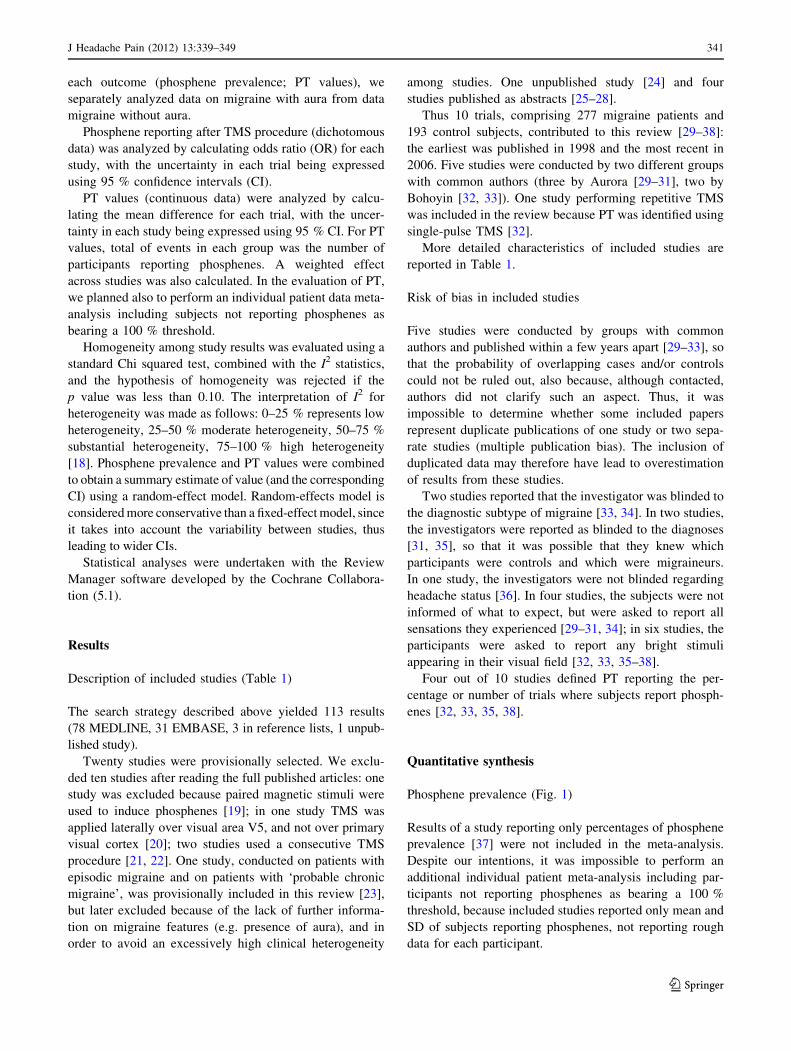
Migraine with aura (Fig. 1a)
There were 9 studies with 300 participants. Significant
statistical heterogeneity among trials was detected.
There was a statistically significant difference in phos-
phene reporting between migraine with aura and con-
trol group, with higher prevalence in migraine group
(111/126 vs. 116/174 participants; OR 4.21; 95 % CI
1.18–15.01).
Migraine without aura (Fig. 1b)
There were 8 studies with 274 participants. No significant
statistical heterogeneity among trials was detected. There
was no statistically significant difference in phosphene
reporting between migraine without aura and control group
(79/111 vs. 113/163 participants; OR 1.04; 95 % CI 0.58–
1.86).
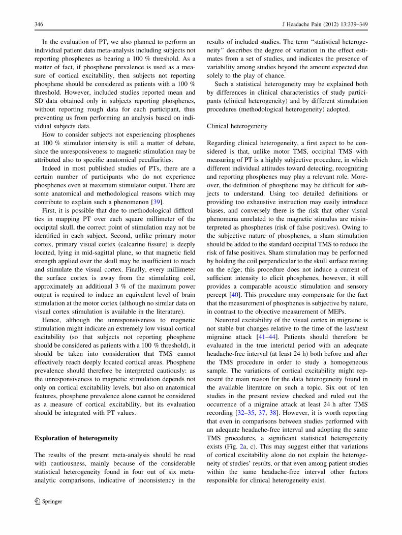
PT values (Fig. 2)
Migraine with aura
Figure-of-eight coil (Fig. 2a) There were 4 studies with
123 participants. Significant statistical heterogeneity among
trials was detected. There was no statistically significant
difference in phosphene threshold between migraine with aura
and control group (mean difference: 2.05; 95 % CI -12.18 to
16.29).
Circular coil (Fig. 2b) There were 5 studies with 104
participants. Significant statistical heterogeneity among
trials was detected. There was a statistically significant
difference between groups, with PT being lower in patients
with migraine with aura than in controls (mean difference:
-28.33; 95 % CI -36.09 to -20.58).
Migraine without aura
Figure-of-eight coil (Fig. 2c) There were 4 studies with
122 participants. Significant statistical heterogeneity among
trials was detected. There was no statistically significant dif-
ference in phosphene threshold between migraine without
aura and control group (mean difference: 5.52; 95 % CI -8.90
to 19.95).
Circular coil (Fig. 2d) There were 3 studies with 68
participants. No significant statistical heterogeneity among
trials was found. There was a statistically significant dif-
ference in phosphene threshold between migraine without
aura and control group (mean difference -17.12; 95 % CI
-23.81 to -10.43).
Migraine with aura Controls Odds Ratio s Total Weight M-H, Random, 95% CI Year
8991]32.1321,35.2[68.55%3.911311118991.latearoruA
9991]48.815,39.2[00.93%1.11824131b9991.latearoruA
1002]30.2,20.0[02.0%2.21615161211002.latesrenelluM
2002]28.4,42.0[70.1%0.6142410162002.latenitohoB
3002]80.5,33.0[92.1%6.6133123193002.latenitohoB
3002]57.6001,10.2[00.54%2.901301013002.latearoruA
5002elbamitsetoN222291915002.lategiwreG
6002]57.25,86.0[00.6%9.21031251416002.latenidyanuG
6002]11.45,95.0[76.5%5.21025181716002.laterdehK
]10.51,81.1[12.4%0.001471621)IC%59(latoT
611111stnevelatoT
Heterogeneity: Tau² = 2.04; Chi² = 19.44, df = 7 (P = 0.007); I² = 64%
)30.0=P(22.2=Z:tceffellarevoroftseT
Migraine without aura Controls Odds Ratio
Study or Subgroup Events Total Event
Study or Subgroup Events Total Events Total Weight M-H, Random, 95% CI Year
9991]02.92,30.0[78.0%0.38210b9991.latearoruA
1002]91.4,30.0[33.0%5.9615121011002.latesrenelluM
2002]49.1,81.0[85.0%1.1342410292002.latenitohoB
3002]03.22,55.0[05.3%3.50130163002.latearoruA
3002]38.2,23.0[59.0%4.92331242513002.latenitohoB
5002elbamitsetoN222291915002.lategiwreG
6002]12.4,41.0[87.0%3.3102510176002.laterdehK
6002]69.41,25.0[97.2%3.8031251316002.latenidyanuG
]68.1,85.0[40.1%0.001361111)IC%59(latoT
31197stnevelatoT
%0=²I;)75.0=P(6=fd,87.4=²ihC:ytienegoreteH
)98.0=P(41.0=Z:tceffellarevoroftseT
a
b
Fig. 1 Phosphene prevalence. a Participants with migraine with aura (MA); b participants with migraine without aura (MwA)
344 J Headache Pain (2012) 13:339–349
123
Page 7
Discussion
In this systematic review, we used systematic and explicit
methods to identify, select and critically appraise studies,
and to extract data, analyzing them with a meta-analysis. A
meta-analysis is the statistical combination of results from
two or more separate studies (pair-wise comparisons of
interventions), allowing an increase in statistical power, an
improvement in precision, sometimes permitting to answer
questions not posed by individual studies and to settle
controversies arising from conflicting claims.
In the present meta-analysis, we found that patients with
migraine with and without aura have a lower PT compared
with controls when a circular coil is used; with a figure-of-
eight coil the difference is not statistically significant. There
was also a statistically significant higher phosphene preva-
lence in migraine with aura compared with controls. No
statistically significant difference was found either in phos-
phene reporting between patients with migraine without aura
and controls, or in PT values obtained by figure-of-eight coil
TMS in subjects with migraine with/without aura versus
controls. Overall considered (and also taking into account the
sample size of each comparison), these results support the
hypothesis of a primary visual cortex hyper-excitability in
migraine with aura, providing not enough evidence for
occipital hyper-excitability in migraine without aura.
Migraine with aura Controls Mean Difference
Migraine with aura Controls Mean Difference
Migraine with aura Controls Mean Difference
Migraine with aura Controls Mean Difference
Study or Subgroup Mean SD Total Mean SD Total Weight IV, Random, 95% CI Year
2002]53.82,55.3[59.51%0.324139.1158.66693.318.282002.latenitohoB
3002]14.52,78.5[46.51%6.421294.2116.86925.2152.483002.latenitohoB
5002]22.6-,83.61-[03.11-%9.62225.014.46917.51.355002.lategiwreG
6002]23.1-,86.71-[05.9-%5.52518.010.27718.215.266002.laterdehK
]92.61,81.21-[50.2%0.0012715)IC%59(latoT
Heterogeneity: Tau² = 189.32; Chi² = 35.08, df = 3 (P < 0.00001); I² = 91%
Test for overall effect: Z = 0.28 (P = 0.78)
Study or Subgroup Mean SD Total Mean SD Total Weight IV, Random, 95% CI Year
8991]23.81-,86.03-[05.42-%0.7231.37.86116.82.448991.latearoruA
9991]40.72-,21.54-[80.63-%6.22242.40.183146.2129.44b9991.latearoruA
1002]74.8-,35.92-[00.91-%3.025170.010.662182.610.741002.latesrenelluM
3002]54.31,54.24-[05.41-%2.639.323.75014.118.243002.latearoruA
6002]27.82-,80.54-[09.63-%9.32124.215.27419.116.536002.latenidyanuG
]85.02-,90.63-[33.82-%0.0014406)IC%59(latoT
Heterogeneity: Tau² = 48.06; Chi² = 12.45, df = 4 (P = 0.01); I² = 68%
Test for overall effect: Z = 7.16 (P < 0.00001)
Study or Subgroup Mean SD Total Mean SD Total Weight IV, Random, 95% CI Year
2002]80.03,27.9[09.91%7.424139.1158.66913.2157.682002.latenitohoB
3002]32.42,16.7[29.51%8.521294.2116.865175.2135.483002.latenitohoB
5002]81.0,85.31-[07.6-%5.62225.014.46918.117.755002.lategiwreG
6002]24.5,24.02-[05.7-%0.32518.010.2778.515.466002.laterdehK
]59.91,09.8-[25.5%0.0012705)IC%59(latoT
Heterogeneity: Tau² = 192.22; Chi² = 29.52, df = 3 (P < 0.00001); I² = 90%
Test for overall effect: Z = 0.75 (P = 0.45)
Study or Subgroup Mean SD Total Mean SD Total Weight IV, Random, 95% CI Year
1002]03.11-,07.82-[00.02-%1.955170.010.660183.110.641002.latesrenelluM
3002]01.72,03.03-[06.1-%4.539.323.7560.217.553002.latearoruA
6002]74.3-,39.52-[07.41-%5.53124.215.27312.818.756002.latenidyanuG
]34.01-,18.32-[21.71-%0.0019392)IC%59(latoT
Heterogeneity: Tau² = 0.00; Chi² = 1.72, df = 2(P = 0.42); I² = 0%
Test for overall effect: Z = 5.02 (P < 0.00001)
a
b
c
d
Fig. 2 PT values. a Patients with MA versus controls (figure-of-eight
coil); b patients with MwA versus controls (figure-of-eight coil);
c patients with MA versus controls (circular coil); d patients with
MwA versus controls (circular coil). Total of events in each group
was the number of participants reporting phosphenes. Standard
deviations in Mulleners et al. [36] were calculated from standard error
and number of participants in each group (standard error 9 Hnumber
of participants)
J Headache Pain (2012) 13:339–349 345
123
Page 8
In the evaluation of PT, we also planned to perform an
individual patient data meta-analysis including subjects not
reporting phosphenes as bearing a 100 % threshold. As a
matter of fact, if phosphene prevalence is used as a mea-
sure of cortical excitability, then subjects not reporting
phosphene should be considered as patients with a 100 %
threshold. However, included studies reported mean and
SD data obtained only in subjects reporting phosphenes,
without reporting rough data for each participant, thus
preventing us from performing an analysis based on indi-
vidual subjects data.
How to consider subjects not experiencing phosphenes
at 100 % stimulator intensity is still a matter of debate,
since the unresponsiveness to magnetic stimulation may be
attributed also to specific anatomical peculiarities.
Indeed in most published studies of PTs, there are a
certain number of participants who do not experience
phosphenes even at maximum stimulator output. There are
some anatomical and methodological reasons which may
contribute to explain such a phenomenon [39].
First, it is possible that due to methodological difficul-
ties in mapping PT over each square millimeter of the
occipital skull, the correct point of stimulation may not be
identified in each subject. Second, unlike primary motor
cortex, primary visual cortex (calcarine fissure) is deeply
located, lying in mid-sagittal plane, so that magnetic field
strength applied over the skull may be insufficient to reach
and stimulate the visual cortex. Finally, every millimeter
the surface cortex is away from the stimulating coil,
approximately an additional 3 % of the maximum power
output is required to induce an equivalent level of brain
stimulation at the motor cortex (although no similar data on
visual cortex stimulation is available in the literature).
Hence, although the unresponsiveness to magnetic
stimulation might indicate an extremely low visual cortical
excitability (so that subjects not reporting phosphene
should be considered as patients with a 100 % threshold), it
should be taken into consideration that TMS cannot
effectively reach deeply located cortical areas. Phosphene
prevalence should therefore be interpreted cautiously: as
the unresponsiveness to magnetic stimulation depends not
only on cortical excitability levels, but also on anatomical
features, phosphene prevalence alone cannot be considered
as a measure of cortical excitability, but its evaluation
should be integrated with PT values.
Exploration of heterogeneity
The results of the present meta-analysis should be read
with cautiousness, mainly because of the considerable
statistical heterogeneity found in four out of six meta-
analytic comparisons, indicative of inconsistency in the
results of included studies. The term ‘‘statistical heteroge-
neity’’ describes the degree of variation in the effect esti-
mates from a set of studies, and indicates the presence of
variability among studies beyond the amount expected due
solely to the play of chance.
Such a statistical heterogeneity may be explained both
by differences in clinical characteristics of study partici-
pants (clinical heterogeneity) and by different stimulation
procedures (methodological heterogeneity) adopted.
Clinical heterogeneity
Regarding clinical heterogeneity, a first aspect to be con-
sidered is that, unlike motor TMS, occipital TMS with
measuring of PT is a highly subjective procedure, in which
different individual attitudes toward detecting, recognizing
and reporting phosphenes may play a relevant role. More-
over, the definition of phosphene may be difficult for sub-
jects to understand. Using too detailed definitions or
providing too exhaustive instruction may easily introduce
biases, and conversely there is the risk that other visual
phenomena unrelated to the magnetic stimulus are misin-
terpreted as phosphenes (risk of false positives). Owing to
the subjective nature of phosphenes, a sham stimulation
should be added to the standard occipital TMS to reduce the
risk of false positives. Sham stimulation may be performed
by holding the coil perpendicular to the skull surface resting
on the edge; this procedure does not induce a current of
sufficient intensity to elicit phosphenes, however, it still
provides a comparable acoustic stimulation and sensory
percept [40]. This procedure may compensate for the fact
that the measurement of phosphenes is subjective by nature,
in contrast to the objective measurement of MEPs.
Neuronal excitability of the visual cortex in migraine is
not stable but changes relative to the time of the last/next
migraine attack [41–44]. Patients should therefore be
evaluated in the true interictal period with an adequate
headache-free interval (at least 24 h) both before and after
the TMS procedure in order to study a homogeneous
sample. The variations of cortical excitability might rep-
resent the main reason for the data heterogeneity found in
the available literature on such a topic. Six out of ten
studies in the present review checked and ruled out the
occurrence of a migraine attack at least 24 h after TMS
recording [32–35, 37, 38]. However, it is worth reporting
that even in comparisons between studies performed with
an adequate headache-free interval and adopting the same
TMS procedures, a significant statistical heterogeneity
exists (Fig. 2a, c). This may suggest either that variations
of cortical excitability alone do not explain the heteroge-
neity of studies’ results, or that even among patient studies
within the same headache-free interval other factors
responsible for clinical heterogeneity exist.
346 J Headache Pain (2012) 13:339–349
123
Page 9
Regarding this last aspect, inter-individual variability
in the anatomy of the occipital region and in skull
thickness may represent another potentially relevant
source of clinical heterogeneity and, to some extent, may
be responsible for the impossibility of perceiving phos-
phenes by some subjects, even when maximum output
stimulation is performed [39]. Furthermore, the ovarian
cycle may represent another factor influencing corti-
cal excitability [45, 46]; only four studies explicitly
assessed such a variable [32, 33, 35, 38], whose effect on
cortical excitability is nevertheless still matter of debate
[47, 48].
Methodological heterogeneity
Regarding methodological heterogeneity, potentially rele-
vant aspects to be taken into account are the coils’ shape,
size and maximum magnetic field strength, and the direc-
tion of the current through the stimulating coil (mono- or
bi-phasic); these stimulator characteristics are sometimes
not explicitly reported in the studies, although they may
deeply influence the results. For example, compared with a
figure-of-eight coil, a circular coil stimulates a larger cor-
tical area [49, 50], and may generate, at least theoretically,
a stronger electric current resulting in a greater probability
of evoking phosphenes.
Another potentially relevant source of methodological
heterogeneity is due to discrepancies in PT definitions, and,
more in general, to the lack of a unique, systematic TMS
protocol in the evaluation of PT in migraine.
Especially in comparison, B (phosphene threshold val-
ues evaluated by means of figure-of-eight coil) heteroge-
neity seems to be related to the presence of some outlying
studies with results that conflict with the rest of the studies.
Repeating pooled analyses on PT values obtained with
figure-of-eight coil and excluding one study at a time to
ensure that the results were not skewed by a single (or a
few) outlier, it may be easily demonstrated that both the
studies conducted by Bohotin [32, 33] are responsible for
the greatest amount of statistical inconsistency among
studies (mean difference -10.80; CI -15.11 to -6.48 with
I2 = 0 %). The limited number of included studies pre-
vented us from performing a more detailed sensitivity
analysis. We did not find any apparent obvious reason (in
terms of clinical and/or methodological differences) for the
outlying results.
Such a high degree of inconsistency among meta-analysis
may be therefore explained not only by clinical or method-
ological differences among studies, but also by significant
variations of cortical excitability among migraine patients. A
higher temporal variability among PT measurements over
time [21, 22], related to unstable excitability levels in these
patients [22, 41–44], might represent the most relevant
clinical factor explaining the high inconsistency among
study results found in the present review.
Conclusions
In this review, we found that patients with migraine with and
without aura have a lower PT compared with controls when a
circular coil single-pulse TMS is used; with a figure-of-eight
coil the difference is not statistically significant. There was
also a statistically significant higher phosphene prevalence in
migraine with aura compared with controls. No statistically
significant difference was found either in phosphene reporting
between patients with migraine without aura and controls, or
in PT values obtained by figure-of-eight coil TMS in subjects
with migraine with/without aura versus controls.
Overall considered, these results support the hypothesis
of a primary visual cortex hyper-excitability in migraine
with aura, providing not enough evidence for occipital
hyper-excitability in migraine without aura.
A significant statistical heterogeneity reflects the pres-
ence of clinical and methodological differences across
studies, and higher temporal variabilities among PT mea-
surements over time, related to unstable excitability levels
in these patients. A unique, shared protocol for future
studies of the PT in migraine patients might overcome such
limitations. Patients should be evaluated in the true inter-
ictal period with an adequate headache-free interval (at
least 24 h) both before and after the TMS procedure in order to
study a homogeneous sample. Furthermore, skull thickness
and ovarian cycle should be assessed as possible confounding
variables, and sham stimulation should be performed to
reduce the rate of false positives. Since the unresponsiveness
to magnetic stimulation depends on cortical excitability levels
and on anatomical peculiarities, phosphene prevalence alone
cannot be considered as an appropriate measure of cortical
excitability, but should be integrated with the parameter of PT.
Further studies conducted with a systematic TMS protocol are
required to confirm whether and to what extent PT values are
reduced in migraine.
Acknowledgments We are in debt with Dr. Stefano Zago
(Department of Neurological Sciences, University of Milan, Italy),
who kindly provided us information on a still unpublished work
(Cappellari et al.). We also thank Dr. Sheena Aurora for providing us
useful additional information on her works. This systematic review
received no specific grant from any funding agency in the public,
commercial, or not-for-profit sectors.
Conflict of interest The authors declare that there is no conflict of
interest.
Open Access This article is distributed under the terms of the
Creative Commons Attribution License which permits any use, dis-
tribution, and reproduction in any medium, provided the original
author(s) and the source are credited.
J Headache Pain (2012) 13:339–349 347
123
Page 10
Appendix: search strategies used in the review
MEDLINE
((‘‘Migraine Disorders’’[Mesh]) OR migrain* OR migraine)
AND (‘‘Phosphenes’’[Mesh]) OR phosphen* OR ‘‘phos-
phene threshold’’) AND ‘‘Transcranial Magnetic Stimula-
tion’’[Mesh])): 78 results.
EMBASE
(migrain* AND phosphen* AND ‘‘Transcranial Magnetic
Stimulation’’): 31 results.
References
1. Welch KMA, DAndrea G, Tepley N, Barkley G, Ramadan NM
(1990) The concept of migraine as a state of central neuronal
hyperexcitability. Neurol Clin 8:817–828
2. Welch KM, Barkeley GL, Tepley N, Ramadan NM (1993)
Central neurogenic mechanisms of migraine. Neurology 43(Suppl
3):S21–S25
3. Shepherd AJ (2001) Increased visual after-effects following
pattern adaptation in migraine: a lack of intracortical excitation?
Brain 124:2310–2318
4. Lehtonen JB (1974) Visual evoked cortical potentials for single
flashes and flickering light in migraine. Headache 14:1–12
5. Connolly JF, Gawel M, Rose FC (1982) Migraine patients exhibit
abnormalities in the visual evoked potential. J Neurol Neurosurg
Psychiatry 45:464–467
6. Diener HC, Scholz E, Dichgans J et al (1989) Central effects of
drugs used in migraine prophylaxis evaluated by visual evoked
potentials. Ann Neurol 25:125–130
7. Amassian VE, Cracco RQ, Maccabee PJ, Rudell A, Eberie L
(1989) Suppression of visual perception by magnetic coil stim-
ulation of human occipital cortex. Electroencephalogr Clin
Neurophysiol 74:458–462
8. Meyer BU, Diehl R, Steinmetz H, Britton TC, Benecke R (1991)
Magnetic stimuli applied over motor and visual cortex: influence
of coil position and field polarity on motor response, phosphenes
and eye movements. Electroencephalogr Clin Neurophysiol
Suppl 43:121–134
9. Hoffken O, Stude P, Lenz M, Bach M, Dinse HR, Tegenthoff M
(2009) Visual paired-pulse stimulation reveals enhanced visual
cortex excitability in migraineurs. Eur J Neurosci 30:714–720
10. Merabet LB, Theoret H, Pascual-Leone A (2003) Transcranial
magnetic stimulation as an investigative tool in the study of
visual function. Optom Vis Sci 80:356–368
11. Ambrosini A, Schoenen J (2003) The electrophysiology of
migraine. Curr Opin Neurol 16:327–331
12. Schoenen J, Ambrosini A, Sandor PS, Maertens de Noordhout A
(2003) Evoked potentials and transcranial magnetic stimulation
in migraine: published data and viewpoint on their pathophysi-
ologic significance. Clin Neurophysiol 114:955–972
13. Fumal A, Bohotin V, Vandenheede M, Schoenen J (2003)
Transcranial magnetic stimulation in migraine: a review of facts
and controversies. Acta Neurol Belg 103:144–154
14. Schoenen J (2006) Neurophysiological features of the migrainous
brain. Neurol Sci 27(Suppl 2):S77–S81
15. Coppola G, Pierelli F, Schoenen J (2007) Is the cerebral cortex
hyperexcitable or hyperresponsive in migraine? Cephalalgia
27:1427–1439
16. International Headache Society (1988) Classification and diag-
nostic criteria for headache disorders, cranial neuralgia and facial
pain. Cephalalgia 8(Suppl. 7):1–96
17. Headache Classification Subcommittee of the International
Headache Society (2004) The International Classification of
Headache Disorders, 2nd edn. Cephalalgia 24 Suppl 1:9–160
18. Higgins JP, Thompson SG, Deeks JJ, Altman DG (2003) Mea-
suring inconsistency in meta-analyses. BMJ 327:557–560
19. Brighina F, Piazza A, Daniele O, Fierro B (2002) Modulation of
visual cortical excitability in migraine with aura: effects of 1 Hz
repetitive transcranial magnetic stimulation. Exp Brain Res
145:177–181
20. Battelli L, Black KR, Wray SH (2002) Transcranial magnetic
stimulation of visual area V5 in migraine. Neurology 58:1066–
1069
21. Young WB, Oshinsky ML, Shechter AL, Gebeline-Myers C,
Bradley KC, Wassermann EM (2004) Consecutive transcranial
magnetic stimulation: phosphene thresholds in migraineurs and
controls. Headache 44:131–135
22. Antal A, Arlt S, Nitsche MA, Chadaide Z, Paulus W (2006)
Higher variability of phosphene thresholds in migraineurs than in
controls: a consecutive transcranial magnetic stimulation study.
Cephalalgia 26:865–870
23. Aurora SK, Barrodale P, Chronicle EP, Mulleners WM (2005)
Cortical inhibition is reduced in chronic and episodic migraine
and demonstrates a spectrum of illness. Headache 45:546–552
24. Cappellari A, Zago S, Valli G, Ferrucci R, De Benedittis G
(2011) Magnetophosphenes in migraineurs: failure to find a
cortical hyper- or hypoexcitability by single-pulse transcranial
magnetic stimulation. Unpublished (as supplied June 2011)
25. Aguggia M, Zibetti M, Febbraro A, Mutani R (1999) Transcranial
magnetic stimulation in migraine with aura: further evidence of
occipital cortex hyperexcitability. Cephalalgia 19:465
26. Aurora SK, Al-Sayed F, Welch KM (1999) The threshold for
magnetophosphenes is lower in migraine. Neurology 52:A472
27. Valli G, Cappellari A, Zago S, Ciammola A, De Benedittis G
(2001) Is migraine associated with hyperexcitability of the
occipital cortex? A transcranial magnetic stimulation controlled
study. Neurology 56:A142
28. Valli G, Cappellari A, Zago S, Ciammola A, De Benedittis G
(2002) Migraine is not associated with hyperexcitability of the
occipital cortex. A transcranial magnetic stimulation controlled
study. In: Proceedings of the 10th World Congress of Pain 2002.
IASP Press, Seattle
29. Aurora SK, Ahmad BK, Welch KM, Bhardhwaj P, Ramadan NM
(1998) Transcranial magnetic stimulation confirms hyperexcit-
ability of occipital cortex in migraine. Neurology 50:1111–1114
30. Aurora SK, Cao Y, Bowyer SM, Welch KM (1999) The occipital
cortex is hyperexcitable in migraine: experimental evidence.
Headache 39:469–476
31. Aurora SK, Welch KM, Al-Sayed F (2003) The threshold for
phosphenes is lower in migraine. Cephalalgia 23:258–263
32. Bohotin V, Fumal A, Vandenheede M, Gerard P et al (2002)
Effects of repetitive transcranial magnetic stimulation on visual
evoked potentials in migraine. Brain 125:912–922
33. Bohotin V, Fumal A, Vandenheede M, Bohotin C, Schoenen J
(2003) Excitability of visual V1-V2 and motor cortices to single
transcranial magnetic stimuli in migraine: a reappraisal using a
figure-of-eight coil. Cephalalgia 23:264–270
34. Gunaydin S, Soysal A, Atay T, Arpaci B (2006) Motor and
occipital cortex excitability in migraine patients. Can J Neurol
Sci 33:63–67
348 J Headache Pain (2012) 13:339–349
123
Page 11
35. Gerwig M, Niehaus L, Kastrup O, Stude P, Diener HC (2005)
Visual cortex excitability in migraine evaluated by single and
paired magnetic stimuli. Headache 45:1394–1399
36. Mulleners WM, Chronicle EP, Palmer JE, Koehler PJ, Vredeveld
JW (2001) Visual cortex excitability in migraine with and with-
out aura. Headache 41:565–572
37. Afra J, Mascia A, Gerard P, Maertens de Noordhout A, Schoenen
J (1998) Interictal cortical excitability in migraine: a study using
transcranial magnetic stimulation of motor and visual cortices.
Ann Neurol 44:209–215
38. Khedr EM, Ahmed MA, Mohamed KA (2006) Motor and visual
cortical excitability in migraineurs patients with or without aura:
transcranial magnetic stimulation. Neurophysiol Clin 36:13–18
39. Brigo F (2012) Occipital TMS in dementia with Lewy bodies:
how to consider subjects not reporting phosphenes? Br J Psy-
chiatry (in press)
40. Sparing R, Mottaghy FM, Ganis G, Thompson WL, Topper R,
Kosslyn SM, Pascual-Leone A (2002) Visual cortex excitability
increases during visual mental imagery—a TMS study in healthy
human subjects. Brain Res 938(1–2):92–97
41. Kropp P, Gerber WD (1998) Prediction of migraine attacks using
a slow cortical potential, the contingent negative variation.
Neurosci Lett 257:73–76
42. Evers S, Quibeldey F, Grotemeyer KH, Suhr B, Husstedt IW
(1999) Dynamic changes of cognitive habituation and serotonin
metabolism during the migraine interval. Cephalalgia 19:485–
491
43. Afra J, Sandor PS, Schoenen J (2000) Habituation of visual and
intensity dependence of auditory evoked cortical potentials tend
to normalize just before and during the migraine attack. Cepha-
lalgia 20:714–719
44. Siniatchkin M, Reich AL, Shepherd AJ, van Baalen A, Siebner
HR, Stephani U (2009) Peri-ictal changes of cortical excitability
in children suffering from migraine without aura. Pain 15:132–
140
45. Smith MJ, Keel JC, Greenberg BD, Adams LF, Schmidt PJ,
Rubinow DA, Wassermann EM (1999) Menstrual cycle effects
on cortical excitability. Neurology 53:2069–2072
46. Smith MJ, Adams LF, Schmidt PJ, Rubinow DR, Wassermann
EM (2002) Effects of ovarian hormones on human cortical
excitability. Ann Neurol 51:599–603
47. Young WB, Oshinsky ML, Shechter AL, Wassermann EM
(2001) Consecutive transcranial magnetic stimulation induced
phosphene thresholds in migraineurs and controls. Neurology
56:A142
48. Boros K, Poreisz C, Paulus W, Antal A (2009) Does the men-
strual cycle influence the motor and phosphene thresholds in
migraine? Eur J Neurol 16:367–374
49. Cohen LG, Roth BJ, Nilsson J, Dang N, Panizza M, Bandinelli S,
Friauf W, Hallett M (1990) Effects of coil design on delivery of
focal magnetic stimulation. Technical considerations. Electroen-
cephalogr Clin Neurophysiol 75:350–357
50. Hallett M (2000) Transcranial magnetic stimulation and the
human brain. Nature 406:147–150
J Headache Pain (2012) 13:339–349 349
123