13

S44 © 2016 Annals of Cardiac Anaesthesia | Published by Wolters Kluwer - Medknow
Transesophageal echocardiography (TEE) can be used to identify risk factors such as aortic atherosclerosis[2] before any sort of surgical manipulations involving aorta and its related structures. TEE has become an important noninvasive tool to diagnose acute thoracic aortic pathologies. TEE evaluation of endoleaks helps early detection and immediate corrective interventions. TEE is an invaluable imaging modality in the management of aortic pathology. TEE has to a large extent improved the patient outcomes.
Key words: Bedside; Mortality; Proximal
Transesophageal echocardiography evaluation of the thoracic aortaT. A. Patil, Arno Nierich1
Seniore Consultant, Department of Anaesthesia, Fortis Hospital, Bengaluru, Karnataka, India, 1Cardiothoracic Anaesthesiologist Intensivist, Isala Sophia, Zwolle, Netherlands
Received: 19-09-16Accepted: 20-09-16
arch, and descending thoracic aorta. Aortic root consists of the aortic annulus, aortic cusps, sinuses of valsalva, and sinotubular junction (STJ). The diameter of the aorta varies between 33 and 35 mm with a wall thickness of about 2 mm. The AA with a length of about 7–11 cm originates from the aortic annulus at the level of STJ crossing beneath the main pulmonary artery, then courses in an anterior and rightward direction over the origin of the right pulmonary artery, which terminates and continues as the aortic arch at the origin of the innominate artery. The aortic arch courses in a posterior and leftward direction giving rise to the innominate (brachiocephalic), left common carotid (LCC), and left subclavian arteries (LSAs). The underlying trachea makes
INTRODUCTION
Atherosclerosis of the ascending aorta (AA) and aortic arch is recognized as one of the important predictors of postoperative stroke after cardiac surgery. Aortic dissection and rupture are emergent situations requiring a rapid and accurate diagnosis to reduce the morbidity and mortality.[1] Transesophageal echocardiography (TEE) can be used to identify risk factors such as aortic atherosclerosis[2] before any sort of surgical manipulations involving aorta and its related structures. TEE has become an important noninvasive tool to diagnose acute thoracic aortic pathologies.
Epiaortic scanning (EAS)[3] is ideal in high-risk patients to identify the sites of severe atherosclerosis on the distal AA and the proximal aortic arch. A novel TEE technique, so-called TEE A-view, by van Zaane et al.,[4] overcomes the “blind spot” of TEE. This allows bedside imaging of the distal AA, the aortic arch, and cerebral branch vessels. With this additional TEE information, appropriate modifications of the surgical techniques can be made to reduce morbidity and mortality.
ANATOMY OF THORACIC AORTA
The thoracic aorta is divided into four segments surgically: Aortic root, AA, aortic
ABSTRACT
Access this article online
Website: www.annals.in
PMID: ***
DOI: 10.4103/0971-9784.192623
Quick Response Code:
Review Article
Address for correspondence: Dr. T. A. Patil, Fortis Hospital, Bengaluru, Karnataka, India. E-mail: [email protected]
Cite this article as: Patil TA, Nierich A. Transesophageal echocardiography evaluation of the thoracic aorta. Ann Card Anaesth 2016;19:S44-55.
This is an open access article distributed under the terms of the Creative Commons Attribution-NonCommercial-ShareAlike 3.0 License, which allows others to remix, tweak, and build upon the work non-commercially, as long as the author is credited and the new creations are licensed under the identical terms.
For reprints contact: [email protected]

Patil and Nierich: TEE evaluation of aorta
Annals of Cardiac Anaesthesia | October 2016 | Vol 19 | Special Issue 1 S45
it difficult to visualize the distal AA and the proximal aortic arch.
The descending thoracic aorta begins distal to the LSA at the level of the ligamentum arteriosum and courses anteriorly and caudally toward the diaphragmatic hiatus. At the site of the ligamentum arteriosum is a relatively fixed narrowing called the aortic isthmus which is the most common site of traumatic decelerating injury. The aorta courses in a smooth curved fashion from the anterolateral side of the 4th thoracic vertebra to the anterior surface of the 11th vertebral body. At distal level, the descending thoracic aorta lies directly posterior to the esophagus [Figure 1].
ECHOCARDIOGRAPHIC EVALUATION OF THE THORACIC AORTA
Primarily insertion of the TEE probe must be performed gently with due consideration to abandon the procedure if resistance is encountered, more so in patients with suspected aortic pathology.
As per the SCA/ASE guidelines, the following imaging planes are recommended for imaging the thoracic aorta.[5] Mid-esophageal (ME) aortic valve (AV) long-axis view, ME ascending (ASC) aortic short-axis (SAX) and long-axis (LAX) view, descending aortic short- and long-axis views, upper esophageal (UE) aortic arch long- and SAX views. The techniques of imaging have been dealt with in the earlier chapter.
ME AV LAX view [Figure 2] is important in evaluating the AV and the proximal AA. Measurements can be made of the left ventricular outflow tract, the aortic annulus, sinuses of valsalva, STJ, and AA if valve replacement and/or root reconstruction is being planned.
ME ASC aortic SAX and LAX views [Figure 3] are crucial to exclude dissections in the aorta. Artifacts are commonly encountered within the AA in this view, hence the priority to distinguish artifacts from true dissections to prevent needless, expensive interventions.
At the mid-esophageal level from the four-chamber view, rotating the probe in a counterclockwise direction or to the left posteriorly and optimizing the depth to 4–6 cm result in descending aortic SAX view. Increasing the multiplane angle to 90°, the descending aortic
LAX view is obtained. This is an important view to evaluate many diagnostic issues such as the presence of an atheromatous plaque, dissection, aneurysm, left pleural effusion, and position of intra-aortic balloon [Figures 4 and 5].
The presence of a pathological lesion in the descending aortic view necessitates additional procedures such as
Figure 1: Anatomical course of the aorta from the aortic root down to its bifurcation (illustrations by Srinivasa Holla)
Figure 3: Mid-esophageal ASC AO SAX, LAX views. RPA: Right pulmonary artery, MPA: Main pulmonary artery, SVC: Superior vena cava, ASC AO: Ascending aorta, SAX: Short-axis, LAX: Long-axis
Figure 2: Mid-esophageal AV LAX view - measurements across root. LV: Left ventricle, RV: Right ventricle, ASC AO: Ascending aorta

Patil and Nierich: TEE evaluation of aorta
S46 Annals of Cardiac Anaesthesia | October 2016 | Vol 19 | Special Issue 1
TEE A-view and/or EAS of the distal AA and aortic arch, especially in the presence of an atheromatous plaque to alter the surgical plan.
A disadvantage of EAS is, however, that it can only be applied during surgery after sternotomy and cannot be used in closed chest procedures (transcatheter aortic valve implantation [TAVI]) or preoperative screening. Since the guidelines do not describe the views of this innovative technique, a short explanation is necessary.
TRANSESOPHAGEAL ECHOCARDIOGRAPHY A-VIEW METHOD
A recently introduced modification of conventional TEE, known as the A-view method, has proven capable of inspecting the distal AA and aortic arch. Assessment of the distal AA sign TEE is disturbed by the interposition of the air-filled trachea between the esophagus and the AA, the so-called “blind spot.”[6] Recently, the A-view (aortic view) method,[7] a modification of conventional TEE, was introduced to overcome this limitation. The A-view method uses an intratracheal balloon (the A-view catheter developed by Stroke2prevent [Figure 6a], Hilversum, the Netherlands) filled with saline to replace the air in the distal trachea and left main bronchus [Figure 6b]. It, therefore, becomes possible to assess the distal AA and aortic arch for the presence and severity of atherosclerosis before cardiac surgery, which provides the surgeon with 30–45 min of additional planning time. Figure 6c shows the TEE A-view distal AA long-axis view with a mobile plaque and Figure 6d shows the Diagrammatic representation of the image. Since visualization will only lead to an improved outcome, if the information is translated to an adjusted surgical approach, further clinical implementation and training is ongoing, but preliminary clinical outcome data are promising.[8]
• A-view catheter• A-view catheter in situ.
Withdrawing the TEE probe and maintaining the descending aortic SAX view on the screen, the aorta changes its appearance from circular to longitudinal at the level of the aortic arch, UE aortic arch LAX view, and increasing the multiplane angle to 90°, the UE aortic arch SAX view is obtained. The diagnostic issues related to this view include recognition of a patent ductus arteriosus, spectral Doppler assessment of the pulmonary artery, and presence of atheromatous plaques in the aortic arch. The origins of the left subclavian, LCC, and brachiocephalic arteries can be visualized frequently by minimal rotation of the TEE probe [Figure 7].
THORACIC AORTIC DISEASES
The primary diseases of the aorta are atherosclerosis, aortic aneurysm, and aortic dissection.
AtherosclerosisAtherosclerosis of the AA and aortic arch is an important predictor of stroke after cardiac surgery occurring in 1%–6% of the patients.[9] The best treatment option is prevention. TEE should be used to identify aortic atherosclerosis before anticipated surgical manipulations. Identification of significant disease by TEE permits alterations in the surgical procedure, such as adopting femoral arterial cannulation, altering the
Figure 5: (a)Descending aortic long‑axis view‑color flowDoppler, (b) Spectral Doppler at the proximal and (c) distal end of the aorta depicted as above and below the baseline, respectively, indicating the flow toward and away from thetransducer
c
baFigure 4: (a) Descending aortic short-axis view, (b) long-axis view - proximal, distal end
ba

Patil and Nierich: TEE evaluation of aorta
Annals of Cardiac Anaesthesia | October 2016 | Vol 19 | Special Issue 1 S47
site of aortic cross-clamp, changing the type of cannulae used for extracorporeal circulation, off-pump coronary artery bypass coronary artery surgery with no touch technique of the aorta, using fibrillatory or hypothermic circulatory arrest, alteration of the site of graft anastomoses, and avoiding antegrade cardioplegia.[10]
Lesions of the AA and arch have been identified as risk factors for stroke, peripheral embolization, perioperative stroke, as well as neuropsychological dysfunction after open heart surgery. Atheroemboli, thromboemboli, and plaque thickness >4 mm correlate with significant embolic risk. Although TEE evaluation of the lesions is sensitive and specific, one of the important limitations is an inadequate visualization of distal AA and proximal aortic arch because of the interposed air-filled trachea. TEE A-view overcomes this problem and gives adequate atherosclerotic information before surgery or during closed chest procedures such as TAVIs.
EAS or TEE A-view imaging should be performed in high-risk patients to further delineate the sites of severe atherosclerosis so that surgical modifications can be made to reduce the morbidity and mortality.
Royse et al.[11] divided the thoracic aorta into six zones in relation to the areas of surgical manipulations which simplified the indications for EAS. Zones 1–3 are the proximal, mid, and distal AA. Zones 1 and 2
are the sites of incision for AV replacement, proximal coronary graft anastomosis, and antegrade cardioplegia cannulation. Zone 3 is the site of aortic cross-clamp, Zone 4 includes the proximal aortic arch which is the site of aortic cannula placement, Zones 5 and 6 include distal aortic arch and proximal descending aorta, respectively, and are not manipulated during cardiac surgery. Atheromatous embolism can occur during aortic cannulation or due to the presence of an intra-aortic balloon. In the study of Royse et al.,[11] Zone 3 was imaged by TT only in 58% of the cases. Manual surgical palpation detects only 50% of those atheromas identified by EAS[11] and might even be dangerous by dislodging the atheroma.[12] When moderate to severe atheroma is identified in Zones 5 and 6 by TEE, the incidence of atheroma in Zones 1–4 is very high. Hence, EAS is recommended. If Zones 1, 2, 3, 5, and 6 are negative, then EAS of Zones 3 and 4 is not recommended in this publication.
A recent meta-analysis by Van Zaane et al.[13] studied the diagnostic accuracy of TEE for estimating AA atherosclerosis. The sensitivity and specificity were 21% (95% confidence interval 13%–32%) and 99% (96%–99%), respectively. Because of the low sensitivity of TEE for the detection of AA atherosclerosis, a negative test result requires verification by additional tests such as TEE A-view and EAS. In case of a positive test result, AA atherosclerosis can be considered as present, and less manipulative strategies might be indicated.
Figure 7: (a) Upper esophageal aortic arch long-axis view, (b) spectral Doppler-aortic arch - proximal end, (c) upper esophageal aortic arch short axis view - PV: Pulmonary valve, MPA: Main pulmonary artery, Left CCA: Left common carotid artery, AO: Aortic arch
c
ba
Figure 6: (a) The A-view catheter. (b) The A-view catheter in the trachea and left main bronchus. (c) The A-view distal ascending aorta long-axis view. An image of the trachea and the ascending aorta as imaged with the A-view method with plaque formation on the posterior and anterior wall (1: A-view catheter in the trachea, 2: The posterior wall of the distal ascending aorta, 3: The anterior wall of the distal ascending aorta, 4: A mobile protruding atheroma indicated by the arrow). (d) Diagrammatic representation of the image
dc
ba

Patil and Nierich: TEE evaluation of aorta
S48 Annals of Cardiac Anaesthesia | October 2016 | Vol 19 | Special Issue 1
Katz et al.[14] recommended a five-point grading system for aortic atheroma. Patients with a mobile atheroma had a 45% incidence of stroke, while the incidence of stroke was only 5% with Grade 3 atheroma [Figures 8 and 9].
Grading of aortic atheromaGrade Description Percentage of
incidence of stroke (%)1 Normal aorta 02 Extensive intimal
thickening <3 mm0
3 Protrudes <5 mm into aortic lumen
5
4 Protrudes >5 mm into aortic lumen
10.5
5 Mobile atheroma 46.5
In comparison to EAS, TEE has been found to be superior for plaque identification in the descending aorta and arch but has a poor predictive value in detecting disease in the AA. A combination of techniques, using EAS, A-view catheter technique and TEE, is recommended in patients at high risk for disease in the distal AA.
Aortic aneurysmsAn aneurysm is defined as dilation of aorta more than 50% of normal aortic diameter involving all the three layers of the vessel wall. The aorta is considered to be dilated if its diameter varies between 3.7 cm and 5.0 cm,
aneurysmal if the diameter is more than 5.0 cm and dissected if it presents with an intimal flap. An aortic aneurysm is either saccular or fusiform. A fusiform aneurysm has a uniform shape with a symmetrical dilatation that involves the entire circumference of the aortic wall whiles saccular aneurysm is a localized outpouching of the aortic wall.
Aneurysms are primarily diseases of aging as a result of degeneration and atherosclerosis. Aging leads to intimal thickening, lipid deposition, and calcification resulting in dilation and weakening of the aortic wall. Connective tissue disorders such as Marfan syndrome, Ehlers-Danlos syndrome trauma, and hypertension are some of the other causes of aneurysms. Thoracic aortic aneurysms are divided into three groups depending on location: AA, aortic arch, and descending thoracic aneurysms or thoracoabdominal aneurysms [Figure 10].
Thoracoabdominal aneurysms are grouped into four types based on the Crawford classification.[15]
Thoracoabdominal aneurysms [Figure 11]• Type 1: Involves the descending aorta just distal to
the origin of LSA to the abdominal aorta above the renal arteries
• Type 2: Involves the descending aorta terminating distal to the renal arteries
Figure 9: (a) Short-axis epiaortic scanning demonstrating the ascending aorta, main pulmonary artery, right pulmonary artery, pulmonary artery catheter in the right pulmonary artery. (b) Long-axis epiaortic scanning demonstrating the ascending aorta and the pulmonary artery. (c) Long-axis epiaortic scanning demonstrating an atheromatous 4 mm plaque (arrow) on the wall of the ascending aorta
c
ba
Figure 8: (a) Upper esophageal aortic arch axis view demonstrating (arrow) a Grade 2 plaque. (b) Upper esophageal aortic arch long-axis view with a (arrow) Grade 3 plaque. (c) Descending aortic long-axis view with a (arrow) Grade 4 plaque
c
ba

Patil and Nierich: TEE evaluation of aorta
Annals of Cardiac Anaesthesia | October 2016 | Vol 19 | Special Issue 1 S49
• Type 3: Involves the distal half of the descending aorta and the abdominal aorta to its bifurcation
• Type 4: Involves the abdominal aorta to its bifurcation.
As per the recommendations by the Society of Thoracic Surgeons, aneurysms with a diameter twice that of the normal aorta need to be surgically corrected.[16] Surgery is indicated if the sinuses are more than 40 mm, AA is more than 50 mm with aortopathy, and more than 60 mm without aortopathy. Patients with connective tissue disorders, strong family history of aortic aneurysms, and symptomatic patients should be considered for early operative therapy because of increased risk of dissection or rupture. Another
alternative for complex cases is the hybrid approach in which an open surgical technique is combined with endovascular aortic repair which has the benefit of complete repair simultaneously reducing the risk of open technique.
TEE can be used to define the size, location, and extent of the aortic aneurysm as well as the presence of a hematoma or thrombus. TEE may also be used to evaluate the patency aortic arch branches to confirm the presence of organ malperfusion. It is an important monitoring tool of cardiac function during aortic aneurysm surgeries.
Aortic dissectionAortic dissection is formed by an intimal tear which is contained by the media leading to the development of a true and false lumen. The false lumen may extend into branches of the aorta in the chest or abdomen, causing malperfusion, ischemia, or occlusion with resultant complications. The dissection can also progress proximally involving the aortic sinus, AV leading to aortic insufficiency, and may also involve the coronary arteries resulting in ischemic events. Aortic dissection is the most common cause of death involving the aorta. The mortality rate is extremely high at the rate of 1%/h–3%/h[17] during the first 48 h. Early and rapid diagnosis is essential to reduce mortality.
Figure 11: Types of thoracoabdominal aneurysms (illustrations by Srinivasa Holla)
Figure 10: (a) Mid-esophageal aortic valve long-axis view demonstrating measurements of a dilated aortic root. (b) The same view with an aneurysmal ascending aorta. (c) Mid-esophageal ascending aortic long-axis view with an aneurysmal ascending aorta. (d) Mid-esophageal aortic valve long-axis view demonstrating a Dacron graft in situ (arrow) at the aortic arch level. (e) upper esophageal aortic valve long axis view demonstrating a Dacron graft in situ (arrow)
d
cba
e
Patil and Nierich: TEE evaluation of aorta
S50 Annals of Cardiac Anaesthesia | October 2016 | Vol 19 | Special Issue 1
a given age and body surface area, for patients above 40 years - predicted sinus dimension (cm) = 1.92 + J 0.74 × BSA (m2). Patients with an aortic root diameter of 50 mm undergoing coronary revascularization are at increased of aortic dissection.
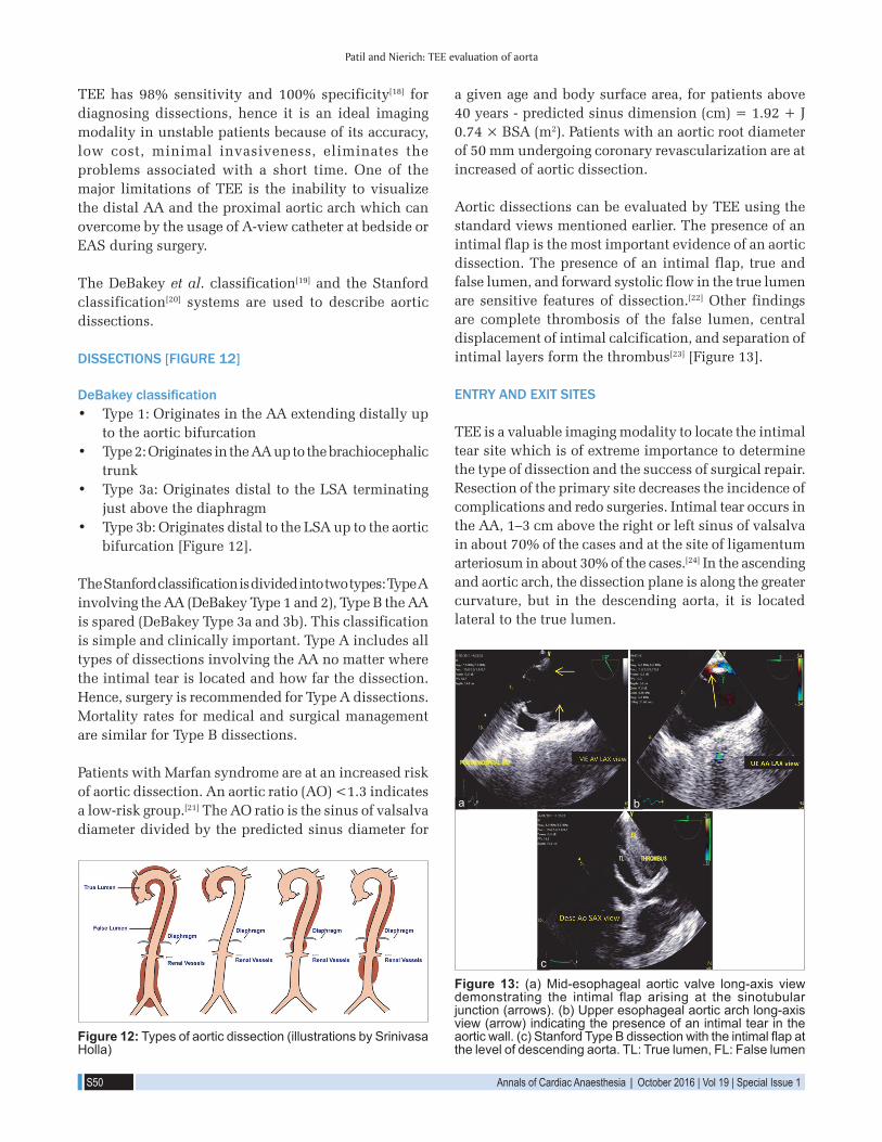
Aortic dissections can be evaluated by TEE using the standard views mentioned earlier. The presence of an intimal flap is the most important evidence of an aortic dissection. The presence of an intimal flap, true and false lumen, and forward systolic flow in the true lumen are sensitive features of dissection.[22] Other findings are complete thrombosis of the false lumen, central displacement of intimal calcification, and separation of intimal layers form the thrombus[23] [Figure 13].
ENTRY AND EXIT SITES
TEE is a valuable imaging modality to locate the intimal tear site which is of extreme importance to determine the type of dissection and the success of surgical repair. Resection of the primary site decreases the incidence of complications and redo surgeries. Intimal tear occurs in the AA, 1–3 cm above the right or left sinus of valsalva in about 70% of the cases and at the site of ligamentum arteriosum in about 30% of the cases.[24] In the ascending and aortic arch, the dissection plane is along the greater curvature, but in the descending aorta, it is located lateral to the true lumen.
TEE has 98% sensitivity and 100% specificity[18] for diagnosing dissections, hence it is an ideal imaging modality in unstable patients because of its accuracy, low cost, minimal invasiveness, eliminates the problems associated with a short time. One of the major limitations of TEE is the inability to visualize the distal AA and the proximal aortic arch which can overcome by the usage of A-view catheter at bedside or EAS during surgery.
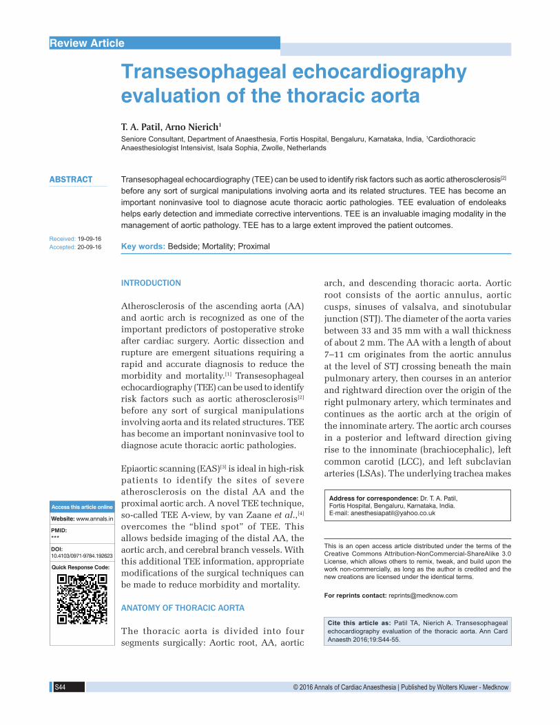
The DeBakey et al. classification[19] and the Stanford classification[20] systems are used to describe aortic dissections.
DISSECTIONS [FIGURE 12]
DeBakey classification• Type 1: Originates in the AA extending distally up
to the aortic bifurcation• Type 2: Originates in the AA up to the brachiocephalic
trunk• Type 3a: Originates distal to the LSA terminating
just above the diaphragm• Type 3b: Originates distal to the LSA up to the aortic
bifurcation [Figure 12].
The Stanford classification is divided into two types: Type A involving the AA (DeBakey Type 1 and 2), Type B the AA is spared (DeBakey Type 3a and 3b). This classification is simple and clinically important. Type A includes all types of dissections involving the AA no matter where the intimal tear is located and how far the dissection. Hence, surgery is recommended for Type A dissections. Mortality rates for medical and surgical management are similar for Type B dissections.
Patients with Marfan syndrome are at an increased risk of aortic dissection. An aortic ratio (AO) <1.3 indicates a low-risk group.[21] The AO ratio is the sinus of valsalva diameter divided by the predicted sinus diameter for
Figure 12: Types of aortic dissection (illustrations by Srinivasa Holla)
Figure 13: (a) Mid-esophageal aortic valve long-axis view demonstrating the intimal flap arising at the sinotubular junction (arrows). (b) Upper esophageal aortic arch long-axis view (arrow) indicating the presence of an intimal tear in the aorticwall.(c)StanfordTypeBdissectionwiththeintimalflapatthe level of descending aorta. TL: True lumen, FL: False lumen
c
ba

Patil and Nierich: TEE evaluation of aorta
Annals of Cardiac Anaesthesia | October 2016 | Vol 19 | Special Issue 1 S51
Artifacts can also occur in the descending aorta. These are mirror image artifacts caused by a highly reflective aorta which occur at twice the distance from the transducer appearing as a double-lumen aorta [Figure 15].
COMPLICATIONS
Complications associated with dissections are aortic insufficiency occurring in about 50%–70% of the cases, coronary dissections in 10%–20%[18] of the cases, pleural and pericardial effusion, and global left ventricular dysfunction. TEE can be used to determine the cause of aortic insufficiency in the presence of dissection in the ME AV SAX view. LV dysfunction may be secondary to ischemia following coronary dissection or due to severe aortic insufficiency presenting in about 10%–15% of the cases. Pericardial effusion is due to transudation of fluid from the wall of the false lumen into the pericardial cavity.[28] Another routine finding is the presence of left pleural effusion causing a hemothorax [Figure 16].
INTRAMURAL HEMATOMA
Intramural hematomas are characterized by thickened aortic walls without an intimal flap involving the ascending or the descending aorta, probably due to rupture of the vasa vasorum leading to hemorrhagic vessel wall. Hematomas involving the AA need emergent surgical therapy, while those involving the descending aorta can be managed medically. Intramural hematomas as described by Mohr-Kahaly et al.[22] are characterized by intimal tear [Figure 17].
TEE plays an important role in the identification of the true and false lumen. True lumen expanded during systole and compressed during diastole. M-mode placed through the dissection helps in identifying the systolic expansion of the true lumen. True lumen is smaller than the false lumen,[25] exhibits a forward systolic flow pattern, has a less echogenic thin layer compared to the false lumen which has a bright echogenic layer. Thickening of the aortic wall more than 15mm is considered to be a sign of dissection,[26] indicating massive thrombosis of the false lumen making it difficult to identify the intimal flap [Figure 14].
It is important to examine the ME AV LAX view carefully to detect proximal AA dissections. It is in this view a Swan-Ganz catheter appears as an artifact. Hence, it is important to differentiate an artifact from a true dissection, lest the patient may be subjected to a needless expensive surgical procedure thereby increasing the morbidity. A linear artifact of the AA can be distinguished from an intimal flap by its indistinct borders, lack of rapid oscillatory movements, extension of the artifact through the aortic wall as a straight line,[27] and the presence of color flow on both sides of the artifact.
Figure 15: Mid-esophageal aortic valve long-axis view demonstrating an artifact (red arrow) in the ascending aorta due to the presence of a pulmonary artery catheter in the right ventricle (yellow arrow)
Figure 14: (a) Descending aortic long-axis view with the systolic color flow Doppler in the true lumen. FL: False lumen. (b) M-mode echocardiography (arrow) indicating the systolic expansion of the true lumen toward the false lumen.(c)Descending aortic short axis view indicating the flowofbloodbycolorflowDopplerfromthetruelumentothefalselumen in the late systolic phase. Note the tracer (red) on the electrocardiogram at the end of systole
c
ba

Patil and Nierich: TEE evaluation of aorta
S52 Annals of Cardiac Anaesthesia | October 2016 | Vol 19 | Special Issue 1
GIANT PENETRATING ULCER
This disease occurs in elderly patients with hypertension, hyperlipidemia, and atherosclerotic disease. It commonly affects the descending aorta in the form of an ulcer, which at a later stage may result in an aneurysm formation.
COARCTATION OF AORTA
This is a congenital narrowing of the aorta at the level of the aortic isthmus. It can be preductal, ductal, or postductal. This condition is commonly associated with a bicuspid AV and a patent ductus arteriosus. Upper esophageal aortic arch shot and LAX views are the recommended views for evaluating this condition by TEE. Findings include narrowing of the aorta distal to LSA and turbulent flow on the color flow Doppler. Transthoracic echocardiography is more favorable due to its anatomical location [Figure 18].
TRAUMATIC AORTIC DISEASE
This disease is associated with a high mortality rate of about 30%, deceleration injury being the most common
cause[29] and aortic isthmus being the most common site of injury apart from descending aorta, aortic arch, and abdominal aorta. TEE with its specificity of 100% remains the imaging modality of choice.
Three types of lesions are seen - sub adventitial aortic rupture, aortic intimal tear, mediastinal hematoma.[30] The subadventitial rupture can be partial or complete (transection) with a thickened highly mobile flap, absence of tear, similar blood flow velocities on both sides of the flap, mosaic color flow Doppler in the area of disruption, and presence of mediastinal hematomas. Mediastinal hematomas appear as an increasing space between the TEE probe and the aortic wall and a bright echogenic space between the aortic wall and the pleura [Figure 19].
TRANSESOPHAGEAL ECHOCARDIOGRAPHY ASSESSMENT OF ENDOVASCULAR REPAIR OF AORTIC ANEURYSMS
Endovascular repair is gaining popularity as an alternative to open surgical repair of aortic aneurysms, thereby minimizing the risk of surgery. TEE is an invaluable tool for identification of aortic pathology, confirmation of placement of the guide wire within the
Figure 19: (a) Descending aortic short-axis view demonstrating thethickenedmedialflapwithanasymmetricalaorticcontourindicating complete subadventitial rupture of the aorta. (b)Similarbloodflowvelocitieswithamosaicappearanceonboth sides of the disruption are seen
ba
Figure 18: (a) Upper esophageal aortic arch short-axis view demonstrating the narrowing (arrow) of the aorta below the level of left subclavianartery. (b)Turbulent flowacross thenarrowed segment
ba
Figure 17: (a) Three-dimensional (zoom) image of the descending aortic short-axis view showing a 5 mm intramural hematoma in the wall of the descending aorta. (b) X-plane color Doppler imaging of the descending aorta showing the intramural hematoma
ba
Figure 16: (a) Descending aortic short-axis view demonstrating the intimal flap with massive left sided pleural effusion, TL: True lumen, FL: False lumen. (b) Mid-esophageal aortic valvelong‑axisviewdemonstratingsevereaorticinsufficiencyfollowing the Stanford Type A dissection
ba
Patil and Nierich: TEE evaluation of aorta
Annals of Cardiac Anaesthesia | October 2016 | Vol 19 | Special Issue 1 S53
SUMMARY
TEE is an invaluable imaging modality in the management of aortic pathology. TEE has to a large extent improved the patient outcomes.
Financial support and sponsorshipNil.
Conflicts of interestThere are no conflicts of interest.
true lumen, helps in stent graft positioning, detecting endoleaks, and evaluating the cardiac function.
The main aim of stent deployment in aneurysms is to exclude the aneurysmal sac so as to prevent further dilation. While in dissections, the aim would be to exclude the intimal tear preventing its evolution.
An endoleak is a common complication after endovascular repair of the aorta. It is characterized by persistent blood flow within the aneurysmal sac occurring in about 20% of the patient.[31] Endoleaks are classified into four types.[32] They may be primary endoleaks usually detected within 30 days or secondary endoleaks occurring after 30 days.
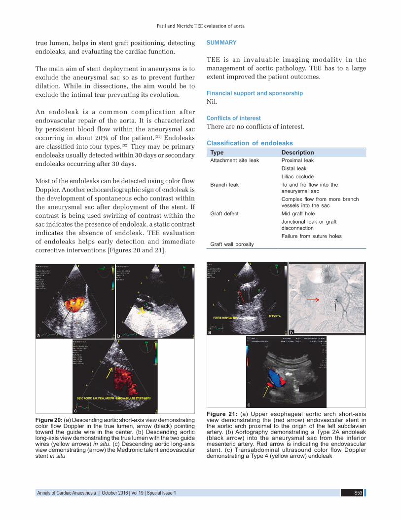
Most of the endoleaks can be detected using color flow Doppler. Another echocardiographic sign of endoleak is the development of spontaneous echo contrast within the aneurysmal sac after deployment of the stent. If contrast is being used swirling of contrast within the sac indicates the presence of endoleak, a static contrast indicates the absence of endoleak. TEE evaluation of endoleaks helps early detection and immediate corrective interventions [Figures 20 and 21].
Figure 21: (a) Upper esophageal aortic arch short-axis view demonstrating the (red arrow) endovascular stent in the aortic arch proximal to the origin of the left subclavian artery. (b) Aortography demonstrating a Type 2A endoleak (black arrow) into the aneurysmal sac from the inferior mesenteric artery. Red arrow is indicating the endovascular stent. (c) Transabdominal ultrasound color flow Doppler demonstrating a Type 4 (yellow arrow) endoleak
c
ba
Figure 20: (a) Descending aortic short-axis view demonstrating color flowDoppler in the true lumen,arrow (black)pointingtoward the guide wire in the center. (b) Descending aortic long-axis view demonstrating the true lumen with the two guide wires (yellow arrows) in situ. (c) Descending aortic long-axis view demonstrating (arrow) the Medtronic talent endovascular stent in situ
c
ba
Classification of endoleaksType DescriptionAttachment site leak Proximal leak
Distal leakLiliac occlude
Branch leak Toand fro flow into theaneurysmal sacComplex flow frommorebranchvessels into the sac
Graft defect Mid graft holeJunctional leak or graft disconnectionFailure from suture holes
Graft wall porosity

Patil and Nierich: TEE evaluation of aorta
S54 Annals of Cardiac Anaesthesia | October 2016 | Vol 19 | Special Issue 1
REFERENCES
1. Olsson C, Thelin S, Ståhle E, Ekbom A, Granath F. Thoracic aortic aneurysm and dissection: Increasing prevalence and improved outcomes reported in a nationwide population-based study of more than 14,000 cases from 1987 to 2002. Circulation 2006;114:2611-8.
2. Nihoyannopoulos P, Joshi J, Athanasopoulos G, Oakley CM. Detection of atherosclerotic lesions in the aorta by transesophageal echocardiography. Am J Cardiol 1993;71:1208-12.
3. Rosenberger P, Shernan SK, Löffler M, Shekar PS, Fox JA, Tuli JK, et al. The influence of epiaortic ultrasonography on intraoperative surgical management in 6051 cardiac surgical patients. Ann Thorac Surg 2008;85:548-53.
4. van Zaane B, Nierich AP, Buhre WF, Brandon Bravo Bruinsma GJ, Moons KG. Resolving the blind spot of transoesophageal echocardiography: A new diagnostic device for visualizing the ascending aorta in cardiac surgery. Br J Anaesth 2007;98:434-41.
5. Shanewise JS, Cheung AT, Aronson S, Stewart WJ, Weiss RL, Mark JB, et al. ASE/SCA guidelines for performing acomprehensive intraoperative multiplane transesophageal echocardiography examination: Recommendations of the American Society of Echocardiography Counci l for Intraoperative Echocardiography and the Society of Cardiovascular Anesthesiologists Task Force for Certification in Perioperative Transesophageal Echocardiography. Anesth Analg 1999;89:870-84.
6. Konstadt SN, Reich DL, Quintana C, Levy M. The ascending aorta: How much does transesophageal echocardiography see? Anesth Analg 1994;78:240-4.
7. Nierich AP, van Zaane B, Buhre WF, Coddens J, Spanjersberg AJ, Moons KG. Visualization of the distal ascending aorta with A-Mode transesophageal echocardiography. J Cardiothorac Vasc Anesth 2008;22:766-73.
8. Nierich A. Clinical outcome after pre-incision assessment of aorticatherosclerosis by A-View echocardiography in 5,886 elective cardiac surgerypatients. J Cardiothorac Vasc Anesth 2011;25:S1.
9. Bucerius J, Gummert JF, Borger MA, Walther T, Doll N, Onnasch JF, et al. Stroke after cardiac surgery: A risk factor analysis of 16,184 consecutive adult patients. Ann Thorac Surg 2003;75:472-8.
10. Ribakove GH, Katz ES, Galloway AC, Grossi EA, Esposito RA, Baumann FG, et al. Surgical implications of transesophageal echocardiography to grade the atheromatous aortic arch. Ann Thorac Surg 1992;53:758-61.
11. Royse C, Royse A, Blake D, Grigg L. Screening the thoracic aorta for atheroma: A comparison of manual palpation, transesophageal and epiaortic ultrasonography. Ann Thorac Cardiovasc Surg 1998;4:347-50.
12. Sylivris S, Calafiore P, Matalanis G, Rosalion A, Yuen HP, Buxton BF, et al. The intraoperative assessment of ascending aortic atheroma: Epiaortic imaging is superior to both transesophageal echocardiography and direct palpation. J Cardiothorac Vasc Anesth 1997;11:704-7.
13. Van Zaane B, Zuithoff NP, Reitsma JB, Bax L, Nierich AP, Moons KG. Meta-analysis of the diagnostic accuracy of transesophageal echocardiography for assessment
of atherosclerosis in the ascending aorta in patients undergoing cardiac surgery. Acta Anaesthesiol Scand 2008;52:1179-87.
14. Katz ES, Tunick PA, Rusinek H, Ribakove G, Spencer FC, Kronzon I. Protruding aortic atheromas predict stroke in elderly patients undergoing cardiopulmonary bypass: Experience with intraoperative transesophageal echocardiography. J Am Coll Cardiol 1992;20:70-7.
15. Svensson LG, Kouchoukos ES. Aortic dissection and aortic aneurysm surgery: Clinical observations, experimental investigations, and statistical analyses. Part II. Curr Probl Surg 1992;29:915-1057.
16. Svensson LG, Kouchoukos NT, Miller DC, Bavaria JE, Coselli JS, Curi MA, et al. Expert consensus document on the treatment of descending thoracic aortic disease using endovascular stent-grafts. Ann Thorac Surg 2008;85 1 Suppl: S1-41.
17. Nienaber CA, Spielman RP, Von Kodolitsch Y, Siglow V, Piepho A, Jaup T, et al. Diagnosis of thoracic aortic dissection: Magnetic resonance imagery versus transesophageal echocardiography. Circulation 1992;85:434-47.
18. Ballal RS, Nanda NC, Gatewood R, D’Arcy B, Samdarshi TE, Holman WL, et al. Usefulness of transesophageal echocardiography in assessment of aortic dissection. Circulation 1991;84:1903-14.
19. DeBakey ME, Henly WS. Cooley DA, Morris GC Jr, Crawford ES, Beall AC Jr. Surgical management of dissecting aneurysms of the aorta. J Thorac Cardiovasc Surg 1965;49:130-49.
20. Daily PO, Trueblood HW, Stinson EB, Wuerflein RD, Shumway NE. Management of acute aortic dissections. Ann Thorac Surg 1970;10:237-47.
21. Legget ME, Unger TA, O’Sullivan EB. Management of acute aortic dissections. Ann Thorac Surg 1970;10:237-47.
22. Mohr-Kahaly S, Erbel R, Kearney P, Puth M, Meyer J. Aortic intramural hemorrhage visualized by transesophageal echocardiography: Findings and prognostic implications. J Am Coll Cardiol 1994;23:658-64.
23. Erbel R, Engberding R, Daniel W, Roelandt J, Visser C, Rennollet H. Echocardiography in diagnosis of aortic dissection. Lancet 1989;1:457-61.
24. Erbel R, Mohr-Kahaly S, Rennollet H, Brunier J, Drexler M, Wittlich N, et al. Diagnosis of aortic dissection: The value of transesophageal echocardiography. Thorac Cardiovasc Surg 1987;35:126-33.
25. Hashimoto S, Kumada T, Osakada G, Kubo S, Tokunaga S, Tamaki S, et al. Assessment of transesophageal Doppler echography in dissecting aortic aneurysm. J Am Coll Cardiol 1989;14:1253-62.
26. Iliceto S, Nanda NC, Rizzon P, Hsuing MC, Goyal RG, Amico A, et al. Color Doppler evaluation of aortic dissection. Circulation 1987;75:748-55.
27. Appelbe AF, Walker PG, Yeoh JK, Bonitatibus A, Yoganathan AP, Martin RP. Clinical significance and origin of artifacts in transesophageal echocardiography of the thoracic aorta. J Am Coll Cardiol 1993;21:754-60.
28. Khan IA, Nair CK. Clinical, diagnostic, and management perspectives of aortic dissection. Chest 2002;122:311-28.
29. Wall MJ Jr, Hirshberg A, LeMaire SA, Holcomb J, Mattox K. Thoracic aortic and thoracic vascular injuries. Surg Clin North Am 2001;81:1375-93.

Patil and Nierich: TEE evaluation of aorta
Annals of Cardiac Anaesthesia | October 2016 | Vol 19 | Special Issue 1 S55
30. V ignon P, Gueret P, edrinne JM, Lagrange P, Semba R, Mitchell S, et al. Transluminal Placement of endovascular stent-graftsfor the treatment of descending thoracic aortic aneurysms. N Engl J Med 1994;331:1729-34.
31. van Marrewijk C, Buth J, Harris PL, Norgren L, Nevelsteen A, Wyatt MG. Significance of endoleaks after endovascular
repair of abdominal aortic aneurysms: The EUROSTAR experience. J Vasc Surg 2002;35:461-73.
32. Veith FJ, Baum RA, Ohki T, Amor M, Adiseshiah M, Blankensteijn JD, et al. Nature and significance of endoleaks and endotension: Summary of opinions expressed at an international conference. J Vasc Surg 2002;35:1029-35.
TAKE HOME MESSAGES
In this detailed review of “TEE evaluation of the thoracic Aorta”, the authors describe based on anatomy of the thoracic aorta, the echo evaluation in six different TEE planes, and the importance of the epiaortic scanning (EUS) of the distal ascending aorta (AA) and aortic arch especially in the case of an atheromatous plaque which changes the surgical plan. The article also describes the six zones in EUS, the aortic aneurysms and aortic dissections with entry and exit sites. Coarctation of aorta and traumatic aortic diseases with EVAR is also described.


