1238 VOLUME 4 NUMBER 12 DECEMBER 2003 NATURE IMMUNOLOGY
Hematopoietic stem cells (HSCs) exist as rare populations in blood-forming tissues that both self-renew and generate all blood cell lin-eages for the life of the host. It is now possible to prospectively isolatemurine HSCs1–3, clonogenic progenitors downstream of HSCs thatgive rise to all cells of the lymphoid4 and myeloerythroid5 lineages, andmost mature blood lineages, by cell surface phenotype. This work hasled to an understanding of the lineage relationships among all bloodcell types and the stages through which each passes in its differentia-tion hierarchy. In contrast to what is known regarding the phylogenyof hematopoietic cells, the molecular mechanisms underlying stemcell specification, self-renewal, amplification and lineal fate decisionsremain poorly understood.
Gene-disruption experiments in mice have shown absolute require-ments for the function of genes such as Tal1 (ref. 6), Lmo2 (refs. 7,8),Runx1 (ref. 9) and Myb10 in the development and/or maintenance offetal HSCs. Microarray approaches have also been used to compareglobal gene expression profiles among prospectively isolated HSCs,committed progenitors and mature blood cell populations11.Although both approaches can be powerful, neither is well suited forunbiased identification or high-throughput functional testing of can-didate blood genes.
The zebrafish has emerged as a unique vertebrate model system forthe analysis of developmental processes because of its larval trans-parency and high fecundity and the ease with which forward geneticand expression screens can demonstrate previously unknown genes ina relatively unbiased way12. Large-scale mutagenesis screens have pro-duced over 50 mutants in blood cell development that fall into at least26 complementation groups13,14. These mutants show defects in a widerange of hematopoietic processes, including HSC specification15–17,
lymphoid and thymic development18 and the generation of functionalmyeloid19 and erythroid13,14 cells.
Because of the early time points analyzed and the visual nature ofthese screens, most mutants have defects in the formation or mainte-nance of embryonic, also known as primitive, red blood cells. As withblood development in mammals, birds and frogs, zebrafish hemato-poiesis occurs in several anatomic sites during embryogenesis and lar-val development20. The first wave of hematopoiesis occurs in astructure called the intermediate cell mass that is similar to the mam-malian yolk sac in that primitive erythrocytes are the main cell typeproduced. Later, hematopoiesis shifts to the ventral wall of the dorsalaorta, the zebrafish equivalent of the aorta-gonad-mesonephrosregion. Based on the expression of markers such as cmyb21 and runx1(refs. 22,23), it seems that the zebrafish aorta-gonad-mesonephrosspecifies HSCs with definitive, multilineage potential. At present,screens aimed at identifying HSC mutants due to alterations in thesemarkers require systematic characterization of steady-state, definitivehematopoiesis and the development of tools to study HSC biologymore precisely.
We have established several tools to characterize the definitiveblood-forming system of adult zebrafish. With the ability to analyzeand isolate each major blood cell lineage, we show that zebrafishembryonic blood mutants often demonstrate previously unrecognizeddefects as adults, even as heterozygous carriers of recessive mutations.Transplantation of adult kidney marrow into prethymic embryonicrecipients generates long-term hematopoietic reconstitution of other-wise lethal gata1–/– mutants in the apparent absence of graft-versus-host disease. Hematopoietic cell transplantation in zebrafish will thusallow future studies to measure HSC activity and to characterize
1Children’s Hospital Boston and the Howard Hughes Medical Institute, 320 Longwood Avenue, Enders 720, Boston, Massachusetts 02115, USA. 2Department of CellBiology, Box 3709, Duke University Medical Center, Durham, North Carolina 27710, USA. 3University of California at Los Angeles, 621 Charles Young Drive South,LS4325, Los Angeles, California 90095, USA. Correspondence should be addressed to L.I.Z. ([email protected]).
Published online 9 November 2003; doi:10.1038/ni1007
Transplantation and in vivo imaging of multilineageengraftment in zebrafish bloodless mutantsDavid Traver1, Barry H Paw1, Kenneth D Poss2, W Todd Penberthy3, Shuo Lin3 & Leonard I Zon1
The zebrafish is firmly established as a genetic model for the study of vertebrate blood development. Here we have characterizedthe blood-forming system of adult zebrafish. Each major blood lineage can be isolated by flow cytometry, and with these linealprofiles, defects in zebrafish blood mutants can be quantified. We developed hematopoietic cell transplantation to study cellautonomy of mutant gene function and to establish a hematopoietic stem cell assay. Hematopoietic cell transplantation canrescue multilineage hematopoiesis in embryonic lethal gata1–/– mutants for over 6 months. Direct visualization of fluorescentdonor cells in embryonic recipients allows engraftment and homing events to be imaged in real time. These results provide acellular context in which to study the genetics of hematopoiesis.
NATURE IMMUNOLOGY VOLUME 4 NUMBER 12 DECEMBER 2003 1239
genetic defects in zebrafish blood mutants. Finally, the development oftransgenic animals with red fluorescent erythrocytes and green fluo-rescent leukocytes has enabled, for the first time to our knowledge,direct visualization of early trafficking events in embryos and multi-lineage engraftment in adults in living transplant recipients.
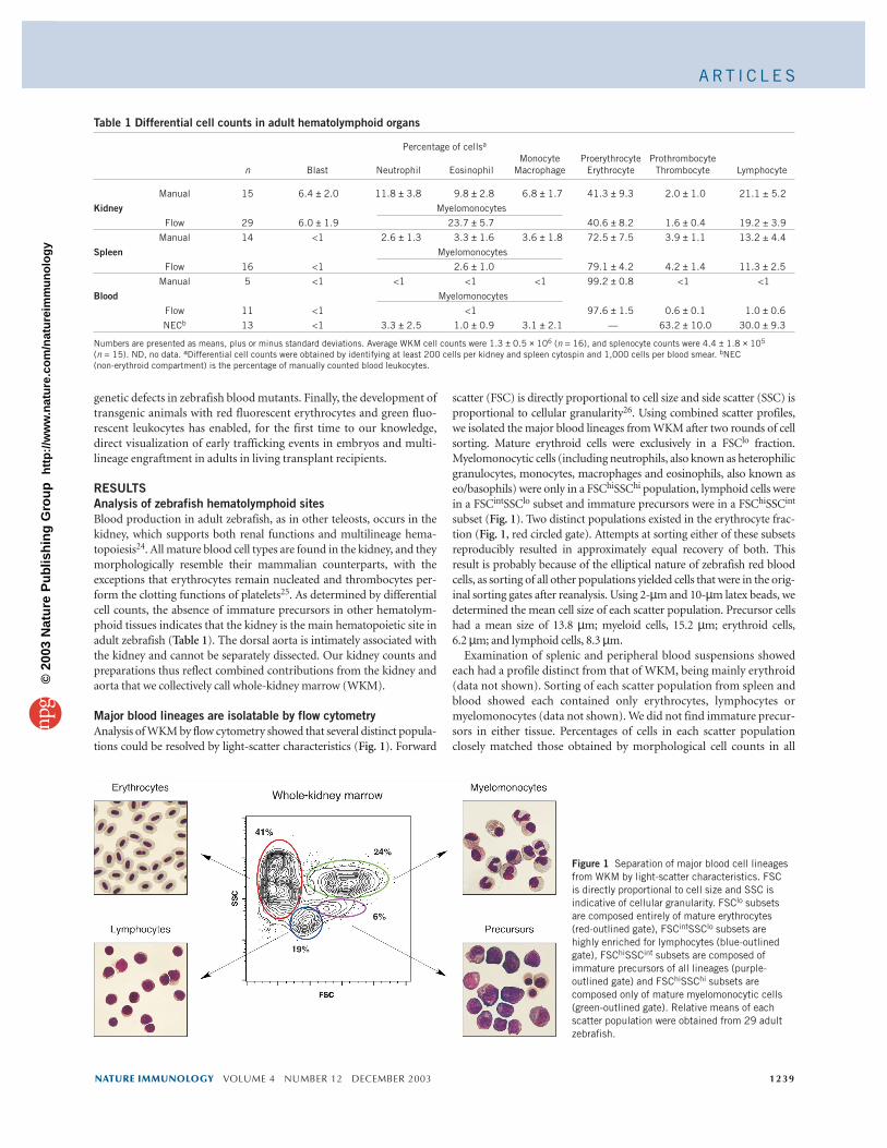
RESULTSAnalysis of zebrafish hematolymphoid sitesBlood production in adult zebrafish, as in other teleosts, occurs in thekidney, which supports both renal functions and multilineage hema-topoiesis24. All mature blood cell types are found in the kidney, and theymorphologically resemble their mammalian counterparts, with theexceptions that erythrocytes remain nucleated and thrombocytes per-form the clotting functions of platelets25. As determined by differentialcell counts, the absence of immature precursors in other hematolym-phoid tissues indicates that the kidney is the main hematopoietic site inadult zebrafish (Table 1). The dorsal aorta is intimately associated withthe kidney and cannot be separately dissected. Our kidney counts andpreparations thus reflect combined contributions from the kidney andaorta that we collectively call whole-kidney marrow (WKM).
Major blood lineages are isolatable by flow cytometryAnalysis of WKM by flow cytometry showed that several distinct popula-tions could be resolved by light-scatter characteristics (Fig. 1). Forward
scatter (FSC) is directly proportional to cell size and side scatter (SSC) isproportional to cellular granularity26. Using combined scatter profiles,we isolated the major blood lineages from WKM after two rounds of cellsorting. Mature erythroid cells were exclusively in a FSClo fraction.Myelomonocytic cells (including neutrophils, also known as heterophilicgranulocytes, monocytes, macrophages and eosinophils, also known aseo/basophils) were only in a FSChiSSChi population, lymphoid cells werein a FSCintSSClo subset and immature precursors were in a FSChiSSCint
subset (Fig. 1). Two distinct populations existed in the erythrocyte frac-tion (Fig. 1, red circled gate). Attempts at sorting either of these subsetsreproducibly resulted in approximately equal recovery of both. Thisresult is probably because of the elliptical nature of zebrafish red bloodcells, as sorting of all other populations yielded cells that were in the orig-inal sorting gates after reanalysis. Using 2-µm and 10-µm latex beads, wedetermined the mean cell size of each scatter population. Precursor cellshad a mean size of 13.8 µm; myeloid cells, 15.2 µm; erythroid cells, 6.2 µm; and lymphoid cells, 8.3 µm.
Examination of splenic and peripheral blood suspensions showedeach had a profile distinct from that of WKM, being mainly erythroid(data not shown). Sorting of each scatter population from spleen andblood showed each contained only erythrocytes, lymphocytes ormyelomonocytes (data not shown). We did not find immature precur-sors in either tissue. Percentages of cells in each scatter populationclosely matched those obtained by morphological cell counts in all
Table 1 Differential cell counts in adult hematolymphoid organs
Numbers are presented as means, plus or minus standard deviations. Average WKM cell counts were 1.3 ± 0.5 × 10⁶ (n = 16), and splenocyte counts were 4.4 ± 1.8 × 10⁵
(n = 15). ND, no data. aDifferential cell counts were obtained by identifying at least 200 cells per kidney and spleen cytospin and 1,000 cells per blood smear. bNEC (non-erythroid compartment) is the percentage of manually counted blood leukocytes.
Figure 1 Separation of major blood cell lineagesfrom WKM by light-scatter characteristics. FSC is directly proportional to cell size and SSC isindicative of cellular granularity. FSClo subsetsare composed entirely of mature erythrocytes(red-outlined gate), FSCintSSClo subsets arehighly enriched for lymphocytes (blue-outlinedgate), FSChiSSCint subsets are composed ofimmature precursors of all lineages (purple-outlined gate) and FSChiSSChi subsets arecomposed only of mature myelomonocytic cells(green-outlined gate). Relative means of eachscatter population were obtained from 29 adultzebrafish.
1240 VOLUME 4 NUMBER 12 DECEMBER 2003 NATURE IMMUNOLOGY
tissues, demonstrating that this flow cytometric assay is accurate inmeasuring the relative percentages of each of the major blood lineages(Table 1). Thus, the kidney seems to be the main site of hematopoiesisin adult zebrafish.
Analysis of zebrafish transgenic lines by flow cytometryBecause we assigned the lineal designations for each kidney scatterpopulation by morphology, we next assessed whether the expressionof lineage-affiliated genes correlated with specific scatter fractions. Weused transgenic zebrafish that express green fluorescent protein (GFP)under the control of lineage-specific gene promoters. GATA2 has beenshown in mammalian studies to be expressed in both eosinophils andbasophils27. Analysis of WKM isolated from transgenic zebrafishexpressing enhanced GFP under control of a gata2 bacterial artificialchromosome construct (gata2eGFP) showed that 3.1 ± 1.0 % (n = 3) ofWKM cells were GFP+. Sorting of this fraction showed nearly all cellswere in the myeloid scatter fraction (FSChiSSChi), and morphologicalanalysis showed all sorted cells had the morphological characteristicsof eosinophils (Fig. 2a). Similar analysis of transgenic zebrafishexpressing GFP under the control of the lymphoid-specific recombi-nation-activating gene 2 (rag2) promoter28 showed that 5.1 ± 0.9 %
(n = 3) of WKM cells were GFP+, and over90% of this population localized in the lym-phoid scatter fraction (FSCintSSClo). Mor-phological analysis showed all cells had thecharacteristics of immature lymphocytes (Fig. 2b). Analysis of zebrafish with an ery-throid-specific gata1eGFP transgene29 showed39.0 ± 5.9% (n = 3) of WKM cells had highexpression of GFP. Isolation of this subsetshowed that over 90% were in the erythroidscatter gate (FSClo), and all purified cells hadthe morphology of mature erythrocytes (Fig. 2c). The gata1eGFP animals also had arare population (1.9 ± 0.3 %; n = 3) of GFPlo
cells. Isolation of this subset showed they werein the precursor, lymphoid and erythroidscatter fractions (Fig. 2d). Sorted GFPlo cellsall had immature erythroid morphologies,
ranging from early proerythroblasts to late polychromatophilic ery-throblasts (Fig. 2d). GFPlo cell populations sorted from the precursorfraction were enriched for the former cell type, whereas cell popula-tions sorted from the lymphoid fraction were enriched for the latter,more mature cell types (Fig. 2d). These data indicate that the erythroidmaturation pathway, in terms of kidney scatter populations, seems totransit from immature proerythroblasts in the precursor scatter frac-tion to maturing late erythroblasts adjacent to and partially in the lym-phoid scatter fraction before their terminal differentiation andappearance in the erythroid gate. CD41, also known as platelet glycoprotein IIb and integrin αIIb (gene name, itga2b), is specificallyexpressed on zebrafish thrombocytes; analysis of itga2beGFP-transgenic animals showed that rare prothrombocytes (CD41lo, 0.8 ± 0.3% of WKM; n = 27) were in the precursor population, andthat mature thrombocytes (CD41hi, 0.8 ± 0.4% of WKM; n = 27) werein the lymphoid fraction (H.F. Lin, D.T., C. Abraham and R. Handin,unpublished data). The GATA-1lo and CD41hi cells account forapproximately 10% of the cells in the lymphoid gate. Although rela-tively minor, these analyses show that the lymphoid scatter fraction,unlike the erythroid and myeloid fractions, contains precursors ofother blood cell lineages.
Figure 2 Analysis of zebrafish transgenic lines byflow cytometry. (a) Analysis of WKM cells markedby a gata2eGFP transgene. GFP+ cells gated fromthe WKM histogram (left) were sorted andreanalyzed by forward and side scatter (middle).Nearly all cells are in the myeloid gate, andmorphological analysis shows all cells areeosinophils (right). (b) Analysis of rag2eGFP WKMshows that more than 90% of GFP+ cells are inthe lymphoid fraction. All sorted cells showlymphoid morphologies (right). (c) Approximately40% of WKM cells express a gata1eGFP transgene.GFPhi cells are in the FSClo erythroid scatterfraction. Morphological analysis shows all cellsare terminally differentiated erythrocytes (right).(d) A minor population of cells in gata1eGFP
animals are GFPlo. Right, double sorting of GATA-1lo cells shows the precursor fraction isenriched for immature proerythroblasts (top),whereas the lymphoid fraction contains smallerpolychromatophilic erythroblasts (bottom).Percents indicate relative percentages of GFP+
NATURE IMMUNOLOGY VOLUME 4 NUMBER 12 DECEMBER 2003 1241
Characterization of zebrafish blood mutants by flow cytometryTo test whether flow cytometric profiling could serve as a diagnostictool, we examined zebrafish blood mutants by flow cytometry (Fig. 3). Nearly all blood mutants identified so far have shown defectsin embryonic erythrocytes12. Most of these mutations are recessive,and many are embryonic lethal when homozygous. These mutantsinclude merlot, retsina and riesling, which have defects in the genesencoding erythrocyte membrane protein band 4.1 (epb41)30, solutecarrier family 4, anion exchanger 1 (slc4a1)31 and erythrocytic β-spectrin (sptb)32, respectively. All three genes are expressed specifi-cally in the erythrocyte membrane, and homozygous mutants showdecreasing blood counts in the larval stages because of failures inmembrane integrity. To determine whether heterozygous carriers ofeach mutation show adult hematopoietic deficiencies, we analyzedWKM suspensions by flow cytometry. Mutants heterozygous for theepb41 mutation had percentages of erythroid and precursor cells sim-ilar to those of their wild-type siblings (Fig. 3c). Mutants heterozy-gous for the slc4a1 mutation, however, showed mild anemia with aconcomitant twofold increase in the precursor scatter population(Fig. 3a,c). Sorting of the precursor fraction showed a preponderanceof immature erythroid cells (Fig. 3a). In contrast, enumeration of celltypes from sorted wild-type precursor fractions showed approxi-mately 40% myeloid precursors, 40% erythroid precursors and 20%lymphoid precursors by morphological criteria. The sptb mutantshowed a similar heterozygous phenotype, with anemia associatedwith precursor increases (Fig. 3c). The phenotypes of both adultslc4a1 and sptb heterozygotes seemed intermediate to those of theirwild-type siblings and embryonic homozygous mutants. For exam-ple, slc4a–/– embryos die because of a block in erythropoiesis at thepolychromatophilic erythroblast stage31. Adult slc4a1+/– animalsshowed a partial but incomplete block at this stage (Fig. 3a). Thisfinding is in agreement with clinical studies showing that human het-erozygous carriers of SLC4A1 mutations can have hematopoieticabnormalities33. These findings indicate that the many gene functionsrequired to make embryonic erythrocytes are similarly required intheir adult counterparts at full gene dosage for normal function.
Occasional homozygous mutants can be raised to adulthood fromcertain lines, including merlot, retsina and pinotage13. Some ofthese mutant animals can survive for several months with an almost
complete absence of erythrocytes. For example, both ebp41–/– andslc4a1 –/– adults had almost no cells in the erythroid scatter popula-tion (Fig. 3b,d). The precursor fraction in ebp41–/– animals wasincreased more than ninefold compared with that of wild-type ani-mals (Fig. 3d). Most cells in this fraction phenotypically resembledpolychromatophilic erythroblasts (Fig. 3b). Adult slc4a1–/– animalshad a very similar phenotype, with a relative precursor increase ofapproximate eightfold compared with that of wild-type (Fig. 3d).The adult phenotypes of both ebp41–/– and slc4a –/– mutants closelyresembled their embryonic phenotypes, indicating that each genehas the same function in both primitive and definitive red blood cellproduction. The pinotage mutant, which has a mutation in an as-yet-unidentified gene, showed no heterozygous phenotype but wasanemic and showed a considerable increase in erythroid precursorswhen homozygous (Fig. 3d). Among all mutants analyzed, we foundno notable alterations reproducibly in the lymphoid or myeloidpopulations.
Hematopoietic cell transplantation rescues lethal gata1–/– mutantsIn mammals, transplantation has been used extensively to functionallytest putative hematopoietic stem and progenitor cell populations, pre-cursor-progeny relationships and cell autonomy of mutant gene func-tion. To address similar issues, we developed hematopoietic celltransplantation in zebrafish. For a donor cell marker of transplantedcells, we used adult gata1eGFP transgenic zebrafish. As zebrafish ery-throcytes have lifespans limited to several weeks14, continued produc-tion of GFP+ cells over several months serves as a surrogate marker ofstem and progenitor cell activity. We used transparent embryos 48 hafter fertilization as transplant recipients to easily visualize donor-derived cells. These recipients are also optimal for the prevention ofgraft rejection, as the onset of lymphopoiesis does not occur until 3–5 dafter fertilization34,35. This is important, as it has not been possible todevelop true inbred strains in zebrafish. We sought to determinewhether transplantation could rescue otherwise lethal embryonicblood mutants. Both the moonshine (mon/mon)13 and vlad tepes(gata1–/–)14,36 blood mutants show a complete absence of erythroidcells and die by 14 days after fertilization. Transplantation of gata1eGFP
WKM into mon/mon mutants showed that donor-derived GFP+ cellshomed to the pronephros and proliferated over a 10-day ‘window’ after
Figure 3 Flow cytometry profiling of zebrafishblood mutants. (a) Scatter profile of WKM in a typical slc4a1 heterozygous mutant (left).Sorted precursors show a preponderance ofimmature erythroid elements (right). (b) WKMscatter profile of a typical ebp41 homozygousmutant (left). Sorted precursors show nearly allcells are immature erythroid cells that rangefrom proerythroblasts to late polychromatophilicerythroblasts (right). Numbers in plots in a andb indicate percent of cells in circled gate. (c) Comparison of mean percentages of WKMerythrocyte and precursor scatter populationsamong heterozygous blood mutants and wild-type (WT) controls. All mutant means (ebp41, n = 18; slc4a1, n = 22; sptb, n = 8) werenormalized to wild-type means (n = 29), whichwere defined as 100%. (d) Relative means ofhomozygous blood mutants (ebp41, n = 6;slc4a1, n = 3; pinotage, n = 9) normalized towild-type (WT) control values.
1242 VOLUME 4 NUMBER 12 DECEMBER 2003 NATURE IMMUNOLOGY
transplantation to reach blood cell numberssimilar to those of wild-type siblings. How-ever, rescue of hematopoiesis failed to rescuemutant survival (D. Ransom, D.T. and L.I.Z.,unpublished data). These findings indicatethat moonshine acts in a cell-autonomous wayin the generation of erythrocytes, but thatnonhematopoietic defects also contribute toembryonic lethality.
Transplantation of the vlad tepes mutant,which results from a defect in gata1 (ref. 36),showed rescue of both erythropoiesis andlong-term survival (Fig. 4). Unlike the resultsobtained with untransplanted gata1–/– mutants, transplantation ofWKM into gata1–/– mutants was sufficient to rescue approximately20% of individual recipients (Fig. 4a). Three independent experimentsin which we assigned scores to embryonic recipients for high-levelreconstitution of GFP+ cells on day 2 after transplantation (between 1 ×10² and 1 × 10³ GFP+ cells per animal) showed approximately 60%,50% and 20% survival over several months (Fig. 4b). Most animals hadcirculating GFP+ cells at numbers indistinguishable from those of age-matched gata1eGFP donors, as assessed by videomicroscopy (Fig. 4c,dand Supplementary Video 1 online) for up to 8 months after trans-plant. In recipients killed at 6 months after transplant, all erythrocytesin WKM were donor derived, based on GFP expression (Fig. 4e).
Hematopoietic stem cells are in the lymphoid fractionTo assess which scatter fraction contains long-term HSCs, we transplanted each from gata1eGFP-transgenic donors into wild-typeembryos at 48 h after fertilization. We found GFP+ cells in the circula-tion 1 week after transplantation of the lymphoid, erythroid and precursor fractions (Table 2). At 5 weeks after transplantation, how-ever, GFP+ cells were present only in lymphoid recipients. At 5 weeks,5 of 110 original transplant recipients were GFP+; 2 of these continuedto produce transgenic erythrocytes at 6 months after transplantation.These data indicate that the lymphoid fraction is the only populationin WKM that contains cells capable of generating erythrocytes formore than 1 month. That GFP+ cells continue to be produced for over6 months also indicates that the lymphoid fraction contains long-termreconstituting HSCs.
Visualization of multilineage engraftment in living recipientsThe optical transparency of zebrafish embryos and larvae, combinedwith transgenic technology, provides an ideal system for studyingblood cells in their natural environment. In the context of transplanta-tion, we sought markers to independently label erythrocytes and
leukocytes to follow multilineage, donor-derived hematopoiesis andearly trafficking events. Analysis of GFP in adult kidney cells isolatedfrom zebrafish carrying a bactineGFP transgene showed that onlyleukocytes were GFP+ (Fig. 5a), with no expression above backgrounddetected in the erythrocyte fraction. GFP was expressed in approxi-mately 97% of the myeloid fraction, 78% of the precursor fraction and52% of the lymphoid fraction. To create a new marker of the erythroidlineage, we used the gata1 promoter to create new germline transgenicanimals expressing the dsRED fluorescent protein. Analysis of kidneyscatter populations showed that approximately 93% of the erythroidfraction, 6% of the lymphoid fraction and 11% of the precursor frac-tion expressed the gata1dsRED transgene. Expression in myeloid cellscould not be detected above background fluorescence. As each of thesepromoters drives expression in nearly mutually exclusive subsets ofblood cells, we generated double-transgenic animals for transplanta-tion experiments. Transplantation of double-positive WKM into wild-type recipients at 48 h after fertilization showed GFP+ cells thattrafficked to the developing thymus and pronephros, whereas dsRED+
cells were seen only in circulation (Supplementary Video 2 online).Examination of recipients under high power showed two types ofGFP+ cells that rolled along the endothelial surfaces of blood vessels.The first were small, round cells that rolled steadily along the vessels(Fig. 5b and Supplementary Video 2 online), similar to the activitydescribed for murine lymphocytes37. The second were large, amoe-boid cells with visible pseudopodia that moved along vessels by anend-over-end motion (Fig. 5b and Supplementary Video 2 online),indicative of myelomonocytic cells. The latter type of cells invadedmany tissues, and occasionally these cells were fluorescent in both theGFP and dsRED channels, possibly resulting from phagocytosis ofdonor-derived erythrocytes (data not shown).
As transplantation into wild-type animals results in competitionfor engraftment by host cells, we did similar transplants into gata1–/–
and bloodless mutant embryos. The bloodless mutation is dominant
Figure 4 Hematopoietic cell transplantationrescues lethality in gata1–/– mutants. (a) Kaplan-Meier survival curves of untransplanted versustransplanted homozygous gata1–/– blood mutants.Untransplanted mutants do not survive past 14 dafter fertilization, whereas transplantation of WKMrescues survival of approximately 20% of mutantrecipients for at least 1 month. (b) Survival oftransiently reconstituted gata1–/– mutants. (c,d) Agata1–/– transplant recipient at 8 weeks (c; stillimage from Supplementary Video 1 online) and 6 months (d) after transplantation. (e) Flow cyto-metry analysis of a gata1–/– transplant recipient at6 months after transplantation. All erythroid cellsare GFP+ (green histogram, right).
NATURE IMMUNOLOGY VOLUME 4 NUMBER 12 DECEMBER 2003 1243
and partially penetrant, and embryos often show complete absence ofprimitive blood cells17. Definitive blood production is delayed butgenerally recovers by late larval stages. Whereas transplantation ofgata1–/– mutants resulted in robust reconstitution of dsRED+ ery-throcytes, reconstitution of GFP+ leukocytes occurred at levels similarto those in wild-type recipients. Analysis of the developing thymusand pronephros in either gata1–/– or wild-type recipients showed onlylow numbers of GFP+ leukocytes (data not shown). The gata1–/–
mutants transplanted with double-positive WKM survived for morethan 8 weeks with continued production of both GFP+ and dsRED+
kidney cells (n = 16; data not shown), demonstrating multilineagereconstitution that probably resulted from HSCs contained in thegraft. In contrast to gata1–/– recipients, transplantation of double-positive WKM into bloodless hosts showed rapid and robust contri-bution of GFP+ cells to the developing thymus and pronephros (n = 13; Fig. 5c and Supplementary Video 3 online). Rapid expansionof dsRED+ cell populations occurred in bloodless recipients over thefirst week after transplantation (data not shown). The robust engraft-ment of double-positive WKM cells in embryonic bloodless recipi-ents led to the continued proliferation of donor-derived cells intoadulthood, indicating the endurance of donor HSC activity in blood-less recipients (n = 7; Fig. 5d and Supplementary Video 4 online).
DISCUSSIONDespite more than 450 million years of evolutionary divergence,hematopoiesis in teleosts is very similar to that in mammals. Asreported here and elsewhere38–41, definitive blood cell lineages inzebrafish show a high degree of conservation at the morphologicallevel to their mammalian counterparts. Gene expression studies12,functional studies23,25,41,42 and elucidation of mutant bloodgenes30–32,36 have indicated that the general mechanisms ofhematopoietic development and effector cell functions are likewiseconserved. These characteristics, combined with the potential of high-throughput genetic screens, make zebrafish unique vertebrates inwhich to study blood cell formation.
At present, however, there is a paucity of reagents available for thestudy of zebrafish blood cells. The ability to separate each hematopoieticlineage from others has been crucial in driving our understanding ofmammalian hematopoiesis. By far the most important technology forlineal purification has been the production of monoclonal antibodies.Generation of monoclonal antibodies to the blood cells of nonmam-malian vertebrates has been difficult, presumably because of differencesin glycosylation patterns that are overwhelmingly recognized by therodent immune system as foreign. Our attempts, as well as those ofother investigators43, to generate mouse monoclonal antibodies toteleost have mostly yielded nonspecific clones that uniformly label allleukocytes. Although attempts in catfish44, carp45,46 and trout47,48 havehad limited success in generating antileukocyte reagents, we have yet toobtain specific clones with zebrafish kidney marrow (D.T, A. Winzeler, J.Sullivan, J. DiCaprio and L. I. Z., unpublished data). It is therefore fortu-nate that the major hematopoietic lineages can be isolated to near purityby flow cytometry with only light-scatter characteristics. With this rela-tively simple technique, we have shown that scatter profiling yields sim-ilar results in all common zebrafish strains, and that aberrantpopulations, such as leukemic T lymphocytes, can be easily visualized
Figure 5 Transplantation of WKM from double-transgenic donors allows independentvisualization of leukocytes and erythrocytes in translucent recipient embryos. (a) Scatterprofile of ungated WKM in a representativegata1dsREDbactineGFP double-transgenic adult(left). The dsRED+ cells are only in the erythrocyte gate (middle), whereas GFP+ cells are nonerythroid (right). Numbers in plots indicate percent of cells in circled gate. (b,c) Transplantation of recipients at 48 h afterfertilization shows transient reconstitution ofdonor-derived erythrocytes and leukocytes. (b) Visualization of the tail vessels in a gata1–/–
transplant recipient shows a slow-moving, roundleukocyte (white arrowheads), a larger leukocyteshowing an end-over-end tumbling migration(arrows), and a rapidly circulating erythrocyte (redarrowheads) at 1 d after transplantation. Eachframe is from Supplementary Video 2 online, andis separated by 300 ms (original magnification,×20; anterior to the left). (c) Dorsal viewscomparing untransplanted (upper) andtransplanted bloodless recipients (lower). Thebloodless recipients show rapid and robustengraftment of the pronephros (arrows) andbilateral thymi (arrowheads) by GFP+ leukocytesby day 5 after transplantation. *, autofluorescence of the eyes and swim bladder in the dsRED channel. DP, double-positive. (d) The bloodless recipients showsustained, multilineage hematopoiesis from donor-derived cells. Top, robust reconstitution of dsRED+ erythrocytes (red arrowheads) and GFP+ leukocytes (whitearrowhead) as seen in the dermal capillaries of a bloodless recipient at 8 weeks after transplantation. Bottom, similar multilineage reconstitution as seen in thetail capillaries of another bloodless recipient at 8 weeks (original magnification, ×20; still image from Supplementary Video 4 online).
a
b c d
Table 2 Transplantation of kidney scatter populations fromgata1eGFP donors
Number of GFP+ recipientsPopulation n 1 Week 5 Weeks 6 Months
1244 VOLUME 4 NUMBER 12 DECEMBER 2003 NATURE IMMUNOLOGY
and purified to homogeneity for further genetic, morphologic andtransplantation studies28. We have also shown that the expression of lin-eage-affiliated genes in zebrafish transgenic lines correlate well with ourlineal scatter populations. Transplantation of each scatter populationhas also shown HSC activity to be contained only in the lymphoid frac-tion, which will aid future HSC enrichment strategies. Flow cytome-try–based lineage analyses should similarly prove useful incharacterizing adult mutants identified in ongoing lymphoid, myeloidand stem cell mutagenesis screens.
We have demonstrated that many of the embryonic zebrafish bloodmutants are anemic as adults, some even as heterozygous carriers ofrecessive mutations. Reductions in red cell numbers are consistentlyassociated with apparently compensatory increases in erythroid pre-cursors, which can be precisely counted and isolated with flow cyto-metry. Animals heterozygous for the slc4a1 mutation, for example, havea phenotype intermediate to that of homozygous mutants and wild-type controls. This indicates that slc4a1 is haploinsufficient when pre-sent in only one copy. Humans with only one functional copy ofSLC4A1also show erythropoietic abnormalities33, supporting our find-ings in the zebrafish and strengthening its utility as a faithful model ofmammalian hematopoiesis. In contrast, heterozygous carriers of theepb41 mutation show no apparent defect. The ebp41 gene encodes pro-tein 4.1R30, which physically interacts with SLC4A1 in the erythrocytecytoskeleton49. Why slc4a1 mutants are not similarly haploinsufficientis unclear, but this may be because of redundant functions of closelyrelated orthologs50. Rare animals homozygous for mutations in eitherslc4a1 or ebp41r that survive to adulthood show similar phenotypes,with few to no erythrocytes and huge increases in immature erythroidprecursors. Cells resembling late basophilic erythroblasts are found incirculation in both mutants30,31, and these may be sufficient for mini-mal oxygenation of adult tissues. Animals with heterozygous mutationsin unknown genes, such as pinotage mutants, showed no notable phe-notypic differences, but as homozygotes showed erythroid-specificdefects similar to those of animals with mutations in slc4a1 or ebp41.This may indicate that pinotage represents a mutation in a gene encod-ing another erythrocyte membrane protein. When candidate geneapproaches are used to identify unknown mutated genes, flow cytome-try–based profiling may thus be helpful in refining candidate choice togenes associated with similar mutant phenotypes.
We developed hematopoietic cell transplantation to provide means totest cell autonomy of mutant gene function, to test for leukemic trans-formation28 and to establish a rigorous assay to test for HSC activity. Asit has not been possible to create inbred zebrafish lines, allogeneic trans-plant rejection is an important concern. Transplantation of gata1eGFP
WKM into nontransgenic, adult siblings resulted in the disappearanceof GFP+ cells in several weeks, indicating that donor cells are rejected bythe host immune system. To lessen histocompatibility issues, we usedembryonic recipients at 48 h after fertilization that had not yet devel-oped lymphocyte subsets34,35,51. Transplantation of transgenic cells inthis setting led to the persistence of GFP+ cells for many months, indi-cating that hosts are tolerized to transplants when transplantation isdone before the development of specific immunity. Although we cannotrule out the possibility of responses against host tissues by lymphocytescontained in transplanted WKM, we found no overt signs of graft-versus-host disease. We next studied hematopoietic cell transplantationin the context of mutant backgrounds, including moonshine, gata1–/–
and bloodless. As discussed above, transplantation into moonshinemutants led to transient and robust reconstitution of GFP+ blood cellsthat approached wild-type levels, yet failed to rescue embryonic lethal-ity. This indicates that moonshine normally functions in a cell-autonomous way in blood cells, and that nonhematopoietic defects
contribute to lethality. Without transplantation, this issue would havebeen difficult to determine, as moonshine mutants die at approximatelythe same stage at which other mutants, such as gata1–/– mutants, diebecause of erythropoietic failure. Given the results of targeted disrup-tion of Gata1 in the mouse52–54, zebrafish gata1 should act in a cell-autonomous and blood-specific way. Hematopoietic cell trans-plantation with transgenic WKM indeed led to rescue of bothhematopoiesis and long-term survival of gata1–/– mutants for at least 6months. This indicates that HSCs in donor WKM can stably engraftgata1–/– mutants and provide long-term reconstitution of at least redblood cells. HSC frequency in the mouse has been estimated to beapproximately 0.02–0.05% of cells within whole bone marrow1,2.Assuming that HSC frequency is similar in the zebrafish kidney, we esti-mate that our transplantations with WKM were probably near limitdilution in terms of long-term reconstitution, as the maximum trans-plantable cell number is approximately 2 × 10³ to 3 × 10³ per embryo. Ifso, this would account for the relatively low percentage of long-termtransplant survival (approximately 20%), and would indicate that long-term survival is mediated by engraftment of single or very few HSCs.Several long-term gata1–/– transplant survivors (>6 months) graduallylost GFP+ erythrocytes until they became severely anemic and died (datanot shown). These findings are very similar to those reported for trans-plantation of mouse c-Kit ‘W’ mutants55. Transplantation of near limit-dilution doses of whole fetal liver suspensions into an allelic series ofc-Kit mutants showed that engraftment efficiency positively correlatedwith increasing mutant severity, and that donor cells occasionally disap-peared over time. In both mouse and zebrafish examples, we found noovert signs of graft-versus-host disease or graft rejection, indicating thatthis loss of engraftment is a normal activity of HSCs when they aretransplanted in very limited numbers. Accordingly, studies in whichhighly purified, single HSCs were transplanted similarly showed manyrecipients lose engraftment over time3,56. These data indicate that thegata1–/– transplant model is sufficiently sensitive for near-limit-dilutiontransplants, and will provide an excellent system in which to test HSCenrichment strategies.
In the transplantation experiments described above, donor deriva-tion was measured by continued production of GFP+ erythrocytesfrom a gata1eGFP transgene. To mark both the erythroid and nonery-throid blood lineages, we subsequently transplanted cells with bothbactineGFP and gata1dsRED transgenes. Visualization of donor-derivedcells over the first few days after transplantation showed dsRED+ cellswere ovoid cells that circulated rapidly, whereas most GFP+ cells werefound rolling along the lumens of blood vessels. We found two classesof rolling cells; their activity was indicative of each consisting of eitherlymphocytes or myelomonocytes. That both GFP+ cell types could befound morphologically in the developing thymus indicates that trans-plantation of wild-type marrow cells, or purified myeloid or lymphoidsubsets, into lymphoid mutants18 may serve as a useful tool to deter-mine whether thymic defects result from aberrant stromal elements orfrom defects intrinsic to T cell precursors. Transplantation into blood-less hosts resulted in more rapid and robust appearance of GFP+ cellsin the developing thymus and pronephros. This is somewhat un-expected, as bloodless has been described to act non-cell auto-nomously17, indicating that the gene mutated in bloodless is requiredas an environmental factor. These non-cell-autonomous require-ments, however, were assayed only for embryonic cell types. Althoughinitially bloodless, these mutants later recover to produce all definitivecell subsets. Based on gene expression profiles, all definitive blood celllineages are considerably delayed in their recovery to near wild-typelevels. This observation, combined with our transplantation resultswith definitive cell types, indicates that bloodless animals may be ideal
NATURE IMMUNOLOGY VOLUME 4 NUMBER 12 DECEMBER 2003 1245
recipients for hematopoietic cell transplantation by providing an envi-ronment that is relatively free of competing host cells over the first fewdays of development. This is also in agreement with findings in whichincreasing transplant engraftment efficiency was determined by avail-able niche space in increasingly severe c-Kit mutant recipients55. It willbe useful to compare engraftment efficiency between gata1–/– andbloodless recipients once HSC-enrichment methods become available.
The advent of assays in which both leukocytes and erythrocytes canbe independently measured in unperturbed, in vivo environmentsprovides a useful tool to study multilineage hematopoiesis from candi-date HSC populations as well as homing of specific cell subsets to thethymus and pronephros, all of which can be visualized in situ in realtime. This system is thus suitable for the study of HSC biology, and willserve as a model to test the enrichment of candidate, prospectively isolated HSC subsets from the zebrafish kidney, and to examine thefunctional defects of known and arising zebrafish blood mutants.With hematopoietic cell transplantation assays and future means toprospectively isolate HSCs, the zebrafish model will be positioned toelucidate the elusive genetics of HSC biology.
METHODSZebrafish. Zebrafish were mated, staged and raised as described57 and weremaintained in accordance with Animal Research at Children’s Hospital guide-lines. Blood mutants, including merlot30, retsina31, riesling32, bloodless17,pinotage and moonshine13, and vlad tepe36 were obtained from crosses of het-erozygous parents and were assigned scores for hematopoietic defects from 2 to4 days after fertilization.
Transgenic gata1dsRED zebrafish were generated by subcloning of a 7-kbzebrafish gata1 promoter fragment into the multiple cloning site of the pDsRed2-1 vector (Clontech). The transgenic construct was excised using XhoI andAflII and was isolated from the vector by electrophoresis and Qiaquick extrac-tion (Qiagen). Purified DNA was resuspended to a concentration of 100 ng/µlin injection buffer containing 5 mM TrisHCl, 0.5 mM EDTA and 100 mM KCl,and was injected into zebrafish embryos at the one-cell stage of development.Animals with resulting transient expression were grouped and incrossed toidentify germline founders.
Zebrafish transgenic for gata2eGFP were generated by insertion of a GFPreporter gene immediately downstream of the ATG start site in gata2 bacterial arti-ficial chromosome (BAC) constructs by Chi-mediated homologous recombina-tion (W.T.P. and S.L., data not shown). The BAC-1 element used in these studiescontained 70–80 kb of genomic zebrafish gata2 DNA, which represents approxi-mately 20.7 kb of promoter sequence upstream of the start site. Unlike gata2germline transgenics made with a 7.3-kb plasmid-based construct, transgenicembryos derived from the BAC-1 founder line have shown this construct faithfullyrecapitulates the endogenous expression of gata2 in both neural and hematopoi-etic tissues (W.T.P. and S.L., data not shown). The gene encoding platelet glyco-protein IIb (integrin aIIb; gene name, itga2b) is found on contig 26,744 of thezebrafish genome array at the Sanger center (http://www.ensembl.org/Danio_rerio, and http://134.174.23.160/CompGenomics/). The itga2beGFP trans-genic line was constructed with 6 kb of promoter linked to eGFP (H.F. Lin and R.Handin, unpublished data). The promoter was cloned from PAC 166I10. Thegata1eGFP and rag2eGFP transgenic lines were generated as described28,29. All exper-iments were done according to the guidelines of Animal Research at Children’sHospital.
Cell collection. Wild-type adult zebrafish were anaesthetized with 0.02% tri-caine before blood, kidney and spleen collection. Blood was obtained by cardiacpuncture with micropipette tips coated with heparin and was immediatelysmeared onto glass slides. After a ventral, midline incision was made, the spleenand kidney were dissected and placed into ice-cold 0.9× PBS containing 5% FCS.Single-cell suspensions were generated by aspiration followed by gentle ‘teasing’of each organ on a 40-µm nylon mesh filter with a plunger from a 1-ml syringe.
Cytology. Cytospin preparations were made with 1 × 10⁵ to 2 × 10⁵ kidney cellsor splenocytes cytocentrifuged at 300 r.p.m. for 3 min onto glass slides in a
Cytospin3 cytocentrifuge (Shandon). Blood smears and cytospin preparationswere processed through May-Grünwald and Giemsa stains (Fluka) for morpho-logical analyses and differential cell counts.
Flow cytometry. Hematopoietic cells isolated from wild-type or mutantzebrafish were processed as described above, washed and resuspended in ice-cold 0.9× PBS plus 5% FBS, and were passed through a filter with a 40-µm poresize. Propidium iodide (Sigma) was added to a concentration of 1 µg/ml toexclude dead cells and debris. Flow cytometry analysis and sorting was based onpropidium iodide exclusion, forward scatter and side scatter, and GFP and/ordsRED fluorescence with a FACSVantage flow cytometer (Beckton Dickinson).Sorted cell populations were run twice to optimize cell purity. Flow cytometrydifferentials of prothrombocytes and thrombocytes were done with transgenicitga2beGFP fish (W.F. Lin, D.T., C. Abraham and R. Handin, unpublished data).Mean cell sizes of scatter populations were determined by linear normalizationto 2-µm and 10-µm latex beads with forward scatter.
Hematopoietic cell transplantation. WKM cells (3 × 10⁶ to 6 × 10⁶) were iso-lated from adult transgenic donors as described above and were filtered andwashed three times before a final resuspension in 10 µl 0.9× PBS containing 5%FCS. To lessen aggregation, 1 U DNaseI (Life Technologies) and 3 U heparin(Sigma) were added. Transplant recipients were anesthetized in tricaine andimmobilized in individual conical wells made in 2% agarose. Approximately 1 × 10² to 1 × 10³ kidney marrow cells were injected into the sinus venosus ofwild-type and mutant embryos at 48 h after fertilization through borosilicateglass capillary needles (1 mm outside diameter, no filament; World PrecisionInstruments) made with a Flaming/Brown micropipette puller (SutterInstruments). Cell suspensions were back-loaded into each needle and injectedinto circulation by forced air with a Narishige injection station and a Narashigemicromanipulator. Recipient embryos were maintained in embryo mediumcontaining penicillin and streptomycin during and for several hours after trans-plantation to prevent infection. Transplanted embryos were visualized dailywith an inverted fluorescent microscope (Leica DM-IRE2) to monitor survivaland GFP+ cells over a span of 7 d, after which larvae were introduced into ourfish system. Imaging of transplant recipients was made with a HammamatsuOrca-ER digital camera and Openlab software.
ACKNOWLEDGMENTSWe thank A. Kiger, N. Trede, and P. Ernst for critical evaluation of the manuscript;H. Stern, J. Amatruda, and M. Fleming for providing sections and review ofhistology; A. Flint and M. Handley for assistance with flow cytometry; and D.Langenau and T. Look, and H.F. Lin and R. Handin, for providing rag2eGFP anditga2beGFP transgenic zebrafish, respectively, prior to publication. We thank R.Wingert and J. Cope for providing mutant lines; H. Zhu for assistance with thegata1dsRED line; and A. Winzeler for technical assistance. Supported by theIrvington Institute for Immunological Research (D.T.); the Howard HughesMedical Institute (B.P. and L.I.Z.); the National Institutes of Health (B.P. andL.I.Z.) and the William Randolph Hearst Foundation (B.P.); and the Grousbeckfamily and Legal Sea Foods (L.I.Z.).
Note: Supplementary information is available on the Nature Immunology website.
COMPETING INTERESTS STATEMENTThe authors declare that they have no competing financial interests.
Received 17 June; accepted 14 October 2003Published online at http://www.nature.com/natureimmunology/
1. Spangrude, G.J., Heimfeld, S. & Weissman, I.L. Purification and characterization ofmouse hematopoietic stem cells. Science 241, 58–62 (1988).
2. Morrison, S.J. & Weissman, I.L. The long-term repopulating subset of hematopoieticstem cells is deterministic and isolatable by phenotype. Immunity 1, 661–673 (1994).
3. Osawa, M., Hanada, K., Hamada, H. & Nakauchi, H. Long-term lymphohematopoieticreconstitution by a single CD34-low/negative hematopoietic stem cell. Science 273,242–245 (1996).
4. Kondo, M., Weissman, I.L. & Akashi, K. Identification of clonogenic common lym-phoid progenitors in mouse bone marrow. Cell 91, 661–672 (1997).
5. Akashi, K., Traver, D., Miyamoto, T. & Weissman, I.L. A clonogenic common myeloidprogenitor that gives rise to all myeloid lineages. Nature 404, 193–197 (2000).
6. Shivdasani, R.A., Mayer, E.L. & Orkin, S.H. Absence of blood formation in mice lacking the T-cell leukaemia oncoprotein tal-1/SCL. Nature 373, 432–434 (1995).
7. Yamada, Y. et al. The T cell leukemia LIM protein Lmo2 is necessary for adult mouse
1246 VOLUME 4 NUMBER 12 DECEMBER 2003 NATURE IMMUNOLOGY
hematopoiesis. Proc. Natl. Acad. Sci. USA 95, 3890–3895 (1998).8. Warren, A.J. et al. The oncogenic cysteine-rich LIM domain protein rbtn2 is essential
for erythroid development. Cell 78, 45–57 (1994).9. Okuda, T., van Deursen, J., Hiebert, S.W., Grosveld, G. & Downing, J.R. AML1, the
target of multiple chromosomal translocations in human leukemia, is essential fornormal fetal liver hematopoiesis. Cell 84, 321–330 (1996).
10. Mucenski, M.L. et al. A functional c-myb gene is required for normal murine fetalhepatic hematopoiesis. Cell 65, 677–689 (1991).
11. Ivanova, N.B. et al. A stem cell molecular signature. Science 298, 601–604 (2002).12. Amatruda, J.F. & Zon, L.I. Dissecting hematopoiesis and disease using the zebrafish.
Dev. Biol. 216, 1–15 (1999).13. Ransom, D.G. et al. Characterization of zebrafish mutants with defects in embryonic
hematopoiesis. Development 123, 311–319 (1996).14. Weinstein, B.M. et al. Hematopoietic mutations in the zebrafish. Development 123,
303–309 (1996).15. Liao, E.C. et al. SCL/Tal-1 transcription factor acts downstream of cloche to specify
hematopoietic and vascular progenitors in zebrafish. Genes Dev. 12, 621–626 (1998).16. Parker, L. & Stainier, D.Y. Cell-autonomous and non-autonomous requirements for the
zebrafish gene cloche in hematopoiesis. Development 126, 2643–2651 (1999).17. Liao, E.C. et al. Non-cell autonomous requirement for the bloodless gene in primitive
hematopoiesis of zebrafish. Development 129, 649–659 (2002).18. Trede, N.S., Zapata, A. & Zon, L.I. Fishing for lymphoid genes. Trends Immunol. 22,
302–307 (2001).19. Herbomel, P., Thisse, B. & Thisse, C. Zebrafish early macrophages colonize cephalic
mesenchyme and developing brain, retina, and epidermis through a M-CSF receptor-dependent invasive process. Dev. Biol. 238, 274–288 (2001).
20. Traver, D. & Zon, L.I. Walking the walk: migration and other common themes in bloodand vascular development. Cell 108, 731–734 (2002).
21. Thompson, M.A. et al. The cloche and spadetail genes differentially affecthematopoiesis and vasculogenesis. Dev. Biol. 197, 248–269 (1998).
22. Burns, C.E. et al. Isolation and characterization of runxa and runxb, zebrafish membersof the runt family of transcriptional regulators. Exp. Hematol. 30, 1381–1389 (2002).
23. Kalev-Zylinska, M.L. et al. Runx1 is required for zebrafish blood and vessel develop-ment and expression of a human RUNX1-CBF2T1 transgene advances a model forstudies of leukemogenesis. Development 129, 2015–2030 (2002).
24. Zapata, A. Ultrastructural study of the teleost fish kidney. Dev. Comp. Immunol. 3,55–65 (1979).
25. Jagadeeswaran, P., Sheehan, J.P., Craig, F.E. & Troyer, D. Identification and charac-terization of zebrafish thrombocytes. Br. J. Haematol. 107, 731–738 (1999).
26. Shapiro, H.M. Parameters and probes. In Practical Flow Cytometry 271–410 (Wiley-Liss, New York, 2002).
27. Zon, L.I. et al. Expression of mRNA for the GATA-binding proteins in humaneosinophils and basophils: potential role in gene transcription. Blood 81,3234–3241 (1993).
28. Langenau, D.M. et al. Myc-induced T-cell leukemia in transgenic zebrafish. Science299, 887–890 (2003).
29. Long, Q. et al. GATA-1 expression pattern can be recapitulated in living transgeniczebrafish using GFP reporter gene. Development 124, 4105–4111 (1997).
30. Shafizadeh, E. et al. Characterization of zebrafish merlot/chablis as non-mammalianvertebrate models for severe congenital anemia due to protein 4.1 deficiency.Development 129, 4359–4370 (2002).
31. Paw, B.H. et al. Cell-specific mitotic defect and dyserythropoiesis associated witherythroid band 3 deficiency. Nat. Genet. 34, 59–64 (2003).
32. Liao, E.C. et al. Hereditary spherocytosis in zebrafish riesling illustrates evolution oferythroid β-spectrin structure, and function in red cell morphogenesis and membranestability. Development 127, 5123–5132 (2000).
33. Tanner, M.J. Band 3 anion exchanger and its involvement in erythrocyte and kidneydisorders. Curr. Opin. Hematol. 9, 133–139 (2002).
34. Trede, N.S. & Zon, L.I. Development of T-cells during fish embryogenesis. Dev. Comp.
Immunol. 22, 253–263 (1998).35. Willett, C.E., Zapata, A.G., Hopkins, N. & Steiner, L.A. Expression of zebrafish rag genes
during early development identifies the thymus. Dev. Biol. 182, 331–341 (1997).36. Lyons, S.E. et al. A nonsense mutation in zebrafish gata1 causes the bloodless phe-
notype in vlad tepes. Proc. Natl. Acad. Sci. USA 99, 5454–5459 (2002).37. Butcher, E.C. Leukocyte-endothelial cell recognition: three (or more) steps to speci-
ficity and diversity. Cell 67, 1033–1036 (1991).38. Al-Adhami, M.A. & Kunz, Y.W. Ontogenesis of haematopoietic sites in Brachydanio
rerio. Dev. Growth Differ. 19, 171–179 (1977).39. Willett, C.E., Cortes, A., Zuasti, A. & Zapata, A.G. Early hematopoiesis and developing
lymphoid organs in the zebrafish. Dev. Dyn. 214, 323–336 (1999).40. Bennett, C.M. et al. Myelopoiesis in the zebrafish, Danio rerio. Blood 98, 643–651
and functional characterization of granulocytes and macrophages in embryonic andadult zebrafish. Blood 98, 3087–3096 (2001).
42. Herbomel, P., Thisse, B. & Thisse, C. Ontogeny and behaviour of early macrophagesin the zebrafish embryo. Development 126, 3735–3745 (1999).
43. Bowden, L.A. et al. Generation and characterisation of monoclonal antibodies againstrainbow trout, Oncorhynchus mykiss, leucocytes. Comp. Biochem. Physiol. CPharmacol. Toxicol. Endocrinol. 117, 291–298 (1997).
44. Miller, N. et al. Functional and molecular characterization of teleost leukocytes.Immunol. Rev. 166, 187–197 (1998).
45. Secombes, C.J., van Groningen, J.J. & Egberts, E. Separation of lymphocyte subpop-ulations in carp Cyprinus carpio L. by monoclonal antibodies: immunohistochemicalstudies. Immunology 48, 165–175 (1983).
46. Rombout, J.H., Taverne-Thiele, A.J. & Villena, M.I. The gut-associated lymphoid tis-sue (GALT) of carp (Cyprinus carpio L.): an immunocytochemical analysis. Dev.Comp. Immunol. 17, 55–66 (1993).
47. Slierendrecht, W.J., Lorenzen, N., Glamann, J., Koch, C. & Rombout, J.H.Immunocytochemical analysis of a monoclonal antibody specific for rainbow trout(Oncorhynchus mykiss) granulocytes and thrombocytes. Vet. Immunol.Immunopathol. 46, 349–360 (1995).
48. Kollner, B., Blohm, U., Kotterba, G. & Fischer, U. A monoclonal antibody recognisinga surface marker on rainbow trout (Oncorhynchus mykiss) monocytes. Fish ShellfishImmunol. 11, 127–142 (2001).
49. Lux, S.E. and Palek, J. Disorders of the red cell membrane. In Blood: Principles andPractice of Hematology (eds. Handin, R.I., Lux, S.E. & Stossel, T.P.) 1701–1818(J.P. Lippincott, Philadelphia, 1995).
50. Hoover, K.B. & Bryant, P.J. The genetics of the protein 4.1 family: organizers of themembrane and cytoskeleton. Curr. Opin. Cell Biol. 12, 229–234 (2000).
51. Danilova, N. & Steiner, L.A. B cells develop in the zebrafish pancreas. Proc. Natl.Acad. Sci. USA 99, 13711–13716 (2002).
52. Pevny, L. et al. Erythroid differentiation in chimaeric mice blocked by a targetedmutation in the gene for transcription factor GATA-1. Nature 349, 257–260 (1991).
53. Weiss, M.J., Keller, G. & Orkin, S.H. Novel insights into erythroid developmentrevealed through in vitro differentiation of GATA-1 embryonic stem cells. Genes Dev.8, 1184–1197 (1994).
54. Fujiwara, Y., Browne, C.P., Cunniff, K., Goff, S.C. & Orkin, S.H. Arrested developmentof embryonic red cell precursors in mouse embryos lacking transcription factor GATA-1. Proc. Natl. Acad. Sci. USA 93, 12355–12358 (1996).
55. Fleischman, R.A., Custer, R.P. & Mintz, B. Totipotent hematopoietic stem cells: nor-mal self-renewal and differentiation after transplantation between mouse fetuses.Cell 30, 351–359 (1982).


![Freimut Gebhard Herbert Hammer · 2020. 9. 17. · Man Renal Transplantation After Ex Vivo Normothermic Perfusion”.In: Transplantation 92 (2011), S. 735–738. [JU+85]R J Uttamsingh](https://static.documents.pub/doc/80x56/611e21fcc9d2713bc41aedee/freimut-gebhard-herbert-hammer-2020-9-17-man-renal-transplantation-after-ex.jpg)


