4/9/2012 1 Contributors: Carolyn Compton, MD, PhD J. Milburn Jessup, MD Leslie H. Sobin, MD Mary Kay Washington, MD, PhD Christian Wittekind, MD AJCC Cancer Staging Manual Seventh Edition UICC TNM Classification of Malignant Tumours Seventh Edition Understanding the definition of Tumor Deposits Guidance for pathologists on documenting TD Utilizing the new N1c category Hindgut Task Force Process for Change The potential importance of satellites or tumor deposits is now defined by the new site-specific factor Tumor Deposits (TD) that describe their texture and number. T1-4 tumors that lack regional lymph node metastasis but have tumor deposit(s) will be classified in addition as N1c.
Transcript
4/9/2012
1
Contributors: Carolyn Compton, MD, PhD J. Milburn Jessup, MD Leslie H. Sobin, MD Mary Kay Washington, MD, PhD Christian Wittekind, MD
AJCC Cancer Staging Manual Seventh Edition
UICC TNM Classification of Malignant Tumours
Seventh Edition
Understanding the definition of Tumor Deposits
Guidance for pathologists on documenting TD
Utilizing the new N1c category
Hindgut Task Force Process for Change
The potential importance of satellites or
tumor deposits is now defined by the new
site-specific factor Tumor Deposits (TD) that
describe their texture and number.
T1-4 tumors that lack regional lymph node
metastasis but have tumor deposit(s) will be
classified in addition as N1c.
4/9/2012
2
Discrete foci of tumor
– Found in the pericolic or perirectal fat or in adjacent mesentery (mesocolic fat) away from the leading edge of the tumor and
– Showing no evidence of residual lymph node tissue but within the lymph drainage area of the primary carcinoma
– Are considered to be peritumoral deposits or satellite nodules, and
– Their number should be recorded in the site-specific Prognostic Markers on the staging form as Tumor Deposits (TD)
Such tumor deposits may revolve from
– Discontinuous spread
– Venous invasion with extravascular spread
– Totally replaced lymph node(s)
However, if a definitive diagnosis of V1 of N1/2 can
be made, then the designation of tumor deposit
does not apply
If tumor deposits are observed with cancer that would otherwise be classified as T1, T2, T3 or T4
– Then the primary tumor classification (especially T1 or T2) is not changed
– The tumor deposit is recorded as N1c and also as a site specific factor in the TD category
– The number of TD also would be recorded
If tumor deposits are observed with cancer that would otherwise be classified as N1/N2
– The tumor deposits are recorded only as a site specific factor in the TD category
– The number of TD also would be recorded
4/9/2012
3
Tumor deposit(s) in the subserosa,
mesentery, or nonperitonealized pericolic or
perirectal tissues without regional nodal
metastasis
If regional nodes are positive for tumor
TD do not affect the T category
TD do not affect the N category either
Data will be analyzed to examine relationship
between depth of invasion and TD
Preoperative or pretreatment carcinoembryonic
antigen (CEA)
Tumor deposits (TD)
Circumferential resection margin (CRM)
Perineural invasion (PN)
Microsatellite instability (MSI)
Tumor regression grade (with neoadjuvant therapy)
KRAS gene analysis
4/9/2012
4
Defined in 7th Edition Chapter 1
Indicates whether microscopic lymph-vascular invasion (LVI) is identified in the pathology report
This term includes lymphatic invasion, vascular invasion, or lymphovascular invasion (synonymous with “lymph-vascular”)
Is there a size criterion for TD?
– No. This was modeled after the N2/3 satellite category
of skin melanoma where no size is given.
How far from the leading tumor edge?
– There is no consensus yet on how far, but must be
discontinuous extension.
Is there variability between pathologists, so that the data
are not consistent?
– Yes. But this is true in many data items.
Can this be recorded after neoadjuvant radiation therapy
or chemotherapy, where tumor regression and isolated
clusters of cells are common?
– Yes. It is vital to document the neoadjuvant treatment to separate
these cases from those receiving surgery without pre-operative
neoadjuvant therapy. The pathologic staging after neoadjuvant
therapy is delineated with ypTNM to further distinguish these
cases.
4/9/2012
5
What is a Tumor Deposit
How Best to Document TD
Lymph-Vascular Invasion (LVI) - Relationship to TD
When to Utilize N1c
Some characteristics of TD:
– Irregular deposits
– Not associated with organized lymphoid tissue
– Not surrounded by thick bundles of parallel collagen fibers
Characteristics of replaced lymph nodes:
– Rounded deposits with organized lymphoid tissue
– Thick collagen capsules
Discretion of the pathologist to make the final decision
Required
– Number of TD
Significant features to document
– Nodule size
– Distance from the main tumor
• <1cm if included in the same block as primary tumor
• >1cm if found further away in perivisceral fat
– Lymphocyte infiltration
4/9/2012
6
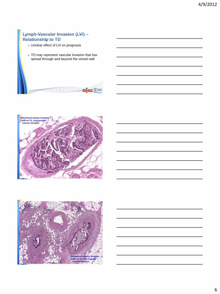
Unclear effect of LVI on prognosis
TD may represent vascular invasion that has
spread through and beyond the vessel wall
Extramural venous invasion
Code as V1, microscopic
venous invasion
Extramural venous invasion
Code as V1, microscopic
venous invasion
4/9/2012
7
Pericolonic adipose tissue with
focus of adenocarcinoma, possible
origin from lymph-vascular invasion
Code as Tumor Deposit (TD)
Pericolonic adipose tissue with
focus of adenocarcinoma, possible
origin from lymph-vascular invasion
Code as Tumor Deposit (TD)
Clear cut LVI – L1 and/or V1 – are not TD
TD is that subset not associated with any specific pathological entity
TD cannot be unequivocally classified as
– Nodes (N)
– Lymphatic invasion (L)
– Venous invasion (V)
– Perineural invasion (PN)
4/9/2012
8
Cancer registrars collect data on
– Nodes (N)
– Lymph-Vascular Invasion (LVI which includes L and V)
– Perineural invasion (PN)
– Tumor deposits (TD)
Tumor deposits have been identified
according to the criteria
and
There is no involvement of regional nodes
Tumor Deposits (discontinuous extramural
extension) (Note L)
___ Not identified
___ Present
___ Indeterminate
Washington MK, Berlin J, Branton PA, Burgart LJ, Carter DK, Fitzgibbons PL, Frankel WL, Halling KC, Jessup JM, Kakar S, Minsky B, Nakhleh RE, Compton CC; Cancer Committee, College of American Pathologists. Protocol for the examination of specimens from patients with primary carcinomas of the colon and rectum. Arch Pathol Lab Med. 2009 October; 133(10):1539–1551.
4/9/2012
9
L. Tumor Deposits (Discontinuous Extramural Extension)
– Irregular discrete tumor deposits in pericolic or perirectal fat away from the leading edge of the tumor and showing no evidence of residual lymph node tissue, but within the lymphatic drainage of the primary carcinoma, are considered peritumoral deposits or satellite nodules and are not counted as lymph nodes replaced by tumor.
– Most examples are due to lymph-vascular or, more rarely, perineural invasion.
– Because these tumor deposits are associated with reduced disease-free and overall survival, their number should be recorded in the surgical pathology report.
– If tumor deposits are observed in lesions that would otherwise be classified as pT1 (tumor confined to submucosa) or pT2 (tumor confined to muscularis propria), then the primary tumor classification is not changed, but the nodule is recorded in a separate N category as N1c.
Washington MK, Berlin J, Branton PA, Burgart LJ, Carter DK, Fitzgibbons PL, Frankel WL, Halling KC, Jessup JM, Kakar S, Minsky B, Nakhleh RE, Compton CC; Cancer Committee, College of American Pathologists. Protocol for the examination of specimens from patients with primary carcinomas of the colon and rectum. Arch Pathol Lab Med. 2009 October; 133(10):1539–1551.
Lymph-Vascular Invasion (Note E)
___ Not identified
___ Present
___ Indeterminate
Perineural Invasion (Note E)
___ Not identified
___ Present
___ Indeterminate
Washington MK, Berlin J, Branton PA, Burgart LJ, Carter DK, Fitzgibbons PL, Frankel WL, Halling KC, Jessup JM, Kakar S, Minsky B, Nakhleh RE, Compton CC; Cancer Committee, College of American Pathologists. Protocol for the examination of specimens from patients with primary carcinomas of the colon and rectum. Arch Pathol Lab Med. 2009 October; 133(10):1539–1551.
E. Lymph-Vascular and Perineural Invasion – Venous invasion has been demonstrated by multivariate analysis
to be an independent adverse prognostic factor.
– Invasion of extramural veins, in particular, has been shown to be an independent indicator of unfavorable outcome and increased risk of occurrence of hepatic metastasis.
– The significance of intramural venous invasion is less clear, because data specific to this issue are lacking.
– In several studies, both lymphatic invasion and perineural invasion have been shown by multivariate analysis to be independent indicators of poor prognosis. The prognostic significance, if any, of the anatomic location of these structures is not defined.
– Furthermore, it is not always possible to distinguish lymphatic vessels from postcapillary venules, because both are small, thin-walled structures.
– Thus, the presence or absence of tumor invasion of small, thin-walled vessels should be reported in all cases.
Washington MK, Berlin J, Branton PA, Burgart LJ, Carter DK, Fitzgibbons PL, Frankel WL, Halling KC, Jessup JM, Kakar S, Minsky B, Nakhleh RE, Compton CC; Cancer Committee, College of American Pathologists. Protocol for the examination of specimens from patients with primary carcinomas of the colon and rectum. Arch Pathol Lab Med. 2009 October; 133(10):1539–1551.
4/9/2012
10
Improvements to staging in 6th & 7th Editions
– 5th Edition did not have substaging for Stages II & III
– 6th & 7th Edition substaging
• Based on available outcomes data
• Accounts for differences in survival
Population-based validation
– Depth of invasion and nodal status interact to
affect survival
SEER survival data
– 35,829 rectal ca pts
– 109,953 colon ca pts
T4N0 stratified by T4a and T4b
– T4a – tumor perforate visceral peritoneum
– T4b – tumor directly invades other organs or
structures
N1 & N2 stratified by number of involved nodes
– N1a/N1b – 1 vs 2-3
– N2a/N2b – 4-6 vs >7
4/9/2012
11
5yr observed and relative survival
T1-2N0 have better survival than T3N0
T3N0 better than T4N0
T1-2N2 better than T3-4N2
T4bN1 similar to T4aN2
T4a better then T4b by N category
Number of N+ affects survival for each T category
SEER population-based analyses supported
– Subdividing T4 (T4a/T4b)
– Subdividing N1 (N1a/N1b) and N2 (N2a/N2b)
– Revised substaging of Stages II/III
• Shift of T1-2N2 lesions from IIIC to IIIA/IIIB
• Shift of T4bN1 lesions from IIIB to IIIC
During time frame of pt accrual and data collection
– SEER definition of extent of disease did not vary for • Degree of penetration of primary tumor (T)
• Number of involved nodes (N)
– Tumor deposits data when identified • Did not affect T
• Affected N in pts with T1/T2 lesion as TD were classified as involved nodes, as now recommended in 7th Edition
– Raw data TN categories stratified and published in JCO
– N1c affects all N0 patients because it moves a Stage group I or II into a IIIA or IIIB
4/9/2012
12
Clear definition of Tumor Deposits in 7th
Edition Colon and Rectum Chapter sections
as follows
– Pathologic staging
– Summary of Changes
– Prognostic Features
– Definition of TNM
Tumor Deposits (TD) cannot be ignored
– Possible negative impact on survival based on
retrospective analyses
Recommend prospective data collection on TD
– Survival impact by TN category may be appropriately
included in subsequent editions of TNM
Strongly support prospective collection of TD data