6 www.psnjournalonline.com Volume 40 Number 1 January–March 2020
Hair loss can be classifi ed by its cause and pre-sentation in all cases and, in some cases, by pa-tient gender. In this article, the author focuses
on four common hair loss disorders that occur in both men and women. The author discusses research fi ndings related to androgenetic alopecia (AGA), telogen effl u-vium (TE), alopecia areata (AA), and scarring alopecia (SA) and provides details on how to approach and man-age these diseases according to patient gender. Notably, there is some overlap in the causes and symptoms of the disorders, which highlights the need for obtaining a clear differential diagnosis ( Malkud, 2015 ). There are a range of tools and tests that can assist with the diagnostic pro-cess and help ensure that relevant and high standards of patient care are provided and maintained. In some cases, the patient may choose not to proceed with medical in-tervention. However, appropriate medical treatments, al-though still relatively limited in some cases, are safe and have proven effi cacy. Hair loss has immense emotional and psychological impact in both genders. It is always
important to consider this when planning hair loss man-agement pathways ( Ruiz-Doblado, Carrizosa, & Garcia-Hernandez, 2003 ). Following is a discussion of common hair loss disorders that occur in both men and women.
ANDROGENETIC ALOPECIA
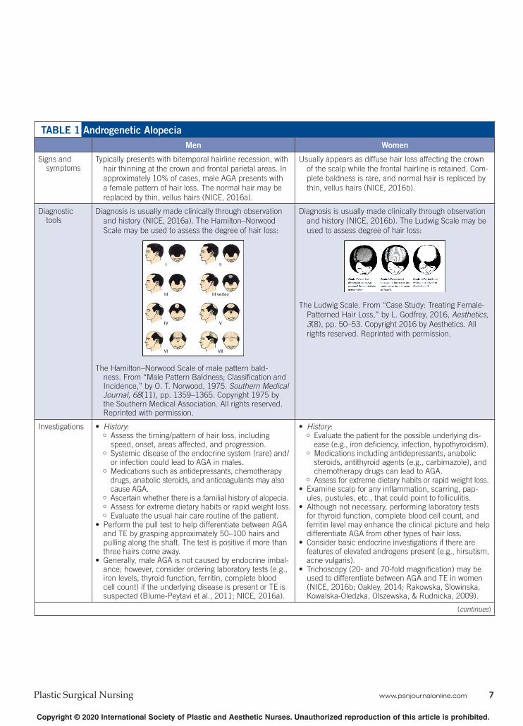
Androgenetic alopecia, as its name suggests, is thought to be androgen-dependent ( National Institute for Health and Care Excellence [NICE], 2016a , 2016b ). This condition presents as diffuse hair loss in both genders but is more likely to occur in postmenopausal women, affecting about 33% of genetically predisposed Caucasian women 70 years or older. In women with AGA, hair loss usually affects the top of the scalp ( NICE, 2016b ). As many as 58% of men aged 30–50 years may experience AGA. In men, hair loss typically starts with bitemporal hairline recession and hair thinning at the crown (i.e., vertex) and frontal parietal ar-eas ( NICE, 2016a ). According to the Hamilton–Norwood Scale ( Norwood, 1975 ), the amount of hair loss increases with age. Men with Grade I–III hair loss can benefi t from medical intervention. Men with Grade IV–VI hair loss gen-erally show good response to hair transplant procedures ( Shankar, Chakravarti, & Shilpakar, 2009 ). In both men and women, the underlying pathological process involves pigmented terminal hairs gradually being replaced by smaller, less pigmented hairs with a similar appearance to the short, thin, barely noticeable vellus hairs that develop on the body during childhood ( NICE, 2016a , 2016b ). See Table 1 for signs and symptoms, diagnostic tools, inves-tigations, and management of AGA in men and women.
TELOGEN EFFLUVIUM
Telogen effl uvium is a nonscarring form of hair loss. It generally occurs about 3 months after a triggering event when up to 70% of the anagen-phase hairs are precipi-tated into telogen hairs, with a bulb or club at their tip. After a few months, new hair pushes the club hairs up and out. This disorder is usually self-limiting, lasting for about 6 months before the hair regrows, provided the trigger is not repeated. There is a diffuse, but temporary loss of these hairs in both genders, and there is often a noticeable change in fi ngernail growth that occurs at the same time ( DermNet NZ, 1997 ; Trüeb, 2008 ).
Types and Treatment of Hair Loss in Men and Women
Emma Coleman , RGN
DOI: 10.1097/PSN.0000000000000285
Emma Coleman, RGN, is an aesthetic and dermatology registered gen-
eral nurse, Kent, United Kingdom .
The author reports no confl icts of interest .
Address correspondence to Emma Coleman, RGN, 12 Meadow Way,
In this article, the author focuses on 4 common hair loss disorders that occur in both men and women. The author discusses research related to androgenetic alopecia, telogen effl uvium, alopecia areata, and scarring alopecia and provides details on how to approach and manage these diseases according to patient gender. There are a range of tools and tests that can assist with the diagnostic process and help ensure that relevant and high standards of patient care are maintained. In some cases, no medical intervention is always a treatment option. However, appro-priate medical treatments, although still relatively limited in some cases, are safe and have proven effi cacy. Hair loss has immense emotional and psychological impact in both genders, and it is always important to consider this when planning hair loss management pathways.
Typically presents with bitemporal hairline recession, with
hair thinning at the crown and frontal parietal areas. In
approximately 10% of cases, male AGA presents with
a female pattern of hair loss. The normal hair may be
replaced by thin, vellus hairs (NICE, 2016a).
Usually appears as diffuse hair loss affecting the crown
of the scalp while the frontal hairline is retained. Com-
plete baldness is rare, and normal hair is replaced by
thin, vellus hairs (NICE, 2016b).
Diagnostic tools
Diagnosis is usually made clinically through observation
and history (NICE, 2016a). The Hamilton–Norwood
Scale may be used to assess the degree of hair loss:
The Hamilton–Norwood Scale of male pattern bald-ness. From “Male Pattern Baldness; Classifi cation and Incidence,” by O. T. Norwood, 1975. Southern Medical Journal, 68 (11), pp. 1359–1365. Copyright 1975 by the Southern Medical Association. All rights reserved. Reprinted with permission.
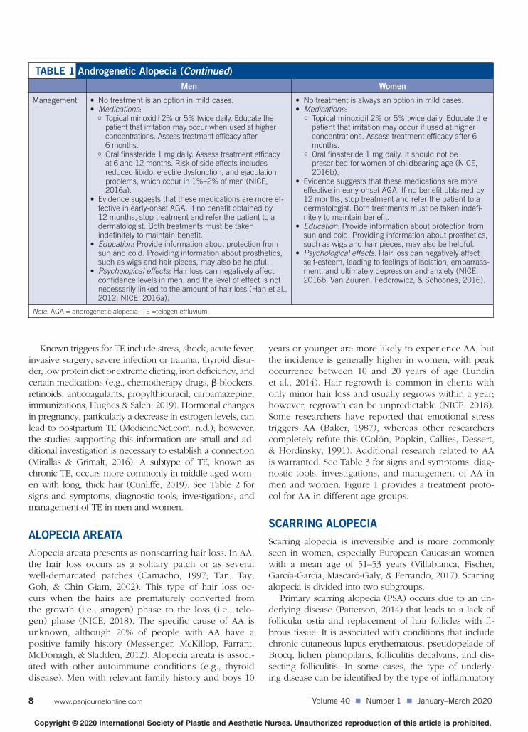
Diagnosis is usually made clinically through observation
and history (NICE, 2016b). The Ludwig Scale may be
used to assess degree of hair loss:
The Ludwig Scale. From “Case Study: Treating Female-
Patterned Hair Loss,” by L. Godfrey, 2016, Aesthetics,
3 (8), pp. 50–53. Copyright 2016 by Aesthetics. All
rights reserved. Reprinted with permission.
Investigations • History : Assess the timing/pattern of hair loss, including
speed, onset, areas affected, and progression. Systemic disease of the endocrine system (rare) and/
or infection could lead to AGA in males. Medications such as antidepressants, chemotherapy
drugs, anabolic steroids, and anticoagulants may also cause AGA.
Ascertain whether there is a familial history of alopecia. Assess for extreme dietary habits or rapid weight loss. Evaluate the usual hair care routine of the patient.
• Perform the pull test to help differentiate between AGA and TE by grasping approximately 50–100 hairs and pulling along the shaft. The test is positive if more than three hairs come away.
• Generally, male AGA is not caused by endocrine imbal-ance; however, consider ordering laboratory tests (e.g., iron levels, thyroid function, ferritin, complete blood cell count) if the underlying disease is present or TE is suspected (Blume-Peytavi et al., 2011; NICE, 2016a).
• History : Evaluate the patient for the possible underlying dis-
ease (e.g., iron defi ciency, infection, hypothyroidism). Medications including antidepressants, anabolic
steroids, antithyroid agents (e.g., carbimazole), and chemotherapy drugs can lead to AGA.
Assess for extreme dietary habits or rapid weight loss. • Examine scalp for any infl ammation, scarring, pap-
ules, pustules, etc., that could point to folliculitis. • Although not necessary, performing laboratory tests
for thyroid function, complete blood cell count, and ferritin level may enhance the clinical picture and help differentiate AGA from other types of hair loss.
• Consider basic endocrine investigations if there are features of elevated androgens present (e.g., hirsutism, acne vulgaris).
• Trichoscopy (20- and 70-fold magnifi cation) may be used to differentiate between AGA and TE in women (NICE, 2016b; Oakley, 2014; Rakowska, Slowinska, Kowalska-Oledzka, Olszewska, & Rudnicka, 2009).
8 www.psnjournalonline.com Volume 40 Number 1 January–March 2020
TABLE 1 Androgenetic Alopecia (Continued)
Men Women
Management • No treatment is an option in mild cases. • Medications :
Topical minoxidil 2% or 5% twice daily. Educate the patient that irritation may occur when used at higher concentrations. Assess treatment effi cacy after 6 months.
Oral fi nasteride 1 mg daily. Assess treatment effi cacy at 6 and 12 months. Risk of side effects includes reduced libido, erectile dysfunction, and ejaculation problems, which occur in 1%–2% of men (NICE, 2016a).
• Evidence suggests that these medications are more ef-fective in early-onset AGA. If no benefi t obtained by 12 months, stop treatment and refer the patient to a dermatologist. Both treatments must be taken indefi nitely to maintain benefi t.
• Education : Provide information about protection from sun and cold. Providing information about prosthetics, such as wigs and hair pieces, may also be helpful.
• Psychological effects : Hair loss can negatively affect confi dence levels in men, and the level of effect is not necessarily linked to the amount of hair loss (Han et al., 2012; NICE, 2016a).
• No treatment is always an option in mild cases. • Medications :
Topical minoxidil 2% or 5% twice daily. Educate the patient that irritation may occur if used at higher concentrations. Assess treatment effi cacy after 6 months.
Oral fi nasteride 1 mg daily. It should not be prescribed for women of childbearing age (NICE, 2016b).
• Evidence suggests that these medications are more effective in early-onset AGA. If no benefi t obtained by 12 months, stop treatment and refer the patient to a dermatologist. Both treatments must be taken indefi -nitely to maintain benefi t.
• Education : Provide information about protection from sun and cold. Providing information about prosthetics, such as wigs and hair pieces, may also be helpful.
• Psychological effects : Hair loss can negatively affect self-esteem, leading to feelings of isolation, embarrass-ment, and ultimately depression and anxiety (NICE, 2016b; Van Zuuren, Fedorowicz, & Schoones, 2016).
Note . AGA = androgenetic alopecia; TE = t elogen effl uvium.
Known triggers for TE include stress, shock, acute fever, invasive surgery, severe infection or trauma, thyroid disor-der, low protein diet or extreme dieting, iron defi ciency, and certain medications (e.g., chemotherapy drugs, β -blockers, retinoids, anticoagulants, propylthiouracil, carbamazepine, immunizations; Hughes & Saleh, 2019 ). Hormonal changes in pregnancy, particularly a decrease in estrogen levels, can lead to postpartum TE ( MedicineNet.com, n.d. ); however, the studies supporting this information are small and ad-ditional investigation is necessary to establish a connection ( Mirallas & Grimalt, 2016 ). A subtype of TE, known as chronic TE, occurs more commonly in middle-aged wom-en with long, thick hair ( Cunliffe, 2019 ). See Table 2 for signs and symptoms, diagnostic tools, investigations, and management of TE in men and women.
ALOPECIA AREATA
Alopecia areata presents as nonscarring hair loss. In AA, the hair loss occurs as a solitary patch or as several well-demarcated patches ( Camacho, 1997 ; Tan, Tay, Goh, & Chin Giam, 2002 ). This type of hair loss oc-curs when the hairs are prematurely converted from the growth (i.e., anagen) phase to the loss (i.e., telo-gen) phase ( NICE, 2018 ). The specifi c cause of AA is unknown, although 20% of people with AA have a positive family history ( Messenger, McKillop, Farrant, McDonagh, & Sladden, 2012 ). Alopecia areata is associ-ated with other autoimmune conditions (e.g., thyroid disease). Men with relevant family history and boys 10
years or younger are more likely to experience AA, but the incidence is generally higher in women, with peak occurrence between 10 and 20 years of age ( Lundin et al., 2014 ). Hair regrowth is common in clients with only minor hair loss and usually regrows within a year; however, regrowth can be unpredictable ( NICE, 2018 ). Some researchers have reported that emotional stress triggers AA ( Baker, 1987 ), whereas other researchers completely refute this ( Colón, Popkin, Callies, Dessert, & Hordinsky, 1991 ). Additional research related to AA is warranted. See Table 3 for signs and symptoms, diag-nostic tools, investigations, and management of AA in men and women. Figure 1 provides a treatment proto-col for AA in different age groups.
SCARRING ALOPECIA
Scarring alopecia is irreversible and is more commonly seen in women, especially European Caucasian women with a mean age of 51–53 years ( Villablanca, Fischer, García-García, Mascaró-Galy, & Ferrando, 2017 ). Scarring alopecia is divided into two subgroups.
Primary scarring alopecia (PSA) occurs due to an un-derlying disease ( Patterson, 2014 ) that leads to a lack of follicular ostia and replacement of hair follicles with fi -brous tissue. It is associated with conditions that include chronic cutaneous lupus erythematous, pseudopelade of Brocq, lichen planopilaris, folliculitis decalvans, and dis-secting folliculitis. In some cases, the type of underly-ing disease can be identifi ed by the type of infl ammatory
Signs and symptoms • Yun and Kim (2007) examined TE presentation
in patients undergoing chemotherapy. Among 20
men with patterned hair loss, occipital hairlines
were preserved in 50% ( n = 10), frontal hairlines
in 15% ( n = 3), and both occipital and frontal
hairlines in 35% ( n = 7).
• Generally, TE presents as a diffuse loss of club
hair in the telogen growth phase around 12
weeks following a trigger (DermNet NZ, 1997).
• Trüeb (2008) reported that 9% of males experi-
enced trichodynia associated with TE.
• The time of the shock or illness may be denoted
by stunted nail growth and a ridge or Beau line
may appear around the time of hair shedding
(DermNet NZ, 1997).
• Hair regrowth is generally spontaneous after 6–9
months (DermNet NZ, 1997).
• Yun and Kim (2007) examined TE presentation in
patients undergoing chemotherapy. Among 25 women
with patterned hair loss, occipital hairlines were pre-
served in 8% ( n = 2), frontal hairlines in 40% ( n = 10),
and both occipital and frontal hairlines in 52%
( n = 13).
• Generally, TE presents as a diffuse loss of club hair in
the telogen growth phase around 12 weeks following a
trigger (DermNet NZ, 1997).
• TE appears to be more common in women, although
this may be because women are more likely to report
and seek treatment of hair loss than men (Malkud,
2015).
• Chronic TE has a higher incidence in middle-aged
women with long hair (Cunliffe, 2019), with shedding
seen in areas normally spared by AGA, especially the
supra-auricular area (Rebora, 2016).
• Willimann and Trüeb (2002) found that women with TE
reported a higher incidence of trichodynia and discom-
fort originating from the hair and scalp than men.
• Up to 90% of women experience postpartum TE due
to hormonal changes (MedicineNet, n.d.). Although
lactation seems to infl uence the hair’s anagen rate, the
studies conducted thus far are too small to provide any
solid evidence about a connection with TE (Mirallas &
Grimalt, 2016).
• The time of the shock or illness may be denoted by
stunted nail growth and a ridge or Beau line may ap-
pear around the time of hair shedding (DermNet NZ,
1997).
• Hair regrowth is generally spontaneous after 6–
9 months (DermNet NZ, 1997).
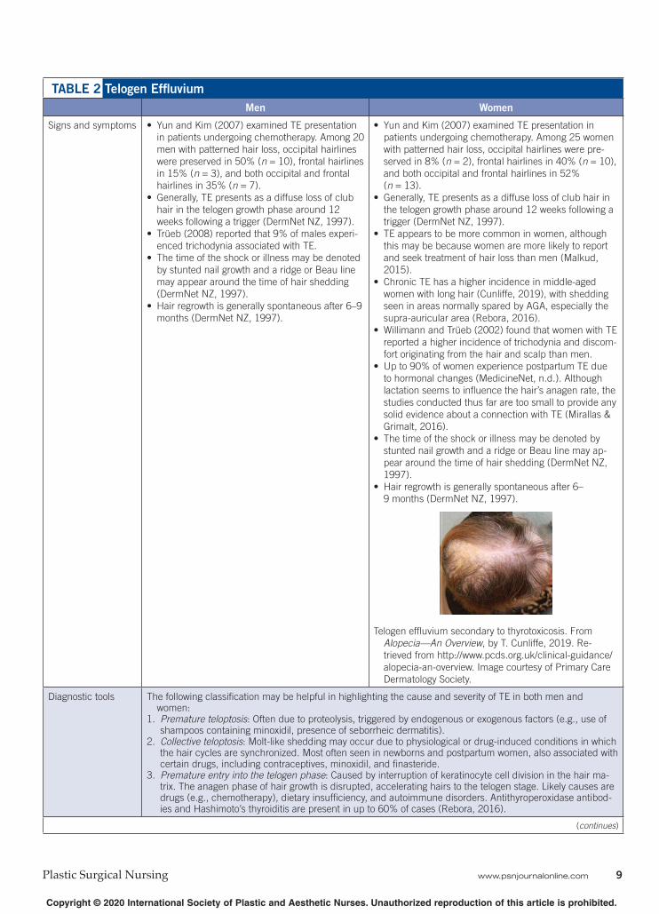
Telogen effl uvium secondary to thyrotoxicosis. From
Alopecia—An Overview , by T. Cunliffe, 2019. Re-
trieved from http://www.pcds.org.uk/clinical-guidance/
alopecia-an-overview. Image courtesy of Primary Care
Dermatology Society .
Diagnostic tools The following classifi cation may be helpful in highlighting the cause and severity of TE in both men and women:
1. Premature teloptosis : Often due to proteolysis, triggered by endogenous or exogenous factors (e.g., use of shampoos containing minoxidil, presence of seborrheic dermatitis).
2. Collective teloptosis : Molt-like shedding may occur due to physiological or drug-induced conditions in which the hair cycles are synchronized. Most often seen in newborns and postpartum women, also associated with certain drugs, including contraceptives, minoxidil, and fi nasteride.
3. Premature entry into the telogen phase : Caused by interruption of keratinocyte cell division in the hair ma-trix. The anagen phase of hair growth is disrupted, accelerating hairs to the telogen stage. Likely causes are drugs (e.g., chemotherapy), dietary insuffi ciency, and autoimmune disorders. Antithyroperoxidase antibod-ies and Hashimoto’s thyroiditis are present in up to 60% of cases (Rebora, 2016).
10 www.psnjournalonline.com Volume 40 Number 1 January–March 2020
TABLE 2 T elogen Effl uvium (Continued)
Men Women
Investigations • Wash test : A reliable pathway for diagnosis of TE. The patient is instructed to wash his or her hair 5 days after the last shampoo in a sink with its drain covered by gauze. The hair entrapped in the gauze is then counted (Amin & Sachdeva, 2013).
• Pluck test (Trichogram) : If there are 25% or more telogen hairs present, the test is positive. Telogen hairs are hairs that have tiny bulbs without sheaths at their roots (Amin & Sachdeva, 2013).
• Punch biopsy with histopathological analysis : Although not necessary, a biopsy shows the number of telogen follicles and can aid in diagnosis (Malkud, 2015). Findings in TE are best seen in transverse sections of a punch biopsy. If 25% of the follicles are in the telogen phase, this confi rms the diagnosis of TE (Amin & Sachdeva, 2013).
• Laboratory testing : Chronic TE sometimes has a metabolic cause, such as hypothyroidism. If symptoms of thyroid malfunction are present, a thyroid function test should be performed. Iron levels should be assessed with complete blood cell count, hemoglobin, and ferritin testing. If syphilis is suspected, a rapid plasma or venereal disease research laboratory test should be performed (Hughes & Saleh, 2019).
Management
• Psychological management : Patients should be reassured that normal
grooming of hair will not increase hair loss or prevent regrowth (Hughes & Selah, 2019).
• Lifestyle : Cigarettes contain heavy metals that can worsen TE; therefore, smoking cessation may be helpful (British Association of Derma-tologists [BAD], 2016).
• Educate patients about normal hair cycles and the relationships between triggers and timing of hair loss (Malkud, 2015).
• Psychological management : Women are more likely to have lowered quality of life
and restricted social contacts as a result of TE (Dinh & Sinclair, 2007).
Patients should be reassured that normal grooming of hair will not increase hair loss or prevent regrowth (Hughes & Selah, 2019).
• Lifestyle : Cigarettes contain heavy metals that can worsen TE; therefore, smoking cessation may be help-ful (BAD, 2016).
• Educate patients about normal hair cycles and relation-ships between triggers and timing of hair loss (Malkud, 2015).
• Hair transplant has no role in the management of TE as it is a temporary disorder (Malkud, 2015). • Once the causative issue has been identifi ed and withdrawn, the TE should also reside. • Although topical minoxidil has not been proven to promote hair regrowth in cases of TE, it may help patients
who wish to take an active role in their treatment (Hughes & Selah, 2019).
Note . AGA = androgenetic alopecia; TE = t elogen effl uvium.
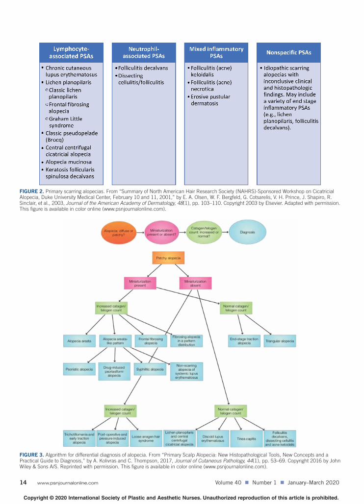
infi ltrate around the hair follicles ( Olsen et al., 2003 ; Villablanca et al., 2017 ). Figure 2 provides a classifi cation system for identifying the underlying disease associated with PSA, which can help determine treatment and man-agement for both men and women.
Secondary scarring alopecia (SSA) is so named be-cause it results from exogenous factors such as trauma caused by burns or radiation or by endogenous infl am-matory processes caused by disorders such as sarcoid-osis, pemphigus vulgaris, or scleroderma ( Villablanca et al., 2017 ). Although controversial, there is evidence to suggest that central centrifugal cicatricial alopecia (CCCA) is a type of SSA largely caused by hair treatments such as straightening with hot irons and using products with chemicals that damage hair follicles. This type of alopecia is seen predominantly in women of Afro-Caribbean de-scent ( Nicholson, Harland, Bull, Mortimer, & Cook, 1993 ; Sperling & Sau, 1992 ). There is some evidence to suggest that CCCA possesses an autosomal mode of inheritance ( Diova, Jordaan, Sarig, & Sprecher, 2014 ). One small study showed some correlation between the incidence of CCCA and diabetes mellitus ( Kyei, Bergfeld, Piliang, & Summers, 2011 ). To correctly classify CCCA, more large-scale and widespread investigation is necessary.
Figure 3 provides a two-step algorithm that can be used with both scarring and nonscarring forms of alope-cia where the presence or absence of follicular miniatur-ization and raised or normal catagen/telogen counts are used as markers for differential diagnosis. See Table 4 for signs and symptoms, diagnostic tools, investigations, and management of SA in men and women.
DIAGNOSTIC TOOLS
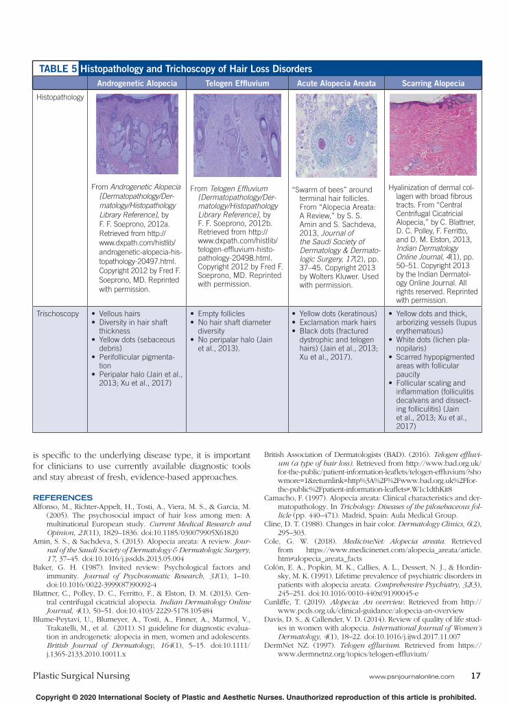
Differentiation in the diagnostic process is diffi cult but es-sential and is achieved with a variety of tools including biopsy with histopathology. Histopathology of AGA will show prominent sebaceous glands, miniaturization of exist-ing hair follicles, and reduced follicular diameter ( Soeprono, 2012a ). Telogen effl uvium histopathology will show abnor-mally high telogen follicles with empty follicular sheaths known as “stele” ( Malkud, 2015 ; Soeprono, 2012b ). Alope-cia areata histopathology typically presents with a “swarm of bees” appearance caused by infl ammatory infi ltrate around terminal hair follicles ( Amin & Sachdeva, 2013 ). Scarring alopecia histopathology will be identical in both genders and is characterized by collagen cells becoming translucent and highlighted by wide, tree trunk-like tracts
• Men are more likely to have a familial history and
may be diagnosed with AA when younger than 10
years (Lundin et al., 2014).
• More large-scale studies investigating gender as-
sociation are needed.
Exclamation mark hairs. From “Alopecia Areata: Clini-
cal Presentation, Diagnosis, and Unusual Cases,”
by A. M. Finner, 2011, Dermatologic Therapy,
24 (3), pp. 348–354. Copyright 2011 by Wiley
Periodicals, Inc. All rights reserved. Reprinted with
permission.
• Incidence is generally higher in females (2.3:1 female vs.
male), with peak diagnosis at 10–20 years of age.
• Women are more likely to have morbid nail symptoms
and autoimmune diseases, usually thyroid disorders (Lun-
din et al., 2014).
• More large-scale studies investigating gender association
are needed.
AA of the nails. From “Alopecia Areata: Clinical Presenta-
tion, Diagnosis, and Unusual Cases,” by A. M. Finner,
2011, Dermatologic Therapy, 24 (3), pp. 348–354. Copy-
right 2011 by Wiley Periodicals, Inc. All rights reserved.
Reprinted with permission.
• Clinical features are the same in both genders. AA most commonly presents as a sudden loss of hair in localized areas. The patch is usually round or oval-shaped and well demarcated at the borders. It may be a solitary patch (AA monolocularis) or numerous patches (AA multilocularis) (Camacho, 1997; Tan, Tay, Goh, & Chin Giam, 2002).
• The patch of alopecia usually has a distinct border, with normal hair demarcating the periphery of the lesion. • The scalp is the most common site affected by AA (90%). • Scalp and body hair such as eyebrows, eyelashes, beard, underarm hair, and pubic hair may be affected (alopecia
totalis), as well as the entire body (alopecia universalis) (Camacho, 1997; Tan et al., 2002). • Clinicians should look for “exclamation point hairs” surrounding the patches. These are hairs that are thick at the
top and become narrower along the length of the strand closer to the base with a root at the bottom (Cline, 1988). • Regrowing hair often lacks pigment, so it is white or blonde in color (Finner, 2011). • Nail changes are seen in 10%–66% of cases.
Red-spotted lunula and periungual erythema are a sign of acute nail involvement. Small shallow pits and trachyonychia are typical (Olsen, 2003).
Diagnostic tools Diagnosis is usually by clinical observation in both genders, although there are diagnostic tools that can be used to help ascertain disease severity:
• One scale presents the clinical signs of AA as follows: Mild : Three or fewer patches of alopecia with a widest diameter of less than 3 cm. Moderate : More than three patches or a patch greater than 3 cm at the widest diameter without alopecia totalis
or universalis. Severe : Alopecia totalis or alopecia universalis.
• Ophiasis : Severe alopecia where the loss of hair occurs in the shape of a wave at the circumference of the head (Kavak, Baykal, Özarmagan, & Akar, 2001).
• SALT The scalp is divided into four areas. The percentage of hair loss in any of the four areas is calculated using the following formula: % of hair loss × %
of surface area of the scalp in that area. The areas are marked out as follows:
Vertex—40% (0.4) of scalp surface area Right profi le of scalp—18% (0.18) of scalp surface area Left profi le of scalp—18% (0.18) of scalp surface area Posterior aspect of scalp—24% (0.24) of scalp surface area.
The SALT score is the sum of percentages of hair loss in all the aforementioned areas (Price & Gummer, 1989).
12 www.psnjournalonline.com Volume 40 Number 1 January–March 2020
TABLE 3 Alopecia Areata (Continued)
Men Women
Investigations • Explore familial history. • Most cases in both men and women can be diagnosed clinically, requiring no investigation, although there are
some protocols that may be helpful when diagnosis is in doubt (Messenger et al., 2012). • Pull test : Evaluates diffuse scalp hair loss. About 40–60 hairs are gently pulled in three different scalp areas. A
positive result occurs when 10 or more hairs are obtained. • Pluck test : The individual pulls out hairs by the roots, which are then examined under a microscope to determine
the phase of growth and differentiate between telogen, anagen, or systemic disease. Anagen hairs have sheaths at-tached to their roots. Anagen effl uvium shows raised broken hairs and a decrease in hairs at telogen growth phase.
• Scalp biopsy and histopathological analysis : These are carried out when alopecia is present, but diagnosis is un-clear. The biopsy allows for differentiation between scarring and nonscarring forms. Hair samples are taken from areas of infl ammation, usually around the border of the patch. Biopsy results will depend on the severity of AA, rather than the patient’s age, race, or gender (Igarashi, Morohashi, Takeuchi, & Sato, 1981). In the subacute stage, increased catagen and telogen hairs are seen. In the acute stage, terminal hairs are encompassed by bulbar lymphocytes or the “swarm of bees” appearance. In the chronic stage, decreased terminal and increased miniaturized hairs are found, with variable amounts of
infl ammation. • Daily hair counts : These are carried out in cases of negative pull test and done by counting the number of hairs
lost each day. Hairs should be counted after the fi rst morning combing or washing. Hairs are collected in a clear plastic bag for 14 days. The number of strands is recorded. If the hair count is more than 100 per day, this is abnormal (except after shampooing, where normal hair counts may reach up to 250).
• Trichoscopy : A noninvasive hair and scalp analysis may be performed using a handheld dermoscope or a video dermoscope. In AA, trichoscopy will highlight yellow dots (i.e., hyperkeratotic plugs), tiny exclamation mark hairs, and black dots (i.e., destroyed hairs in follicles) (Amin & Sachdeva, 2013).
Management • Management tends to be adjusted according to age rather than sex. • There is no defi nitive cure, and the main gender difference in management will be in the use of appropriate pros-
thetic types (e.g., women may prefer a head scarf to a hair piece) (NICE, 2018). • Intralesional corticosteroids : These are indicated as fi rst-line treatment in AA, with hair loss effecting less than
50% of the scalp. 10 mg/mL of triamcinolone acetonide is injected with a 0.5-in. needle in 0.1-ml injections, ap-proximately 1 cm apart. Review treatment effect at 4–8 weeks; repeat every 4–6 weeks if needed (Pascher, Kurtin, & Andrade, 1970).
• Topical corticosteroids : Painless and present a good option with children (e.g., fl uocinolone acetonide cream, fl uo-cinolone scalp gel, betamethasone valerate lotion, clobetasol propionate ointment); however, results are sporadic (Amin & Sachdeva, 2013).
• Systemic corticosteroids : Consider only in severe cases, as the side effects of these drugs may be limiting. One small-scale study involving 20 participants provided evidence that a tapering course of prednisolone improved hair regrowth by more than 25% in 30%–47% ( n = 9–14) of the participants with mild to severe AA (Sharma & Gupta, 1999). Another study showed some success using oral mini-pulse therapy with corticosteroids, which is a type of systemic treatment where large amounts of medications are administered to patients in short intervals to achieve stronger medication effects and avoid long-term use. The researchers found the treatment minimized side effects (Pasricha & Kumrah, 1996).
• Glucocorticoids : These may be prescribed for their anti-infl ammatory effects (Ross & Shapiro, 2005). • Minoxidil : A topical agent that stimulates follicle proliferation at the root and allows for differentiation above the
dermal papilla, independent of its vascular infl uences (Fiedler, Wendrow, Szpunar, Metzler, & DeVillez, 1990). • Anthralin : A topical agent that works by promoting free radical production over the scalp, leading to erythema or
pruritus after application of 0.5%–1% once daily over a 2-week period. The treatment is continued for 3–6 months, (Ross & Shapiro, 2005). Anthralin is particularly effective when used in combination with minoxidil (Fiedler et al., 1990).
• Topical immunomodulators : One recent study assessed the use of inosiplex in nine participants with AA (i.e., totalis). All participants developed enhanced T-cell function, and seven participants displayed signifi cant hair regrowth (Galbraith, Thiers, & Fundenberg, 1984). Additional studies are necessary to further assess effi cacy and side effect risks.
• PUVA : Reduces infl ammation around the hair follicles caused by Langerhans and mononuclear cells (Amin & Sachdeva, 2013). One study investigated the effect of PUVA with 26 participants; 54% displayed greater than 90% hair regrowth; those with alopecia areata universalis had more successful outcomes, whereas those with a positive familial history and alopecia areata totalis were less likely to respond to this treatment (Whitmont & Cooper, 2003).
• Oral cyclosporine : Inhibits T-cell activity; however, its risk for hepatotoxicity, nephrotoxicity, and other side effects may outweigh its benefi ts (Gupta et al., 1990).
• Topical tacrolimus : A calcineurin inhibitor that prevents activation of many cytokines in the infl ammatory process including interleukin-2, interferon, and tumor necrosis factor. There have been confl icting reports of its effi cacy in promoting hair regrowth in AA cases (Amin & Sachdeva, 2013).
• Oral sulfasalazine : It has immunosuppressive and immunomodulatory effects. One small study with 39 partici-pants showed good hair regrowth in 26% of patients ( n = 10) following treatment with 3 g of oral sulfasalazine. A moderate response was seen in 31% of participants ( n = 12) (Ellis, Brown, & Voorhees, 2002). Because of its positive safety profi le, this drug may present a preferable choice over tacrolimus and long-term steroids.
Note . AA = alopecia areata; PUVA = psoralen plus ultraviolet light therapy; SALT = Severity of Alopecia Tool.
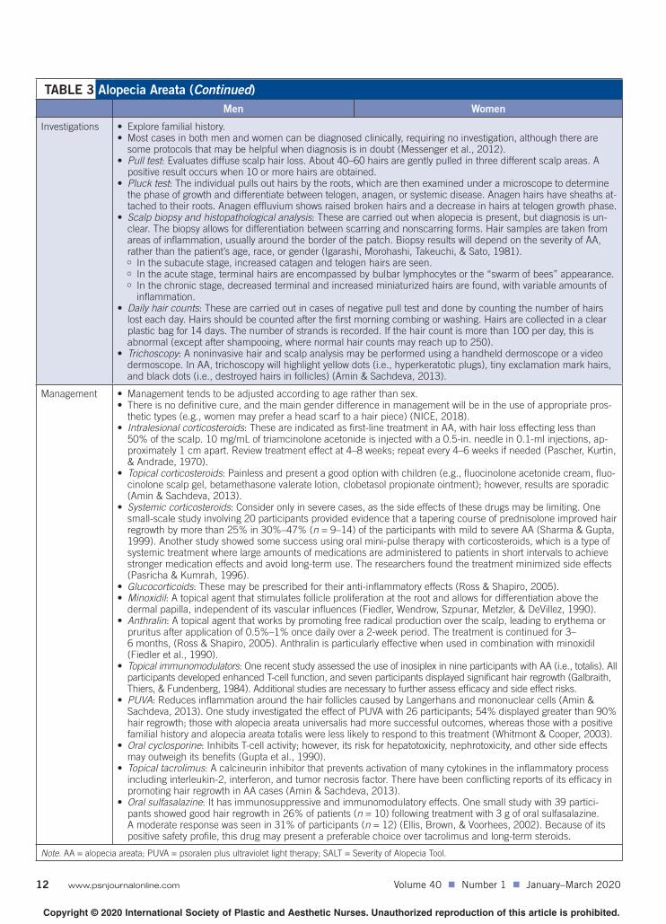
FIGURE 1. Treatment protocol for alopecia areata is licensed under Creative Commons Attribution-NonCommercial-NoDerivs 3.0 Unported [no
modifi cations]. From “Alopecia Areata: A Review,” by S. S. Amin and S. Sachdeva, 2013, Journal of the Saudi Society of Dermatology & Dermatologic
Surgery, 17 , pp. 37–45. Copyright 2013 by Wolters Kluwer. Used with permission. This fi gure is available in color online (www.psnjournalonline.com).
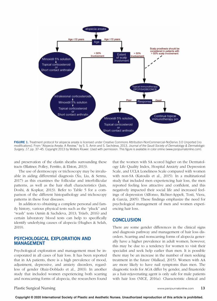
and preservation of the elastin sheaths surrounding these tracts ( Blattner, Polley, Ferritto, & Elston, 2013 ).
The use of dermoscopy or trichoscopy may be invalu-able in aiding differential diagnosis ( Xu, Liu, & Senna, 2017 ) as this examines the follicular and interfollicular patterns, as well as the hair shaft characteristics ( Jain, Doshi, & Kopkar, 2013 ). Refer to Table 5 for a com-parison of the different histopathology and trichoscopy patterns in these four diseases.
In addition to obtaining a complete personal and fam-ily history, various physical tests such as the “pluck” and “wash” tests ( Amin & Sachdeva, 2013 ; Trüeb, 2016 ) and certain laboratory blood tests can help to specifi cally identify underlying causes of alopecia ( Hughes & Selah, 2019 ).
PSYCHOLOGICAL EXPLORATION AND MANAGEMENT
Psychological exploration and management must be in-corporated in all cases of hair loss. It has been reported that in AA patients, there is a high prevalence of mood, adjustment, depressive, and anxiety disorders, regard-less of gender ( Ruiz-Doblado et al., 2003 ). In another study that included women experiencing both scarring and nonscarring forms of alopecia, the researchers found
that the women with SA scored higher on the Dermatol-ogy Life Quality Index, Hospital Anxiety and Depression Scale, and UCLA Loneliness Scale compared with women with non-SA ( Katoulis et al., 2015 ). In a multinational study that included men experiencing hair loss, the men reported feeling less attractive and confi dent, and this negatively impacted their social life and increased feel-ings of depression ( Alfonso, Richter-Appelt, Tosti, Viera, & Garcia, 2005 ). These fi ndings emphasize the need for psychological management of men and women experi-encing hair loss.
CONCLUSION
There are some gender differences in the clinical signs and diagnosis pathway and management of hair loss dis-orders. Scarring and nonscarring forms of alopecia gener-ally have a higher prevalence in adult women; however, this may be due to a tendency for women to visit their specialist and seek help earlier than men. It is possible there may be an increase in the number of men seeking treatment in the future ( Malkud, 2015 ). Women with AA are more likely to have nail symptoms than men. The diagnostic tools for AGA differ by gender, and fi nasteride as a hair-rejuvenating agent is only safe for male patients with hair loss ( NICE, 2016a ). Characteristic clinical and
16 www.psnjournalonline.com Volume 40 Number 1 January–March 2020
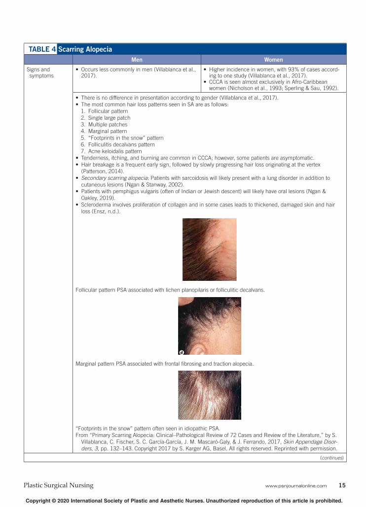
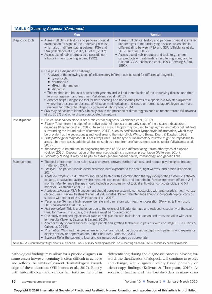
TABLE 4 Scarring Alopecia (Continued)
Men Women
Diagnostic tools
• Assess full clinical history and perform physical examination for signs of the underlying disease, which aids in differentiating between PSA and SSA (Villablanca et al., 2017; Xu et al., 2017).
• Assess use of hair products as a possible con-tributor in men (Sperling & Sau, 1992).
• Assess full clinical history and perform physical examina-tion for signs of the underlying disease, which aids in differentiating between PSA and SSA (Villablanca et al., 2017; Xu et al., 2017).
• Assess use of hair products and tools (e.g., chemi-cal products or treatments, straightening irons) and to rule out CCCA (Nicholson et al., 1993; Sperling & Sau, 1992).
• PSA poses a diagnostic challenge. Analysis of the following types of infl ammatory infi ltrate can be used for differential diagnosis:
This method can be used across both genders and will aid identifi cation of the underlying disease and there-fore management and treatment (Villablanca et al., 2017).
Another helpful diagnostic tool for both scarring and nonscarring forms of alopecia is a two-step algorithm where the presence or absence of follicular miniaturization and raised or normal catagen/telogen count are markers for differential diagnosis (Kolivras & Thompson, 2016).
• SSA may be easier to identify clinically due to the presence of direct triggers such as recent trauma (Villablanca et al., 2017) and other disease-associated symptoms.
Investigations • Clinical observation alone is not suffi cient for diagnosis (Villablanca et al., 2017). • Biopsy : Taken from the edge of an active patch of alopecia at an early stage of the disease aids accurate
diagnosis (Villablanca et al., 2017). In some cases, a biopsy may be used to highlight infl ammatory cell infi ltrate surrounding the infundibulum (Patterson, 2014), such as perifollicular lymphocytic infl ammation, which may be prevalent at the sebaceous gland level around the mid-follicle (Wilson, Burge, Dean, & Dawber, 1992).
• Histopathological diagnosis : It is not always useful as the type of infl ammatory infi ltrate might be impossible to assess. In these cases, additional studies such as direct immunofl uorescence can be useful (Villablanca et al., 2017).
• Trichoscopy : A helpful tool in diagnosing the type of PSA and differentiating it from other types of alopecia (Oakley, 2015). Desquamation of the inner root sheath is a common presentation (Patterson, 2014).
• Laboratory testing : It may be helpful to assess general patient health, immunology, and genetic links.
Management • The goal of treatment is to halt disease progress, prevent further hair loss, and reduce psychological impact (Patterson, 2014).
• Lifestyle : The patient should avoid excessive heat exposure to the scalp, tight weaves, and braids (Patterson, 2014).
• Acute neutrophilic PSA : Patients should be treated with a combination therapy incorporating systemic antibiot-ics (e.g., tetracycline, azithromycin), systemic corticosteroids, and isotretinoin. Review treatment effect at 2–6 months. Maintenance therapy should include a combination of topical antibiotics, corticosteroids, and 5% minoxidil (Villablanca et al., 2017).
• Acute lymphocytic PSA : Management should combine systemic corticosteroids with antimalarials (i.e., hydroxy-chloroquine). Review treatment effect at 2–6 months. Patient maintenance should incorporate topical cortico-steroids with minoxidil 5% (Villablanca et al., 2017).
• Recurrence : SA has a high recurrence rate and can return with treatment cessation (Kolivras & Thompson, 2016; Villablanca et al., 2017).
• Hair transplant : This is a challenge due to the extent of follicular damage and reduced vascularity of the scalp. Plus, for maximum success, the disease must be “burned out.”
• One study combined injections of platelet-rich plasma with follicular extraction and transplantation with excel-lent results (Saxena, Saxena, & Savant, 2016).
• Another study showed success using a punch hair grafting technique in patients with end-stage CCCA (Davis & Callender, 2014).
• Prosthetics : Wigs and hair pieces are an option and should be discussed in depth with patients who express or display anxiety or depression about their hair loss (Patterson, 2014).
• Support : Refer the patient to local and online support groups as appropriate.
pathological fi ndings may allow for a precise diagnosis in some cases; however, certainty is often diffi cult to achieve and refl ects the limits of current dermatological knowl-edge of these disorders ( Villablanca et al., 2017 ). Biopsy with histopathology and various hair tests are helpful in
differentiating during the diagnostic process. Moving for-ward, the classifi cation of alopecia will continue to evolve and change, with diagnostic clarity based primarily on trichoscopy fi ndings ( Kolivras & Thompson, 2016 ). As successful treatment of hair loss disorders in many cases
is specifi c to the underlying disease type, it is important for clinicians to use currently available diagnostic tools and stay abreast of fresh, evidence-based approaches.
REFERENCES Alfonso , M. , Richter-Appelt , H. , Tosti , A. , Viera , M. S. , & Garcia , M.
( 2005 ). The psychosocial impact of hair loss among men: A multinational European study . Current Medical Research and Opinion , 21 ( 11 ), 1829 – 1836 . doi:10.1185/030079905X61820
Amin , S. S. , & Sachdeva , S. ( 2013 ). Alopecia areata: A review . Jour-nal of the Saudi Society of Dermatology & Dermatologic Surgery , 17 , 37 – 45 . doi:10.1016/j.jssdds.2013.05.004
Baker , G. H. ( 1987 ). Invited review: Psychological factors and immunity . Journal of Psychosomatic Research , 31 ( 1 ), 1 – 10 . doi:10.1016/0022-3999(87)90092-4
Blattner , C. , Polley , D. C. , Ferritto , F. , & Elston , D. M. ( 2013 ). Cen-tral centrifugal cicatricial alopecia . Indian Dermatology Online Journal , 4 ( 1 ), 50 – 51 . doi:10.4103/2229-5178.105484
Blume-Peytavi , U. , Blumeyer , A. , Tosti , A. , Finner , A. , Marmol , V. , Trakatelli , M. , et al. ( 2011 ). S1 guideline for diagnostic evalua-tion in androgenetic alopecia in men, women and adolescents . British Journal of Dermatology , 164 ( 1 ), 5 – 15 . doi:10.1111/j.1365-2133.2010.10011.x
British Association of Dermatologists (BAD) . ( 2016 ). Telogen effl uvi-um (a type of hair loss) . Retrieved from http://www.bad.org.uk/for-the-public/patient-information-leafl ets/telogen-effl uvium/?showmore = 1&returnlink = http%3A%2F%2Fwww.bad.org.uk%2Ffor-the-public%2Fpatient-information-leafl ets#.W1c1dthKit8
Camacho , F. ( 1997 ). Alopecia areata: Clinical characteristics and der-matopathology . In Trichology: Diseases of the pilosebaceous fol-licle (pp. 440 – 471 ). Madrid, Spain : Aula Medical Group .
Cline , D. T. ( 1988 ). Changes in hair color . Dermatology Clinics , 6 ( 2 ), 295 – 303 .
Cole , G. W. ( 2018 ). MedicineNet: Alopecia areata . Retrieved from https://www.medicinenet.com/alopecia_areata/article.htm#alopecia_areata_facts
Colón , E. A. , Popkin , M. K. , Callies , A. L. , Dessert , N. J. , & Hordin-sky , M. K. ( 1991 ). Lifetime prevalence of psychiatric disorders in patients with alopecia areata . Comprehensive Psychiatry , 32 ( 3 ), 245 – 251 . doi:10.1016/0010-440x(91)90045-e
Cunliffe , T. ( 2019 ). Alopecia: An overview . Retrieved from http://www.pcds.org.uk/clinical-guidance/alopecia-an-overview
Davis , D. S. , & Callender , V. D. ( 2014 ). Review of quality of life stud-ies in women with alopecia . International Journal of Women’s Dermatology , 4 ( 1 ), 18 – 22 . doi:10.1016/j.ijwd.2017.11.007
From Telogen Effl uvium [Dermatopathology/Der-matology/Histopathology Library Reference] , by F. F. Soeprono, 2012b. Retrieved from http://www.dxpath.com/histlib/telogen-effl uvium-histo-pathology-20498.html. Copyright 2012 by Fred F. Soeprono, MD. Reprinted with permission.
“Swarm of bees” around terminal hair follicles. From “Alopecia Areata: A Review,” by S. S. Amin and S. Sachdeva, 2013, Journal of the Saudi Society of Dermatology & Dermato-logic Surgery, 17 (2), pp. 37–45. Copyright 2013 by Wolters Kluwer. Used with permission.
Hyalinization of dermal col-lagen with broad fi brous tracts. From “Central Centrifugal Cicatricial Alopecia,” by C. Blattner, D. C. Polley, F. Ferritto, and D. M. Elston, 2013, Indian Dermatology Online Journal, 4 (1), pp. 50–51. Copyright 2013 by the Indian Dermatol-ogy Online Journal . All rights reserved. Reprinted with permission.
Trischoscopy • Vellous hairs • Diversity in hair shaft
thickness • Yellow dots (sebaceous
debris) • Perifollicular pigmenta-
tion • Peripalar halo (Jain et al.,
2013; Xu et al., 2017)
• Empty follicles • No hair shaft diameter
diversity • No peripalar halo (Jain
et al., 2013).
• Yellow dots (keratinous) • Exclamation mark hairs • Black dots (fractured
dystrophic and telogen hairs) (Jain et al., 2013; Xu et al., 2017).
• Yellow dots and thick, arborizing vessels (lupus erythematous)
• White dots (lichen pla-nopilaris)
• Scarred hypopigmented areas with follicular paucity
• Follicular scaling and infl ammation (folliculitis decalvans and dissect-ing folliculitis) (Jain et al., 2013; Xu et al., 2017)
Diova , N. C. , Jordaan , F. H. , Sarig , O. , & Sprecher , E. ( 2014 ). Autoso-mal dominant inheritance of central centrifugal cicatricial alope-cia in Black South Africans . Journal of the American Academy of Dermatology , 70 ( 4 ), 679 – 682 . doi:10.1016/j.jaad.2013.11.035
Ellis , C. N. , Brown , M. F. , & Voorhees , J. J. ( 2002 ). Sulfasalazine for alopecia areata . Journal of the American Academy of Dermatol-ogy , 46 ( 4 ), 541 – 544 . doi:10.1067/mjd.2002.119671
Ensz , S. ( n.d. ). Scleroderma skin involvement: Alopecia (hair loss) . Retrieved from https://sclero.org/scleroderma/symptoms/skin/alopecia/a-to-z.html
Fiedler , V. C. , Wendrow , A. , Szpunar , G. J. , Metzler , C. , & DeVillez , R. L. ( 1990 ). Treatment resistant alopecia areata response to combination therapy with minoxidil plus anthralin . Archives of Dermatology , 126 ( 6 ), 756 – 759 . doi:10.1001/archderm.126.6.756
Finner , A. M. ( 2011 ). Alopecia areata: Clinical presentation, diagno-sis, and unusual cases . Dermatologic Therapy , 24 ( 3 ), 348 – 354 . doi:10.1111/j.1529-8019.2011.01413.x
Galbraith , G. M. P. , Thiers , B. H. , & Fundenberg , H. H. ( 1984 ). An open label trial of immunomodulation therapy with inosiplex (Isoprinosine) in patients with alopecia totalis and cell medi-ated immunodefi ciency . Journal of the American Academy of Dermatology , 11 , 224 – 230 . doi:10.1016/S0190-9622(84)70153-8
Godfrey , L. ( 2016, July 19 ). Case study: Treating female-patterned hair loss . Aesthetics . Retrieved from https://aestheticsjournal.com/feature/case-study-treating-female-patterned-hair-loss
Goel , A. ( 2017, June 10 ). Androgenetic alopecia—Male and fe-male pattern . Retrieved from https://www.linkedin.com/pulse/androgenetic-alopecia-dr-anupriya-goel/
Gupta , A. K. , Ellis , C. N. , Nickoloff , B. J. , Goldfarb , M. T. , Ho , V. C. , Rocher , L. L. , et al. ( 1990 ). Oral cyclosporine in the treatment of infl ammatory and noninfl ammatory dermatoses: A clinical and im-munopathologic analysis . Archives of Dermatology , 126 , 339 – 350 .
Hall , M. ( 2019 ). 10 effective ways to treat telogen effl uvium you need to know . Retrieved from https://www.hairlosscureguide.com/10-effective-ways-to-treat-telogen-effl uvium-you-need-to-know/
Han , S. H. , Byun , J. W. , Lee , W. S. , Kang , H. , Kye , Y. C. , Kim , K. H. , et al. ( 2012 ). Quality of life assessment in male patients with androgenetic alopecia: Result of a prospective, multicenter study . Annals of Dermatology , 24 ( 3 ), 311 – 318 . doi:10.5021/ad.2012.24.3.311
Hughes , E. , & Selah , D. ( 2019 ). Telogen effl uvium . Retrieved from https://www.ncbi.nlm.nih.gov/books/NBK430848/
Igarashi , R. , Morohashi , M. , Takeuchi , S. , & Sato , Y. ( 1981 ). Immu-nofl uorescence studies on complement components in hair fol-licles of normal scalp and of scalp affected by alopecia areata . Acta Demato-venereologica , 61 ( 2 ), 131 – 135 .
Jain , N. , Doshi , B. , & Kopkar , U. ( 2013 ). Trichoscopy in alopecias: Diagnosis simplifi ed . International Journal of Trichology , 5 ( 4 ), 170 – 178 . doi:10.4103/0974-7753.130385
Katoulis , A. , Christodoblou , C. , Liakou , A. , Kouris , A. , Korkoliakou , P. , Kaloudi , E. , et al. ( 2015 ). Quality of life and psychosocial impact of scarring and non-scarring alopecia in women . Jour-nal of the German Society of Dermatology , 13 ( 2 ), 137 – 142 . doi:10.1111/ddg.12548
Kavak , A. , Baykal , C. , & Özarmagan , G. , & Akar, U. ( 2001 ). HLA in alopecia areata . International Journal of Dermatology , 39 ( 8 ), 598 – 592 . doi:10.1046/j.1365-4362.2000.00921.x
Kolivras , A. , & Thompson , C. ( 2016 ). Primary scalp alopecia: New histopathological tools, new concepts and a practical guide to diagnosis . Journal of Cutaneous Pathology , 44 , 53 – 69 . doi:10.1111/cup.12822
Kyei , A. , Bergfeld , W. F. , Piliang , M. , & Summers , P. ( 2011 ). Medical and environmental risk factors for the development of central centrifugal cicatricial alopecia . Archives of Dermatology , 147 ( 8 ), 909 – 914 . doi:10.1001/archdermatol.2011.66
Lundin , M. , Chawa , S. , Sachdev , A. , Bhanusali , D. , Seiffert-Sinha , K. , & Sinha , A. ( 2014 ). Gender differences in alopecia areata . Journal of Drugs & Dermatology , 13 ( 4 ), 409 – 413 .
Malkud , S. ( 2015 ). Telogen effl uvium: A review . Journal of Clini-cal Diagnosis and Research , 9 ( 9 ), WE01 – WE03 . doi:10.7860/JCDR/2015/15219.6492
MedicineNet.com . ( n.d. ). Scalp, hair and nails; Telogen effl u-vium . Retrieved from https://www.medicinenet.com/image-collection/telogen_effl uvium_picture/picture.htm
Messenger , A. G. , McKillop , J. , Farrant , P. , McDonagh , A. J. , & Slad-den , M. ( 2012 ). British Association of Dermatologists’ guidelines for the management of alopecia areata . British Journal of Der-matology , 166 , 916 – 926 . doi:10.1111/j.1365-2133.2012.10955.x
National Institute for Health and Care Excellence (NICE) . ( 2016a ). Clinical knowledge summary: Alopecia—Androgenetic—Male . Retrieved from https://cks.nice.org.uk/alopecia-androgenetic-male#!topicsummary
National Institute for Health and Care Excellence (NICE) . ( 2016b ). Clinical knowledge summary: Alopecia—Androgenetic—Female . Retrieved from https://cks.nice.org.uk/alopecia-andro-genetic-female#!scenario
National Institute for Health and Care Excellence (NICE) . ( 2018 ). Clinical knowledge summary: Alopecia areata . Retrieved from https://cks.nice.org.uk/alopecia-areata#!scenario
Ngan , V. , & Oakley , A. ( 2019 ). Pemphigus vulgaris . Retrieved from https://www.dermnetnz.org/topics/pemphigus-vulgaris
Ngan , V. , & Stanway , A. ( 2002 ). Sarcoidosis . Retrieved from https://www.dermnetnz.org/topics/sarcoidosis
Nicholson , A. , Harland , C. , Bull , R. , Mortimer , P. , & Cook , M. ( 1993 ). Chemically induced cosmetic alopecia . British Journal of Derma-tology , 128 ( 5 ), 537 – 541 . doi:10.1111/j.1365-2133.1993.tb00231.x
Norwood , O. T. ( 1975 ). Male pattern baldness: Classifi cation and incidence . Southern Medical Journal , 68 ( 11 ), 1359 – 1365 . doi:10.1097/00007611-197511000-00009
Oakley , A. ( 2014 ). Scalp folliculitis . Retrieved from https://www.dermnetnz.org/topics/scalp-folliculitis
Oakley , A. ( 2015 ). Alopecia areata . Retrieved from https://www.dermnetnz.org/topics/alopecia-areata
Olsen , E. A. ( 2003 ). Hair . In I. M. Freedberg , A. Z. Eisen , K. Wolff , K. F. Austen , L. A. Goldsmith , & S. I. Katz (Eds.), Fitzpatrick’s dermatology in general medicine ( 6th ed ., Vol. I , pp. 348 – 354 ). New York : McGraw-Hill .
Olsen , E. A. , Bergfeld , W. F. , Cotsarelis , G. , Prince , V. H. , Shapiro , J. , Sinclair , R. , et al. ( 2003 ). Summary of North American Hair Research Society (NAHRS)-sponsored workshop on cicatricial alopecia, Duke University Medical Center, February 10 and 11, 2001 . Journal of the American Academy of Dermatology , 48 ( 1 ), 103 – 110 . doi:10.1067/mjd.2003.68
Pascher , F. , Kurtin , S. , & Andrade , R. ( 1970 ). Assay of 0.2 percent fl uocinolone acetonide cream for alopecia areata and totalis: Ef-fi cacy and side effects including histologic study of the ensuing localized acneiform response . Dermatologica , 141 , 193 – 202 . doi:10.1159/000252466
Pasricha , J. , & Kumrah , L. ( 1996 ). Alopecia totalis treated with oral mini-pulse (OMP) therapy with betamethasone . Indian Journal of Dermatology, Venereology and Leprology , 62 ( 2 ), 106 – 109 .
Patterson , S. ( 2014, March ). Central centrifugal cicatricial alope-cia . Retrieved from https://www.dermnetnz.org/topics/central-centrifugal-cicatricial-alopecia
Price , V. H. , & Gummer , C. L. ( 1989 ). Loose anagen syndrome . Jour-nal of the American Academy of Dermatology , 20 ( 2 ), 249 – 256 . doi:10.1016/S0190-9622(89)70030-X
Rakowska , A. , Slowinska , M. , Kowalska-Oledzka , E. , Olszewska , M. , & Rudnicka , L. ( 2009 ). Dermoscopy in female androgenic alopecia: Method standardization and diagnostic criteria . International Jour-nal of Trichology , 1 ( 2 ), 123 – 130 . doi:10.4103/0974-7753.58555
Rebora , A. ( 2016 ). Proposing a simpler classifi cation of telo-gen effl uvium . Skin Appendage Disorders , 2 ( 1–2 ), 35 – 38 . doi:10.1159/000446118
Ross , E. K. , & Shapiro , J. ( 2005 ). Management of hair loss . Derma-tology Clinics , 23 ( 2 ), 227 – 243 . doi:10.1016/j.det.2004.09.008
Ruiz-Doblado , S. , Carrizosa , A. , & Garcia-Hernandez , M. J. ( 2003 ). Alopecia areata: Psychiatric comorbidity and adjustment to ill-ness . International Journal of Dermatology , 42 ( 6 ), 434 – 437 . doi:10.1046/j.1365-4362.2003.01340.x
Saxena , K. , Saxena , D. K. , & Savant , S. S. ( 2016 ). Successful hair transplant outcome in cicatrix lichen planus of the scalp by combining beard and hair along with platelet rich plasma . Journal of Cutaneous and Aesthetic Medicine , 9 ( 1 ), 51 – 55 . doi:10.4103/0974-2077.178562
Shankar , D. S. K. , Chakravarti , M. , & Shilpakar , R. ( 2009 ). Male androgenetic alopecia: Population-based study in 1,005 sub-jects . International Journal of Trichology , 1 ( 2 ), 131 – 133 . doi:10.4103/0974-7753.58556
Sharma , V. K. , & Gupta , S. ( 1999 ). Twice weekly 5 mg dexamethasone oral pulse in the treatment of extensive alopecia areata . Journal of Dermatology , 26 ( 9 ), 562 – 565 . doi:10.1111/j.1346-8138.1999.tb02049.x
Soeprono , F. F. ( 2012a ). Androgenetic alopecia [ Dermatopathology/Dermatology/Histopathology Library Reference ]. Retrieved from http://www.dxpath.com/histlib/androgenetic-alopecia-histopathology-20497.html
Soeprono , F. F. ( 2012b ). Telogen effl uvium [ Dermatopathology/Dermatology/Histopathology Library Reference ]. Retrieved from http://www.dxpath.com/histlib/telogen-effl uvium-histopathology-20498.html
Sperling , L. C. , & Sau , P. ( 1992 ). The follicular degeneration syn-drome in black patients. “Hot comb alopecia” revisited and re-vised . Archives of Dermatology , 128 ( 1 ), 68 – 74 .
Tan , E. , Tay , Y. K. , Goh , C. L. , & Chin Giam , Y. ( 2002 ). The pattern and profi le of alopecia areata in Singapore—A study of 219
Asians . International Journal of Dermatology , 41 ( 11 ), 748 – 753 . doi:10.1046/j.1365-4362.2002.01357.x
Trüeb , R. M. ( 2008 ). Diffuse hair loss . In U. Blume-Paytavi , A. Tosti , D. A. Whiting , & R. M. Trüeb (Eds.), Hair growth and disorders ( 1st ed. , pp. 259 – 272 ). Berlin : Springer .
Trüeb , R. M. ( 2016 ). Chemotherapy-induced hair loss . Skin Therapy Letter , 15 ( 7 ), 5 – 7 . Retrieved from https://www.perunavitacomeprima.org/contentValue627L5.aspx
Van Zuuren , E. J. , Fedorowicz , Z. , & Schoones , J. ( 2016 ). Interven-tions for female pattern hair loss . Cochrane Database of System-atic Reviews , 5 , CD007628 . doi:10.1002/14651858.CD007628.pub4
Villablanca , S. , Fischer , C. , García-García , S. C. , Mascaró-Galy , J. M. , & Ferrando , J. ( 2017 ). Primary scarring alopecia: Clinical-pathological review of 72 cases and review of the literature . Skin Appendage Disorders , 3 , 132 – 143 . doi:10.1159/000467395
Whitmont , K. J. , & Cooper , A. J. ( 2003 ). PUVA treatment of alope-cia areata totalis and universalis: A retrospective study . Austral-asian Journal of Dermatology , 44 , 106 – 109 . doi:10.1046/j.1440-0960.2003.00654.x
Willimann , B. , & Trüeb , R. ( 2002 ). Hair pain (trichodynia): Frequency and relationship to hair loss and patient gender . Dermatology , 205 ( 4 ), 374 – 377 . doi:10.1159/000066437
Wilson , C. L. , Burge , S. M. , Dean , D. , & Dawber , R. P. ( 1992 ). Scar-ring alopecia in discoid lupus erythematosus . British Journal of Dermatology , 126 ( 4 ), 307 – 314 . doi:10.1111/j.1365-2133.1992.tb00670.x
Xu , L. , Liu , K. X. , & Senna , M. M. ( 2017 ). A practical approach to the diagnosis and management of hair loss in children and adoles-cents . Frontiers in Medicine (Lausanne) , 4 , 112 . doi:10.3389/fmed.2017.00112
Yun , S. J. , & Kim , S. J. ( 2007 ). Hair loss pattern due to chemotherapy-induced anagen effl uvium: A cross-sectional observation . Der-matology , 215 , 36 – 40 . doi:10.1159/000102031
For more than 61 additional continuing education articles related to dermatologic conditions, go to NursingCenter.com.
Instructions:• Read the article on page 6.
• The test for this CE activity is to be taken online at
www.NursingCenter.com. Find the test under the
article title. Tests can no longer be mailed or faxed.
• You will need to create and login to your personal CE
Planner account before taking online tests. Your
planner will keep track of all your Lippincott
Professional Development online CE activities for you.
• There is only one correct answer for each question.
A passing score for this test is 12 correct answers.
If you pass, you can print your certificate of earned
contact hours and access the answer key. If you fail,
you have the option of taking the test again at no
additional cost.
• For questions, contact Lippincott Professional
Development: 1-800-787-8985.
Registration Deadline: March 4, 2022
Disclosure Statement: The authors and planners
have disclosed that they have no financial relationships
related to this article.
Provider Accreditation:
Lippincott Professional Development will award 1.5 con-
tact hours for this continuing nursing education activity.
Lippincott Professional Development is accredited
as a provider of continuing nursing education by the
American Nurses Credentialing Center’s Commission on
Accreditation.
This activity is also provider approved by the California
Board of Registered Nursing, Provider Number CEP
11749 for 1.5 contact hours. Lippincott Professional
Development is also an approved provider of continuing
nursing education by the District of Columbia, Georgia,
and Florida CE Broker #50-1223.
Payment:• The registration fee for this test is $17.95.