1521-009X/48/3/160–168$35.00 https://doi.org/10.1124/dmd.119.089284 DRUG METABOLISM AND DISPOSITION Drug Metab Dispos 48:160–168, March 2020 Copyright ª 2020 by The Author(s) This is an open access article distributed under the CC BY-NC Attribution 4.0 International license. UDP-Glycosyltransferase 3A Metabolism of Polycyclic Aromatic Hydrocarbons: Potential Importance in Aerodigestive Tract Tissues s Ana G. Vergara, Christy J.W. Watson, Gang Chen, and Philip Lazarus Department of Pharmaceutical Sciences, College of Pharmacy and Pharmaceutical Sciences, Washington State University, Spokane, Washington Received September 5, 2019; accepted November 27, 2019 ABSTRACT Polycyclic aromatic hydrocarbons (PAHs) are potent carcinogens and are a primary risk factor for the development of lung and other aerodigestive tract cancers in smokers. The detoxification of PAHs by glucuronidation is well-characterized for the UDP- glycosyltransferase (UGT) 1A, 2A, and 2B subfamilies; however, the role of the UGT3A subfamily in PAH metabolism remains poorly understood. UGT3A enzymes are functionally distinct from other UGT subfamilies (which use UDP-glucuronic acid as a cosub- strate) due to their utilization of alternative cosubstrates (UDP-N- acetylglucosamine for UGT3A1, and UDP-glucose and UDP-xylose for UGT3A2). The goal of the present study was to characterize UGT3A glycosylation activity against PAHs and examine their expression in human aerodigestive tract tissues. In vitro metabolism assays using UGT3A2-overexpressing cell microsomes indicated that UGT3A2 exhibits glycosylation activity against all of the simple and complex PAHs tested. The V max /K m ratios for UGT3A2 activity with UDP-xylose versus UDP-glucose as the cosubstrate ranged from 0.65 to 4.4 for all PAHs tested, demonstrating that PAH glycosylation may be occurring at rates up to 4.4-fold higher with UDP-xylose than with UDP-glucose. Limited glycosylation activity was observed against PAHs with UGT3A1-overexpressing cell microsomes. While UGT3A2 exhibited low levels of hepatic expres- sion, it was shown by western blot analysis to be widely expressed in aerodigestive tract tissues. Conversely, UGT3A1 exhibited the high- est expression in liver with lower expression in aerodigestive tract tissues. These data suggest that UGT3A2 plays an important role in the detoxification of PAHs in aerodigestive tract tissues, and that there may be cosubstrate-dependent differences in the detoxifica- tion of PAHs by UGT3A2. SIGNIFICANCE STATEMENT UGT3A2 is highly active against PAHs with either UDP-glucose or UDP-xylose as a cosubstrate. UGT3A1 exhibited low levels of activity against PAHs. UGT3A1 is highly expressed in liver while UGT3A2 is well expressed in extrahepatic tissues. UGT3A2 may be an important detoxifier of PAHs in humans. Introduction Tobacco smoke contains over 4800 compounds with at least 69 identifiable carcinogens (Hoffmann et al., 2001). One of the most potent and abundant groups of tobacco carcinogens are the polycyclic aromatic hydrocarbons (PAHs), which are classified as group 1, 2A, or 2B carcinogens by the International Agency for Research on Cancer (Hecht, 1999). In addition to being present in tobacco smoke, PAHs are produced by incomplete combustion of organic compounds including wood, coal, oil, and gasoline, and many food sources, and humans are exposed to PAHs on a regular basis through air, water, soil, and food sources by ingestion, inhalation, and dermal contact (Mumtaz et al., 1996). The amount of carcinogenic PAHs found in the smoke of a single nonfiltered cigarette varies from 80 to 160 mg/cigarette, with one of the highest concentrations observed for benzo(a)pyrene (BaP): 20–40 ng/ cigarette (Hoffmann et al., 2001). While other PAHs, including dibenzo(a,l)pyrene (DBalP) and 5-methylchrysene (5-MeC), are stron- ger lung tumorigens than BaP in rodent models, they are far less abundant in tobacco smoke (1.7–3.2 ng/cigarette for DBalP and 0.6 ng/ cigarette for 5-MeC) (Sellakumar and Shubik, 1974; Nesnow et al., 1995; Prahalad et al., 1997; Hecht, 1999; Hoffmann et al., 2001). Nonsmokers can receive up to 70% of their PAH exposure through their diet, with high levels found in meat products (13–26 mg/kg), seafood (8.0–71 mg/kg), cereals (15–44 mg/kg), and oils/fats (24 mg/kg) (Choi et al., 2010). Smokers are exposed to higher levels of PAHs than nonsmokers, with urinary PAH metabolites 1.5- to 6.9-fold higher compared with nonsmokers (Suwan-ampai et al., 2009). This work was supported by the National Institutes of Health National Institutes of Environmental Health Sciences [Grants R01-ES025460 and R01-ES025460- 02S1]; the Health Sciences and Services Authority of Spokane, WA [Grant WSU002292]; and the synthesis of UDP-xylose by CarboSource Services, supported in part by the National Science Foundation Research Coordination Networks [Grant 0090281]. Part of this work was presented as follows: Vergara AG, Watson CJW, and Lazarus P (2017) Characterization of UDP-glycosyltransferase 3A (UGT3A) variants in tobacco carcinogen metabolism. FASEB J. 31 (1 Suppl):821.2; and Vergara AG, Watson CJW, Chen G, and Lazarus P (2019) Glycosylation of polycyclic aromatic hydrocarbons by UDP-glycosyltransferase 3A2 (UGT3A2) and aerodigestive tract tissues. FASEB J. 33 (1 Suppl):673.9. https://doi.org/10.1124/dmd.119.089284. s This article has supplemental material available at dmd.aspetjournals.org. ABBREVIATIONS: BaP, benzo(a)pyrene; BaP-7,8-diol, trans-7,8-dihydroxy-7,8-dihydro- benzo(a)pyrene; BaP-9,10-diol, benzo(a)pyrene-trans- 9,10-dihydrodiol; DBalP, dibenzo(a,l)pyrene; DBalP-11,12-diol, trans-11,12-dihydroxy-11,12-dihydrodibenzo(a,l)pyrene; HEK293, human embryonic kidney 293; 5-MeC, 5-methylchrysene; 5-MeC-1,2-diol, 1,2-dihydro-1,2-dihydroxy-5-methylchrysene; 1-OH-pyrene, 1-hydroxypyrene; PAH, polycyclic aromatic hydrocarbon; PCR, polymerase chain reaction; TBS, Tris-buffered saline; UDP-Glc, UDP-glucose; UDP-GlcNAc, UDP- N-acetylglucosamine; UDP-GlcUA, UDP-glucuronic acid; UDP-Xyl, UDP-xylose; UGT, UDP-glycosyltransferase. 160 http://dmd.aspetjournals.org/content/suppl/2019/12/13/dmd.119.089284.DC1 Supplemental material to this article can be found at: at ASPET Journals on February 21, 2022 dmd.aspetjournals.org Downloaded from at ASPET Journals on February 21, 2022 dmd.aspetjournals.org Downloaded from at ASPET Journals on February 21, 2022 dmd.aspetjournals.org Downloaded from at ASPET Journals on February 21, 2022 dmd.aspetjournals.org Downloaded from
Transcript
1521-009X/48/3/160–168$35.00 https://doi.org/10.1124/dmd.119.089284DRUG METABOLISM AND DISPOSITION Drug Metab Dispos 48:160–168, March 2020Copyright ª 2020 by The Author(s)This is an open access article distributed under the CC BY-NC Attribution 4.0 International license.
UDP-Glycosyltransferase 3A Metabolism of Polycyclic AromaticHydrocarbons: Potential Importance in Aerodigestive Tract Tissues s
Ana G. Vergara, Christy J.W. Watson, Gang Chen, and Philip Lazarus
Department of Pharmaceutical Sciences, College of Pharmacy and Pharmaceutical Sciences, Washington State University,Spokane, Washington
Received September 5, 2019; accepted November 27, 2019
ABSTRACT
Polycyclic aromatic hydrocarbons (PAHs) are potent carcinogensand are a primary risk factor for the development of lung andother aerodigestive tract cancers in smokers. The detoxification ofPAHs by glucuronidation is well-characterized for the UDP-glycosyltransferase (UGT) 1A, 2A, and 2B subfamilies; however, therole of the UGT3A subfamily in PAH metabolism remains poorlyunderstood. UGT3A enzymes are functionally distinct from otherUGT subfamilies (which use UDP-glucuronic acid as a cosub-strate) due to their utilization of alternative cosubstrates (UDP-N-acetylglucosamine for UGT3A1, and UDP-glucose and UDP-xylosefor UGT3A2). The goal of the present study was to characterizeUGT3A glycosylation activity against PAHs and examine theirexpression in human aerodigestive tract tissues. In vitrometabolismassays using UGT3A2-overexpressing cell microsomes indicatedthat UGT3A2 exhibits glycosylation activity against all of the simpleand complex PAHs tested. The Vmax/Km ratios for UGT3A2 activitywith UDP-xylose versus UDP-glucose as the cosubstrate rangedfrom 0.65 to 4.4 for all PAHs tested, demonstrating that PAH
glycosylation may be occurring at rates up to 4.4-fold higher withUDP-xylose than with UDP-glucose. Limited glycosylation activitywas observed against PAHs with UGT3A1-overexpressing cellmicrosomes. While UGT3A2 exhibited low levels of hepatic expres-sion, it was shown bywestern blot analysis to bewidely expressed inaerodigestive tract tissues. Conversely, UGT3A1 exhibited the high-est expression in liver with lower expression in aerodigestive tracttissues. These data suggest that UGT3A2 plays an important role inthe detoxification of PAHs in aerodigestive tract tissues, and thatthere may be cosubstrate-dependent differences in the detoxifica-tion of PAHs by UGT3A2.
SIGNIFICANCE STATEMENT
UGT3A2 is highly active against PAHs with either UDP-glucose orUDP-xylose as a cosubstrate. UGT3A1 exhibited low levels ofactivity against PAHs. UGT3A1 is highly expressed in liver whileUGT3A2 is well expressed in extrahepatic tissues. UGT3A2 may bean important detoxifier of PAHs in humans.
Introduction
Tobacco smoke contains over 4800 compounds with at least 69identifiable carcinogens (Hoffmann et al., 2001). One of the most potentand abundant groups of tobacco carcinogens are the polycyclic aromatichydrocarbons (PAHs), which are classified as group 1, 2A, or 2Bcarcinogens by the International Agency for Research on Cancer (Hecht,
1999). In addition to being present in tobacco smoke, PAHs areproduced by incomplete combustion of organic compounds includingwood, coal, oil, and gasoline, and many food sources, and humans areexposed to PAHs on a regular basis through air, water, soil, and foodsources by ingestion, inhalation, and dermal contact (Mumtaz et al.,1996). The amount of carcinogenic PAHs found in the smoke of a singlenonfiltered cigarette varies from 80 to 160 mg/cigarette, with one of thehighest concentrations observed for benzo(a)pyrene (BaP): 20–40 ng/cigarette (Hoffmann et al., 2001). While other PAHs, includingdibenzo(a,l)pyrene (DBalP) and 5-methylchrysene (5-MeC), are stron-ger lung tumorigens than BaP in rodent models, they are far lessabundant in tobacco smoke (1.7–3.2 ng/cigarette for DBalP and 0.6 ng/cigarette for 5-MeC) (Sellakumar and Shubik, 1974; Nesnow et al.,1995; Prahalad et al., 1997; Hecht, 1999; Hoffmann et al., 2001).Nonsmokers can receive up to 70% of their PAH exposure through theirdiet, with high levels found in meat products (13–26 mg/kg), seafood(8.0–71 mg/kg), cereals (15–44 mg/kg), and oils/fats (24 mg/kg) (Choiet al., 2010). Smokers are exposed to higher levels of PAHs thannonsmokers, with urinary PAH metabolites 1.5- to 6.9-fold highercompared with nonsmokers (Suwan-ampai et al., 2009).
This work was supported by the National Institutes of Health National Institutesof Environmental Health Sciences [Grants R01-ES025460 and R01-ES025460-02S1]; the Health Sciences and Services Authority of Spokane, WA [GrantWSU002292]; and the synthesis of UDP-xylose by CarboSource Services,supported in part by the National Science Foundation Research CoordinationNetworks [Grant 0090281].
Part of this work was presented as follows: Vergara AG, Watson CJW, andLazarus P (2017) Characterization of UDP-glycosyltransferase 3A (UGT3A)variants in tobacco carcinogen metabolism. FASEB J. 31 (1 Suppl):821.2; andVergara AG, Watson CJW, Chen G, and Lazarus P (2019) Glycosylation ofpolycyclic aromatic hydrocarbons by UDP-glycosyltransferase 3A2 (UGT3A2) andaerodigestive tract tissues. FASEB J. 33 (1 Suppl):673.9.
https://doi.org/10.1124/dmd.119.089284.s This article has supplemental material available at dmd.aspetjournals.org.
The major carcinogen activation pathway for PAHs is via thecytochrome P450 (CYP) class of enzymes, with biotransformation ofBaP by several CYP enzymes, including 1A1 and 1B1, to formhydroxylated or epoxide forms (Shimada et al., 1996; Kim et al.,1998). BaP epoxides are hydrolyzed by microsomal epoxide hydrolaseto form BaP-diols, which can undergo further metabolism by CYP1A1,1B1, and 3A4 to BaP-diol-epoxides, many of which are capable offorming PAH-DNA adducts (Thakker et al., 1977; Levin et al., 1980;Trushin et al., 2012). The carcinogenicity of PAHs is dependent on thenumber of benzenoid rings, their ring structure (fjord vs. bay regions),and having metabolites that can form DNA adducts (Moorthy et al.,2015; Gao et al., 2018). DBalP is the most carcinogenic PAH because ithas a fjord region that is nonplanar, reactive, and binds preferentially toadenine nucleotides (Ewa and Danuta, 2017). In contrast, 5-MeC andBaP have a bay region that is planar, less reactive, and binds to guaninenucleotides; 5-MeC is more carcinogenic than BaP because it hasa methylated bay region and an additional bay region (Palackal et al.,2002; Ewa and Danuta, 2017).A major mode of detoxification of PAHs is by the phase II family of
UDP-glycosyltransferases (UGTs). While extensive studies have exam-ined the UGT1A, 2A, and 2B enzyme subfamilies for activity againstPAHs (Jin et al., 1993; Fang et al., 2002; Uchaipichat et al., 2004; Finelet al., 2005; Luukkanen et al., 2005; Dellinger et al., 2006; Itäaho et al.,2010; Bushey et al.,2011, 2013; Olson et al., 2011), few studies haveexamined the activity of UGT3A enzymes against these carcinogeniccompounds. Unlike other UGT enzymes which use UDP-glucuronicacid (UDP-GlcUA) as a cosubstrate, the UGT3A enzymes are uniquefrom other UGTs in that they use alternative sugars as cosubstrates, withUGT3A1 using UDP-N-acetylglucosamine (UDP-GlcNAc) andUGT3A2 using UDP-glucose (UDP-Glc) or UDP-xylose (UDP-Xyl)(Mackenzie et al., 2008, MacKenzie et al., 2011). While UGT3A1 wasshown to be expressed in liver and kidney and to a lesser extent in testes,colon, and duodenum, UGT3A2 was found to be primarily anextrahepatic enzyme (Mackenzie et al., 2008, MacKenzie et al., 2011).In a screening of their activity against a variety of substrates, UGT3A1
and 3A2 both exhibited glycosylic activity against the simple PAHs 1-naphthol and 1-hydroxypyrene (1-OH-pyrene) (Mackenzie et al., 2008,MacKenzie et al., 2011; Meech and Mackenzie, 2010; Meech et al.,2012). The goals of the present study were to better characterize UGT3Aactivity against more complex PAHs and to examine their expression inhuman aerodigestive tract tissues, which are target sites of PAHcarcinogenicity.
Materials and Methods
Chemicals and Reagents. The RNeasy Mini Kit was purchased from Qiagen(Valencia, CA). SuperScript II reverse transcriptase, Platinum Taq DNA poly-merase, the pcDNA3.1/V5-His TOPO TA Expression Kit, One Shot TOP10competent Escherichia coli, Lipofectamine 2000, the PureLink Genomic DNAMini Kit, Invitrolon Polyvinylidene diflouride/Filter Paper Sandwich, and theNovex ECL Chemiluminescent Substrate Reagent Kit were obtained fromInvitrogen (Carlsbad, CA). Oligonucleotides for polymerase chain reaction(PCR) were purchased from Integrated DNA Technologies (Coralville, IA).The GeneJet Gel Extraction and DNA Cleanup Micro Kit, GeneJet Plasmid Miniand Midi Kit, Pierce BCA Protein Assay Kit, and Gelcode Blue Stain Reagentwere purchased from Thermo Scientific (Waltham, MA). Dulbecco’s modifiedEagle’s medium, Dulbecco’s PBS, and geneticin were purchased from Gibco(Grand Island, NY). Premium grade FBS was purchased from Seradigm (Radnor,PA) and ChromatoPur bovine albumin was purchased from MB Biomedicals(Santa Ana, CA).
The UGT3A2 antibody was purchased from Santa Cruz Biotechnology(Dallas, TX), while donkey anti-goat IgG horseradish peroxidase conjugate,anti-b-actin, rabbit anti-mouse IgG horseradish peroxidase conjugate, and goatanti-rabbit horseradish peroxidase conjugate were purchased from Thermo Fisher
Scientific (Rockford, IL). UDP-Glc and anti-UGT3A1 antibody were purchasedfrom Abcam (Cambridge, MA). UDP-Xyl was purchased from CarbosourceServices (Athens, GA). 1-OH-pyrene, 1-naphthol, alamethicin, ampicillin, UDP-GlcNAc, and b-N-acetylglucosaminidase were purchased from Sigma-Aldrich(St. Louis, MO). b-Glucosidase and exo-1,4-b-D-xylosidase were purchasedfromMegazyme (Bray, Ireland). 1-OH-BaP, 7-OH-BaP, 8-OH-BaP, 9-OH-BaP,B(a)P-trans-9,10-dihydrodiol (BaP-9,10-diol), trans-11,12-dihydroxy-11,12-dihydrodibenzo(a,l)pyrene (DBalP-11,12-diol), and 1,2-dihydro-1,2-dihydroxy-5-methylchrysene (5-MeC-1,2-diol) were purchased from MRI Global (KansasCity, MO). 3-OH-BaP and trans-7,8-dihydroxy-7,8-dihydro-B(a)P (BaP-7,8-diol) were purchased from Toronto Research Chemicals (North York, ON,Canada). High-performance liquid chromatography grade ammonium acetate andOptima acetonitrile were purchased from Fisher Scientific (Pittsburgh, PA).
Tissues. Normal human tissue specimens were obtained from the Banner SunHealth Research Institute (Sun City, AZ), the Cooperative Human TissueNetwork Eastern Division (Philadelphia, PA), or the H. Lee Moffitt CancerCenter (Tampa, FL), and were isolated postsurgery or postmortem within 3 hoursand frozen at280�C.Mixed tissues selected for this studywere primarily from therespiratory tract [lung (n 5 5), trachea (n 5 4), tongue (n 5 3), floor of mouth(n 5 2), tonsil (n 5 5), larynx (n 5 4), and esophagus (n 5 5)], digestive tract[jejunum (n 5 5), colon (n 5 5), and liver (n 5 5)], and breast (n 5 5). Thedemographic information for these human tissues is described in SupplementalTable 1. Of the tissue samples where demographic information was obtained,51% were female, with 80% from Whites and 20% from Blacks.
Normal human kidney total RNA was purchased from Stratagene (La Jolla,CA); total RNA was extracted using standard protocols from normal human livertissue obtained from the Penn State University College of Medicine Tissue Bank.All protocols involving the analysis of tissue specimens from these tissue bankswere approved by the institutional review board atWashington State University inaccordance with assurances filed with and approved by the U.S. Department ofHealth and Human Services.
Generation of UGT3A-Overexpressing Cell Lines. A stable humanembryonic kidney 293 (HEK293) cell line overexpressing UGT3A1 wasgenerated using standard protocols. Normal human liver total RNA (2 mg) wasextracted using an RNeasy Mini Kit from normal human liver tissue, whichwas used as a template in a reverse transcription reaction containing SuperScript IIreverse transcriptase (200 U). cDNA corresponding to 200 ng total liver RNAwasused with 2.5 U of Platinum Taq DNA polymerase for the PCR amplification ofUGT3A1. The primers used to amplify UGT3A1 from liver cDNA were 59-TGCTTCTGTGGAAGTGAGCATGGT-39 (sense) and 59-AGCCTCATGTCTTCTTCACCTTC-39 (antisense), corresponding to nucleotides219 to15 and11576to 11554, respectively, relative to the UGT3A1 translation start site. PCR wasperformed with an initial denaturation temperature of 94�C for 2 minutes,40 cycles of 94�C for 30 seconds, 57�C for 40 seconds, and 72�C for 1 minute and45 seconds, followed by a final cycle of 10 minutes at 72�C. The UGT3A1sequence was verified by Sanger sequencing (Genewiz, South Plainfield, NJ) andcompared with that described for UGT3A1 in GenBank (NM_152404.3). Thesequencing results revealed that the UGT3A1 insert contained a synonymousC1320T nucleotide change, which maintained the alanine at amino acid residue430. The UGT3A1 insert was cloned into a pcDNA3.1/V5-His-TOPO vectorusing standard protocols. After transformation using One Shot TOP10 competentE. coli, transformants were grown on plates containing LB agar and ampicillin(100 mg/ml) and confirmed by Sanger sequencing. Lipofectamine 2000 was usedto transfect 8 mg of pcDNA3.1/V5-His-TOPO/UGT3A1 plasmid into HEK293cells purchased from American Type Culture Collection (Manassas, VA). TheHEK293 cell line was authenticated by American Type Culture Collection usingshort-tandem repeat polymorphisms analysis in December 2017. Stable cell lineswere grown in Dulbecco’s modified Eagle’s medium supplemented with10% FBS and 700 mg/ml of geneticin. Genomic DNA was extracted from thestable cell line using the PureLink Genomic DNA Mini Kit and Sangersequencing was used to confirm the presence and identity of the UGT3A1 cDNAsequence.
The UGT3A2 overexpressing HEK293 cell line was generated by reversetranscription PCR as described previously using normal human total kidney RNA(2 mg) as the template for reverse transcription. The primers used to amplifyUGT3A2 from kidney cDNA were 59-GGCTTCCGTAGAAGTGAGCATG-39(sense) and 59-CCTGGCCTTATGTCTCCTTCACC-39 (antisense), correspond-ing to nucleotides219 to13 and11579 to 11557, respectively, relative to the
UGT3A2 translation start site. PCR was performed with an initial denaturationtemperature of 94�C for 2 minutes, 40 cycles of 94�C for 30 seconds, 57�C for 40seconds, and 72�C for 2 minutes, followed by a final cycle of 10 minutes at 72�C.The PCR product was excised and purified from an agarose gel using the GeneJetGel Extraction Kit. The purified PCR product was verified by Sanger sequencingand was found to be identical to the reference UGT3A2 cDNA sequence(NM_174914.3). The verified UGT3A2 cDNA was cloned into the pcDNA3.1/V5-His-TOPOvector and overexpressed in HEK293 cells as described previouslyfor UGT3A1. The UGT3A2-overexpressing HEK293 cell line was verified bySanger sequencing.
Analysis of UGT3A Protein Expression. For UGT-overexpressing cell lines,whole cell homogenates and S9 and microsomal fractions were prepared throughdifferential centrifugation utilizing methods adapted from a previous study(Dellinger et al., 2007). Briefly, cell homogenates were prepared by resuspendingpelleted cells in Tris-buffered saline [(TBS); 25mMTris base, 138 mMNaCl, and2.7 mM KCl (pH 7.4)], followed by five rounds of freeze-thaw cycles prior togentle homogenization. The S9 fraction was prepared by centrifuging the cellhomogenate at 9000g for 30minutes at 4�C. The S9 fraction was further processedby ultracentrifugation at 105,000g for 1 hour at 4�C, and the microsomal pelletwas resuspended in TBS. Total protein concentrations were determined using thePierce BCA Protein Assay Kit.
Western blot analysis was performed using 20 mg of total protein homogenateutilizing a 10% SDS-polyacrylamide gel, which was subsequently transferred toan Invitrolon Polyvinylidene difluoride membrane. For UGT3A1, membraneswere blocked with a 5% solution of milk in TBS containing 0.1% Tween 20 andprobed with a rabbit monoclonal UGT3A1 antibody (1:1500 dilution), followedby a goat anti-rabbit secondary antibody (1:1000 dilution). For UGT3A2, themembrane was blocked with a 5% solution of ChromatoPur bovine albumin inTBS containing 0.1% Tween 20 and probed with goat polyclonal UGT3A2antibody (1:1000 dilution), followed by a donkey anti-goat secondary antibody(1:2500 dilution). The b-actin antibody (1:5000 dilution) was used to verify equalloading using the rabbit anti-mouse secondary antibody (1:10,000 dilution) forboth the UGT3A1 and UGT3A2 western blots. Immunocomplexes werevisualized with the Novex ECL Chemiluminescent Kit following the manufac-turer’s protocols.
Tissues were homogenized with a Qiagen TissueLyser II (Hilden, Germany) in2 ml tubes with a 5 mm bead at 22 Hz for 2 minutes. S9 fractions were preparedusing TBS by centrifugation at 9000g for 30minutes at 4�C.Western blot analysiswas performed using 20 mg of S9 fractions that were analyzed as describedpreviously for the UGT-overexpressing cell lines. Loading variability wasmonitored by Coomassie blue staining. Gelcode Blue Stain Reagent was usedto detect total protein for normalization by densitometry analysis using Image Jsoftware (https://imagej.nih.gov/ij/; National Institutes of Health, Bethesda, MD).
Glycosylation Assays and Analysis. To screen for glycosylation activity forboth UGT3A enzymes, incubations were performed with alternative sugars usinga method adapted from a previous study (Bushey et al., 2011). Microsomes(10–100 mg total protein) from either the UGT3A1- or UGT3A2-overexpressingHEK293 cell lines were incubated with alamethicin (50 mg/mg total protein) for15 minutes on ice. Glycosylation reactions were performed with 200–800 mMsubstrate, 50 mM Tris-HCl (pH 7.4), 10 mM MgCl2, and 4 mM UDP-GlcNAc,UDP-Glc, or UDP-Xyl in a final reaction volume of 25 ml at 37�C for 1.5 hours.Reactions were terminated by the addition of 25 ml cold acetonitrile. Reactionmixtures were centrifuged for 10 minutes at 16,100g, and supernatants werecollected for ultra-performance liquid chromatography analysis.
Glycoside metabolite formation was quantified using an ACQUITY UPLCSystem (Waters, Milford, MA) utilizing an ACQUITY UPLC BEH C18 column(1.7 mm, 2.1� 100 mm; Waters) at a constant temperature of 25�C. Using a flowrate of 0.4 ml/min, glycosides were eluted and separated from parent substrateusing various gradients for different PAHs. For the glycoside of 1-OH-pyrene, theinitial elution was 90% buffer A [5 mM NH4OAc (pH 5.0) and 10% acetonitrile]and 10% buffer B (100% acetonitrile) for 2 minutes, subsequent linear gradient to75% B for 2 minutes, subsequent linear gradient to 100% B for 2 minutes, andreturn to the initial condition from 6 to 7.5 minutes. A similar gradient was usedfor other substrates, but the initial ratio of buffers A to B varied slightly. Thesubstrates with an initial elution concentration of 85% A and 15% B were 1-OH-BaP, 3-OH-BaP, 7-OH-BaP, 8-OH-BaP, 9-OH-BaP, BaP-7,8-diol, BaP-9,10-diol, DBalP-11,12-diol, and 5-MeC-1,2-diol; 1-naphthol was examined with aninitial elution concentration of 80% A and 20% B. The UV absorbance for each
substrate and glycoside were as follows: 1-OH-pyrene and 1-naphthol weredetected at 240 nm; BaP-7,8-diol, BaP-9,10-diol, and 5-MeC-1,2-diol weredetected at 254 nm; and 1-OH-BaP, 3-OH-BaP, 7-OH-BaP, 8-OH-BaP, 9-OH-BaP, and DBalP-11,12-diol were detected at 305 nm. If a metabolite peak wasidentified, kinetic analysis was performed for the enzyme against the activesubstrate. Kinetic analysis was performed for UGT3A-overexpressing micro-somes (100 mg for UGT3A1; 0.20–75 mg for UGT3A2) using 0.25–2800 mMsubstrate. For glycosylation rate determinations, total protein and incubation timesfor each substrate were optimized experimentally to ensure that substrateutilization was less than 10% and to maximize the levels of detection while ina linear range of glycoside formation. Reactions were terminated after 1.5 hoursfor UGT3A1 and 45minutes or 2 hours for UGT3A2 by the addition of 25ml coldacetonitrile.
The area under the curve for the substrate and glycoside peaks were determinedusing the MassLynx 4.1 software and quantified using the ratio of glycosidecompared with unconjugated substrate. 1-OH-pyrene, a common UGT substrateand a known substrate for UGT3A1 and 3A2 (Mackenzie et al., 2008, MacKenzieet al., 2011), was used as a positive control for activity. Reactions withuntransfected HEK293 cell microsomes, no substrate added, or substrate only,were used as negative controls. In addition, UGT2A1-overexpressingmicrosomeswere used as a positive control for all substrates (Bushey et al., 2011). Themetabolites were confirmed by sensitivity to glycosidases (b-N-acetylglucosa-minidase for reactions with microsomal UGT3A1 protein and b-glucosidase andb-xylosidase for reactions with microsomal UGT3A2 protein) by incubating 2 mlof glycosidase in a reconstituted 10 ml reaction (with water) at 37�C overnight.Reactions were terminated by the addition of 12ml cold acetonitrile and processedas described previously. Kinetic parameters (Km and Vmax) were calculated fromtriplicate experiments using GraphPad Prism 7.
Statistical Analysis. A two-tailed t test was used to compare the kinetics (Km,Vmax, and Vmax/Km) of glycoside formation for the UGT3A2-overexpressingHEK293 cell line for UDP-Xyl when comparing to UDP-Glc. A P value of lessthan 0.05 was considered statistically significant.
Results
Expression of UGT3A Enzymes in Human Tissues. In the currentstudy, UGT3A1 and UGT3A2 expression was analyzed in a compre-hensive panel of aerodigestive tract tissues. As shown by western blotsof UGT3A1- and UGT3A2-overexpressing cell lines, there was nocrossreactivity of the UGT3A1 (Fig. 1A) or UGT3A2 (Fig. 1B)antibodies with any of the other UGTs tested. The molecular weightof both recombinant UGT3A proteins was approximately 53 kDa, asreported previously (Mackenzie et al., 2008, MacKenzie et al., 2011).Representative western blots showed that both UGT3A1 and 3A2
were expressed in all tissues tested (Fig. 1, C and D, respectively).Densitometry analysis showed that the relative expression of UGT3A1was highest in liver (used as the reference at 1.0), followed by tongue,jejunum, and larynx (approximately 0.30 for each) . trachea (0.20) .lung, breast, and colon (approximately 0.14 for each) . tonsil andesophagus (approximately 0.040 for both) . floor of mouth (0.025)(Fig. 1E). The relative expression for UGT3A2 was highest in the floorof the mouth (used as the reference at 1.0), followed by trachea andlarynx (approximately 0.70 for both) . breast, lung, and tongue(approximately 0.60 for each) . esophagus, tonsil, and colon (approx-imately 0.50 for each) . jejunum (0.30) . liver (0.21) (Fig. 1F).Glycosylation of PAHs by UGT3A Enzymes. Microsomal protein
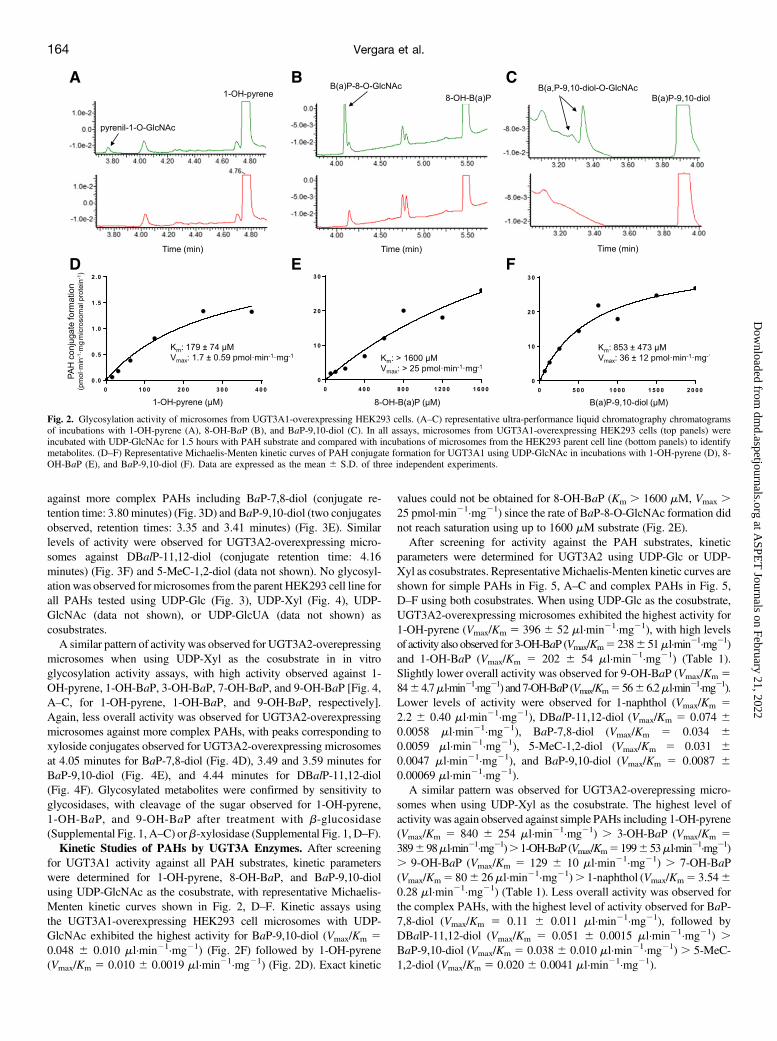
from the UGT3A1- and UGT3A2-overexpressing HEK293 celllines was used to screen for activity against the following PAHs: 1-OH-pyrene, 1-naphthol, 1-OH-BaP, 3-OH-BaP, 7-OH-BaP, 8-OH-BaP, 9-OH-BaP, BaP-7,8-diol, BaP-9,10-diol, DBalP-11,12-diol,and 5-MeC-1,2-diol. In vitro glycosylation assays using UDP-GlcNAcas cosubstrate showed UGT3A1 activity against 1-OH-pyrene,a known UGT3A1 substrate (Meech and Mackenzie, 2010), to formthe pyrenil-1-O-GlcNAc conjugate (retention time: 3.78 minutes)(Fig. 2A). UGT3A1-overexpressing microsomes also demonstrated
activity against 8-OH-BaP (Fig. 2B) and BaP-9,10-diol (Fig. 2C). TwoGlcNAc conjugates were observed for BaP-9,10-diol (retention times:3.28 and 3.34 minutes), likely representing N-acetylglucosaminides atthe 9- and 10-diol positions. Detectable glycosylation activity forUGT3A1-overexpressing microsomes was not observed for any otherPAH tested using up to 100 mg microsomal protein. No glycosylationwas observed for microsomes from the parent HEK293 cell line for 1-OH-pyrene, 8-OH-BaP, or BaP-9,10-diol using UDP-GlcNAc ascosubstrate (Fig. 2, A–C) or when using either UDP-Glc, UDP-Xyl,or UDP-GlcUA as cosubstrate (data not shown).
In vitro glycosylation assays with UGT3A2-overexpressing micro-somes showed UGT3A2 activity against all of the PAHs tested usingUDP-Glc as cosubstrate. In addition to 1-OH-pyrene, UGT3A2-overexpressing microsomes exhibited high activity against the simplePAHs (1-OH-BaP, 3-OH-BaP, 7-OH-BaP, and 9-OH-BaP) to formglucoside metabolites with a range of retention times from 3.84 to4.26 minutes (Fig. 3, A–C, for 1-OH-pyrene, 1-OH-BaP, and 9-OH-BaP, respectively). More moderate activity was observed for UGT3A2-overexpressing microsomes against 1-naphthol (data not shown). Lessoverall activity was observed for UGT3A2-overexpressing microsomes
Fig. 1. Western blot analysis of UGT3A1 and 3A2 protein expression in HEK293 overexpressing cell lines and human tissues. (A) Antibody against UGT3A1 was analyzedfor specificity for the UGT3A1-overexpressing HEK293 cell line, and possible crossreactivity with the empty HEK293 parent cell line and cell lines overexpressingUGT1A1, 1A9, 3A2, 2B7, 2B17, and 2A1 using total protein homogenate (20 mg). b-Actin was used as a loading control. (B) Antibody against UGT3A2 was analyzed forspecificity for the UGT3A2-overexpressing HEK293 cell line, and possible crossreactivity with empty HEK293 parent cell line and cell lines overexpressing UGT3A1, 1A1,1A9, 2A1, 2B7, and 2B17 using total protein homogenate (20 mg). b-Actin was used as a loading control. (C) Representative western blot of UGT3A1 protein expression ofS9 fractions of various human tissues (n5 2–5 specimens for each tissue site). The S9 fraction of UGT3A1-overexpressing HEK293 cells was used as a positive control, andthe S9 fraction of the HEK293 parent cell line was used as a negative control. Total protein stain was used to normalize expression in tissues. (D) Representative western blotof UGT3A2 protein expression of S9 fraction in various human tissues (n 5 2–5 specimens for each tissue site). The S9 fraction of UGT3A2-overexpressing HEK293 cellswas used as a positive control, and the S9 fraction of the HEK293 parent cell line was used as a negative control. Total protein stain was used to normalize expression intissues. (E) Relative UGT3A1 protein expression was quantified by comparing protein levels in each tissue with the tissue exhibiting the highest UGT3A1 expression(i.e., liver). (F) Relative UGT3A2 protein expression was quantified by comparing protein levels in each tissue with the tissue exhibiting the highest UGT3A2 expression(i.e., floor of mouth). (E and F) Relative amounts are expressed as the mean 6 S.E. to account for the number of tissues analyzed in each group (n 5 2–5 specimens for eachtissue site).
against more complex PAHs including BaP-7,8-diol (conjugate re-tention time: 3.80 minutes) (Fig. 3D) and BaP-9,10-diol (two conjugatesobserved, retention times: 3.35 and 3.41 minutes) (Fig. 3E). Similarlevels of activity were observed for UGT3A2-overexpressing micro-somes against DBalP-11,12-diol (conjugate retention time: 4.16minutes) (Fig. 3F) and 5-MeC-1,2-diol (data not shown). No glycosyl-ation was observed for microsomes from the parent HEK293 cell line forall PAHs tested using UDP-Glc (Fig. 3), UDP-Xyl (Fig. 4), UDP-GlcNAc (data not shown), or UDP-GlcUA (data not shown) ascosubstrates.A similar pattern of activity was observed for UGT3A2-overepressing
microsomes when using UDP-Xyl as the cosubstrate in in vitroglycosylation activity assays, with high activity observed against 1-OH-pyrene, 1-OH-BaP, 3-OH-BaP, 7-OH-BaP, and 9-OH-BaP [Fig. 4,A–C, for 1-OH-pyrene, 1-OH-BaP, and 9-OH-BaP, respectively].Again, less overall activity was observed for UGT3A2-overexpressingmicrosomes against more complex PAHs, with peaks corresponding toxyloside conjugates observed for UGT3A2-overexpressing microsomesat 4.05 minutes for BaP-7,8-diol (Fig. 4D), 3.49 and 3.59 minutes forBaP-9,10-diol (Fig. 4E), and 4.44 minutes for DBalP-11,12-diol(Fig. 4F). Glycosylated metabolites were confirmed by sensitivity toglycosidases, with cleavage of the sugar observed for 1-OH-pyrene,1-OH-BaP, and 9-OH-BaP after treatment with b-glucosidase(Supplemental Fig. 1, A–C) orb-xylosidase (Supplemental Fig. 1, D–F).Kinetic Studies of PAHs by UGT3A Enzymes. After screening
for UGT3A1 activity against all PAH substrates, kinetic parameterswere determined for 1-OH-pyrene, 8-OH-BaP, and BaP-9,10-diolusing UDP-GlcNAc as the cosubstrate, with representative Michaelis-Menten kinetic curves shown in Fig. 2, D–F. Kinetic assays usingthe UGT3A1-overexpressing HEK293 cell microsomes with UDP-GlcNAc exhibited the highest activity for BaP-9,10-diol (Vmax/Km 50.048 6 0.010 ml·min21·mg21) (Fig. 2F) followed by 1-OH-pyrene(Vmax/Km 5 0.010 6 0.0019 ml·min21·mg21) (Fig. 2D). Exact kinetic
values could not be obtained for 8-OH-BaP (Km . 1600 mM, Vmax .25 pmol·min21·mg21) since the rate of BaP-8-O-GlcNAc formation didnot reach saturation using up to 1600 mM substrate (Fig. 2E).After screening for activity against the PAH substrates, kinetic
parameters were determined for UGT3A2 using UDP-Glc or UDP-Xyl as cosubstrates. RepresentativeMichaelis-Menten kinetic curves areshown for simple PAHs in Fig. 5, A–C and complex PAHs in Fig. 5,D–F using both cosubstrates. When using UDP-Glc as the cosubstrate,UGT3A2-overexpressing microsomes exhibited the highest activity for1-OH-pyrene (Vmax/Km 5 396 6 52 ml·min21·mg21), with high levelsof activity also observed for 3-OH-BaP (Vmax/Km5 2386 51ml·min21·mg21)and 1-OH-BaP (Vmax/Km 5 202 6 54 ml·min21·mg21) (Table 1).Slightly lower overall activity was observed for 9-OH-BaP (Vmax/Km 58464.7ml·min21·mg21) and7-OH-BaP (Vmax/Km55666.2ml·min21·mg21).Lower levels of activity were observed for 1-naphthol (Vmax/Km 52.2 6 0.40 ml·min21·mg21), DBalP-11,12-diol (Vmax/Km 5 0.074 60.0058 ml·min21·mg21), BaP-7,8-diol (Vmax/Km 5 0.034 60.0059 ml·min21·mg21), 5-MeC-1,2-diol (Vmax/Km 5 0.031 60.0047 ml·min21·mg21), and BaP-9,10-diol (Vmax/Km 5 0.0087 60.00069 ml·min21·mg21).A similar pattern was observed for UGT3A2-overepressing micro-
somes when using UDP-Xyl as the cosubstrate. The highest level ofactivity was again observed against simple PAHs including 1-OH-pyrene(Vmax/Km 5 840 6 254 ml·min21·mg21) . 3-OH-BaP (Vmax/Km 5389698ml·min21·mg21).1-OH-BaP(Vmax/Km5199653ml·min21·mg21). 9-OH-BaP (Vmax/Km 5 129 6 10 ml·min21·mg21) . 7-OH-BaP(Vmax/Km5 806 26ml·min21·mg21). 1-naphthol (Vmax/Km5 3.5460.28 ml·min21·mg21) (Table 1). Less overall activity was observed forthe complex PAHs, with the highest level of activity observed for BaP-7,8-diol (Vmax/Km 5 0.11 6 0.011 ml·min21·mg21), followed byDBalP-11,12-diol (Vmax/Km 5 0.051 6 0.0015 ml·min21·mg21) .BaP-9,10-diol (Vmax/Km 5 0.038 6 0.010 ml·min21·mg21) . 5-MeC-1,2-diol (Vmax/Km 5 0.020 6 0.0041 ml·min21·mg21).
Fig. 2. Glycosylation activity of microsomes from UGT3A1-overexpressing HEK293 cells. (A–C) representative ultra-performance liquid chromatography chromatogramsof incubations with 1-OH-pyrene (A), 8-OH-BaP (B), and BaP-9,10-diol (C). In all assays, microsomes from UGT3A1-overexpressing HEK293 cells (top panels) wereincubated with UDP-GlcNAc for 1.5 hours with PAH substrate and compared with incubations of microsomes from the HEK293 parent cell line (bottom panels) to identifymetabolites. (D–F) Representative Michaelis-Menten kinetic curves of PAH conjugate formation for UGT3A1 using UDP-GlcNAc in incubations with 1-OH-pyrene (D), 8-OH-BaP (E), and BaP-9,10-diol (F). Data are expressed as the mean 6 S.D. of three independent experiments.
Except for 1-naphthol, theKm value was at least an order ofmagnitudelower for the simple PAHs compared with the more complex PAHs,reaching 379-fold lower for 1-OH-pyrene compared with 5-MeC-1,2-diol when using UDP-Glc as the cosubstrate, and 140-fold lower for 1-OH-pyrene compared with BaP-7,8-diol when using UDP-Xyl as thecosubstrate. A significantly (P, 0.05) higher level of activity (Vmax/Km)was observed for UGT3A2-overexpressing microsomes with UDP-Xyl as the cosubstrate compared with assays with UDP-Glc as the
cosubstrate for 1-naphthol, 9-OH-BaP, BaP-7,8-diol, BaP-9,10-diol,and DBalP-11,12-diol, with the UDP-Xyl/UDP-Glc Vmax/Km ratioreaching up to 4.4-fold for BaP-9,10-diol (Table 1).
Discussion
The role of the UGT3A subfamily in carcinogen metabolism has beenunderstudied when compared with members of the UGT1A, 2A, and 2B
Fig. 3. Representative ultra-performance liquid chromatography (UPLC) chromatograms showing glycosylation activity of microsomes from UGT3A2-overexpressingHEK293 cells with UDP-Glc as the cosubstrate. (A–F) Representative UPLC chromatograms of incubations with 1-OH-pyrene (A), 1-OH-BaP (B), 9-OH-BaP (C), BaP-7,8-diol (D), BaP-9,10-diol (E), and DBalP-11,12-diol (F). In all assays, microsomes from the UGT3A2-overexpressing HEK293 cells (top panels) were incubated with UDP-Glc for 1.5 hours with PAH substrate and compared with incubations of microsomes from the HEK293 parent cell line (bottom panels) to identify metabolites.
Fig. 4. Representative ultra-performance liquid chromatography (UPLC) chromatograms showing glycosylation activity of microsomes from UGT3A2-overexpressingHEK293 cells with UDP-Xyl as the cosubstrate. (A–F) Representative UPLC chromatograms of incubations with 1-OH-pyrene (A), 1-OH-BaP (B), 9-OH-BaP (C), BaP-7,8-diol (D), BaP-9,10-diol (E), and DBalP-11,12-diol (F). In all assays, microsomes from the UGT3A2-overexpressing HEK293 cells (top panels) were incubated with UDP-Xyl for 1.5 hours with PAH substrate and compared with incubations of microsomes from the HEK293 parent cell line (bottom panels) to identify metabolites.
subfamilies, with UGT3A1 and 3A2 having previously been shownto exhibit activity against the simple PAHs 1-naphthol and 1-OH-pyrene (Mackenzie et al., 2008, MacKenzie et al., 2011; Meech andMackenzie, 2010; Meech et al., 2012). In the present study, UGT3A1was confirmed to exhibit activity against 1-OH-pyrene, and it alsoexhibited glycosylation activity against 8-OH-BaP and BaP-9,10-diol.However, no detectable activity was observed for UGT3A1 againstany other PAH tested. While UGT3A1 exhibited low activity againstthe three PAHs, this activity was approximately 5-fold higher(i.e., Vmax/Km) against the more complex PAH, BaP-9,10-diol thanagainst 1-OH-pyrene.A different pattern was observed for UGT3A2, with relatively high
glycosylation activity against all of the PAHs tested when either UDP-Glc or UDP-Xyl was used as the cosubstrate. The activity of UGT3A2was higher against the simple PAHs, with the Vmax/Km ratios ranging
from 644- to 12,774-fold higher for 1-OH-pyrene, 1-OH-BaP, 3-OH-BaP, 7-OH-BaP, and 9-OH-BaP compared with the more complexPAHs including BaP-7,8-diol, BaP-9,10-diol, DBalP-11,12-diol, and 5-MeC-1,2-diol when UDP-Glc was used as the cosubstrate, and 727- to42,000-fold higher when UDP-Xyl was used as the cosubstrate. Theonly simple PAH that UGT3A2 exhibited modest activity against was 1-naphthol, which exhibited a Vmax/Km ratio that was 23- to 25-fold lowerwith either UDP-Glc or UDP-Xyl as the cosubstrate than that observedfor 7-OH-BaP, the simple PAH against which UGT3A2 exhibited thenext lowest activity.UGT3A2 using UDP-Xyl as the cosubstrate exhibited approximately
equivalent or slightly lower Km values than when using UDP-Glc as thecosubstrate against all PAHs tested, except for DBalP-11,12-diol.Similarly, the Vmax/Km ratios observed for UGT3A2 with UDP-Xyl asthe cosubstrate were similar to or higher than assays with UDP-Glc as
Fig. 5. Representative Michaelis-Menten kinetic curves of PAH conjugate formation for UGT3A2 using either UDP-Glc or UDP-Xyl as the cosubstrate. (A–F)Representative Michaelis-Menten kinetic curves of PAH conjugate formation for UGT3A2 with 1-OH-pyrene (A), 1-OH-BaP (B), 9-OH-BaP (C), BaP-7,8-diol (D), BaP-9,10-diol (E), and DBalP-11,12-diol (F). Michaelis-Menten kinetic curves are represented by solid black circles and black lines for UDP-Glc; the open blue circles and bluedashed lines represent UDP-Xyl.
TABLE 1
Kinetic analysis of UGT3A2 activity against PAH substrates using alternative sugars as cosubstratesa
Substrate
UDP-Glucose UDP-Xylose UDP-Xyl/UDP-Glc
Km Vmax Vmax/Km Km Vmax Vmax/Km Km RatioVmax/Km
RatiomM pmol·min2l·mg21 ml·min21·mg21 mM pmol·min2l·mg21 ml·min21·mg21
aData are expressed as milligrams of total microsomal protein. The Km, Vmax, and Vmax/Km values represent the mean 6 S.D. of three independent experiments.*P , 0.05 vs. corresponding value for UGT3A2-overexpressing microsomes using UDP-Glc as the cosubstrate.
the cosubstrate. These data suggest that both sugars may be used equallyefficiently by UGT3A2 for the conjugation of PAHs.In the present study, modest relative expression was observed for
UGT3A1 protein in aerodigestive tract tissues including tongue, lung,larynx, jejunum, trachea, and colon. The expression observed forUGT3A1 protein in human lung in the present study contrasts withthe lack of UGT3A1 mRNA expression found in human lung ina previous study (Mackenzie et al., 2008). Relatively low UGT3A1protein expression was observed in several other aerodigestive tracttissues including tonsil, esophagus, and floor of mouth. The relativelyhigh expression of UGT3A1 found in human liver in the present studyconfirms the relatively high hepatic expression found for UGT3A1mRNA in a previous study (Mackenzie et al., 2008).Relatively high expression of UGT3A2 protein was observed in all
aerodigestive tract tissues examined in the present study, with thehighest expression observed in floor of mouth, trachea, larynx andtongue. The lowest relative expression of UGT3A2 protein wasobserved in human liver. This pattern was similar to the higher levelsof UGT3A2 mRNA detected in trachea, lung, and colon than observedin liver in a previous study (MacKenzie et al., 2011). However, whileUGT3A2 was found to be expressed in both liver and esophagus in thepresent study, UGT3A2 mRNA was not detected in either tissue inprevious studies, potentially due to issues involving mRNA quality, lackof homogeneity between different tissue specimens, or the sensitivity ofmethods used for the different studies (MacKenzie et al., 2011).Large differences in expression were observed between specimens for
several tissue sites in this study. While this could be due to in-terindividual expression differences, which could potentially play a rolein susceptibility to PAH-induced carcinogenesis, this could also be dueto differences in cell composition between samples. For example, the188-fold range in UGT3A1 expression for breast could be due tocomposition differences in epithelial and stromal cells, collagen, and fat(Boyd et al., 2010). Further studies using laser-dissected specimens willbe required to better analyze this possibility.UDP sugars are used in glycosylation reactions in the lumen of the
endoplasmic reticulum andGolgi apparatus, but in addition they can alsobe used to form proteoglycans and glycoproteins and participate in cellsignal transduction, protein targeting, intercellular communication, andrecognition of pathogens (Bertozzi and Kiessling, 2001; Arase et al.,2009; Lazarowski and Harden, 2015). While differences in tissue orcirculating UDP-sugar concentrations could potentially affect theactivities of the different UGT enzymes against PAHs and othersubstrates, only limited studies have reported on the concentrations ofUDP sugars in humans. UDP-Glc is converted by UDP-Glc-6-de-hydrogenase to UDP-GlcUA, which can then be converted to UDP-Xylby UDP-glucuronate decarboxylase (Harper and Bar-Peled, 2002).UDP-Xyl potentially inhibits UDP-Glc-6-dehydrogenase, which couldaffect the conversion of UDP-Glc to UDP-GlcUA in some tissues(Gainey and Phelps, 1972). UDP-Glc and UDP-GlcNAc exhibit higherconcentrations than UDP-GlcUA in normal human breast tissue, with allUDP sugars increasing in concentration in breast cancer tissue (Oikariet al., 2018). Higher concentrations were observed for UDP-Glc than forUDP-Xyl in several animal tissues (Hardingham and Phelps, 1968;Handley and Phelps, 1972). An additional study reported levels of UDP-Glc (73 mM) . UDP-GlcUA (28 mM) . UDP-galactose (24 mM) .UDP-Xyl (7.0 mM) in sheep nasal septum cartilage (Gainey and Phelps,1972).Previous studies have examined UGT2B expression in lung, showing
that UGT2B11 and 2B17 exhibit the highest levels of expression,accounting for 49% and 30% of total lung UGT2B expression,respectively (Jones and Lazarus, 2014). Other studies suggested thatUGT1A6 exhibited the highest level of expression in lung of any UGT
enzyme, accounting for 39% of total UGT expression, with UGT1A1,1A8, and 2A1 also accounting for 10%–25% of total lung expression(Nishimura and Naito, 2006). The UGTs that have shown some level ofexpression in lung that exhibit PAH activity are 1A1, 1A4, 1A5, 1A6,1A9, 1A10, 2A1, 2A3, 2B7, 2B15, and 2B17, with 1A4 and 1A5 onlyshown to exhibit activity against 1-OH-pyrene (Jin et al., 1993; Münzelet al., 1996; Fang et al., 2002; Uchaipichat et al., 2004; Finel et al., 2005;Luukkanen et al., 2005; Dellinger et al., 2006; Nishimura and Naito,2006; Nakamura et al., 2008; Itäaho et al., 2010; Bushey et al., 2011,2013; Olson et al., 2011; Jones and Lazarus, 2014). Of these, UGT1A10and 2A1 exhibited some of the lowest Km values against PAHs(Dellinger et al., 2006; Bushey et al., 2011). UGT3A2-mediatedglycosylation with UDP-Xyl exhibited lower or similar Km values thanthese UGTs against many of the PAHs tested in the present study. A 9-fold lower Km value (1.2 mM) for 1-OH-pyrene and a 4-fold lower Km
value (9.6 mM) for 9-OH-BaP were observed for UGT3A2 with UDP-Xyl as the cosubstrate than that observed for UGT1A10 with UDP-GlcUA as the cosubstrate (11 and 38 mM, respectively) (Dellinger et al.,2006). UGT3A2 also exhibited comparable Km values for 3-OH-BaP(7.2 mM vs. 9.7 mM), 7-OH-BaP (8.5 mM vs. 9.8 mM), and BaP-7,8-diol (168mMvs. 183–189mM) compared with that observed previouslyfor UGT1A10 (Fang et al., 2002; Dellinger et al., 2006). Similarly, theKm values for UGT3A2-mediated glycosylation of 1-OH-BaP and5-MeC-1,2-diol with UDP-Xyl as the cosubstrate were 40- and 2.2-foldlower than that observed previously for UGT2A1 with UDP-GlcUAas the cosubstrate (6.1 mM vs. 247 mM and 124 mM vs. 270 mM,respectively) (Bushey et al., 2011). With UDP-Glc as the cosubstrate,the Km value was lower for UGT3A2 for five PAHs when comparedwith other UGTs (using UDP-GlcUA as the cosubstrate), including 1-OH-pyrene, 1-OH-BaP, 3-OH-BaP, 7-OH-BaP, and 9-OH-BaP (Del-linger et al., 2006; Bushey et al., 2011).Of all of the UGT enzymes, previous studies had shown that
UGT1A10 exhibited the lowest Km values against PAHs, and thesevalues were in general very comparable to that observed for UGT3A2 inthe present study. UGT1A10, like UGT3A2, is well expressed ina variety of aerodigestive tract tissues, suggesting that both UGT3A2and 1A10 may be important enzymes for the detoxification of PAHs inthese tissues (Mojarrabi and Mackenzie, 1998; Strassburg et al., 1999;Zheng et al., 2002; Dellinger et al., 2006; Nakamura et al., 2008).However, UGT3A2 is well expressed in lung while only one study hasshown UGT1A10 to be expressed in lung (Dellinger et al., 2006). Theother UGT enzyme that is well expressed in lung and exhibits relativelyhigh glycosylating activity against PAHs is UGT2A1 (Bushey et al.,2011). Therefore, both UGT3A2 and 2A1 may be important in thedetoxification of PAHs in lung.In summary, UGT3A1 and 3A2 were shown be expressed in all of the
aerodigestive tract tissues tested. UGT3A2was significantly more activethan UGT3A1 against all PAHs tested and exhibited the lowest Km
values against seven of the 10 PAHs tested in this study compared withthat observed in previous studies for other UGTs. This high level ofactivity was observed when using either UDP-Glc or UDP-Xyl as thecosubstrate. These data suggest that UGT3A2 plays an important role inthe detoxification of PAHs in target tissues like tissues of theaerodigestive tract. These data also suggest that PAHs could potentiallybe detoxified by various UGT enzymes using different cosubstrates.
Authorship ContributionsParticipated in research design: Vergara, Watson, Chen, Lazarus.Conducted experiments: Vergara.Performed data analysis: Vergara, Watson, Chen, Lazarus.Wrote or contributed to the writing of the manuscript: Vergara, Watson,
Arase T, Uchida H, Kajitani T, Ono M, Tamaki K, Oda H, Nishikawa S, Kagami M, Nagashima T,Masuda H, et al. (2009) The UDP-glucose receptor P2RY14 triggers innate mucosal immunity inthe female reproductive tract by inducing IL-8. J Immunol 182:7074–7084.
Bertozzi CR and Kiessling LL (2001) Chemical glycobiology. Science 291:2357–2364.Boyd NF, Martin LJ, Bronskill M, Yaffe MJ, Duric N, and Minkin S (2010) Breast tissue com-position and susceptibility to breast cancer. J Natl Cancer Inst 102:1224–1237.
Bushey RT, Chen G, Blevins-Primeau AS, Krzeminski J, Amin S, and Lazarus P (2011) Char-acterization of UDP-glucuronosyltransferase 2A1 (UGT2A1) variants and their potential role intobacco carcinogenesis. Pharmacogenet Genomics 21:55–65.
Bushey RT, Dluzen DF, and Lazarus P (2013) Importance of UDP-glucuronosyltransferases 2A2and 2A3 in tobacco carcinogen metabolism. Drug Metab Dispos 41:170–179.
Choi H, Harrison R, Komulainen H, and Delgado Saborit JM (2010) WHO Guidelines for IndoorAir Quality: Selected Pollutants (PAHs). 289-345.
Dellinger RW, Chen G, Blevins-Primeau AS, Krzeminski J, Amin S, and Lazarus P (2007)Glucuronidation of PhIP and N-OH-PhIP by UDP-glucuronosyltransferase 1A10. Carcinogen-esis 28:2412–2418.
Dellinger RW, Fang JL, Chen G, Weinberg R, and Lazarus P (2006) Importance of UDP-glucuronosyltransferase 1A10 (UGT1A10) in the detoxification of polycyclic aromatic hydro-carbons: decreased glucuronidative activity of the UGT1A10139Lys isoform. Drug Metab Dispos34:943–949.
Ewa B and Danuta MS (2017) Polycyclic aromatic hydrocarbons and PAH-related DNA adducts.J Appl Genet 58:321–330.
Fang JL, Beland FA, Doerge DR, Wiener D, Guillemette C, Marques MM, and Lazarus P (2002)Characterization of benzo(a)pyrene-trans-7,8-dihydrodiol glucuronidation by human tissue micro-somes and overexpressed UDP-glucuronosyltransferase enzymes. Cancer Res 62:1978–1986.
Finel M, Li X, Gardner-Stephen D, Bratton S, Mackenzie PI, and Radominska-Pandya A (2005)Human UDP-glucuronosyltransferase 1A5: identification, expression, and activity. J PharmacolExp Ther 315:1143–1149.
Gainey PA and Phelps CF (1972) Uridine diphosphate glucuronic acid production and utilization invarious tissues actively synthesizing glycosaminoglycans. Biochem J 128:215–227.
Gao P, da Silva E, Hou L, Denslow ND, Xiang P, and Ma LQ (2018) Human exposure topolycyclic aromatic hydrocarbons: metabolomics perspective. Environ Int 119:466–477.
Handley CJ and Phelps CF (1972) The concentrations of sugar nucleotides in bovine cornealepithelium and endothelium. Biochem J 127:911–912.
Hardingham TE and Phelps CF (1968) The tissue content and turnover rates of intermediates in thebiosynthesis of glycosaminoglycans in young rat skin. Biochem J 108:9–16.
Harper AD and Bar-Peled M (2002) Biosynthesis of UDP-xylose. Cloning and characterization ofa novel Arabidopsis gene family, UXS, encoding soluble and putative membrane-bound UDP-glucuronic acid decarboxylase isoforms. Plant Physiol 130:2188–2198.
Hecht SS (1999) Tobacco smoke carcinogens and lung cancer. J Natl Cancer Inst 91:1194–1210.Hoffmann D, Hoffmann I, and El-Bayoumy K (2001) The less harmful cigarette: a controversialissue. A tribute to Ernst L. Wynder. Chem Res Toxicol 14:767–790.
Itäaho K, Laakkonen L, and Finel M (2010) How many and which amino acids are responsible forthe large activity differences between the highly homologous UDP-glucuronosyltransferases(UGT) 1A9 and UGT1A10? Drug Metab Dispos 38:687–696.
Jin CJ, Miners JO, Burchell B, and Mackenzie PI (1993) The glucuronidation of hydroxylatedmetabolites of benzo[a]pyrene and 2-acetylaminofluorene by cDNA-expressed human UDP-glucuronosyltransferases. Carcinogenesis 14:2637–2639.
Jones NR and Lazarus P (2014) UGT2B gene expression analysis in multiple tobacco carcinogen-targeted tissues. Drug Metab Dispos 42:529–536.
Kim JH, Stansbury KH, Walker NJ, Trush MA, Strickland PT, and Sutter TR (1998) Metabolism ofbenzo[a]pyrene and benzo[a]pyrene-7,8-diol by human cytochrome P450 1B1. Carcinogenesis19:1847–1853.
Lazarowski ER and Harden TK (2015) UDP-sugars as extracellular signaling molecules: cellularand physiologic consequences of P2Y14 receptor activation. Mol Pharmacol 88:151–160.
Levin W, Buening MK, Wood AW, Chang RL, Kedzierski B, Thakker DR, Boyd DR, Gadagi-namath GS, Armstrong RN, Yagi H, et al. (1980) An enantiomeric interaction in the metabolismand tumorigenicity of (1)- and (2)-benzo[a]pyrene 7,8-oxide. J Biol Chem 255:9067–9074.
Luukkanen L, Taskinen J, Kurkela M, Kostiainen R, Hirvonen J, and Finel M (2005) Kineticcharacterization of the 1A subfamily of recombinant human UDP-glucuronosyltransferases.Drug Metab Dispos 33:1017–1026.
MacKenzie PI, Rogers A, Elliot DJ, Chau N, Hulin JA, Miners JO, and Meech R (2011) The novelUDP glycosyltransferase 3A2: cloning, catalytic properties, and tissue distribution. Mol Phar-macol 79:472–478.
Mackenzie PI, Rogers A, Treloar J, Jorgensen BR, Miners JO, and Meech R (2008) Identificationof UDP glycosyltransferase 3A1 as a UDP N-acetylglucosaminyltransferase. J Biol Chem 283:36205–36210.
Meech R and Mackenzie PI (2010) UGT3A: novel UDP-glycosyltransferases of the UGT super-family. Drug Metab Rev 42:45–54.
Meech R, Miners JO, Lewis BC, and Mackenzie PI (2012) The glycosidation of xenobiotics andendogenous compounds: versatility and redundancy in the UDP glycosyltransferase superfamily.Pharmacol Ther 134:200–218.
Mojarrabi B and Mackenzie PI (1998) Characterization of two UDP glucuronosyltransferases thatare predominantly expressed in human colon. Biochem Biophys Res Commun 247:704–709.
Moorthy B, Chu C, and Carlin DJ (2015) Polycyclic aromatic hydrocarbons: from metabolism tolung cancer. Toxicol Sci 145:5–15.
Mumtaz MM, George JD, Gold KW, Cibulas W, and DeRosa CT (1996) ATSDR evaluation ofhealth effects of chemicals. IV. Polycyclic aromatic hydrocarbons (PAHs): understandinga complex problem. Toxicol Ind Health 12:742–971.
Münzel PA, Bookjans G, Mehner G, Lehmköster T, and Bock KW (1996) Tissue-specific 2,3,7,8-tetrachlorodibenzo-p-dioxin-inducible expression of human UDP-glucuronosyltransferaseUGT1A6. Arch Biochem Biophys 335:205–210.
Nakamura A, Nakajima M, Yamanaka H, Fujiwara R, and Yokoi T (2008) Expression of UGT1Aand UGT2B mRNA in human normal tissues and various cell lines. Drug Metab Dispos 36:1461–1464.
Nesnow S, Ross JA, Stoner GD, and Mass MJ (1995) Mechanistic linkage between DNA adducts,mutations in oncogenes and tumorigenesis of carcinogenic environmental polycyclic aromatichydrocarbons in strain A/J mice. Toxicology 105:403–413.
Nishimura M and Naito S (2006) Tissue-specific mRNA expression profiles of human phase Imetabolizing enzymes except for cytochrome P450 and phase II metabolizing enzymes. DrugMetab Pharmacokinet 21:357–374.
Oikari S, Kettunen T, Tiainen S, Häyrinen J, Masarwah A, Sudah M, Sutela A, Vanninen R,Tammi M, and Auvinen P (2018) UDP-sugar accumulation drives hyaluronan synthesis in breastcancer. Matrix Biol 67:63–74.
Olson KC, Sun D, Chen G, Sharma AK, Amin S, Ropson IJ, Spratt TE, and Lazarus P(2011) Characterization of dibenzo[a,l]pyrene-trans-11,12-diol (dibenzo[def,p]chrysene)glucuronidation by UDP-glucuronosyltransferases. Chem Res Toxicol 24:1549–1559.
Palackal NT, Lee SH, Harvey RG, Blair IA, and Penning TM (2002) Activation of polycyclicaromatic hydrocarbon trans-dihydrodiol proximate carcinogens by human aldo-keto reductase(AKR1C) enzymes and their functional overexpression in human lung carcinoma (A549) cells.J Biol Chem 277:24799–24808.
Prahalad AK, Ross JA, Nelson GB, Roop BC, King LC, Nesnow S, and Mass MJ (1997) Dibenzo[a,l]pyrene-induced DNA adduction, tumorigenicity, and Ki-ras oncogene mutations in strain A/Jmouse lung. Carcinogenesis 18:1955–1963.
Sellakumar A and Shubik P (1974) Carcinogenicity of different polycyclic hydrocarbons in therespiratory tract of hamsters. J Natl Cancer Inst 53:1713–1719.
Shimada T, Hayes CL, Yamazaki H, Amin S, Hecht SS, Guengerich FP, and Sutter TR (1996)Activation of chemically diverse procarcinogens by human cytochrome P-450 1B1. Cancer Res56:2979–2984.
Strassburg CP, Strassburg A, Nguyen N, Li Q, Manns MP, and Tukey RH (1999) Regulation andfunction of family 1 and family 2 UDP-glucuronosyltransferase genes (UGT1A, UGT2B) inhuman oesophagus. Biochem J 338:489–498.
Suwan-ampai P, Navas-Acien A, Strickland PT, and Agnew J (2009) Involuntary tobacco smokeexposure and urinary levels of polycyclic aromatic hydrocarbons in the United States, 1999 to2002. Cancer Epidemiol Biomarkers Prev 18:884–893.
Thakker DR, Yagi H, Levin W, Lu AY, Conney AH, and Jerina DM (1977) Stereospecificity ofmicrosomal and purified epoxide hydrase from rat liver. Hydration of arene oxides of polycyclichydrocarbons. J Biol Chem 252:6328–6334.
Trushin N, Alam S, El-Bayoumy K, Krzeminski J, Amin SG, Gullett J, Meyers C, and Prokopczyk B(2012) Comparative metabolism of benzo[a]pyrene by human keratinocytes infected with high-riskhuman papillomavirus types 16 and 18 as episomal or integrated genomes. J Carcinog 11:1.
Uchaipichat V, Mackenzie PI, Guo XH, Gardner-Stephen D, Galetin A, Houston JB, and MinersJO (2004) Human UDP-glucuronosyltransferases: isoform selectivity and kinetics of 4-methylumbelliferone and 1-naphthol glucuronidation, effects of organic solvents, and in-hibition by diclofenac and probenecid. Drug Metab Dispos 32:413–423.
Zheng Z, Fang JL, and Lazarus P (2002) Glucuronidation: an important mechanism for de-toxification of benzo[a]pyrene metabolites in aerodigestive tract tissues. Drug Metab Dispos 30:397–403.
Address correspondence to: Philip Lazarus, Department of PharmaceuticalSciences, College of Pharmacy and Pharmaceutical Sciences, Washington StateUniversity, 412 E. Spokane Falls Blvd., Spokane, WA, 99202. E-mail: [email protected]
Supplemental Figures and Tables MS ID#: DMD/2019/089284 TITLE: UDP-glycosyltransferase 3A (UGT3A) metabolism of polycyclic aromatic hydrocarbons: potential importance in aerodigestive tract tissues AUTHORS: Ana G. Vergara, Christy J. W. Watson, Gang Chen, and Philip Lazarus
Supplemental Figure 1. Cleavage of metabolites by glycosidases. Cleavage of the glycosylated metabolites for 1-OH-pyrene (A and D), 1-OH-BaP (B and E), and 9-OH-BaP (C and F) by β-glucosidase (A-C) and β-xylosidase (D-F). The top panels are assays incubated with microsomes from the UGT3A2-overexpressing HEK293 cells with UDP-Glc (A-C) or UDP-Xyl (D-F) for 1.5 hours and the lower panels are the same assays incubated overnight with their respective glycosidase.


![User Datagram Protocol (UDP) UDP [RFC 768] UDP Socket](https://static.documents.pub/doc/80x56/586e022b1a28ab3c168b57c2/user-datagram-protocol-udp-udp-rfc-768-udp-socket.jpg)


