Understanding Human Leishmaniasis: The Need for an Integrated Approach
M. Hide,1 B. Bucheton,1 S. Kamhawi,2 R. Bras-Gonçalves,3
S. Sundar,4 J.-L. Lemesre,3 and A.-L. Ba~nuls1
1Génétique et Evolution des Maladies Infectieuses (UMR CNRS/IRD 2724), IRD de Montpellier,911 av Agropolis BP 64501, 34394 Montpellier Cedex 5, France
2Laboratory of Parasitic Diseases, National Institutes of Allergy and Infectious Diseases,NIH, Bethesda, MD 20892, USA
3Pathogenie des Trypanosomatidés (UR008), IRD de Montpellier,911 av Agropolis BP 64501, 34394 Montpellier Cedex 5, France
4Infectious Diseases Research Laboratory, Department of Medicine, Institute of Medical Sciences,Banaras Hindu University, 6 SK Gupta, Nagar, Lanka,Varanasi 221005, India
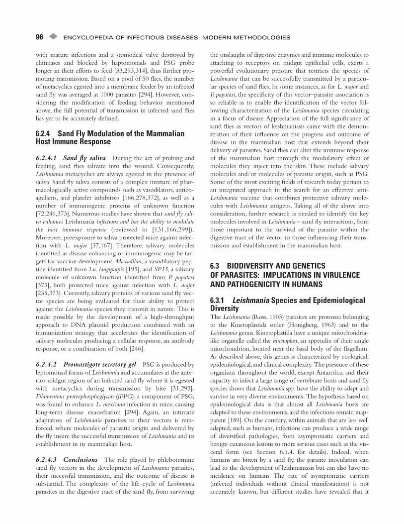
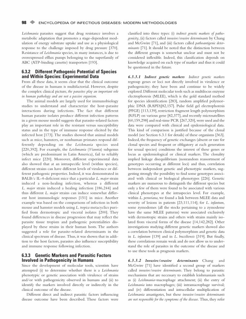
is 12 million people. Most of the affected countries are in thetropics and subtropics: more than 90% of the world’s cases ofvisceral leishmaniasis are in India, Bangladesh, Nepal, Sudan,and Brazil (Fig. 6.2), 90% of all cases of mucocutaneous leish-maniasis (Fig. 6.3) occur in Bolivia, Brazil, and Peru, where-as 90% of all cases of cutaneous leishmaniasis (Fig. 6.3) occurin Afghanistan, Brazil, Iran, Peru, Saudi Arabia, and Syria (forfurther detail, see http://www.who.int/leishmaniasis/en/).
6.1.2 The Players in LeishmaniasisLeishmania parasites are responsible for cutaneous forms aswell as visceral forms of the disease. Healing or progression ofthis infection is related to the genetic and immune status ofthe host, the virulence and pathogenicity of different speciesand strains of Leishmania, and the vector involved. The hostscan be humans but also rodents, dogs, and other mammals[16,307], and great diversity of immune response existsdepending on the host considered (see Section 6.4 fordetails). Similarly, within 500 known phlebotomine species,only 31 have been positively identified as vectors of theLeishmania pathogenic species and 43 as probable vectors[181]. Among them, some vectors such as PhlebotomusPhlebotomus papatasi and P. Paraphlebotomus sergenti can only be
Leishmaniasis has been known for many hundreds of years,with one of the first clinical descriptions made in 1756 byAlexander Russell and called Aleppo boil. Many names cor-respond to this group of diseases: kala-azar, Dum-dum fever,white leprosy, espundia, pian bois, and so on. Leishmaniasesare parasitic diseases spread by the bite of the infected femalephlebotomine sand fly (Fig. 6.1). Leishmaniases are caused byapproximately 20 species, pathogenic for humans, belongingto the genus Leishmania (kinetoplastids order, Honigberg,1963) and within 500 known phlebotomine species, ofwhich only some 30 have been positively identified as vectorsof these pathogenic species.
6.1.1 Geographic DistributionHuman leishmaniases are found on all continents, exceptAntarctic and Australia.However, cutaneous leishmaniasis wasrecently revealed in Australian red kangaroos [296].Approximately 350 million people live in endemic areas,thereby comprising populations at risk, and annual incidenceis estimated at 1–1.5 million cases of cutaneous leishmaniasisplus 500,000 cases of visceral leishmaniasis; overall prevalence
infected by one Leishmania species, whereas Lutzomyia longi-palpis is a permissive vector, able to transmit differentLeishmania species (see Section 6.2 for details). Finally, the 20species described as pathogenic for humans belong to theLeishmania genus (Ross, 1903). They are divided into twosubgenera (Leishmania in the Old World (Saf ’Janova, 1983)and Viannia in the New World (Lainson and Shaw, 1987)), theLeishmania subgenus is composed of several species or speciescomplexes (Leishmania donovani complex, L. mexicana com-plex, L. major, L. tropica, etc.) and the Viannia subgenus con-tains species of the L. braziliensis complex (L. braziliensis(Viannia, 1911), L. peruviana (Velez, 1913), and the L. guya-nensis complex (L. guyanensis (Floch, 1954), L. panamensis(Lainson and Shaw, 1972)), L. lainsoni, etc.). These Leishmaniaspecies are associated with different diseases (see Section 6.3for details). For example, infections by Leishmania donovanicomplex species are associated with visceral leishmaniasis andL. braziliensis infections are responsible for mucocutaneous
leishmaniasis. However, the first species complex is able togenerate benign cutaneous lesions, and L. braziliensis has beenisolated from simple cutaneous lesions but also from visceralforms. It is clear that the clinical outcome of infection depends ona multifaceted association of factors among the three main playersinvolved: hosts, parasites, and vectors.
6.1.3 The Life Cycle of the Leishmania ParasiteLeishmania parasites are transmitted to their host by the bite ofan infected female phlebotomine sand fly (Psychodidae fami-ly, Phlebotominae subfamily), which needs a blood meal toproduce its eggs (Fig. 6.4). The sand fly vectors are primarilyinfected when feeding on the blood of an infected individualor a vertebrate reservoir host. Many mammal species couldact as a reservoir host, for example, rodents or dogs [16,307].
During feeding, host macrophages, containing amastigotes(Fig. 6.5), are ingested by the vector. These parasite forms,round and nonmotile (3–7 �m in diameter), are released intothe posterior abdominal midgut of the insect, where theytransform into promastigotes to begin their extracellular lifecycle in the vector. This form is motile, elongated (10–20�m), and flagellated (Fig. 6.6).
The promastigotes then migrate to the anterior part of thealimentary tract of the sand fly where they multiply by bina-ry fission.Approximately 7 days after feeding, the promastig-otes undergo metacyclogenesis and become infectious(metacyclic promastigotes). They are released into the hosttogether with saliva when the sand fly lacerates the skin withits proboscis during feeding. The sand flies usually feed atnight while the host is asleep.
These metacyclic promastigotes are taken up by hostmacrophage, where they metamorphose into the amastigoteform. They increase in number by binary fission within thephagolysosome until the cell eventually bursts, then infectother phagocytic cells and continue the cycle. In cases of vis-ceral leishmaniasis, all organs, containing macrophages andphagocytes, can be infected, especially the lymph nodes,spleen, liver, and bone marrow.
88 ◆ ENCYCLOPEDIA OF INFECTIOUS DISEASES: MODERN METHODOLOGIES
Fig. 6.2. Distribution of visceral leishmaniasis (WHO website:http://www.who.int/leishmaniasis/leishmaniasis_maps/en/index.html).
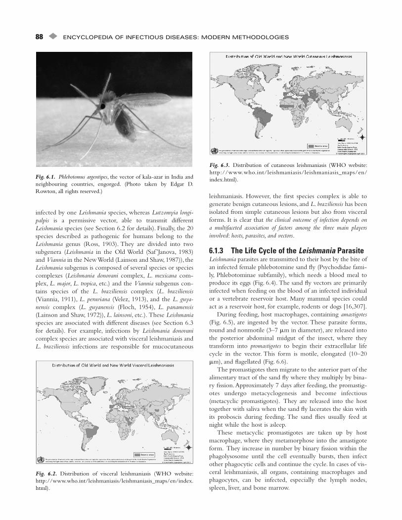
Fig. 6.3. Distribution of cutaneous leishmaniasis (WHO website:http://www.who.int/leishmaniasis/leishmaniasis_maps/en/index.html).Fig. 6.1. Phlebotomus argentipes, the vector of kala-azar in India and
neighbouring countries, engorged. (Photo taken by Edgar D.Rowton, all rights reserved.)
6.1.4 SymptomsA high rate of infected people remain asymptomatic, but forothers, the infection by Leishmania can produce very differentclinical symptoms. Indeed, several forms of leishmaniasis exist:cutaneous leishmaniasis and mucocutaneous leishmaniasis,which cause skin sores, and visceral leishmaniasis, whichaffects some of the internal organs of the body (e.g., spleen,liver, bone marrow). People with cutaneous leishmaniasis usu-ally develop skin sores a few weeks (sometimes as long asmonths) after being bitten, whereas people with visceral leish-maniasis usually become sick within several weeks or months(rarely as long as years).
The most severe form of the disease is visceral leishmani-asis (VL) (Fig. 6.7), which has a mortality rate of almost 100%if untreated. It is characterized by irregular bouts of fever,substantial weight loss, swelling of the spleen and liver, andanemia. Leishmania species responsible for this form mainlybelong to the Leishmania donovani complex. VL caused byL. infantum especially affects children. Other symptoms, calledpost-kala-azar dermal leishmaniasis (PKDL), can appear sev-eral months (or years) after VL treatment.This complicationof VL is characterized by a macular, maculopapular, and
nodular rash in a patient who has recovered from VL and whois otherwise well [400].
Mucocutaneous leishmaniasis (MCL) (Fig. 6.8.), mainlycaused by L. braziliensis and more rarely by the L. guyanensiscomplex, produces lesions that can lead to extensive and dis-figuring destruction of mucous tissues of the nose, mouth,and body, including the face, arms, and legs, causing seriousdisability.
The cutaneous leishmaniases (CL) (Fig. 6.9) are the mostcommon and represent 50–75% of all new cases. CL alsoresult in a variety of clinical manifestations, in terms of thenumber of lesions (up to 200 on the exposed part of thebody) and with selfhealing lesions compared with lesionsrequiring specific anti-Leishmania treatment. The lesion islocalized at the site of the sand fly bite and satellite lesions inthe vicinity of the original lesion can sometimes be observed.CL are mainly attributable to L. amazonensis, L. braziliensis, L.
CHAPTER 6 UNDERSTANDING HUMAN LEISHMANIASIS ◆ 89
Fig. 6.4. Leishmania life cycle (WHO website: http://www.who.int/tdr/diseases/leish/leish.htm).
Fig. 6.5. Two human macrophages infected by L. donovani amastig-otes, all rights reserved. Fig. 6.6. L. infantum promastigotes, all rights reserved.
Antiretroviral Therapy (HAART)). These cases are mostlylocalized in Europe where intravenous drug users have beenidentified as the main population at risk. In this case, theimmunological status of these people creates a favorableground for the Leishmania parasite.
6.1.5 Prevention, Diagnosis, and TreatmentsLeishmaniases are a diverse and complex group of disorders.Unfortunately, strict rules cannot be applied for a type ofLeishmania causing a typical disease, as even subtle changes inhost immunity, the environment, and the parasite itself mightresult in completely different clinical manifestations; there-fore, various approaches to disease control are necessary.Hence, prevention, diagnosis, and treatments depend onLeishmania species diagnostics and on the disease form; theydiffer for CL,VL, and MCL.
6.1.5.1 Prevention of leishmaniases6.1.5.1.1 Zoonotic cutaneous leishmaniasis (ZCL) In the
Old World, identification and control of animal reservoirs (smallrodents) consist of deep plowing to destroy the burrows (breed-ing and resting sites) and plant (Chenopodiacae) sources of foodfor the rodents. Poisoning is no longer used, as it is consideredtoo dangerous for other animals. In New World, especially inLatin America, large mammals living in forests or around hous-es can help contain the disease. In recent years, there has beenan increase in the incidence of ZCL attributable to urbaniza-tion and deforestation, leading to domestication of transmissioncycles, and the building of dams and new irrigation schemes,which have increased the population of animal reservoirs.Because populations living close to or at the edge of forests areparticularly vulnerable, such habitats should be moved awayfrom the forests. Limited clearance of peridomestic forest canreduce the risk of intradomiciliary transmission [101,102].
90 ◆ ENCYCLOPEDIA OF INFECTIOUS DISEASES: MODERN METHODOLOGIES
Fig. 6.8. Mucocutaneous leishmaniasis. (Photo taken by PhilippeDesjeux.WHO website: http://www.who.int/leishmaniasis/disease_epidemiology/en/index.html.)
Fig. 6.9. Cutaneous leishmaniasis. (Photo taken by PhilippeDesjeux.WHO website: http://www.who.int/leishmaniasis/disease_epidemiology/en/index.html.)
guyanensis, L. mexicana (Biagi, 1953), L. panamensis, L. naiffi,L. venezuelensis, L. lainsoni, and L. shawi in the New World andL. major (Yakimoff and Schockor, 1914), L. aethiopica (Ashfordand Bray, 1973),L. tropica (Wright, 1903),L. arabica, and L. ger-billi (Wang, Qu, and Guan, 1964) in the Old World, even ifother species such as L. donovani (Laveran and Mesnil, 1903),L. infantum (Nicolle, 1908) have also been isolated from cuta-neous lesions. Diffuse CL, mainly caused by L. amazonensisand L. aethiopica, never heals spontaneously and tends torelapse after treatment.This form is characterized by dissem-inated nodular lesions that resemble lepromatous leprosy.
Finally, these diseases have not only been found in devel-oping countries since 1985, when the first co-infected patientwas detected [93], even if the Leishmania–HIV co-infectioncases are decreasing in Europe (introduction of Highly Active
Fig. 6.7. Visceral leishmaniasis. (Photo taken by Philippe Desjeux.WHO website: http://www.who.int/leishmaniasis/disease_epidemiology/en/index.html.)
6.1.5.1.2 Anthroponotic cutaneous leishmaniasisAnthroponotic cutaneous leishmaniasis (ACL) is confined tourban or suburban areas of the Old World. Early diagnosis andtreatment of recurring cases are necessary to avoid an increase intransmission risk, as they reduce morbidity, mortality, and trans-mission (reduction of human reservoir).The best prevention forACL is the use of long-lasting impregnated bed nets in order toprevent infected sand flies from infecting healthy people andreduce untreated cases that continue infecting sand flies.Residual insecticide house spraying is another important pre-vention and intervention strategy. Mosquito repellents can becombined with pyrethroid-impregnated clothes (e.g., uniformsfor military personnel) for individual protection [78,99].Mosquito coils and the electrically heated fumigation mats con-taining pyrethroids are also helpful in protection.
6.1.5.1.3 Zoonotic visceral leishmaniasis In zoonoticvisceral leishmaniasis (ZVL) endemic areas, the dog is a majorreservoir. Several preventive measures are advocated: insecti-cide-impregnated dog collars, vaccination of pets againstleishmaniasis, and elimination of infected stray dogs candecrease the incidence of infection. Canine and indirectlyhuman leishmaniasis (because dogs are the Leishmania reser-voir) is prevented by using deltamethrin-treated collars toprotect dogs against L. infantum infection [127].
6.1.5.1.4 Anthroponotic visceral leishmaniasis (AVL)Elimination of the human reservoir by early diagnosis andtreatment of PKDL and VL can reduce the transmissioneffectively. Furthermore, in anthroponotic foci, vector controlthrough residual insecticide spray and improvement of theenvironment to control the growth of sand flies are the majortools for prevention.
6.1.5.2 Diagnosis of leishmaniases6.1.5.2.1 Visceral leishmaniasis Typical clinical features
of VL such as fever followed by splenomegaly (enlargement ofthe spleen) and lymphadenopathy (swelling of the lymph nodes) ina patient living in the endemic area should arouse suspicion ofVL. Presence of antileishmanial antibodies, detected throughconventional ELISA, IFAT, or DAT or the popular rapid rK39strip test, indicates infection [2,12,20,117,149,331,354,385].This is usually confirmed through demonstration of amastig-otes in tissue smears mostly from the spleen, bone marrow, orlymph nodes. Polymerase chain reaction (PCR) is employedfor demonstration of parasitic DNA in peripheral blood fordiagnosis [249,309].
In India, a rapid strip test based on rK39 antigen hasbecome available and should improve the diagnostic situa-tion [42,136,250,353,354,375,401]. However, there is aneed to develop a diagnostic test that has a high degree ofspecificity for active disease. Detection of antigen in urine(KAtex) is a promising tool, provided its format is improved[17,116,150, 279,312,345]. DNA detection by PCR isanother powerful tool that could be established at severalnodal centers in endemic areas serving the entire endemicregion for diagnosis and evaluation of cure [214,248,249,
265, 308,327,394]. Both KAtex and PCR correlate wellwith disease activity and thus have a clear edge over toolsbased on antibody detection.
6.1.5.2.2. Cutaneous leishmaniasis In areas of endemic-ity without sufficient laboratory infrastructure, CL is oftendiagnosed on the basis of clinical characteristics of the lesions.However, parasitological confirmation is important, becauseclinical manifestations may be mimicked by other infectionsand granulomatous diseases: lupus vulgaris, leprosy, and so on.Species identification may be important in predicting the course of thedisease and selecting therapy.
Leishmania may be isolated in up to 80% of sores duringthe first half of their natural course [273]. Parasites seem to beparticularly difficult to isolate from sores caused by L.braziliensis, responsible for the vast majority of cases in Brazil.Touch preparations from biopsies and histopathology usuallyhave a low sensitivity [81,389]. Slit-skin smears taken fromthe nodular edge of the lesion, or scrapings from within theulcer [273] examined microscopically are positive in32.7–84% [242,389]. Culture of fine needle aspiration mate-rial has been reported to be the most sensitive method[242,389]. Mucocutaneous leishmaniasis (MCL) is more dif-ficult to diagnose parasitologically; even hamster inoculationonly brings the yield up to 50% [389].
PCR introduced to determine the parasite species is usedincreasingly for diagnosis, greatly improving the diagnosticrates for CL and MCL [92,203]. For CL in Ecuador, using cul-ture as standard, PCR was 97% sensitive as compared withmicroscopy (42%) and histology (33%) [18],whereas in Brazil,71% of MCL cases were detected by PCR compared to 17%detected by conventional method [203]. Clinically, speciesidentification may be important for epidemiological and ther-apeutic reasons, for example to identify the dominant speciesin a CL focus in Brazil [91]. Isoenzyme methods [283] andmonoclonal antibodies [15,158] have been employed forspecies typing as well as analysis of amplified minicircle kine-toplast DNA (kDNA), by choosing primers from variableregions of different Leishmania species kDNA minicircle [327].
6.1.5.3 Leishmaniasis treatments Treatment of leish-maniases has centered around pentavalent antimonials (SbV) forsix decades except in North Bihar, India, where large-scaleantimony resistance is emerging and where SbV, even withthe higher doses, is able to cure only 35–50% of patients[342,352,356,359,360]. In the Old World (L. major, L. tropi-ca, and L. donovani complex) and the New World (L. mexi-cana and L. braziliensis complexes), CL and PKDL arecommonly treated with SbV. A species-based approach totreatment has been advocated, especially in countries whereseveral species may cause CL [245,295]. Intralesional SbV hasbeen used with encouraging results in the Old Worldselfhealing CL [5,371].
A second-line drug, pentamidine isethionate, is expensive andtoxic, beacuse it can be responsible for irreversible insulin-dependent diabetes mellitus and death. It was used to treatSbV-refractory patients with VL, but its efficacy has declined
CHAPTER 6 UNDERSTANDING HUMAN LEISHMANIASIS ◆ 91
and its use for VL has been abandoned [160,162,164]. Duringthe late 1980s and the early 1990s, many Indian patients diedfor want of treatment after failing therapy with SbV and pen-tamidine.Though for some forms of CL, pentamidine is stillattractive because very few doses are needed [9,335].
Due to increasing SbV-unresponsive VL, especially in Indiaover the last decade, amphotericin B has become the drug ofchoice [230]. However, it is toxic and requires close monitor-ing. Though the cure rate with amphotericin B is approxi-mately 100% and relapses are rare, the need for hospitalizationlasting 5–6 weeks, infusion reactions, occasional seriousadverse reactions such as hypokalemia, myocarditis, and deathprecludes its widespread application in peripheral health postswhere monitoring facilities are limited. Thus, a large numberof patients have to wait several weeks to months for hospital-ization and treatment [132,229,230,361]. In South America,many regard amphotericin as the drug of choice for MCL,because of the low relapse rate [80,291]. The introduction oflipid-associated amphotericin, i.e., liposomal amphotericin B(AmBisome), amphotericin B lipid complex (ABLC; Abelcet) andamphotericin B colloidal dispersion (Amphocil), has been one of themost important developments in the chemotherapy of leish-maniasis. In these formulations, deoxycholate has beenreplaced by other lipids that mask amphotericin B from sus-ceptible tissues, thus reducing toxicity, and are preferentiallytaken up by reticuloendothelial cells, thus targeting drug deliv-ery to the parasite and increasing efficacy. Three lipid formu-lations are commercially available, but their cost is prohibitive[88,89,106,188,227,351]. In India, all three formulations, withcomparable efficacy, have been used, with AmBisome beingthe safest [89,105,188,343,344,346,348,349,351].
Paromomycin, an aminoglycoside, is well tolerated andeffective for VL, but less so for CL [74,76,163].Topical paro-momycin ointment has been used for the treatment of CL[187,252,305]. The search for an effective oral antileishma-nial drug spans two decades. Allopurinol, the azoles,rifampicin, and atovaquone showed activity in experimentalsystems, but proved disappointing in clinical trials. Oral mil-tefosine, an alkyllysophospholipid, originally developed as ananti-cancer agent, is now approved for the treatment of VLin India [347]. In several clinical trials, miltefosine curedmore than 90% of patients with only minor gastrointestinalside effects such as vomiting in about half of the patients andless commonly diarrhea [165,347,350,355]. An asympto-matic transient rise in hepatic transaminases occurs duringthe second week of treatment, returning back to baselinevalues on continued treatment. It induces rapid cure, with amajority of patients becoming afebrile within the first week,quick regression of spleen, and recovery of blood counts.However, due to the risk of teratogenicity, Miltefosineshould not be given to child-bearing age women except ifcontraception can be secured during and after treatment.Oral sitamaquine, an 8-aminoquinoline derivative, has beenshown to have clinically significant antileishmanial activity.This effective oral antileishmanial compound has been test-ed in Kenya, Brazil, and India [104,161,325,387].
6.1.5.4 Vaccines? There is no vaccine available against anyform of leishmaniasis for prophylaxis. Control of leishmani-asis remains a source of grave concern worldwide. As mostof the available methods for leishmaniasis treatment andcontrol are of limited effectiveness, there is now an urgentneed for new low-cost drugs and/or new therapeutic inter-ventions such as a vaccine, which would be the most practi-cal and efficient tool for the control of these parasiticdiseases [90].
Although considerable progress has been made over thelast decade in understanding the immune mechanisms under-lying protective responses, identifying potential candidateantigens, and implementing these principles in animal mod-els, very few candidate vaccines have progressed beyond theexperimental stage.
In recent years, great interest has been focused on thedevelopment of vaccines against localized cutaneous disease.Comparatively, VL has received limited attention. Indeed,only studies to identify the immunological factors of VLpatients after chemotherapy and in asymptomatic subjectshave been reported so far [231]. In regions where VL isendemic, such as the Mediterranean area, severe disease onlyoccurs in a small population of around 10–33%, whereas themajority of infected individuals show no clinical symptomsand a significant part have self-resolving infection [21].Furthermore, patients who have recovered from kala-azar areusually immune to reinfection, suggesting that vaccinationagainst VL should be possible. The fact that a large proportion ofthe people living in endemic areas has self-resolving subclini-cal infections and the immunological mechanisms that controlparasite multiplication in asymptomatic subjects are not welldefined provides a rationale for designing immunoprophylac-tic strategies against VL.
Historically, “leishmanization’’ with live organisms wasused to protect against disfiguring CL, because of the knowl-edge that individuals whose skin lesions had healed wereimmune. Knowledge of pathogenesis fortified by immuno-logical understanding and genetic sequencing studies havegradually led to rational approaches toward the induction ofprotective immunity to Leishmania in animal models. Thusfar, attempts at human vaccination have been unsuccessful,but several promising candidate vaccines are being exploredin mouse models and in dogs.
In humans, measurement of cytokines in culture super-natants of Leishmania antigen-activated PBMCs and T-cellclone analysis support the view that (i) cell-mediated immuni-ty, regulated by Th1 CD4� lymphocytes, was required for thedestruction of Leishmania parasites in macrophage phagolyso-somes [179]; (ii) control of infection in asymptomatic subjectswas partially associated with the expansion of parasite-specificCD8� lymphocytes [211]; and (iii) these measurementsrevealed a coexistence of Th1 and Th2 responses in kala-azarpatients as well as in cured individuals [253].Therefore, even inhumans, it is difficult to demarcate the responses leading toeither visceral disease (“susceptible’’) or protective immunity(“resistance’’) against Leishmania parasites. Successful resistance
92 ◆ ENCYCLOPEDIA OF INFECTIOUS DISEASES: MODERN METHODOLOGIES
is probably the result of cooperation between the various armsof the immune system.
Recently, a vaccine against canine VL involving Leishmaniaexcreted–secreted antigen has been developed (LiESAp)[226]. It proved efficient in both experimentally and natural-ly L. infantum-exposed dogs in southern France [147,194]. Indogs, the vaccine-induced protection correlates with an earlyproduction of IFN-� by a Th1 subset of CD4� T cells, whichactivate macrophages to destroy intracellular amastigotesthrough NO production. This was demonstrated by anti-LiESAp IgG2 reactivity, LiESAp-specific lymphocyte prolif-eration assays, and enhanced NO-mediated anti-leishmanialactivity of canine monocyte-derived macrophages (CM-DM).In vaccinated dogs, NO-mediated Leishmania killing wasassociated with higher IFN-� production by T cells when L. infantum-infected CM-DMs were co-cultured with autol-ogous lymphocytes [147,194]. The main scientific issues inthe design of a Leishmania vaccine are no different from thosefor any other vaccine. On a positive note, there is currentlyrapid progress in our understanding of the molecularnature of potential vaccine candidates and the mechanismsthat determine infection-preventing immune responses.Multidisciplinary approaches integrating studies on parasiteand host factors would facilitate our understanding of thedisease and help in the design of a vaccine against human VL.
6.1.6 Why an Integrated Approach?Even if we can generalize the life cycle of Leishmaniabecause it always contains one vector, one parasite, and onehost, the outcome of transmission, infection, and disease aredependent on the intrinsic characteristics of these threeplayers. Indeed, the epidemiology of leishmaniasis will be reflec-tive of the particular combination of interactions among all players:parasite, vector, reservoir host, and environmental conditions. Inmany endemic areas, the exact role of these players and theirrelations to human infections are unknown and it is difficultto generalize. Integrated analysis of both parasite genetics,parasite virulence factors, host immune responses, vectorcompetence, host genetics, socioeconomic, and environ-mental risk factors is necessary for a better understandingof the interplay between these different factors and the riskof developing leishmaniasis. This approach could also pro-vide information on the critical biological pathwaysinvolved in the host resistance or susceptibility to leishma-niasis and therefore help in orienting new therapeuticor vaccine strategies. Indeed, factors determining thehost resistant/susceptible status are complex and largelyunknown. Environmental factors acting on the phle-botomine and/or animal reservoir populations could mod-ulate exposure of the human host to infected sand fly bites.Moreover, it has been suggested that the host immuneresponse may also depend on the parasite strain, and differ-ent parasitic factors directly or indirectly responsible for thedisease outcome have been described. Factors affecting thepatient immune competence such as HIV infection or mal-nutrition have also been described to mediate susceptibility
to VL. Immunity in leishmaniasis is considered mainly T-cellmediated, but more and more nonspecific factors acting inthe early stage of infection are now considered as importantfor either the progression or control of the disease.Therefore, we will first expose the advances in the identifi-cation of the factors involved, due to the vector (Section6.2), parasite (Section 6.3), and host (Section 6.4), and inthe interactions between these players.The last section willfocus on kala-azar in India, and we will demonstrate thenecessity of this integrated approach to better understandthis complex epidemiologic focus.
6.2 IMPACT OF SAND FLY VECTORS ON LEISHMANIASIS
Phlebotomine sand flies belong to the order Diptera, subor-der Nymatocera, and family Psychodidae. They are small,about 3 mm in length, hairy flies characterized by a “hop-ping’’ flight and wings that remain erect above the abdomenwhen at rest. Sand flies are widely distributed and occupytropical, subtropical, and temperate biotopes [4].
Phlebotomine sand flies are biological vectors ofLeishmania in which the parasites undergo a complex devel-opmental cycle beginning with ingested amastigotes and ter-minating with transmission of infective metacyclicpromastigotes. Not all sand fly species transmit Leishmaniaparasites, however, with the genera Phlebotomus (Old World)and Lutzomyia (New World) accounting for all incriminatedvectors to date. The bite of an infective sand fly vector is theonly means by which any Leishmania species can be transmit-ted at a sustained and significant level. Importantly, the impactof sand flies on the establishment and spread of leishmaniasisextends beyond the transmission of Leishmania parasites to adirect effect on the host response to infection. In this section,the complexity of sand fly—Leishmania and sand fly—mammalian host interactions is outlined.
6.2.1 The Life Cycle of Leishmaniain a Competent Sand Fly VectorThe life cycle of Leishmania parasites is contained within thedigestive tract of the sand fly and begins with the ingestion of aninfected blood meal containing amastigotes. Around 4 h after blood feeding, a chitinous peritrophic matrix (PM) issecreted, surrounding the blood meal within 24 h. The PM acts as a barrier that slows the diffusion of digestive enzymessecreted by the sand fly in response to blood ingestion and indi-rectly protects the parasites from the harmful effects of theenzymes [260].This provides the opportunity for amastigotes todifferentiate into sluggishly dividing procyclics, and by day 2 intolarge flagellated nectomonads (Fig.6.10). The blood meal isdigested around 3–4 days after feeding. At this point, the PMbreaks down,permitting escape of nectomonads and their attach-ment to the midgut epithelium.The degradation of the PM wasinitially attributed in full to the secretion of chitinases byLeishmania parasites [315]. Recently, however, Ramalho-Ortigao
CHAPTER 6 UNDERSTANDING HUMAN LEISHMANIASIS ◆ 93
et al. [272] showed that sand flies secrete their own chitinases afterinduction by the bloodmeal.The activity of sand fly chitinasespeaks at about 48 h post blood feeding, coinciding with the timeof the escape of nectomonads from the confinement of the PM[269]. Once in the gut lumen, attachment to the epitheliumallows the nectomonads to persist in the midgut and preventstheir expulsion with remnants of the undigested blood meal.Thereafter, nectomonads differentiate into leptomonads thatdivide rapidly as they migrate anteriorly to the thoracic part ofthe midgut [135,293].Around day 7 after feeding, leptomonadsgive rise to infective metacyclics that accumulate in the anteriormidgut below the stomodeal valve [135,293]. Metacyclics arecharacterized morphologically by their small cell body and longflagellum, and functionally by their free and rapid motility [311].Simultaneously, haptomonads, highly specialized forms thatadhere to each other and to the stomodeal valve, form a concen-tric parasite plug that blocks the opening of the valve (Fig. 6.10).
With such a complex life cycle, the parasites have to over-come several adverse conditions before they can successfullycomplete their development in the fly [180,299]. Such obsta-cles include digestive enzymes secreted by the sand fly[50,108,270], midgut lectins [381,382, 384], excretion ofbloodmeal remnants [182,261], and sand fly innate immuneresponses [51,271]. As a result, different species of Leishmaniaclosely evolved to fit distinct sand fly species, overcoming theseobstacles and giving rise to the term “vector competence.’’
6.2.2 Vector CompetenceA major determinant of vector competence is the ability ofparasites to attach to the midgut epithelium of the sand fly toavoid expulsion with the blood meal remnants. Numerous
studies, some involving mutants specifically deficient inlipophosphoglycan (LPG), a large and abundant molecule on thesurface of Leishmania promastigotes, have implicated LPG asthe ligand that mediates this attachment [62,262,302,303].LPG is a tripartite GPI-anchored molecule with a backboneof conserved disaccharide repeats consisting of phosphorylat-ed galactose-mannose sugars –6Gal�1,4Man�1-PO4– cappedwith a neutral sugar. The LPG of different Leishmania speciesis highly polymorphic where the backbone can be unsubsti-tuted (L. donovani, Sudan; and L. chagasi), partially substituted(L. donovani, India), or completely substituted (L. major and L. tropica) by side chains varying in the number and nature oftheir sugar residues [206,216,217,332,368] (Fig. 6.11A). Thedriving force for the observed LPG side chain substitutions isthought to be dependent on the complexity of the receptorpresent on the midgut epithelium of the targeted sand fly vec-tor. Experimental infections showed that some sand flyspecies, such as Lutzomyia longipalpis and Phlebotomus argentipes,developed mature transmissible infections when infected withseveral foreign Leishmania species [168,261,294,304]. Thesespecies were termed permissive vectors. Others, including P. papatasi and P. sergenti, can only support the growth of theLeishmania species they are found infected with in nature (L. major and L. tropica, respectively) [168,261]; as such, theyare considered restricted vectors. It is important to note that thisspecies-restricted vectorial competence can also be strain spe-cific. Certain natural variants of L. major, such as the WestAfrican Seidman strain, which lacks galactose side chains, donot maintain infection in P. papatasi but do maintain infectionin another, closely related species P. duboscqi [206]. The strain-specific variability of LPG galactosylation in L. major was
94 ◆ ENCYCLOPEDIA OF INFECTIOUS DISEASES: MODERN METHODOLOGIES
Fig. 6.10. Life cycle of Leishmania in a competent sand fly vector. See color plates.
attributed to the differential expression of a family of six genesencoding L. major galactosyltransferases that vary in their expres-sion and activity [110]. Additionally, in the north of Israel, astrain of L. tropica whose LPG terminates with galactoseinstead of glucose residues, known to decorate the LPG ofpreviously characterized L. tropica, was isolated from P. arabicusand not the classical vector P. sergenti [332]. As for sand flymidgut receptors, the first and only identified receptor to dateis PpGalec, a tandem repeat galectin responsible for theobserved specificity of P. papatasi for L. major [169].
Though appropriate LPG polymorphisms are necessary,vector competence has also been associated with the abilityof certain Leishmania species to overcome other adverse con-ditions in the midgut of their respective competent vectors.For example, Leishmania species are able to overcome theharmful effects of digestive enzymes in a competent vector,but not in a foreign sand fly species, by specifically inhibitingor retarding the peak activity of these enzymes [50,107,316].Secreted glycoconjugates, a family of LPG-related moleculescharacteristic of Leishmania, were implicated in this protec-tion [302,317], highlighting the degree of adaptation neces-sary for parasite survival in competent vectors.
6.2.3 Metacyclogenesis and TransmissionTransmission of the parasites from the sand fly to the mam-malian host requires detachment of the parasites from themidgut epithelium. This event is again mediated by LPG,which undergoes stage-specific modifications involving elongationof the molecule and/or changes to the nature of sugar residueson its side chains or neutral cap [206,217,262,301,303]. Forexample, during metacyclogenesis, the LPG of L. major elon-gates to approximately twice its procyclic length, and themajority of terminal galactose sugars get capped by arabinose
residues (Fig. 6.11B). This modified LPG cannot bind toPpGalec, the midgut receptor for L. major procyclic LPG in P. papatasi [169]. In L. chagasi, metacyclics downregulate theglucose substitutions in their LPG, which, in contrast to pro-cyclic parasites and procyclic LPG, becomes unable to bind tothe midgut of its natural vector L. longipalpis [333]. Thisdetachment frees the metacyclics and ensures their availabilityfor transmission to the mammalian host. The trigger thatinitiates metacyclogenesis is not well understood. The onlyavailable evidence to date is a negative regulation by tetrahy-drobiopterin, a byproduct of pteridine metabolism, whose lev-els are high following a bloodmeal and decline with timeelevating metacyclogenesis [84].
To further enhance their chances for successful transmis-sion, Leishmania parasites evolved the haptomonad stage,whose specific function is to block the stomodeal valve sepa-rating the midgut from the foregut.These parasites are non-motile and adhere to the chitinous lining of the valve. Thephysical blockage of the valve is compounded by the secretionby the parasites of a proteophosphoglycan-rich gel termed thepromastigote secretory gel (PSG) [156,340]. Both act in concertto obstruct the intake of blood during feeding, requiring morebites and a longer period to feed, and promoting regurgitationof metacyclics into the skin of the mammalian host[33,183,294]. In addition, parasite chitinases destroy the chiti-nous lining of the stomodeal valve, further contributing to thedefective feeding mechanism in infected flies [314,380].Another aspect of sand fly feeding that promotes transmissionis sand fly probing. Due to their small mouth parts, sand fliesneed to lacerate multiple skin-surface capillaries to create thepool of blood upon which they feed [277,278]. Beach et al.[32] have shown that infected sand flies can transmitLeishmania parasites while probing. Moreover, infected flies
CHAPTER 6 UNDERSTANDING HUMAN LEISHMANIASIS ◆ 95
Fig. 6.11. Illustration of (A) Leishmania major (completely substituted) and L. donovani “Sudan’’(unsubstituted) LPGs and (B) the changes during metacyclogenesis of L. major LPG.
with mature infections and a stomodeal valve destroyed bychitinases and blocked by haptomonads and PSG probelonger in their efforts to feed [33,293,314], thus further pro-moting transmission. Based on a pool of 50 flies, the numberof metacyclics egested into a membrane feeder by an infectedsand fly was averaged at 1000 parasites [294]. However, con-sidering the modification of feeding behavior mentionedabove, the full potential of transmission in infected sand flieshas yet to be accurately defined.
6.2.4 Sand Fly Modulation of the MammalianHost Immune Response
6.2.4.1 Sand fly saliva During the act of probing andfeeding, sand flies salivate into the wound. Consequently,Leishmania metacyclics are always egested in the presence ofsaliva. Sand fly saliva consists of a complex mixture of phar-macologically active compounds such as vasodilators, antico-agulants, and platelet inhibitors [166,278,372], as well as anumber of immunogenic proteins of unknown function[72,246,373]. Numerous studies have shown that sand fly sali-va enhances Leishmania infections and has the ability to modulatethe host immune response (reviewed in [131,166,299]).Moreover, preexposure to saliva protected mice against infec-tion with L. major [37,167]. Therefore, salivary moleculesidentified as disease enhancing or immunogenic may be tar-gets for vaccine development. Maxadilan, a vasodilatory pep-tide identified from Lu. longipalpis [195], and SP15, a salivarymolecule of unknown function identified from P. papatasi[373], both protected mice against infection with L. major[235,373]. Currently, salivary proteins of various sand fly vec-tor species are being evaluated for their ability to protectagainst the Leishmania species they transmit in nature. This ismade possible by the development of a high-throughputapproach to DNA plasmid production combined with animmunization strategy that accelerates the identification ofsalivary molecules producing a cellular response, an antibodyresponse, or a combination of both [246].
6.2.4.2 Promastigote secretory gel PSG is produced byleptomonad forms of Leishmania and accumulates at the ante-rior midgut region of an infected sand fly where it is egestedwith metacyclics during transmission by bite [31,293].Filamentous proteophosphoglycan (fPPG), a component of PSG,was found to enhance L. mexicana infection in mice, causinglong-term disease exacerbation [294]. Again, an intimateadaptation of Leishmania parasites to their vectors is rein-forced, where molecules of parasitic origin and delivered bythe fly insure the successful transmission of Leishmania and itsestablishment in its mammalian host.
6.2.4.3 Conclusions The role played by phlebotominesand fly vectors in the development of Leishmania parasites,their successful transmission, and the outcome of disease issubstantial. The complexity of the life cycle of Leishmaniaparasites in the digestive tract of the sand fly, from surviving
the onslaught of digestive enzymes and immune molecules toattaching to receptors on midgut epithelial cells, exerts apowerful evolutionary pressure that restricts the species ofLeishmania that can be successfully transmitted by a particu-lar species of sand flies. In some instances, as for L. major andP. papatasi, the specificity of this vector–parasite association isso reliable as to enable the identification of the vector fol-lowing characterization of the Leishmania species circulatingin a focus of disease. Appreciation of the full significance ofsand flies as vectors of leishmaniasis came with the demon-stration of their influence on the progress and outcome ofdisease in the mammalian host that extends beyond theirdelivery of parasites. Sand flies can alter the immune responseof the mammalian host through the modulatory effect ofmolecules they inject into the skin. These include salivarymolecules and/or molecules of parasite origin, such as PSG.Some of the most exciting fields of research today pertain toan integrated approach in the search for an effective anti-Leishmania vaccine that combines protective salivary mole-cules with Leishmania antigens. Taking all of the above intoconsideration, further research is needed to identify the keymolecules involved in Leishmania – sand fly interactions, fromthose important to the survival of the parasite within thedigestive tract of the vector to those influencing their trans-mission and establishment in the mammalian host.
6.3 BIODIVERSITY AND GENETICS OF PARASITES: IMPLICATIONS IN VIRULENCEAND PATHOGENICITY IN HUMANS
6.3.1 Leishmania Species and EpidemiologicalDiversityThe Leishmania (Ross, 1903) parasites are protozoa belongingto the Kinetoplastida order (Honigberg, 1963) and to theLeishmania genus. Kinetoplastida have a unique mitochondria-like organelle called the kinetoplast, an appendix of their singlemitochondrion, located near the basal body of the flagellum.As described above, this genus is characterized by ecological,epidemiological, and clinical complexity. The presence of theseorganisms throughout the world, except Antarctica, and theircapacity to infect a large range of vertebrate hosts and sand flyspecies shows that Leishmania spp. have the ability to adapt andsurvive in very diverse environments. The hypothesis based onepidemiological data is that almost all Leishmania hosts areadapted to these environments, and the infections remain inap-parent [189]. On the contrary, within animals that are less welladapted, such as humans, infections can produce a wide rangeof diversified pathologies, from asymptomatic carriers andbenign cutaneous lesions to more serious cases such as the vis-ceral form (see Section 6.1.4. for details). Indeed, whenhumans are bitten by a sand fly, the parasite inoculation canlead to the development of leishmaniasis but can also have noincidence on humans. The rate of asymptomatic carriers(infected individuals without clinical manifestations) is notaccurately known, but different studies have revealed that it
96 ◆ ENCYCLOPEDIA OF INFECTIOUS DISEASES: MODERN METHODOLOGIES
seems to be higher than expected. For example, on theBalearic Islands, L. infantum was amplified by PCR in 22% ofblood donors [280] and asymptomatic carriers were alsorevealed in Brazil [77], southern France [192], and India [323].
This great phenotypic variability is also expressed by thehigh number of Leishmania species described in the literature.A large part of these species has been defined on the basis ofepidemiological, clinical, geographical, and biological data,for example, L. guyanensis (isolated in Guyana), L. peruviana(isolated in Peru), L. infantum (isolated from a child inTunisia), L. gerbilli (isolated from gerbils), and so on. Theseextrinsic characteristics were first used to determine thespecies because morphological characteristics cannot be usedfor species identification. Even if differences in length havebeen observed among Leishmania spp. [125,174], the differentspecies are indistinguishable in morphology in both the pro-mastigote and amastigote stages. The development of geneticand phenotypic tools has provided means to reconsider theLeishmania taxonomy more rigorously. The first problemnoted was that these organisms could not be defined on the basis ofthe biological concept of species [215]. Indeed, the studies of pop-ulation genetics published show a basic clonal populationstructure in different species [25,23,362,364,365]. However,this model is not as simple as it appears because these organ-isms have been shown to use different multiplication strate-gies, with several hybridization events between speciesevidenced in the literature [24,41,112,120,175]. For exam-ple, in the New World, hybrids between L. braziliensis and L.peruviana, and L. guyanensis and L. braziliensis were described[24,112], and in the Old World, hybrids have been shownbetween L. major and L. arabica [120]. However, these recom-bination events do not seem frequent enough to disturb theclonal propagation of clones stable in space and time. Thus, thespecies definition of these “agamospecies’’ (a group of individ-uals in which reproduction is almost exclusively done by asexualmeans) still remains arbitrary and is based on a mix of intrin-sic and extrinsic characteristics considered together. In thisframework, different analyses clearly showed that the speciesstatus of some taxa was not taxonomically valid or question-able [23,26,85,137,212,213,283,396].
It must be kept in mind that there is a need for a rigorousand clear nomenclature for efficient communication betweenthe scientific and medical professions. Indeed, first the vari-ous Leishmania species require different medical posologies totreat patients (see Section 6.1.5.3 for details) and second,clinical data suggest a close association between the clinical outcomeof the disease in humans and the species responsible for the infection.Concerning the second point, for examples, (i) the L. donovanicomplex is mainly responsible for visceral forms; (ii) mucos-al lesions are generally associated with L. braziliensis; (iii) L. major, L. tropica, L. mexicana, L. guyanensis, and L. peruvianaproduce a variety of Leishmania skin lesions in humans; and(iv) L. amazonensis is generally associated with diffuse cuta-neous leishmaniasis. But once again, the clinical picture ismore complex since at an intraspecific level, we can observedifferent disease outcomes: for example, L. amazonensis was
isolated from six patients, three with cutaneous lesions, onewith mucosal lesions, and two with diffuse cutaneous forms[205]; L. infantum can cause both cutaneous and visceralforms; and L. braziliensis produces cutaneous lesions and inaround 10% of cases metastasizes.
Other points complicate the clinical picture: the existenceof hybrids (see above) and mixed infections with differentLeishmania strains. Concerning hybrids, L. braziliensis can pro-duce cutaneous or mucocutaneous lesions in humans requir-ing care, whereas L. peruviana is responsible for dry benigncutaneous lesions that heal spontaneously. The hybridsbetween these two species found in Peru were isolated frompatients either with mucocutaneous lesions or with benignlesions typical of the L. peruviana species [112].These strainsare thus capable of producing the different pathologies foundin each species. Concerning mixed infections by differentLeishmania species, few cases have been described in the Newand Old World in the literature [13,30,154,210,341].However, the molecular epidemiology studies evidenced thatmany foci exist in which several species circulate simultane-ously [205]. It is hypothesized that the number of mixedinfections is underestimated because of a selection problemduring the parasite culture required by molecular techniques.This is confirmed by a study conducted in Bolivia [30] andalso presents a problem for Leishmania diagnosis, prognosis,and for the understanding of the real role of parasites in path-ogenicity in humans.
Moreover, it seems important to note and to consider thecases of co-infection of Leishmania with other pathogens. Thisis relatively frequent according to the literature and variouspathogens in association with Leishmania such as Mycobacteriumtuberculosis [94,386], Trypanosoma cruzi [30], Salmonella andSchistosoma [109], and of course HIV (for reviews see[97,103,234,268]) have been studied. Furthermore, in somecases, these co-infections can produce unusual clinical forms ofleishmaniasis [66,75].
Another aspect of the incredible environmental adaptationof Leishmania parasites is their ability to become drug resist-ant. Indeed, drug and multidrug resistance has emerged as amajor problem in treating both VL and CL. In particular, theappearance of antimonial resistance has changed the patternof leishmaniasis treatment in the world. Indeed, pentavalentantimony has long been the cornerstone of anti-Leishmaniachemotherapy, but resistance to this drug class is so high insome parts of the world, particularly in northeast India (seeSection 6.1.3 for details), that it is quickly becoming obsolete[251,352]. There are many factors that can influence the effi-cacy of drugs in the treatment of leishmaniasis.These includeboth an intrinsic variation in the sensitivity of Leishmaniaspecies, described for pentavalent antimonials, but also paro-momycin, azoles, and other drugs that have reached clinicaltrials, as well as acquired drug resistance to antimonials [79].Thus, the understanding of the molecular mechanisms that the par-asite adopts or may adopt in the future is of high clinical relevance.We know that the parasite is able to adapt itself to becomeresistant. For example, some results on glibenclamide-resistant
CHAPTER 6 UNDERSTANDING HUMAN LEISHMANIASIS ◆ 97
Leishmania parasites suggest that drug resistance involves ametabolic adaptation that promotes a stage-dependent mod-ulation of energy substrate uptake and use as a physiologicalresponse to the challenge imposed by drug pressure [370].Resistance of Leishmania species, in many instances, is due tooverexpressed efflux pumps belonging to the superfamily ofABC (ATP-binding cassette) transporters [193].
6.3.2 Different Pathogenic Potential of Speciesand Within Species: Experimental DataFrom all these data, it seems clear that the clinical outcomeof the disease in humans is multifactorial. However, despitethe complex clinical picture, the parasites play an important rolein human pathology and are not a passive organism.
The animal models are largely used for immunobiologystudies to understand and characterize the host–parasiteinteractions during infections. The fact that differenthuman parasite isolates produce different infection patternsin a given mouse model suggests that parasite-related factorsplay an important role in the resistant versus susceptibilitystatus and in the type of immune response elicited by theinfected host [173]. The studies showed that animal modelssuch as mice, hamsters, or nonhuman primates respond dif-ferently depending on the Leishmania species used[220,392]. For example, the Leishmania (Viannia) subgenus(which are predominant in Latin America), fail to reliablyinfect mice [220]. Moreover, different experimental dataalso showed that at an intraspecific level (within species),different strains can have different levels of virulence or dif-ferent pathogenic properties. Indeed, it was demonstrated inBALB/c IL-4-deficient mice that a particular L. major straininduced a non-healing infection, whereas a different L. major strain induced a healing infection [186,244] andthus different L. major strains can induce somewhat differ-ent host immunologic responses [151] in mice. Anotherexample was based on the comparisons of infection in bothmouse and hamster models using L. tropica metacyclics puri-fied from dermotropic and visceral isolates [200]. Theyfound differences in disease progression that may reflect theparasite tissue tropism and pathogenic potentialities dis-played by these strains in their human hosts. The authorssuggested a role for parasite-related determinants in theclinical spectrum of disease. Thus, it was shown that in addi-tion to the host factors, parasites also influence susceptibilityand immune response following infection.
6.3.3 Genetic Markers and Parasitic FactorsInvolved in Pathogenicity in HumansSince the development of molecular tools, scientists haveattempted (i) to determine whether there is a Leishmaniaphenotypic or genetic association with virulence of strainsand/or with pathogenicity observed in humans and (ii) toidentify the markers involved directly or indirectly in theclinical outcome of the disease.
Different direct and indirect parasitic factors influencingdisease outcome have been described. These factors were
classified into three types: (i) indirect genetic markers of patho-genicity, (ii) factors called invasive/evasive determinants by Changand McGwire [71], and (iii) factors called pathoantigenic deter-minants [71]. It should be noted that the distinction betweenthe different groups is somewhat unclear and must not beconsidered inflexible. Indeed, this classification depends onknowledge acquired on each type of marker and thus it couldbe questioned in the future.
6.3.3.1 Indirect genetic markers Indirect genetic markersregroup genes or loci not directly involved in virulence orpathogenicity; they have been and continue to be widelyexplored.Different molecular tools such as multilocus enzymeelectrophoresis (MLEE), which is the gold standard methodfor species identification [283], random amplified polymor-phic DNA (RAPD)[82,157]. Pulse field gel electrophoresis(PFGE) [113,139], restriction fragment length polymorphism(RFLP) on various gene [82,377], and recently microsatellites[60,159,298] and real-time PCR [267,320],were used and thedata were compared with clinical and epidemiological data.This kind of comparison is justified because of the clonalmodel (see Section 6.3.1 for details) of these organisms [363].Indeed, the frequency of genetic exchanges (absent or rare forclonal species and frequent or obligatory at each generationfor sexual species) conditions the interest of these genes orlocus as epidemiological or clinical markers. The clonalityimplied linkage disequilibrium (nonrandom reassortment ofgenotypes occurring at different loci) and thus, correlationbetween independent genetic and phenotypic markers, sug-gesting strongly the possibility to find some genotypes associ-ated with clinical or biological phenotypes [226]. Geneticmarkers are numerous to distinguish the different species butonly a few of them were found to be associated with variousclinical phenotypes at the intraspecies level. For example,within L. peruviana, we found a link between MLEE data andseverity of lesions in patients [23,111,114]; for L. infantum,some zymodemes (all the stocks pertaining to a zymodemehave the same MLEE patterns) were associated exclusivelywith dermotropic strains and others with strains mainly iso-lated from visceral forms of the disease [14,142,282]. Otherinvestigations studying different genetic markers showed alsoa correlation between clinical polymorphism and genetic datain L. infantum [139] and in L. braziliensis [319]. But finally,these correlations remain weak and do not allow us to under-stand the role of parasites in the outcome of the disease andto use these tools as prognosis markers.
6.3.3.2 Invasive/evasive determinants Chang andMcGwire [71] have identified a second group of markerscalled invasive/evasive determinants. They belong to parasiticmechanisms that are necessary to establish leishmaniasis suchas (i) Leishmania–macrophage attachment; (ii) the entry ofLeishmania into macrophages; (iii) intramacrophage survival;and (iv) differentiation and intracellular multiplication ofLeishmania amastigotes, but these invasive/evasive determinantsare not responsible for the symptoms of the disease. Thus, they refer
98 ◆ ENCYCLOPEDIA OF INFECTIOUS DISEASES: MODERN METHODOLOGIES
to all determinants that help successfully establish Leishmaniainfection in the host such as glycosylphosphatidylinositol (GPI),glycosylphospholipid (GIPL), lipophosphoglycan (LPG), leish-manolysin (GP63), cysteine proteases (CPs), among others. Thesemolecules have been widely studied, especially LPG, GP63,and CPs. LPG is the dominant surface molecule of pro-mastigotes involved in (i) binding, migration, and release ofthe parasite in the sand fly midgut but also in (ii) the modu-lation of resistance to lysis by the host’s complement. It isalmost completely absent from amastigotes [83,217,247,262].LPG is not involved in virulence within all Leishmania species.For example, it is not required for infection by L. mexicana[155], whereas it is needed for L. donovani and L. major infec-tion [221,337]. Its structure varies between Leishmania speciesand also differs between procyclic and metacyclic promastig-otes (see Section 6.2.2 for details). Some analyses showed, inaddition to stage-specific and interspecies variability, anintraspecies polymorphism in lipophosphoglycan structure[206].This diversity may be linked to the Leishmania adapta-tion to the sand fly species rather than related to the clinicaldiversity observed in humans.
Another important surface molecule,GP63, is an ecto-met-alloprotease particularly abundant in promastigotes and alsoreleased by this stage of Leishmania [219]. Like LPG, GP63 isdownregulated in the amastigote form [318]. These moleculesmay be involved in the evasion from humoral lytic factors andin the attachment of parasites to macrophages followed by theirintracellular entry into these phagocytes [395]. GP63 protein isencoded by a multigene family repeated in tandem. Geneticand structural diversity was extensively studied and showed ahigh polymorphism at both inter- and intraspecific levels[119,140,289,339,376]. Like LPG, this protein seems to besubjected to strong host-selection pressure by the vector as wellas by the vertebrate host [141].But no link was found betweenthe genetic or phenotypic diversity of GP63 and theintraspecies clinical polymorphism of strains [139].
Scientists have also shown increased interest in cysteine pro-teases because of the key roles some of them play in infectionand expression of the disease, making them potential drug tar-gets or vaccinal antigen. In L. major, a total of 65 CPs mayexist, many of which are likely to play crucial roles in host-parasite interactions, particularly in facilitating survival andgrowth of parasites in mammals by destruction of host pro-teins, nutrition, evasion of the host immune response, andLeishmania survival within host macrophages [4,237,240,297].The functional studies of the most widely studied CPs, CPB,allowed to explore the ways in which these molecules influ-ence the interactions between parasite and mammalian host(see reviews [4,237,240,297] for details). Indeed, the genera-tion of Leishmania cp-deficient mutants and inoculation onmice showed the involvement of these proteins in virulenceand pathogenicity. For example, the L. mexicana strain defi-cient in the cpb array reduced virulence in BALB/c mice[4,238]. As for the gp63 array, the genetic studies showed ahigh level of polymorphism, among species as well as withinspecies. Nevertheless, only one publication showed a statisti-
cal correlation between gene organization of cpb in the L.infantum population and the strain tropism (cutaneous versusvisceral) [67].
This list is far from exhaustive: other molecules such asPSA (GP46), an abundant surface glycoprotein of the pro-mastigote form [35,357], or A2 protein, shown to have aninfluence on the outcome of the disease [398], appear to playan important role in the invasive/evasive phases of theLeishmania cycle. For example, A2 is an important gene for L. donovani virulence but is not expressed in L. major[397,398]. Nevertheless, we can note once again the highlevel of heterogeneity depending on the considered species.
6.3.3.3 Pathoantigenic determinants The third group offactors comprises Leishmania pathoantigenic determinants [71].This group includes all the molecules described in the literature capa-ble of inducing host immunopathology as the principal cause of clini-cal symptoms. Thus, all Leishmania antigens eliciting antibodiesat high titers compared to antibody titers against the otherdeterminants (invasive/evasive determinants) can be classifiedin this category. These pathoantigenic determinants are allconserved structural or soluble cytoplasmic proteins, whichare often complexed with other molecules to form subcellu-lar particles [71]. Moreover, they have been found to containimmunogenic B-cell epitopes.The list of candidate moleculesis based on data obtained from kala-azar patients (the visceralform of the disease as described above) [276]; thus, they clear-ly differ from those obtained from cutaneous leishmaniasis.For example, the unique 117-basepair repeat, encoding for a39-amino acid peptide (recombinant products � rK39) in theLeishmania kinesin-like gene, is expressed by the amastigotesof visceralizing Leishmania (L. donovani, L. chagasi) and not bydermotropic species (L. major, L. amazonensis, and L. brazilien-sis) [61]. Indeed, sera from kala-azar patients contains antibod-ies specific to this 39-amino acid peptide called anti-rK39 athigh titers [331]. It is interesting to note that this antigen hasbeen successfully used for serodiagnosis of active kala-azarcases.
To date, the interactions between these molecules and thehuman immune system as well as activation of specific anti-bodies production remain unknown. All these molecules arelocalized in amastigote cytoplasm and are thus beyond thereach of their specific antibodies [71]. However, their potentialcontributions to immunopathology are apparent. In a study onprotective immunity in Leishmania [266],Chang and McGwire[71] suggest that some Leishmania-specific T-cell epitopes mayalso exist and cause additional immunopathology.
6.3.3.4 Conclusions In summary, all the experimentaland epidemiological data show that the identity of the para-site responsible for infection plays a fundamental role in theclinical diversity observed in humans, as it does when we con-sider the different species as different parasites of a singlespecies. As described above, factors or factor groups from theLeishmania parasite could clearly be involved in this clinical diversi-ty at both interspecific and intraspecific levels.
CHAPTER 6 UNDERSTANDING HUMAN LEISHMANIASIS ◆ 99
Unfortunately, their true roles and the biological pathwaysin which they participate remain unknown because of theimmunopathological complexity involved. All these studiesare based mainly on the comparison of strains responsible fordifferent degrees of pathogenicity. Nevertheless, it seems thatthe majority of infections remain inapparent in natural pop-ulations considering all the vertebrate hosts, but this is alsotrue in humans, as described in the literature [77,209,255,280]. Although it is known that leishmaniasis is the resultof a complex association of host and parasite factors, we donot know what occurs in asymptomatic carriers. We do notknow whether strains from patients and from asymptomaticcarriers are genetically different. To explore the pathogenicpotential of strains and identify the parasite factors involvedin pathogenicity, it is fundamental to compare isolates fromasymptomatic carriers with parasites from patients.
As described above, leishmaniasis results from apparentlymultiple factors of Leishmania origin but also host and envi-ronmental origin combined (see the other parts of thisreview for details).Thus, all these data illustrate the value ofmechanistic approaches focusing on both parasite and hostdefense pathways in dissecting the specific biological roles ofthe different complex virulence factors and pathoantigenicdeterminants [338].
6.4 THE IMMUNE RESPONSE AND GENETICFACTORS FROM THE MAMMALIAN HOST
In endemic area populations, it is striking to observe, for agiven parasite species, a wide range of interindividual variabil-ity in susceptibility/resistance to disease. Furthermore, epi-demiological studies have shown that infection by Leishmaniaparasites remains asymptomatic in most cases [21,57,146,399].These subjects (detected either by a positive serology, theLeishmanin skin test, or detection of parasite by PCR [330])are either able to clear infection or can remain asymptomaticcarriers for years (as evidenced by the development of leish-maniasis in immunosuppressed patients several years after theirlast stay in endemic areas). Other subjects, however, are unableto control parasite dissemination and/or multiplication anddevelop clinical symptoms of diverse severity. Malnutrition,immunosuppression (AIDS, malignancy), pregnancy, age, aswell as immunological capacities and genetic factors are riskfactors associated with the development of leishmaniasis.Malnutrition alters the immune response and leads to increasedparasite visceralization during Leishmania donovani infection[11,143].Leishmaniasis in HIV-infected individuals is often theconsequence of a reactivation of a latent infection. Acceleratemultiplication of parasites and the invasion of multiple visceralsites stems from progressive T-cell immunosuppression [6,393].Leishmania-HIV co-infections appear to be accompanied bychanging nonpathogenic into pathogenic strains, and der-motropic strains are seen to induce viscerotropic behavior [7].
Although the general state of health and physiological con-ditions of the host can and do influence disease progression,
genetic predisposition indubitably plays a major role in deter-mining disease outcomes. Thus, the aim of this section is toanalyze how the host response to parasite infection mediatessusceptibility/resistance to leishmaniasis. First, the differenthost immunological responses to infection and their relationto susceptibility/resistance to disease will be presented, andthen we will focus on how these observed response differ-ences are related to genetic factors from the mammalian host.
6.4.1 The Host Immune Response to LeishmaniaIn their mammalian host, Leishmania species are obligateintracellular parasites of hematopoietic cells of the mono-cyte/macrophage lineage. As such they infect and multiplywithin cells having a central role in the host immuneresponse, as they are both involved in innate immunity (as anti-Leishmania effector cells) and in presenting parasite antigensto lymphocytes, and thus in initiating the acquired immuneresponse [95,96,300,334] (Fig. 6.12).
6.4.1.1 Early events On infection, Leishmania parasitesare first confronted with the host’s innate immune response (seeFig. 6.12). Mechanisms of the innate response leading to thecontrol of infection are mediated by the intrinsic capacity ofmacrophages [133] to become infected by promastigotes andthen by amastigotes and to activate on infection to limit par-asite multiplication. The ability of macrophages and dendriticcells [40] to produce interleukin-12 (IL-12) and other pro-inflammatory cytokines (tumor necrosis factor-� [TNF-�],IL-1) early during the course of infection is also a critical step[358,366,367]. IL-12 has a key role in the development ofcell-mediated immunity through the induction of naiveT cells to differentiate into Th1 cells (see acquired immunitybelow) and through the activation of NK cells to secreteinterferon-� (IFN-�) [36,313]. IFN-� and TNF-� arecytokines involved in the activation of infected macrophages,which is characterized by an increased production of radicaloxygen and nitric oxide (NO), which are potent anti-Leishmania molecules [49,121,122,148,196,241]. Intramac-rophagic radical oxygen (ROS) is produced by the NADPHoxidase complex, whereas NO is produced by the induciblenitric oxide synthase (iNOS).
However, in no way can the parasite be seen as a passivepartner in the establishment of the immune response. Indeed,several studies on macrophage gene expression have shownthat the pattern of gene expression in infected macrophagesis profoundly modified upon infection [54,73,292]: a numberof genes encoding molecules involved in the macrophageanti-microbial response are down-modulated [53,68,100,144,259,275,369,388],whereas fewer genes coding immuno-suppressive molecules such as TGF-�, IL-10, IL-10R, areselectively up-regulated [29,48,59,123,138,383].
6.4.1.2 Acquired immunity Acquired response develops withthe surface parasite peptide-presentation by infected macrophages anddendritic cells (see Fig. 6.12). These peptides are the result of
100 ◆ ENCYCLOPEDIA OF INFECTIOUS DISEASES: MODERN METHODOLOGIES
intracellular processing of Leishmania antigens and are present-ed to T-cell receptors by the major histocompatibility complex(MHC) molecules [185,402]. Depending on the peptide pre-sented and the cytokine context (i.e., presence of IL-12 or IL-4), this will lead or not to the activation and proliferation ofCD8� cytotoxic T cells and to the differentiation of naiveCD4� T helper (Th) cells into Th1 or Th2 subtypes[98,184,236]. Th1 cells secrete cytokines usually associated withinflammation such as IL-2, IFN-�, and granulocyte-macrophagecolony-stimulating factor (GM-CSF) and induce Leishmaniacell-mediated immune responses (induction of macrophage micro-bicide activities and activation of cytotoxic T cells). In contrast,Th2 cells help in the development of the humoral response (pro-duction of antibodies by B cells) and produce cytokines (IL-4,IL-5, IL-10, IL-13, etc.) that inhibit both development of Th1responses and macrophage activation [274].
Other T-cell populations were shown to be involved inlong-term protection. IFN-� producing CD8� T cells orCD8� T cytotoxic cells play a role in immunity to reinfection[39,86,239]. More recently, CD4� CD25� regulatory T cells[306, 326] (Treg cells) were proved to mediate persistence of L.major parasites at a low level in healed cutaneous lesions [38].Thus, Treg cells seem to suppress the ability of the immune
response to completely eliminate parasite infection.This mightreflect a Leishmania parasite adaptive strategy to maintain itstransmission cycle in nature; such persistence can lead to dis-ease reactivation;however, it could also contribute to the main-tenance of a lifelong immunity against reinfection [38,225].
6.4.1.3 Anti-Leishmania immunity in differentLeishmania species and hosts The fact that resistantinbreed strains of mice (self-resolution of lesions) develop aTh1 response, whereas susceptible strains (progressive non-healing lesions) develop a Th2 response upon experimentalinfection by L. major provides an exquisite demonstration thatTh1 and Th2 subsets can influence the course of diseasetoward opposite poles [145,152,191,202,321] (Fig. 6.12). Inhumans, (i) the observation of a strong humoral response(characterized by high anti-Leishmania antibody titers) duringthe course of disease; (ii) the fact that a delayed-type hyper-sensitivity (DTH; detected by the leishmanin skin test)response, which is a marker of cellular immunity, develops incured patients; and (iii) the fact that DTH positivity is alsodetected in exposed healthy subjects (asymptomatic infection)are also compatible with the Th1/Th2 model of resistance/susceptibility established in mice infected by L. major.
CHAPTER 6 UNDERSTANDING HUMAN LEISHMANIASIS ◆ 101
Fig. 6.12. Immunological determinants influencing parasite multiplication. During blood meal infectedsand flies transmit metacyclic promastigotes to the vertebrate host, which convert to the amastigoteform on entering macrophages and dendritic cells. IL-12 production from infected cells induces NKcells activation, CD4� T helper cell differentiation, activation of CD8� cytotoxic T-cells and INF-gproduction. INF-g stimulates iNOS expression and NO production in the macrophage, which mediatesparasite killing. Failure to produce IL-12, to respond to INF-g or alternatively IL-4/IL-13 productionresults in unregulated parasite replication within the infected cells facilitated by host cell IL-10 produc-tion. IL-10 production by CD4� CD25� regulatory T-cells can both facilitate disease development aswell as maintaining latent infection and concomitant immunity. See color plates.
However, in mice and other animal models, the importanceof the Th1/Th2 dichotomy in determining the course ofinfection is less clear when animals are experimentally infect-ed with other Leishmania species such as the “visceralizing’’species of the L. donovani complex [28,197,228]. Similar to L. major infection, resistant mouse strains such as C57BL/6develop a Th1 response with CD4� cells, producing IFN-�and IL-2 during L. donovani or L. infantum infections, whereassusceptible strains (BALB/C) exhibit a decrease in IFN-� pro-duction. In contrast to L. major infection, the IL-12 effect isdelayed for 4 weeks after infection. Furthermore, susceptiblestrains lack Th2 immune response despite disease progression[172,224,228]. Thus, in “visceralizing’’ Leishmania infection,Th1 response is not suppressed by Th2 response, in contrast toL. major infection.
Mice have also been used to evaluate the immune responsedirected toward other New World Leishmania spp. (L. mexi-cana, L. amazonensis) causing cutaneous leishmaniasis and itrevealed striking differences with the L. major model. Overall,although a protective response is quite clearly Th1 mediatedin all species studied to date, it has become apparent that therelevant importance of the specific Th2 response in diseaseprogression is clearly Leishmania species dependent.Old Worldspecies (L. major and L. donovani) diverged from the NewWorld species some 40–80 million years ago. It is thereforenot surprising that these different parasite species have devel-oped different strategies to survive within different tissue sitesand/or a different range of mammalian hosts [220].
It is worth noting that even susceptible mice experimental-ly infected, for example, by L. donovani complex parasites, areable to finally resolve infection spontaneously, which makethem a better model of subclinical infection rather than pro-gressive disease. In contrast, hamsters infected by L. donovani develop progressive disease that mimics human vis-ceral leishmaniasis more closely [87,129,130,290,374].Surprisingly, there are significant amounts of Th1 cytokinesexpressed in the spleen of hamsters, although little or substan-tial amount of IL-4 and IL-10 is present. Instead, susceptibilityto L.donovani in hamster seems more to be mediated by a defectof NO production by iNOS in infected macrophages ratherthan the development of a Th2 response [224]. In dog also, anatural reservoir of L. infantum, studies done so far have notbeen able to clearly establish the existence of a Th1/Th2dichotomy in susceptibility to canine leishmaniasis[8,68,243,263].Although resistance is associated with a Th1 response(production of IL-2, TNF-�, and IFN-� able to stimulatemacrophage leishmanicidal activity [254,263,310], susceptibilityhas not been shown to be associated with a Th2 response [70,264].
6.4.1.4 The immune response in human leishmaniasisIn humans as in experimental models, different patterns ofimmunological response are observed according to the clinicalmanifestation and exposure to the different Leishmania species.Indeed, different T-cell type responses are observed among thedifferent cutaneous forms of leishmaniasis.An absence of a Th1response (rather than presence of Th2) is seen in diffuse cuta-
neous leishmaniasis, whereas patients with self-healing lesionsdevelop a Th1 response [1,65,176,177]. High IFN-� levels arealso detected in chronic lesions and mucocutaneous leishma-niasis, which are rather characterized by a mixed Th1/Th2response [19,69,204,223]. In the case of the immune responsedirected against visceralizing Leishmania spp., it was shown thatperipheral blood mononuclear cells (PBMCs) from individualswith asymptomatic or subclinical infection respond toLeishmania antigens with proliferation and production of IL-2,IFN-�, and IL-12. However, visceral leishmaniasis displays acytokine profile of mixed Th1/Th2 characteristics such asIFN-� along with IL-10 readily detected [128,170,179].Furthermore, both Th1 and Th2 clones producing IFN-� andIL-4 have been isolated from cured patients [178]. Thus, it hasnot been possible to clearly associate a Th2 polarity with non-healing, systemic, or reactivation forms of leishmaniasis.Overall, IFN-�-producing cells or mRNA remain readilydetectable in patients with visceral leishmaniasis, PKDL [126],or chronic cutaneous leishmaniasis, and the opposing cytokinemost commonly found in these clinical settings is not IL4 as inmice but IL-10 [10]. Interestingly, IL-10 is not a “pure’’Th2cytokine, as it can also be produced by alternatively activatedmacrophages and, as shown recently in mice, by Treg cells thatalso produce IFN-�. In contrast to IL-4, which inhibits Th1expansion, IL-10 action serves more to down-regulate the acti-vation of macrophage microbicidal activity by IFN-� produc-ing cells.The role of Treg cells has been proposed to be bothto control the severity of inflammation (which occurs withinTh1-type responses and can be harmful to the host) and topromote long-term low parasite persistence in order to main-tain a memory pool necessary to resist reinfection [38].
6.4.1.5 Conclusions Although the control of infectionis almost always associated with the development of a Th1response,mechanisms promoting disease susceptibility are notyet fully understood and determinants other than the Th2cell subset are likely involved depending on the parasitespecies. Although macrophages are the primary host cell forLeishmania, the role of these cells has not been well charac-terized either in disease prevention or in disease progression.The effector functions of macrophages have always beendescribed in a T-cell-dependent manner and the fate ofinfected macrophages in the pre-T-cell phase is not wellknown. It is also obvious that the parasites modulate themacrophage in terms of their antigen-presenting and intra-cellular signaling capacity. In this regard, intramacrophagicinteraction (which needs to be further explored) betweenhost and parasite molecules could regulate the capacity ofmacrophage to respond to IFN-� (possibly through thesecretion of IL-10 or other mechanisms) and explain theprogression of disease in the context of a Th1 response.
6.4.2 Host Genetic Factors in Resistance/Susceptibility to LeishmaniasisAs stated above, immunological studies in experimentalmodels of infection and in endemic area populations have
102 ◆ ENCYCLOPEDIA OF INFECTIOUS DISEASES: MODERN METHODOLOGIES
associated certain clinical manifestations with qualitative orquantitative changes in the host-specific immune responseto Leishmania. Although these studies identified severalimmunological components that are markers of disease ver-sus asymptomatic or subclinical infections, these studies didnot identify the primary effects causing the observed com-plex immunological phenotypes. Growing evidence frommouse and human studies suggests that they are in part relat-ed to the genetic make-up of the host.This raises the hopethat the analysis of host genetic susceptibility will help inidentifying these defects and in demonstrating the causal linkbetween immunological phenotypes and clinical diseases.
6.4.2.1 Genetic studies in mouse The existence ofgenetic host factors involved in resistance/susceptibility wasfirst suggested by the fact that genetically distinct inbreedstrains of mice exhibited substantial differences in the infec-tion outcome in experimental infection by a single strain ofLeishmania. The possibility of intercrossing or backcrossingbetween resistant and sensitive inbreed strains of mice hasmade it possible to start unraveling the basis of genetic sus-ceptibility to leishmaniasis in these experimental models ofinfection.The control of the early stages of L. donovani infec-tion (innate immunity) is associated with a mutation in thetransmembrane domain of Nramp1 (Natural resistance-asso-ciated macrophage protein 1) [52,378]. This gene, nowrenamed Sl11a1, codes for a macrophage-restricted divalentcation transporter that is recruited to the phagosome uponphagocytosis [322].Although the mechanisms by which thistransporter limits the replication of intracellular pathogens isnot yet fully understood, it could be through the alterationof the phagolysosome environment, especially iron concen-tration, which is critical for the generation of oxygen-freeradicals [43,46,390]. Furthermore, late control of L. donovaniinfection in susceptible mouse strains, concomitant with thedevelopment of acquired immunity, has been associated withalleles at the MHC locus [44].
It is worth noting that genetic determinants of murine cuta-neous leishmaniasis (L. major) map to different regions of themouse genome and appear to be more complex because 10–15loci were implicated in the control of diverse clinical orimmunological phenotypes [34,134,199,284,285,288,379].Although the causative genes in these regions remain to be iden-tified, they pointed to interesting positional candidate genes: (i)a susceptibility locus encoding a number of Th2 cytokines onmouse chromosome 11 is syntenic with human 5q31-33,whichwas shown to influence infection levels by Schistosoma mansoni[207],Plasmodium falciparum [124,281], and susceptibility to asth-ma [208]; other loci include genes encoding (ii) other cytokinesor cytokine receptors (Ifng, Ifngr1, etc.); (iii) macrophage effectors(Nos2); (iv) transcription factors (Stat6); and so on.
Analysis of genetic susceptibility (control of lesion growth)of mice to infection by L. mexicana (causing human cutaneousleishmaniasis), also revealed important differences with L. majorbecause it was mapped to a single locus on mouse chromo-some 4 [287]. On the contrary, in L. mexicana infections,
visceralization seems to be influenced by the Nramp1 and H2loci (MHC) [286], as in L. donovani, whereas with L. major thisphenotype is controlled by different genes (which remain to beidentified) located on chromosomes 2 and 11. In mice, suscep-tibility to disease caused by different Leishmania species thusappears to be regulated by multiple, distinct genetic loci; there-fore, it is not surprising that the immune regulation of diseaseand healing to each species also differs.
Infection of BALB/c-susceptible and C57BL/6-resistantmice by L. major has been extensively used to study theimmunological determinants of a Th2 versus Th1 response.Thishuge body of work led to the widely admitted idea that Th1 isprotective in leishmaniasis, whereas Th2 is associated with susceptibili-ty. However, it is not yet clear how the association occurs: doesthe host control the parasite because it develops a Th1 response or is itbecause parasite multiplication is controlled that a Th1 response eventu-ally develops? Genetic dissection in mice of clinical, immunolog-ical, or infection phenotypes [198] will help answer thisquestion. Interestingly, in C57BL/6 and BALB/c mice con-genic for three L. major susceptibility loci, the cytokine profile(Th1/Th2) correlated with the parental genetic background(C57BL/6 or BALB/c) but not with disease severity [118].
Experimental infection in inbreed strains of mice has madeit possible to characterize the host immune response directedagainst Leishmania parasites and to identify genetic determi-nants responsible for the differences in resistance versus sus-ceptibility. In mice, resistance/susceptibility was found to becontrolled by different sets of genes according to the parasitespecies involved. Moreover, experimental infection by a givenparasite species was found to elicit different immune respons-es according to the animal model under scrutiny. The ques-tion now is to validate these experimental observations inhumans living in endemic areas, because experimental condi-tions of infection in mice are very far from natural infectionin which only a few promastigotes are delivered in the hostderma along with Phlebotomus salivary antigens.
6.4.2.2 Human genetic factors involved inresistance/susceptibility Evidence is now emerging thathost genetic factors also influence the outcome of human infection byLeishmania. Epidemiological studies have shown familialaggregation of clinical phenotypes that are consistent with theexistence of inherited factors in susceptibility to CL and VL inhumans [58,63,399]. Furthermore, some studies indicated thatthe distribution of disease phenotypes (CL or VL) in extend-ed pedigrees living in endemic areas are statistically bestexplained by the segregation of one or two major susceptibility loci[3,256,324]. Together with the observation of profound eth-nic differences in the ratio of asymptomatic to symptomaticinfections [58,153], these epidemiological observationsstrongly suggest that human susceptibility to cutaneous orvisceral leishmaniasis is mediated by host genetic factors.
6.4.2.3 The candidate gene approach Identificationof genes or genetic loci in mice accounting for the differ-ences in susceptibility/resistance among the different
CHAPTER 6 UNDERSTANDING HUMAN LEISHMANIASIS ◆ 103
inbreed strains of mice opened the way to a mouse-to-manstrategy to identify human susceptibility/ resistance genes[45]. Thus, early attempts to identify genetic factors inhuman leishmaniasis have first focused on polymorphismsin candidate genes tested by means of either case–controlstudies (association) or family-based studies (transmissiondisequilibrium test [TdT] or linkage analysis). Associationshave been reported between some HLA alleles and thediverse forms of cutaneous leishmaniasis (CL and MCL)[27,190,258]. In contradiction with the mouse model, how-ever, all attempts to demonstrate such an association withvisceral leishmaniasis – in Brazil [45,257], India [329],Sudan [57], and the Mediterranean area [222] – have failed,pointing out the existence of important differences betweenhumans and mice. Polymorphisms in the TNFA and TNFBgenes in the MHC class III region, which encode TNF-�and lymphotoxin-�, respectively, have been associated withmucocutaneous leishmaniasis [64] (some of the promoterpolymorphisms such as the TNFA-308 SNP were shown tobe associated with cerebral malaria [218] and/or to drivehigher transcription of the gene [391]). Polymorphisms of
the TNFA promoter were also examined in asymptomaticL. chagasi infection and patients with visceral leishmaniasis[171]. Interestingly, in this study, genetic association wasobserved with the development of Leishmania-specific cellu-lar immunity rather than disease itself. This suggests thatalthough TNFA polymorphisms may be a risk factor for VL,they cannot alone explain the development of disease.
6.4.2.4 Genetic control of visceral leishmaniasis in twopopulations of eastern sudan Two recent studies havestarted to study the genetic control of visceral leishmaniasisin Eastern Sudan, an area endemic for L. donovani and wherean upsurge of cases has occurred over the last decade. One ofthe studies was carried out during an outbreak (1995–2000)that caused infection in almost all inhabitants of a villageof the Sudanese–Ethiopian border (Fig. 6.13). Though morethan 90% of the villagers showed immunological evidence ofinfection, 25% developed visceral disease. Substantial differ-ences in disease prevalence were observed between ethnicgroups living in sympatry (the Haoussa and Fellata having adecreased risk of developing VL compared to the Aringa), and
104 ◆ ENCYCLOPEDIA OF INFECTIOUS DISEASES: MODERN METHODOLOGIES
Fig. 6.13. The influence of host factors in an outbreak of VL in the village of Barbar El Fugarra(Eastern Sudan).
certain families among the Aringa ethnic group were moreaffected than others, suggesting the existence of a familialcomponent in susceptibility to VL [57,58,115]. An attempt todemonstrate that this familial component was attributable toa major gene segregating in the Aringa pedigree was unsuc-cessful, indicating that the hypothesis of a single major locusis probably not tenable. However, a linkage study carried outon affected sib-pairs, a strategy that accommodates a multi-genic control and scanning the entire genome, showed thatone locus (chromosome 22q12, p � 3.10–5) and possiblyanother one (chromosome 2q22-q23, p � 0.0006) controlledsusceptibility to disease in the Aringa population [55]. Thetwo loci identified by this study do not contain classicallytested candidate genes in leishmaniasis and therefore allowthe formulation of a new hypothesis on susceptibility/resistancegenes important in human infection by L. donovani.Identification of these genes is now required to characterizethe critical steps in the pathological process involved in thislethal disease. Several interesting genes are present at 22q12,such as CSF2RB, encoding the � chain of the granulocyte-macrophage stimulating factor (GM-CSF) receptor, andNCF4, encoding the soluble P40-PHOX subunit of theNADPH oxidase complex involved in the generation ofsuperoxide in phagocytic cells. Another gene, IL2RB, encod-ing the IL-2 receptor β chain, could also be involved in sus-ceptibility through the modulation the T-cell response byIL-2. However, it is not yet known if a single gene or sever-al genes at the 22q12 locus contribute to the linkageobserved in the frame of this study. It is interesting to notethat two other genes, NMI (N-myc interactor; OMIM603525) and STAM2 (signal transducing adapter molecule 2;OMIM 606244), located in the 2q22-q23 region, are alsocoding proteins, involved in modulating signal transductiondownstream of the IL-2 receptor, reinforcing the hypothesisthat the IL-2 signaling pathway plays an important role indetermining susceptibility to VL. Furthermore, the study car-ried out in this village population also suggests that hostgenetics might play an important role in outbreaks deter-mining to a large extent which subjects are at risk of severedisease. The reduction in the number of susceptible subjects,due to patient death and to the induction of protectiveimmunity in cured patients, has probably played a significantrole in the termination of the outbreak. Furthermore, thearrival of genetically susceptible immigrants and childrenborn after the outbreak from cured susceptible parents couldcreate the conditions of a new outbreak and account for theperiodic occurrence of KA outbreaks in this region of Sudan.In this regard, the Haoussa and Fellata, who first settled in thisvillage in the 1940s, had experienced an outbreak of KAbefore the one documented in this study and were less affect-ed by the present outbreak than the Aringa who had immi-grated to the village more recently.
The other study was carried out in an area located 70 kmaway from the area covered by the above-described study andinvolved KA patients from the Massalit ethnic group (a groupclosely related to the Aringa in that they originate from the
same geographic area in Western Sudan and Chad). Only acandidate gene approach was reported from this study [47].Suggestive linkage (Lodscore � 1.8, p � 0.002) and signifi-cant association with VL (TdT, p � 0.008) were obtained withan intragenic microsatellite of the IL4 gene in the 5q31-33region (syntenic with the locus controlling visceralization ofL. major on mouse chromosome 11). These results are consis-tent with a functional polymorphism in the close vicinity ofthe IL4 gene controlling susceptibility to VL in the Massalittribe. Another microsatellite marker in the IFNGR1 locusindicated a possible implication of this gene (Linkage:Lodscore � 0.73, p � 0.035;TdT, p � 0.007) in the develop-ment of PKDL, although a larger sample size is required toconfirm this result [232]. Interestingly, the 5q31-33 region andthe IFNGR1 locus showed no linkage in the Aringa study[57].The reason for this may be that (i) although the Aringaand Massalit are a closely related ethnic group, subtle geneticdifferences may exist between these two groups;or that (ii) theexpression of genetic susceptibility at the population level willdepend on the environment, that is, transmission intensity orparasite diversity. In this regard, we can note that the Aringawere submitted to particularly high transmission levels duringthe Barbar El Fugarra outbreak, which may have driven high-er infection rates compared to the Massalit.
Both studies in Sudan tested linkage with SL11A1(NRAMP1) polymorphisms. Suggestive linkage (Lodscore �1.29) was observed in the Aringa [57], linkage was confirmedin the Massalit tribe (Lodscore � 1.41), and involvement ofthe NRAMP1 gene was further confirmed by transmissiondisequilibrium test analysis [233]. Thus, the gene encodingNRAMP1, or a closely linked gene, is involved in determin-ing genetic susceptibility to VL. Given the large body of lit-erature on the role of NRAMP1 in both mouse and humansusceptibility to diverse intracellular pathogens [46], theseresults are consistent with a role of NRAMP1 in susceptibil-ity to VL. However, given the low strength of genetic linkageor association at this locus, its role in determining the infec-tion outcome in humans is minor compared to the strongeffect observed in mice.
6.4.2.5 Conclusions Overall studies in mice and inpopulations living in endemic areas suggest that the risk ofclinical leishmaniasis (CL, MCL, and VL) is markedlyincreased by allelic variants at specific genetic loci. Loci asso-ciated or linked with human leishmaniasis are summarized inTable 6.1. Some loci seem specific to particular clinical man-ifestations such as HLA/cutaneous leishmaniasis (CL, MCL)or NRAMP1/VL, whereas others may act across differentclinical presentations (cytokines and cytokine receptorgenes). Additional work in distinct populations is required todefinitively establish the involvement of certain of thesegenes in susceptibility/resistance to leishmaniasis. It is worthnoting that different ethnic groups display different suscepti-bilities to disease, which seems a common feature and shouldbe carefully taken into account in order to avoid type I errors(false-positive associations) due to population admixture.
CHAPTER 6 UNDERSTANDING HUMAN LEISHMANIASIS ◆ 105
As evidenced by the genome-wide scan described above,the candidate gene approach, relying heavily on the resultsobtained in experimental models, is likely to miss importantgenes specific to natural infection in humans.Other such glob-al approaches on different populations (linkage genome-wideanalysis, microarray transcriptom analysis, etc.) in which nohypotheses are made on the genes involved will be required inthe future and constitute the first step toward their identifica-tion. High-throughput genotyping methods are now becom-ing available that make it possible to characterize individualgenotypes at thousands of polymorphic sites in the humangenome in a very short time. Although the related cost of suchapproaches are today too high to be used extensively (especial-ly for the limited budgets in infectious disease research), wehope that these will become increasingly available in the com-ing years and help analysis of human susceptibility to leishma-niasis on a global scale. However, the possibility of genotypinghundreds of subjects for thousands of genetic markers shouldnot override the fact that the selection of the population sam-ple to be used in the analysis is the most critical step.
As we have stated earlier in this chapter,not only genetic fac-tors from the host are involved in determining susceptibility/resistance to leishmaniasis.The possibility of integrating other
risk factors (from the environment or the parasite) at theindividual level should thus help select leishmaniasis patientsfor whom genetic susceptibility is the most likely and in turnincrease the power to detect and identify host genetic effects.The asymptomatic phenotype is also probably quite hetero-geneous, with subjects being asymptomatic carriers, thosedeveloping unnoticed subclinical disease, and others clearinginfection totally. Characterization of the quality of theimmune response, demonstration and quantification of theparasite in these “asymptomatic’’ individuals could help splitthis complex phenotype into more homogeneous sub-phe-notypes for genetic analysis.
In summary, genetics of the host could provide criticalinformation for the discovery of key steps in the pathogenesisof Leishmania infections and allow the identification of newtargets (targeted on the host response rather than the parasite)for chemotherapy and vaccination. We can also hope thatgenetic studies will allow the identification of subjects at highrisk of severe disease. Such subjects could benefit from target-ed prophylactic measures; they will also be evaluated carefullyin drug and vaccine trials, as different proportions of suscepti-ble/resistant subjects in the vaccinated or placebo group couldbe important confounding factors in the analysis.
106 ◆ ENCYCLOPEDIA OF INFECTIOUS DISEASES: MODERN METHODOLOGIES
TABLE 6.1. Unraveling Human Genetic Susceptibility to Leishmaniasis: a Review of the Literature
GeneticPhenotype Candidate association/
Country Population (Leishmania sp.) Locus genes or alleles linkage References
Results of human genetic studies carried out on diverse populations of the world exposed to diverse Leishmania species. Studies are ordered bygenetic loci and approaches (candidate gene testing vs. genome-wide analysis). Positive results are given in bold and negative results are in italics.
of parasite genetics, parasite virulence factors, host immuneresponses, host genetics, as well as socioeconomic and envi-ronmental risk factors will provide a better understanding ofthe interplay between these different factors and the risk of
CHAPTER 6 UNDERSTANDING HUMAN LEISHMANIASIS ◆ 107
Fig. 6.14. Transmission of kala-azar in Bihar, India.
Fig. 6.15. Children with Indian kala-azar with burn marks on theabdomen in an effort to cure by traditional healers (all rights reserved).
6.5 THE NEED FOR AN INTEGRATEDAPPROACH: THE KALA-AZAR EXAMPLE IN INDIA
In all the previous sections, we have detailed the differentknown factors involved in leishmaniasis. In this last section,we will focus on the need for an integrated approach con-sidering parasite, vector, and host involvement with theexample of Indian VL.
In India, millions are at risk; the state of Bihar accountsfor nearly one-fifth of worldwide cases (Fig. 6.14). The cur-rent episode of leishmaniasis in India is unique: the diseasestarted in the early 1970s, and for more than 30 years therehas been incessant transmission spreading in all directions.There have been efforts to control the disease, mostly knee-jerk reactions, and this has hardly had an impact on trans-mission. Affected populations (Fig. 6.15) are among thepoorest in the world and are not much informed on exist-ing preventive measures. Furthermore, misuse of the first-line drugs in these communities is widespread [342], and thelack of response to pentavalent antimonials has been increas-ing sharply over the last few years in India, up to more than50% of the patients in the hyperendemic areas of Bihar[201,328,352].
In these hyperendemic areas, it is now well established thatmost exposed individuals are asymptomatic [22,336,399]. Amultidisciplinary approach, combining parasite, host, and vec-tor studies, could help (i) to understand why in an endemicarea in Bihar different clinical outcomes (asymptomatic/pau-cisymptomatic vs severe visceral disease) result from infectionby L. donovani and (ii) to identify factors determining resist-ance or susceptibility to the disease. The integrated analysis
developing VL, the critical biological pathways involved inhost resistance or susceptibility to VL, and therefore help ori-ent new therapeutic or vaccine strategies.
Vector control should include not only insecticide spray-ing but also household vector control measures such as insec-ticide-impregnated bed nets and curtains, sanitationimprovement, and elimination of the sand fly breeding sites.There is an absolute lack of awareness regarding the etiology,transmission, and factors favoring growth of sand flies. Thus,the piecemeal efforts are not likely to succeed. A well-coor-dinated effort with a combined IEC (information, education,and communication) approach, multipronged vector-controlmeasures, including elimination of breeding sites, personalprotection, and insecticide spraying, early diagnosis and effec-tive treatment accessible to all, either free or at a subsidizedcost, can only make a favorable impact on transmission.
Nevertheless, to be efficient, these measures require anaccurate knowledge of the vector’s ecology (geographical dis-tribution, species involved, habitat, transmission rate).
Lack of a vaccine is one of the strongest drawbacks incontrolling VL in India and other endemic regions. Intensiveefforts toward vaccine development with fast-track clinicaldevelopment and approval are crucial. Exact immunologicalaberration in VL has yet to be unraveled, and continuedresearch in human VL can improve the understanding of thedisease and provide important clues toward immunotherapyas well as vaccine development. Concerning a VL vaccine,evaluation of the different players’ involvement is a criticalstep in a phase III vaccine evaluation trial. Indeed, inclusionin the vaccinated and placebo groups of different propor-tions of resistant and susceptible subjects could seriouslyimpair the results of the study. Concerning this last point, amultidisciplinary approach would help in identyfing resistantand susceptible populations in a given endemic area.Moreover, the study of the vaccine antigen-specific antibod-ies and the cellular immune responses induced ex vivo in T-cell stimulation assays in VL patients before and aftertreatment and in asymptomatic subjects will provide theidentification of a number of immunologic parameters insubjects exhibiting patterns of progressive disease or appar-ent resistance. This is a fundamental prerequisite to identify-ing the interactions between various cell types that areinvolved both in processing and effector responses. Thiswould facilitate our understanding of the disease and help inthe design of a vaccine against VL.
6.6 CONCLUSION
In this chapter, we attempted to demonstrate the multifacto-rial aspect of leishmaniasis. The exact involvement of hosts,vectors and parasites, and interactions among them in theoutcome of the disease remains unknown. Although weknow that all of them have an impact on the manifestation ofthe disease: (i) there is a strong vector–parasite specificity;(ii) Leishmania species are statistically associated with certain
clinical forms and some factors have been described asassociated with clinical diversity; and (iii) the risk of leishmani-asis is markedly increased by allelic variants at specific hostgenetic loci – it is necessary to study all of these factors in a popula-tion of a single focus and it is especially important to cross the results.The integrated analysis on the same subjects (epidemiological,parasitological, immunological, and genetic studies) wouldtherefore provide a clear picture of the interplay between envi-ronmental, parasitic, and host factors in the development of thedisease. For example, in the case of the Indian focus, an inte-grated approach could help us to better understand (i) theincreasingly worrying problem of drug resistance (Is it dueevolution of the host-parasite system? What are the biochemi-cal mechanisms involved?) and (ii) asymptomatic carriers (Arethese people infected by a “particular’’ parasite and/or are theyable to contain the infection by themselves? In both cases,whatare the molecular processes involved?
ACKNOWLEDGMENTS
We are grateful to the IRD (Institut de Recherche pour ledévepoppement) and the CNRS (Centre National de laRecherche Scientifique) for financial support.
reducedNK: Natural killerNMI: N-myc interactorNO: Nitric oxydeNramp: Natural resistance-associated macrophage proteinPBMC: Peripheral blood mononuclear cellPCR: Polymerase chain reactionPFGE: Pulse field gel electrophoresisPHOX: Phagocyte oxidasePKDL: Post-kala-azar leishmaniasisPM: Peritrophic matrixPSA: Protein surface antigenPSG: Promastigote secretory gelRAPD: Random amplified polymorphic DNARFLP: Restriction fragment length polymorphismSbV: Stibogluconate (pentavalent antimonials)SNP: Single nucleotide polymorphismSTAM: Signal transducing adapter moleculeTdT: Transmission disequilibrium testTGF: Transforming growth factorTreg: Regulatory T cellsTh: T helper cellsTNF: Tumor necrosis factorVL: Visceral leishmaniasisZCL: Zoonotic cutaneous leishmaniasisZVL: Zoonotic visceral leishmaniasis
GLOSSARY
Acquired immune response: Immunity mediated by lympho-cytes and characterized by antigen specificity and memory. Itis a specific, inducible immune response to pathogens.
Affected sib-pair study: This is the most familiar form of non-parametric linkage analysis.This is observed if affected siblingpairs inherit the same marker allele from their parents morefrequently than would be expected by chance.
Allele: An allele is a variant of a single gene, inherited at aparticular genetic locus; it is a particular sequence ofnucleotides, coding for messenger RNA.
Antibodies: Any of numerous molecules of immunoglobulinsuperfamily produced by the B cells as a primary immunedefense in response to specific proteins (antigens).
Antigen: Any substance recognized by the body as beingforeign, that stimulates the production of antibodies. Theseantigens produce an immune response by the organism inresponse to their presence.
Biological concept of species: It defines species in terms of inter-breeding. For instance, Ernst Mayr defined a species as fol-lows:“species are groups of interbreeding natural populations
that are reproductively isolated from other such groups.’’Thebiological species concept explains why the members of aspecies resemble one another, that is, form phenetic clusters,and differ from other species.
Case–control studies: They are based on the comparison ofgenotypes or allele frequencies between affected cases andunaffected controls groups.
Chemotherapy: The treatment of disease by means ofchemicals that have a specific toxic effect on the disease-producing microorganisms (e.g., antibiotics) or that selec-tively destroy cancerous tissue (anticancer therapy).
Clone, clonal, clonality: From a genetic point of view, this termrefers to all cases in which the daughter cells are geneticallyidentical to the parental cell, whatever the actual matingsystem.
Cytokines: Small proteins or biological factors (in the range of8-30 kDa) that are released by cells and have specific effectson cell–cell interaction, communication, and behavior ofother cells.
Endemic disease: Present or usually prevalent in a populationor geographical area at all times.
Epidemiology: The study of the distribution and determinantsof health-related states and events in populations and thecontrol of health problems.
Genetic association: It is related to observing if a particulargene polymorphism is statistically associated with disease, itcan be carried out by different methods (case–control studiesor family-based association tests).
Genetic locus: The site in a linkage map or on a chromosomewhere the gene for a particular trait is located. Any one of thealleles of a gene may be present at this site.
Glycoconjugates: Carbohydrates covalently linked to a non-sugar moiety (lipids or proteins). The major glycoconjugatesare glycoproteins, glycopeptides, peptidoglycans, glycolipids,and lipopolysaccharides.
Immune response: Alteration in the reactivity of an organismimmune system in response to an antigen; in vertebrates, thismay involve antibody production, induction of cell-mediatedimmunity, complement activation or development ofimmunological tolerance.
Immunization: The act of inducing antibody formation lead-ing to immunity.
Immunogenic: Producing immunity, evoking an immuneresponse.
Immunosuppression: This occurs when T and/or B lympho-cytes are depleted in size or suppressed in their reactivity,expansion, or differentiation.
Incidence: The frequency of new infections during adesignated time period expressed.
CHAPTER 6 UNDERSTANDING HUMAN LEISHMANIASIS ◆ 109
Innate immunity: Early nonspecific immune response topathogens.Also called natural immunity, it is immediate first lineof defense, including the complement system, natural killer cellsand phagocytic cells (e.g., macrophages, dendritic cells).
Kinetoplast: Mass of mitochondrial DNA,usually adjacent to theflagellar basal body, in flagellate protozoa (these microorganismsbelong to the group of kinetoplastids (Kinetoplastida order).
Leishmanization: An ancient practice of immunization to pro-tect against infection to Leishmania by inoculating live parasites.
Linkage analysis: A mean for determining the localization in thegenome of an unknown susceptibility gene with respect to posi-tionally known genetic markers. This is based on the tendencyfor closely positioned sequenced to be inherited together.
Lodscore: It represents the intensity of linkage between twomarkers on the genome (the logarithm of the likelihood ratiofor the odds in favor of linkage over no linkage). It providesa statistical test of the null hypothesis of free recombination(no linkage) over the alternative hypothesis of linkage.
Lutzomyia: A genus of New World sand flies or bloodsuck-ing midges (family Psychodidae) that serve as vectors of leish-maniasis and Oroyo fever; formerly combined with the OldWorld sand fly genus Phlebotomus.
Major histocompatibility complex (MHC): The set of gene locispecifying major histocompatibility antigens. The MHCmolecules display antigenic peptides to T lymphocyte recep-tors and initiate the specific immunity.
Metacyclogenesis: Process by which noninfective procyclic pro-mastigotes are transformed into metacyclic promastigotes, theinfectious form. This process is characterized by morphologicalchanges of the parasite and also biochemical transformations.
Multilocus Enzyme Electrophoresis (MLEE): Protein extractsfrom given samples, for example various pathogen stocks, areseparated by electrophoresis.The gel is then subjected to a his-tochemical reaction involving the specific substrate of a givenenzyme, and the zone of activity of this enzyme is specificallystained. The same enzyme from different samples may migrateat different rates.These different electrophoretic forms of thesame enzyme are referred to as isoenzymes or isozymes.
Pathogenicity: The ability of a pathogen to inflict damage onthe host.
Pedigree: A multigenerational family health history dia-grammed with a set of international symbols to indicate theindividuals in the family, their relationships to one another,those with a disease, and so on.
Phagocytosis: Phagocytosis involves the ingestion and diges-tion by phagocyte cells of microorganisms, insoluble parti-cles, damaged or dead host cells, cell debris or activatedclotting factors. The principal phagocytes include the neu-trophils and monocytes (types of white blood cells).
Phenotype: The observable characteristics of an organism, theexpression of gene alleles (genotype) as an observable physi-cal or biochemical trait. It is the result from interactionbetween the genotype and the environment.
Phlebotomus: A genus of psychodidae that functions as thevector of a number of pathogenic organisms, includingLeishmania.
Polymerase chain reaction (PCR): A technique to amplify aspecific region of double-stranded DNA. An excess of twoamplimers, oligonucleotide primers complementary to twosequences that flank the region to be amplified, are annealedto denatured DNA and subsequently elongated, usually by aheat-stable DNA polymerase from Thermus aquaticus (Taqpolymerase).
Prevalence: The proportion of individuals in a populationhaving a disease.
Promastigote: Term now generally used instead of “lep-tomonad’’ or “leptomonad stage,’’ to avoid confusion withthe flagellate genus Leptomonas. It denotes the flagellatestage of a trypanosomatid protozoan in which the flagellumarises from a kinetoplast in front of the nucleus and emergesfrom the anterior end of the organism; usually an extracel-lular phase, as in the insect intermediate host (or in culture)of Leishmania parasites.
Prophylaxis: The administration of chemicals or drugs tomembers of a community to reduce the number of carri-ers of a disease and to prevent others contracting thedisease.
Reservoir host: A reservoir is the source of an infectingmicroorganism. It serves as a source from which other indi-viduals can be infected. For example, a zoonosis is a commu-nicable disease that is transmitted from a nonhuman animal(reservoir) to a human.
Sib-pairs: See affected sib-pair study.
Taxonomy: The theory and practice of biological classifica-tion.The theories and techniques of naming, describing, andclassifying organisms, the study of the relationships of taxa,including positional changes that do not involve changes inthe names of taxa.
Transmission disequilibrium Test (TdT): It is a family-basedassociation test. In this case, only cases and their parentsare included in the analysis. The TdT is used to lookfor bias in transmission of alleles from heterozygous par-ents to affected offspring (different from 0.5 if there isassociation).
Vector:An agent, usually an animal or an insect, that transmitsa pathogen form one host to another.
Zymodeme: Regroups all the Leishmania strains that have thesame MLEE patterns for all the loci.
110 ◆ ENCYCLOPEDIA OF INFECTIOUS DISEASES: MODERN METHODOLOGIES
REFERENCES
1. Ajdary S, Alimohammadian MH, Eslami MB, Kemp K,Kharazmi A. Comparison of the immune profile of nonheal-ing cutaneous Leishmaniasis patients with those with activelesions and those who have recovered from infection. InfectImmun 2000;68(4):1760–4.
2. Alam MJ, Rahman KM, Asna SM, Muazzam N, Ahmed I,Chowdhury MZ. Comparative studies on IFAT, ELISA &DAT for serodiagnosis of visceral leishmaniasis in Bangladesh.Bangladesh Med Res Counc Bull 1996;22(1):27–32.
3. Alcais A, Abel L, David C, Torrez ME, Flandre P, Dedet JP.Evidence for a major gene controlling susceptibility to tegu-mentary leishmaniasis in a recently exposed Bolivian popula-tion. Am J Hum Genet 1997;61(4):968–79.
4. Alexander J, Coombs GH, Mottram JC. Leishmania mexicanacysteine proteinase-deficient mutants have attenuated viru-lence for mice and potentiate a Th1 response. J Immunol1998;161(12):6794–801.
5. Alkhawajah AM, Larbi E, al-Gindan Y, Abahussein A, Jain S.Treatment of cutaneous leishmaniasis with antimony: intra-muscular versus intralesional administration. Ann Trop MedParasitol 1997;91(8):899–905.
6. Alvar J. Leishmaniasis and AIDS co-infection: the Spanishexample. Parasitol Today 1994;10(4):160–3.
7. Alvar J, Canavate C, Gutierrez-Solar B, et al. Leishmania andhuman immunodeficiency virus coinfection: the first 10 years.Clin Microbiol Rev 1997;10(2):298–319.
9. Amato V, Amato J, Nicodemo A, Uip D, Amato-Neto V,Duarte M. Treatment of mucocutaneous leishmaniasis withpentamidine isothionate. Ann Dermatol Venereol 1998;125(8):492–5.
10. Anderson CF, Mendez S., Sacks DL. Nonhealing infectiondespite Th1 polarization produced by a strain of Leishmaniamajor in C57BL/6 mice. J Immunol 2005;174(5):2934–41.
11. Anstead GM, Chandrasekar B, Zhao W, Yang J, Perez LE,Melby PC. Malnutrition alters the innate immune responseand increases early visceralization following Leishmania dono-vani infection. Infect Immun 2001;69(8):4709–18.
12. Anthony RL, Christensen HA, Johnson CM. Micro enzyme-linked immunosorbent assay (ELISA) for the serodiagnosis ofNew World leishmaniasis.Am J Trop Med Hyg 1980;29(2):190–4.
13. Antoniou M, Doulgerakis C, Pratlong F, Dedet JP,Tselentis Y.Short report: treatment failure due to mixed infection by dif-ferent strains of the parasite Leishmania infantum. Am J Trop MedHyg 2004;71(1):71–2.
14. Aoun K, Bouratbine A, Harrat Z,et al. Epidemiologic and par-asitologic data concerning sporadic cutaneous leishmaniasis innorthern Tunisia. Bull Soc Pathol Exot 2000;93(2):101–3.
15. Ardehali S, Moattari A., Hatam GR, Hosseini SM, Sharifi I.Characterization of Leishmania isolated in Iran. 1. Serotypingwith species specific monoclonal antibodies. Acta Trop2000;75(3):301–7.
16. Ashford RW. The leishmaniases as emerging and reemergingzoonoses. Int J Parasitol 2000;30(12–13):1269–81.
17. Attar ZJ, Chance ML, el-Safi S, et al. Latex agglutination testfor the detection of urinary antigens in visceral leishmaniasis.Acta Trop 2001;78(1):11–6.
18. Aviles H, Belli A, Armijos R, Monroy FP, Harris E. PCRdetection and identification of Leishmania parasites in clinicalspecimens in Ecuador: a comparison with classical diagnosticmethods. J Parasitol 1999;85(2):181–7.
19. Bacellar O, Lessa H, Schriefer A, et al. Up-regulation of Th1-type responses in mucosal leishmaniasis patients. Infect Immun2002;70(12):6734–40.
20. Badaro R, Benson D, Eulalio MC, et al. rK39: a cloned anti-gen of Leishmania chagasi that predicts active visceral leishma-niasis. J Infect Dis 1996;173(3):758–61.
21. Badaro R, Jones TC, Carvalho EM, et al. New perspectives ona subclinical form of visceral leishmaniasis. J Infect Dis1986;154(6):1003–11.
22. Badaro R, Jones TC, Lorenco R, et al. A prospective study ofvisceral leishmaniasis in an endemic area of Brazil. J Infect Dis1986;154(4):639–49.
23. Bañuls AL, Dujardin JC, Guerrini F, et al. Is Leishmania(Viannia) peruviana a distinct species? A MLEE/RAPD evo-lutionary genetics answer. J Eukaryot Microbiol 2000;47(3):197–207.
24. Bañuls AL, Guerrini F, Le Pont F, et al. Evidence for hybridiza-tion by multilocus enzyme electrophoresis and random ampli-fied polymorphic DNA between Leishmania braziliensis andLeishmania panamensis/guyanensis in Ecuador. J EukaryotMicrobiol 1997;44(5):408–11.
25. Bañuls AL, Hide M, Tibayrenc, M. Molecular epidemiologyand evolutionary genetics of Leishmania parasites. Int J Parasitol1999;29:1137–47.
26. Bañuls AL, Jonquieres R, Guerrini F, et al. Genetic analysis ofLeishmania parasites in Ecuador: are Leishmania (Viannia) pana-mensis and Leishmania (V.) guyanensis distinct taxa? Am J TropMed Hyg 1999;61(5):838–45.
27. Barbier D, Demenais F, Lefait JF, David B, Blanc M, Hors J,Feingold N. Susceptibility to human cutaneous leishmaniasisand HLA, Gm, Km markers. Tissue Antigens 1987;30(2):63–7.
28. Barbosa Junior AA, Andrade ZA, Reed SG. The pathologyof experimental visceral leishmaniasis in resistant and sus-ceptible lines of inbred mice. Braz J Med Biol Res 1987;20(1):63–72.
29. Barral-Netto M, Barral A, Brownell CE, et al. Transforminggrowth factor-beta in leishmanial infection: a parasite escapemechanism. Science 1992;257(5069):545–8.
30. Bastrenta B, Mita N, Buitrago R, et al. Human mixed infec-tions of Leishmania spp. and Leishmania-Trypanosoma cruzi in asub Andean Bolivian area: identification by polymerase chainreaction/hybridization and isoenzyme. Mem Inst Oswaldo Cruz2003;98(2):255–64.
31. Bates PA, Rogers ME. New insights into the developmentalbiology and transmission mechanisms of Leishmania. Curr MolMed 2004;4(6):601–9.
CHAPTER 6 UNDERSTANDING HUMAN LEISHMANIASIS ◆ 111
32. Beach R, Kiilu G, Hendricks L, Oster C, Leeuwenburg J.Cutaneous leishmaniasis in Kenya: transmission of Leishmaniamajor to man by the bite of a naturally infected Phlebotomusduboscqi. Trans R Soc Trop Med Hyg 1984;78(6):747–51.
33. Beach R, Kiilu G, Leeuwenburg J. Modification of sand flybiting behavior by Leishmania leads to increased parasite trans-mission. Am J Trop Med Hyg 1985;34(2):278–82.
34. Beebe AM, Mauze S, Schork NJ, Coffman RL. Serial backcrossmapping of multiple loci associated with resistance toLeishmania major in mice. Immunity 1997;6(5):551–7.
35. Beetham JK, Donelson JE, Dahlin RR. Surface glycoproteinPSA (GP46) expression during short- and long-term culture ofLeishmania chagasi. Mol Biochem Parasitol 2003;131(2):109–17.
36. Belkaid Y, Butcher B, Sacks DL. Analysis of cytokine produc-tion by inflammatory mouse macrophages at the single-celllevel: selective impairment of IL-12 induction in Leishmania-infected cells. Eur J Immunol 1998;28(4):1389–400.
37. Belkaid Y, Kamhawi S, Modi G, et al. Development of a natu-ral model of cutaneous leishmaniasis: powerful effects of vec-tor saliva and saliva preexposure on the long-term outcome ofLeishmania major infection in the mouse ear dermis. J Exp Med1998;188(10):1941–53.
38. Belkaid Y, Piccirillo CA, Mendez S, Shevach EM, Sacks DL.CD4�CD25� regulatory T cells control Leishmania majorpersistence and immunity. Nature 2002;420(6915):502–7.
39. Belkaid Y,Von Stebut E, Mendez S, et al. CD8+ T cells arerequired for primary immunity in C57BL/6 mice followinglow-dose, intradermal challenge with Leishmania major.J Immunol 2002;168(8):3992–4000.
40. Bell D,Young JW, Banchereau J. Dendritic cells. Adv Immunol1999;72:255–324.
41. Belli AA, Miles MA, Kelly JM. A putative Leishmania panamensis/Leishmania braziliensis hybrid is a causative agent of human cuta-neous leishmaniasis in Nicaragua. Parasitology 1994;109: 435–442.
42. Bern C, Jha SN, Joshi AB,Thakur GD, Bista MB. Use of therecombinant K39 dipstick test and the direct agglutination testin a setting endemic for visceral leishmaniasis in Nepal. Am JTrop Med Hyg 2000;63(3–4):153–7.
43. Biggs TE, Baker ST, Botham MS, Dhital A, Barton CH, PerryVH. Nramp1 modulates iron homoeostasis in vivo and in vitro:evidence for a role in cellular iron release involving de-acidifica-tion of intracellular vesicles. Eur J Immunol 2001;31(7):2060–70.
44. Blackwell J, Freeman J, Bradley D. Influence of H-2 complexon acquired resistance to Leishmania donovani infection inmice. Nature 1980;283(5742):72–4.
45. Blackwell JM. Genetic susceptibility to leishmanial infections:studies in mice and man. Parasitology 1996;112(Suppl):S67–74.
46. Blackwell JM, Goswami T, Evans CA, et al. SLC11A1 (for-merly NRAMP1) and disease resistance. Cell Microbiol2001;3(12):773–84.
47. Blackwell JM, Mohamed HS, M. E. Ibrahim ME. Genetics andvisceral leishmaniasis in the Sudan: seeking a link. TrendsParasitol 2004;20(6):268–74.
48. Bogdan C, Gessner A, Rollinghoff M. Cytokines in leishmani-asis: a complex network of stimulatory and inhibitory interac-tions. Immunobiology 1993;189(3–4):356–96.
49. Bogdan C, Rollinghoff M, Diefenbach A. The role of nitricoxide in innate immunity. Immunol Rev 2000;173:17–26.
50. Borovsky D, Schlein Y. Trypsin and chymotrypsin-likeenzymes of the sandfly Phlebotomus papatasi infected withLeishmania and their possible role in vector competence. MedVet Entomol 1987;1(3):235–42.
51. Boulanger N, Lowenberger C,Volf P, et al. Characterization ofa defensin from the sand fly Phlebotomus duboscqi induced bychallenge with bacteria or the protozoan parasite Leishmaniamajor. Infect Immun 2004;72(12):7140–6.
52. Bradley DJ, Taylor BA, Blackwell J, Evans EP, Freeman J.Regulation of Leishmania populations within the host. III.Mapping of the locus controlling susceptibility to visceral leish-maniasis in the mouse. Clin Exp Immunol 1979;37(1):7–14.
53. Brittingham A, Mosser DM. Exploitation of the complementsystem by Leishmania promastigotes. Parasitol Today1996;12(11):444–7.
54. Buates S, Matlashewski G. General suppression of macrophagegene expression during Leishmania donovani infection. JImmunol 2001;166(5):3416–22.
55. Bucheton B,Abel L,El-Safi S, et al.A major susceptibility locuson chromosome 22q12 plays a critical role in the control ofkala-azar. Am J Hum Genet 2003;73(5):1052–60.
56. Bucheton B,Abel L, Kheir MM, et al. Genetic control of vis-ceral leishmaniasis in a Sudanese population: candidate genetesting indicates a linkage to the NRAMP1 region. GenesImmun 2003;4(2):104–9.
57. Bucheton B, El-Safi SH, Hammad A, et al.Antileishmanial anti-bodies in an outbreak of visceral leishmaniasis in eastern Sudan:high antibody responses occur in resistant subjects and are notpredictive of disease. Trans R Soc Trop Med Hyg 2003;97(4):463–8.
58. Bucheton B, Kheir MM, El-Safi SH, et al. The interplaybetween environmental and host factors during an outbreak ofvisceral leishmaniasis in eastern Sudan. Microbes Infect2002;4(14):1449–57.
59. Buelens C,Willems F, Delvaux A, et al. Interleukin-10 differ-entially regulates B7-1 (CD80) and B7-2 (CD86) expressionon human peripheral blood dendritic cells. Eur J Immunol1995;25(9):2668–72.
60. Bulle B, Millon L, Bart JM, et al. Practical approach for typingstrains of Leishmania infantum by microsatellite analysis. J ClinMicrobiol 2002;40(9):3391–7.
61. Burns JM, Jr, Shreffler WG, Benson DR, Ghalib HW, BadaroR, Reed SG. Molecular characterization of a kinesin-relatedantigen of Leishmania chagasi that detects specific antibody inAfrican and American visceral leishmaniasis. Proc Natl Acad SciUSA 1993;90(2):775–9.
62. Butcher BA, Turco SJ, Hilty BA, Pimenta PF, Panunzio M,Sacks DL. Deficiency in beta1,3-galactosyltransferase of aLeishmania major lipophosphoglycan mutant adversely influ-ences the Leishmania-sand fly interaction. J Biol Chem1996;271(34):20573–9.
63. Cabello PH, Lima AM, Azevedo ES, Krieger H. Familialaggregation of Leishmania chagasi infection in northeasternBrazil. Am J Trop Med Hyg 1995;52(4):364–5.
64. Cabrera M, Shaw MA, Sharples C, et al. Polymorphism intumor necrosis factor genes associated with mucocutaneousleishmaniasis. J Exp Med 1995;182(5):1259–64.
65. Caceres-Dittmar G, Tapia FJ, Sanchez MA, et al.Determination of the cytokine profile in American cutaneous
112 ◆ ENCYCLOPEDIA OF INFECTIOUS DISEASES: MODERN METHODOLOGIES
leishmaniasis using the polymerase chain reaction. Clin ExpImmunol 1993;91(3):500–5.
66. Calza L, D’Antuono A, Marinacci G, et al. Disseminated cuta-neous leishmaniasis after visceral disease in a patient withAIDS. J Am Acad Dermatol 2004;50(3):461–5.
67. Campos-Ponce M, Ponce C, Ponce E, Maingon RD.Leishmania chagasi/infantum: further investigations on Leishmaniatropisms in atypical cutaneous and visceral leishmaniasis foci inCentral America. Exp Parasitol 2005;109(4):209–19.
68. Carrera L, Gazzinelli RT, Badolato R, et al. Leishmania pro-mastigotes selectively inhibit interleukin 12 induction in bonemarrow-derived macrophages from susceptible and resistantmice. J Exp Med 1996;183(2):515–26.
69. Carvalho EM, Johnson WD, Barreto E, et al. Cell mediatedimmunity in American cutaneous and mucosal leishmaniasis. JImmunol 1985;135(6):4144–8.
70. Chamizo C, Moreno J, Alvar J. Semi-quantitative analysis ofcytokine expression in asymptomatic canine leishmaniasis. VetImmunol Immunopathol 2005;103(1–2):67–75.
71. Chang KP, McGwire BS. Molecular determinants and regula-tion of Leishmania virulence. Kinetoplastid Biol Dis 2002;1(1):1.
72. Charlab R,Valenzuela JG, Rowton ED, Ribeiro JM.Toward anunderstanding of the biochemical and pharmacological com-plexity of the saliva of a hematophagous sand fly Lutzomyialongipalpis. Proc Natl Acad Sci USA 1999;96(26):15155–60.
73. Chaussabel D, Semnani RT, McDowell MA, Sacks D, Sher A,Nutman TB. Unique gene expression profiles of humanmacrophages and dendritic cells to phylogenetically distinctparasites. Blood 2003;102(2):672–81.
74. Chunge CN, Owate J, Pamba HO, Donno L.Treatment of vis-ceral leishmaniasis in Kenya by aminosidine alone or com-bined with sodium stibogluconate. Trans R Soc Trop Med Hyg1990;84(2):221–5.
75. Colebunders R, Depraetere K, Verstraeten T, et al. Unusualcutaneous lesions in two patients with visceral leishmaniasisand HIV infection. J Am Acad Dermatol 1999;41(5 Pt 2):847–50.
76. Correia D,Macedo VO,Carvalho EM,et al.Comparative studyof meglumine antimoniate, pentamidine isethionate andaminosidine sulfate in the treatment of primary skin lesionscaused by Leishmania (Viannia) braziliensis. Rev Soc Bras MedTrop 1996;29(5):447–53.
77. Costa CH, Stewart JM, Gomes RB, et al. Asymptomatichuman carriers of Leishmania chagasi. Am J Trop Med Hyg2002;66(4):334–7.
78. Croft AM, Baker D, von Bertele MJ. An evidence-based vec-tor control strategy for military deployments: the British Armyexperience. Med Trop (Mars) 2001;61(1):91–8.
79. Croft SL. Monitoring drug resistance in leishmaniasis. TropMed Int Health 2001;6(11):899–905.
80. Crofts MA. Use of amphotericin B in mucocutaneous leish-maniasis. J Trop Med Hyg 1976;79(5):111–3.
81. Cuba Cuba CA, Marsden PD, Barreto AC, Rocha R, SampaioRR, Patzlaff L. Parasitologic and immunologic diagnosis ofAmerican (mucocutaneous) leishmaniasis. Bull Pan Am HealthOrgan 1981;15(3):249–59.
82. Cuervo P, Cupolillo E, Nehme N, Hernandez V, Saravia N,Fernandes O. Leishmania (Viannia): genetic analysis of cuta-
neous and mucosal strains isolated from the same patient. ExpParasitol 2004;108(1–2):59–66.
84. Cunningham ML, Titus RG, Turco SJ, Beverley SM.Regulation of differentiation to the infective stage of the pro-tozoan parasite Leishmania major by tetrahydrobiopterin. Science2001;292(5515):285–7.
85. Cupolillo E, Grimaldi G, Jr, Momen H. A general classificationof New World Leishmania using numerical zymotaxonomy.Am J Trop Med Hyg 1994;50(3):296–311.
86. Da Conceicao-Silva F, Perlaza BL, Louis JA, Romero P.Leishmania major infection in mice primes for specific majorhistocompatibility complex class I-restricted CD8+ cytotoxicT cell responses. Eur J Immunol 1994;24(11):2813–7.
87. Dasgupta S, Mookerjee A, Chowdhury SK, Ghose AC.Immunosuppression in hamsters with progressive visceralleishmaniasis: an evaluation of the role of nitric oxide towardimpairment of the lymphoproliferative response. Parasitol Res1999;85(7):594–6.
88. Davidson RN, Croft SL, Scott A, Maini M, Moody AH,Bryceson AD. Liposomal amphotericin B in drug-resistant vis-ceral leishmaniasis. Lancet 1991;337(8749):1061–2.
89. Davidson RN, Di Martino L, Gradoni L, et al. Liposomalamphotericin B (AmBisome) in Mediterranean visceral leish-maniasis: a multi-centre trial. Q J Med 1994;87(2):75–81.
90. Davies CR, Kaye P, Croft SL, Sundar S. Leishmaniasis: newapproaches to disease control. Br Med J 2003;326(7385):377–82.
91. De Andrade AS, Gomes RF, Fernandes O, de Melo MN. Useof DNA-based diagnostic methods for human leishmaniasis inMinas Gerais, Brazil. Acta Trop 2001;78(3):261–7.
92. De Brujin MH, Labrada LA, Smyth AJ, Santrich C,Barker DC.A comparative study of diagnosis by the polymerase chainreaction and by current clinical methods using biopsies fromColombian patients with suspected leishmaniasis. Trop MedParasitol 1993;44(3):201–7.
93. De la Loma A, Alvar J, Martinez Galiano E, Blazquez J, AlcalaMunoz A, Najera R. Leishmaniasis or AIDS? Trans R Soc TropMed Hyg 1985;79(3):421–2.
95. Delves PJ, Roitt IM. The immune system. First of two parts.N Engl J Med 2000;343(1):37–49.
96. Delves PJ, Roitt IM. The immune system. Second of twoparts. N Engl J Med 2000;343(2):108–17.
97. Deniau M, Canavate C, Faraut-Gambarelli F, Marty P. Thebiological diagnosis of leishmaniasis in HIV-infected patients.Ann Trop Med Parasitol 2003;97(Suppl 1):115–33.
98. Dennert G. Evidence for non-identity of T killer and T helpercells sensitised to allogeneic cell antigens. Nature 1974;249(455):358–360.
99. Deparis X, Boutin JP, Michel R, et al. Disease vector controlstrategy in the French army. Med Trop (Mars) 2001;61(1):87–90.
CHAPTER 6 UNDERSTANDING HUMAN LEISHMANIASIS ◆ 113
100. Descoteaux A, Matlashewski G. c-fos and tumor necrosis factorgene expression in Leishmania donovani-infected macrophages.Mol Cell Biol 1989;9(11):5223–7.
101. Desjeux P. The increase in risk factors for leishmaniasis world-wide. Trans R Soc Trop Med Hyg 2001;95(3):239–43.
102. Desjeux P. Worldwide increasing risk factors for leishmaniasis.Med Microbiol Immunol (Berl) 2001;190(1–2):77–9.
103. Desjeux P,Alvar J. Leishmania/HIV co-infections: epidemiolo-gy in Europe. Ann Trop Med Parasitol 2003;97(Suppl 1):3–15.
104. Dietze R,Carvalho SF,Valli LC, et al. Phase 2 trial of WR6026,an orally administered 8-aminoquinoline, in the treatment ofvisceral leishmaniasis caused by Leishmania chagasi. Am J TropMed Hyg 2001;65(6):685–9.
105. Dietze R, Fagundes SM, Brito EF, et al. Treatment of kala-azar in Brazil with Amphocil (amphotericin B cholesteroldispersion) for 5 days. Trans R Soc Trop Med Hyg 1995;89(3):309–11.
106. Dietze R, Milan EP, Berman JD, et al.Treatment of Braziliankala-azar with a short course of amphocil (amphotericin Bcholesterol dispersion). Clin Infect Dis 1993;17(6):981–6.
107. Dillon RJ, Lane RP. Bloodmeal digestion in the midgut ofPhlebotomus papatasi and Phlebotomus langeroni. Med Vet Entomol1993;7(3):225–32.
108. Dillon RJ, Lane RP. Influence of Leishmania infection onblood-meal digestion in the sandflies Phlebotomus papatasi andP. langeroni. Parasitol Res 1993;79(6):492–6.
109. Djidingar D, Chippaux JP, Gragnic G,Tchani O, Meynard D,Julvez J. Visceral leishmaniasis in Niger: six new parasitologi-cally confirmed cases. Bull Soc Pathol Exot 1997;90(1):27–9.
110. Dobson DE, Scholtes LD,Valdez KE, et al. Functional identifica-tion of galactosyltransferases (SCGs) required for species-specificmodifications of the lipophosphoglycan adhesin controllingLeishmania major-sand fly interactions. J Biol Chem 2003;278(18):15523–31.
111. Dujardin JC, Bañuls AL, Dujardin JP,Arevalo J,Tibayrenc M, LeRay D. Comparison of chromosome and isoenzyme polymor-phism in geographical populations of Leishmania (Viannia) peru-viana. Parasitology 1998;117(Pt 6): 547–54.
112. Dujardin JC, Bañuls AL, Llanos-Cuentas A, et al. PutativeLeishmania hybrids in the Eastern Andean valley of Huanuco,Peru. Acta Trop 1995;59(4):293–307.
113. Dujardin JC, Gajendran N, Arevalo J, et al. Karyotype poly-morphism and conserved characters in the Leishmania(Viannia) braziliensis complex explored with chromosome-derived probes. Ann Soc Belg Med Trop 1993;73(2):101–18.
114. Dujardin JC, Llanos-Cuentas A, Caceres A, et al. Molecularkaryotype variation in Leishmania (Viannia) peruviana: indica-tion of geographical populations in Peru distributed along anorth-south cline. Ann Trop Med Parasitol 1993;87(4):335–47.
115. El-Safi S, Bucheton B, Kheir MM, Musa HA, El-Obaid M,Hammad A, Dessein A. Epidemiology of visceral leishmaniasisin Atbara River area, eastern Sudan: the outbreak of Barbar ElFugara village (1996–1997). Microbes Infect 2002;4(14):1439–47.
116. El-Safi SH, Abdel-Haleem A, Hammad A, et al. Field evalua-tion of latex agglutination test for detecting urinary antigensin visceral leishmaniasis in Sudan. East Mediterr Health J2003;9(4):844–55.
117. El Harith A, Kolk AH, Leeuwenburg J, et al. Improvement ofa direct agglutination test for field studies of visceral leishma-niasis. J Clin Microbiol 1988;26(7):1321–5.
118. Elso CM, Roberts LJ, Smyth GK, et al. Leishmaniasis hostresponse loci (lmr1-3) modify disease severity through aTh1/Th2-independent pathway. Genes Immun 2004;5(2):93–100.
119. Espinoza JR, Skinner AC, Davies CR, et al. Extensive poly-morphism at the Gp63 locus in field isolates of Leishmaniaperuviana. Mol Biochem Parasitol 1995;72(1–2):203–13.
120. Evans DA, Kennedy WPK, Elbihari S, Chapman CJ, Smith V,Peters W. Hybrid formation within the genus Leishmania?Parassitologia 1987;29:165–73.
121. Fonseca SG, Romao PR, Figueiredo F, et al. TNF-alpha medi-ates the induction of nitric oxide synthase in macrophages butnot in neutrophils in experimental cutaneous leishmaniasis.Eur J Immunol 2003;33(8):2297–306.
122. Gantt KR, Goldman TL, McCormick ML, et al. Oxidativeresponses of human and murine macrophages duringphagocytosis of Leishmania chagasi. J Immunol 2001;167(2):893–901.
123. Gantt KR, Schultz-Cherry S, Rodriguez N, et al.Activation ofTGF-beta by Leishmania chagasi: importance for parasite sur-vival in macrophages. J Immunol 2003;170(5):2613–20.
124. Garcia A, Marquet S, Bucheton B, et al. Linkage analysis ofblood Plasmodium falciparum levels: interest of the 5q31-q33chromosome region. Am J Trop Med Hyg 1998;58(6):705–9.
125. Gardener PJ, Shchory L, Chance ML. Species differentiationin the genus Leishmania by morphometric studies with theelectron microscope. Ann Trop Med Parasitol 1977;71(2):147–55.
126. Gasim S,Elhassan AM,Khalil EA,et al.High levels of plasma IL-10 and expression of IL-10 by keratinocytes during visceralleishmaniasis predict subsequent development of post-kala-azardermal leishmaniasis. Clin Exp Immunol 1998;111(1):64–9.
127. Gavgani AS, Hodjati MH, Mohite H, Davies CR. Effect ofinsecticide-impregnated dog collars on incidence ofzoonotic visceral leishmaniasis in Iranian children: amatched-cluster randomised trial. Lancet 2002;360(9330):374–9.
128. Ghalib HW, Piuvezam MR, Skeiky YA, et al. Interleukin 10production correlates with pathology in human Leishmaniadonovani infections. J Clin Invest 1993;92(1):324–9.
129. Ghosh AK, Ghosh DK. Infection pattern of leishmaniasis inhamsters produced by recent isolates from kala-azar patients inIndia. Indian J Med Res 1987;86:14–9.
130. Gifawesen C,Farrell JP.Comparison of T-cell responses in self-limiting versus progressive visceral Leishmania donovani infec-tions in golden hamsters. Infect Immun 1989;57(10):3091–6.
131. Gillespie RD, Mbow ML,Titus RG. The immunomodulatoryfactors of bloodfeeding arthropod saliva. Parasite Immunol2000;22(7):319–31.
132. Giri OP, Singh AN.Experience with amphotericin B in sodiumstibogluconate – unresponsive cases of visceral Leishmaniasis innorth Bihar. J Assoc Phys India 1994;42(9): 690–1.
133. Gordon S. Macrophages and theimmune response. InFundamental Immunology (ed. W. Paul). Lippincott Raven,Philadelphia, 2003, pp. 481–95.
114 ◆ ENCYCLOPEDIA OF INFECTIOUS DISEASES: MODERN METHODOLOGIES
134. Gorham JD, Guler ML, Steen RG, et al. Genetic mapping of amurine locus controlling development of T helper 1/T helper 2type responses. Proc Natl Acad Sci USA 1996;93(22): 12467–72.
135. Gossage SM, Rogers ME, Bates PA. Two separate growthphases during the development of Leishmania in sand flies:implications for understanding the life cycle. Int J Parasitol2003;33(10):1027–34.
136. Goswami RP, Bairagi B, Kundu PK. K39 strip test-easy, reli-able and cost-effective field diagnosis for visceral leishmaniasisin India. J Assoc Phys India 2003;51:759–61.
137. Grimaldi G, Jr,Tesh RB.Leishmaniases of the New World: cur-rent concepts and implications for future research. ClinMicrobiol Rev 1993;6(3):230–50.
138. Groux H, Cottrez F, Rouleau M, et al. A transgenic model toanalyze the immunoregulatory role of IL-10 secreted by anti-gen-presenting cells. J Immunol 1999;162(3):1723–1729.
139. Guerbouj S, Guizani I, Speybroeck N, Le Ray D, Dujardin JC.Genomic polymorphism of Leishmania infantum: a relationshipwith clinical pleomorphism? Infect Genet Evol 2001;1(1):49–59.
140. Guerbouj S, Guizani I, Victoir K, Le Ray D, Dujardin JC.Parasite candidate vaccines: a warning from polymorphicLeishmania populations. Parasitol Today 2000;16(6):265.
141. Guerbouj S,Victoir K, Guizani I, et al. Gp63 gene polymor-phism and population structure of Leishmania donovani com-plex: influence of the host selection pressure? Parasitology2001;122(Pt 1):25–35.
142. Harrat Z, Pratlong F, Belazzoug S, et al. Leishmania infantum andL.major in Algeria.Trans R Soc Trop Med Hyg 1996;90(6):625–9.
143. Harrison LH, Naidu TG, Drew JS, de Alencar JE, Pearson RD.Reciprocal relationships between undernutrition and the para-sitic disease visceral leishmaniasis. Rev Infect Dis 1986;8(3):447–53.
144. Hawn TR, Ozinsky A, Underhill DM, Buckner FS, Akira S,Aderem A. Leishmania major activates IL-1 alpha expression inmacrophages through a MyD88-dependent pathway. MicrobesInfect 2002;4(8):763–71.
145. Heinzel FP, Sadick MD, Holaday BJ, Coffman RL, LocksleyRM. Reciprocal expression of interferon gamma or inter-leukin 4 during the resolution or progression of murine leish-maniasis. Evidence for expansion of distinct helper T cellsubsets. J Exp Med 1989;169(1):59–72.
146. Ho M, Siongok TK, Lyerly WH, Smith DH. Prevalence anddisease spectrum in a new focus of visceral leishmaniasis inKenya. Trans R Soc Trop Med Hyg 1982;76(6):741–6.
147. Holzmuller P, Cavaleyra M, Moreaux J, et al. Lymphocytes ofdogs immunised with purified excreted-secreted antigens ofLeishmania infantum co-incubated with Leishmania infectedmacrophages produce IFN gamma resulting in nitric oxide-mediated amastigote apoptosis. Vet Immunol Immunopathol2005;106(3–4):247–57.
148. Holzmuller P, Sereno D, Cavaleyra M., et al. Nitric oxide-mediated proteasome-dependent oligonucleosomal DNAfragmentation in Leishmania amazonensis amastigotes. InfectImmun 2002;70(7):3727–35.
149. Hommel M, Peters W, Ranque J, Quilici M, Lanotte G. Themicro-ELISA technique in the serodiagnosis of visceral leish-maniasis. Ann Trop Med Parasitol 1978;72(3):213–18.
150. Hommel M, Sarkari B, Carney J, Chance ML. Katex for thediagnosis of human visceral leishmaniasis. Med Trop (Mars)2001;61(6):503–5.
151. Hondowicz B, Scott P. Influence of host and parasite factorson the innate immune response and Th2 stability followinginfection with Leishmania major. Microbes Infect 1999;1(1):65–71.
152. Howard JG, Hale C, Liew FY. Immunological regulation ofexperimental cutaneous leishmaniasis. IV. Prophylactic effectof sublethal irradiation as a result of abrogation of suppressorT cell generation in mice genetically susceptible to Leishmaniatropica. J Exp Med 1981;153(3):557–68.
153. Ibrahim ME, Lambson B,Yousif AO, et al. Kala-azar in a hightransmission focus: an ethnic and geographic dimension. Am JTrop Med Hyg 1999;61(6):941–4.
154. Ibrahim ME, Smyth AJ, Ali MH, Barker DC, Kharazmi A.The polymerase chain reaction can reveal the occurrence ofnaturally mixed infections with Leishmania parasites. Acta Trop1994;57(4):327–32.
155. Ilg T. Lipophosphoglycan is not required for infection ofmacrophages or mice by Leishmania mexicana. EMBO J2000;19(9):1953–62.
156. Ilg T, Stierhof YD, Craik D, Simpson R, Handman E, Bacic A.Purification and structural characterization of a filamentous,mucin-like proteophosphoglycan secreted by Leishmania para-sites. J Biol Chem 1996;271(35):21583–96.
157. Indiani de Oliveira C, Teixeira MJ, Teixeira CR, et al.Leishmania braziliensis isolates differing at the genome level dis-play distinctive features in BALB/c mice. Microbes Infect2004;6(11):977–84.
158. Jaffe CL, Bennett E, Grimaldi G, Jr, McMahon-Pratt D.Production and characterization of species-specific mono-clonal antibodies against Leishmania donovani for immunodiag-nosis. J Immunol 1984;133(1):440–7.
159. Jamjoom MB, Ashford RW, Bates PA, Kemp SJ, Noyes HA.Towards a standard battery of microsatellite markers for theanalysis of the Leishmania donovani complex. Ann Trop MedParasitol 2002;96(3):265–70.
160. Jha SN, Singh NK, Jha TK. Changing response to diamidinecompounds in cases of kala-azar unresponsive to antimonial. JAssoc Phys India 1991;39(4):314–6.
161. Jha T, Sundar S, Thakur CP, Felton J, Sabin A, Horton J. Aphase II dose-ranging study of sitamaquine for the treatmentof visceral leishmaniasis in India. Am J Trop Med Hyg2005;73(6): 1005–11.
162. Jha TK. Evaluation of diamidine compound (pentamidineisethionate) in the treatment resistant cases of kala-azar occur-ring in North Bihar, India. Trans R Soc Trop Med Hyg 1983;77(2):167–70.
163. Jha TK, Olliaro P, Thakur CP, et al. Randomised controlledtrial of aminosidine (paromomycin) vs sodium stibogluconatefor treating visceral leishmaniasis in North Bihar, India. Br MedJ 1998;316(7139):1200–5.
164. Jha TK, Sharma VK. Pentamidine-induced diabetes mellitus.Trans R Soc Trop Med Hyg 1984;78(2):252–3.
165. Jha TK, Sundar S,Thakur CP, et al. Miltefosine, an oral agent,for the treatment of Indian visceral leishmaniasis. N Engl J Med1999;341(24):1795–800.
CHAPTER 6 UNDERSTANDING HUMAN LEISHMANIASIS ◆ 115
166. Kamhawi S. The biological and immunomodulatory proper-ties of sand fly saliva and its role in the establishment ofLeishmania infections. Microbes Infect 2000;2:1–9.
167. Kamhawi S, Belkaid Y, Modi G, Rowton E, Sacks D.Protection against cutaneous leishmaniasis resulting from bitesof uninfected sand flies. Science 2000;290(5495):1351–4.
168. Kamhawi S, Modi GB, Pimenta PF, Rowton E, Sacks DL. Thevectorial competence of Phlebotomus sergenti is specific forLeishmania tropica and is controlled by species-specific,lipophosphoglycan-mediated midgut attachment. Parasitology2000;121(Pt 1):25–33.
169. Kamhawi S, Ramalho-Ortigao M, Pham VM, et al. A role forinsect galectins in parasite survival. Cell 2004;119(3):329–41.
170. Karp SE, Farber A, Salo JC, et al. Cytokine secretion by genet-ically modified nonimmunogenic murine fibrosarcoma.Tumor inhibition by IL-2 but not tumor necrosis factor.J Immunol 1993;150(3):896–908.
171. Karplus TM, Jeronimo SM, Chang H, et al.Association betweenthe tumor necrosis factor locus and the clinical outcome ofLeishmania chagasi infection. Infect Immun 2002; 70(12):6919–25.
172. Kaye PM, Curry AJ, Blackwell JM. Differential production ofTh1- and Th2-derived cytokines does not determine thegenetically controlled or vaccine-induced rate of cure inmurine visceral leishmaniasis. J Immunol 1991;146(8):2763–70.
173. Kebaier C, Louzir H, Chenik M, Ben Salah A, Dellagi K.Heterogeneity of wild Leishmania major isolates in experimen-tal murine pathogenicity and specific immune response. InfectImmun 2001;69(8):4906–15.
174. Kellina OI. On the dimensions of the leishmanial forms ofLeishmania tropica major and Leishmania tropica minor. MedParazitol (Mosk) 1962;31:716–8.
175. Kelly JM, Law JM, Chapman CJ, Van Eys GJ, Evans DA.Evidence of genetic recombination in Leishmania. Mol BiochemParasitol 1991;46(2):253–63.
176. Kemp K. Cytokine-producing T cell subsets in human leish-maniasis. Arch Immunol Ther Exp (Warsz) 2000;48(3):173–6.
177. Kemp M, Hey AS, Kurtzhals JA, et al. Dichotomy of thehuman T cell response to Leishmania antigens. I. Th1-likeresponse to Leishmania major promastigote antigens in individ.uals recovered from cutaneous leishmaniasis. Clin Exp Immunol1994;96(3):410–5.
178 Kemp M, Kurtzhals JA, Bendtzen K, et al. Leishmania donovani-reactive Th1- and Th2-like T-cell clones from individuals whohave recovered from visceral leishmaniasis. Infect Immun1993;61(3):1069–73.
179 Kenney RT, Sacks DL, Gam AA, Murray HW, Sundar S.Splenic cytokine responses in Indian kala-azar before and aftertreatment. J Infect Dis 1998;177(3):815–8.
180 Killick-Kendrick R.Some epidemiological consequences of theevolutionary fit between Leishmaniae and their phlebotominevectors. Bull Soc Pathol Exot Filiales 1985;78(5):747–55.
181. Killick-Kendrick R. The biology and control of phle-botomine sand flies. Clin Dermatol 1999;17(3):279–89.
182. Killick-Kendrick R, Killick-Kendrick M, Tang Y.Anthroponotic cutaneous leishmaniasis in Kabul,Afghanistan:the low susceptibility of Phlebotomus papatasi to Leishmaniatropica. Trans R Soc Trop Med Hyg 1994;88(2):252–3.
183. Killick-Kendrick R, Molyneux DH. Transmission of leishma-niasis by the bite of phlebotomine sandflies: possible mecha-nisms. Trans R Soc Trop Med Hyg 1981;75(1):152–4.
184. Kisielow P, Hirst JA, Shiku H, et al. Ly antigens as markers forfunctionally distinct subpopulations of thymus-derived lym-phocytes of the mouse. Nature 1975;253(5488):219–20.
185. Klein J, Sato A. The HLA system. First of two parts. N Engl JMed 2000;343(10):702–9.
186. Kopf M, Brombacher F, Kohler G, et al. IL-4-deficient Balb/cmice resist infection with Leishmania major. J Exp Med 1996.184(3):1127–36.
187. Krause G, Kroeger A. Topical paromomycin/methylbenzetho-nium chloride plus parenteral meglumine antimonate as treat-ment of American cutaneous leishmaniasis: controlled study.Clin Infect Dis 1999;29(2):466–7.
188. Laguna F, Videla S, Jimenez-Mejias ME, et al. Amphotericin Blipid complex versus meglumine antimoniate in the treatmentof visceral leishmaniasis in patients infected with HIV: a ran-domized pilot study. J Antimicrob Chemother 2003;52(3):464–8.
189. Lainson R, Shaw JJ. Evolution, classification and geographicaldistribution. In The Leishmaniases in Biology and Medicine (edsW. Peters and R. Killick-Kendrick). Academic Press, London,1987, pp. 1–120.
190. Lara ML, Layrisse Z, Scorza JV, et al. Immunogenetics of humanAmerican cutaneous leishmaniasis. Study of HLA haplotypes in24 families from Venezuela. Hum Immunol 1991;30(2):129–35.
191. Launois P, Himmelrich H,Tacchini-Cottier F, Milon G, LouisJA. New insight into the mechanisms underlying Th2 celldevelopment and susceptibility to Leishmania major in BALB/cmice. Microbes Infect 1999;1(1):59–64.
192. Le Fichoux Y, Quaranta JF, Aufeuvre JP, et al. Occurrence ofLeishmania infantum parasitemia in asymptomatic blood donorsliving in an area of endemicity in southern France. J ClinMicrobiol 1999;37(6):1953–7.
193. Leandro C, Campino L. Leishmaniasis: efflux pumps andchemoresistance. Int J Antimicrob Agents 2003;22(3):352–7.
194. Lemesre JL, Holzmuller P, Cavaleyra M, Goncalves RB, HottinG, Papierok G. Protection against experimental visceral leish-maniasis infection in dogs immunized with purified excretedsecreted antigens of Leishmania infantum promastigotes. Vaccine2005;23(22):2825–40.
195. Lerner EA, Ribeiro JM, Nelson RJ, Lerner MR. Isolation ofmaxadilan, a potent vasodilatory peptide from the salivaryglands of the sand fly Lutzomyia longipalpis. J Biol Chem 1991;266(17):11234–6.
196. Liew FY, Millott S, Parkinson C, Palmer RM, Moncada S.Macrophage killing of Leishmania parasite in vivo is mediatedby nitric oxide from L-arginine. J Immunol 1990;144(12):4794–4797.
197. Liew FY, O’Donnell CA. Immunology of leishmaniasis. AdvParasitol 1993;32:161–259.
198. Lipoldova M, Svobodova M, Havelkova H, et al. Mouse genet-ic model for clinical and immunological heterogeneity ofleishmaniasis. Immunogenetics 2002;54(3):174–83.
199. Lipoldova M, Svobodova M, Krulova M, et al. Susceptibility toLeishmania major infection in mice: multiple loci and hetero-geneity of immunopathological phenotypes. Genes Immun2000;1(3):200–6.
116 ◆ ENCYCLOPEDIA OF INFECTIOUS DISEASES: MODERN METHODOLOGIES
200. Lira R, Mendez S, Carrera L, Jaffe C, Neva F, Sacks D.Leishmania tropica: the identification and purification of meta-cyclic promastigotes and use in establishing mouse and ham-ster models of cutaneous and visceral disease. Exp Parasitol1998;89(3):331–42.
201. Lira R, Sundar S, Makharia A, et al. Evidence that the highincidence of treatment failures in Indian kala-azar is due to theemergence of antimony-resistant strains of Leishmania dono-vani. J Infect Dis 1999;180(2):564–7.
202. Locksley RM, Pingel S, Lacy D,Wakil AE, Bix M, Fowell DJ.Susceptibility to infectious diseases: Leishmania as a paradigm.J Infect Dis 1999;179(Suppl 2):S305–8.
203. Lopez M, Inga R, Cangalaya M, et al. Diagnosis of Leishmaniausing the polymerase chain reaction: a simplified procedure forfield work. Am J Trop Med Hyg 1993;49(3):348–56.
204. Louzir H, Melby PC, Ben Salah A, et al. Immunologic deter-minants of disease evolution in localized cutaneous leishmani-asis due to Leishmania major. J Infect Dis 1998;177(6):1687–95.
205. Lucas CM, Franke ED, Cachay MI, et al. Geographic distribu-tion and clinical description of leishmaniasis cases in Peru. AmJ Trop Med Hyg 1998;59(2):312–7.
206. Mahoney AB, Sacks DL, Saraiva E, Modi G, Turco SJ. Intra-species and stage-specific polymorphisms in lipophosphogly-can structure control Leishmania donovani-sand fly interactions.Biochemistry 1999;38(31):9813–23.
207. Marquet S, Abel L, Hillaire D, et al. Genetic localization of alocus controlling the intensity of infection by Schistosoma man-soni on chromosome 5q31-q33. Nat Genet 1996;14(2):181–4.
208. Marsh DG, Neely JD, Breazeale DR, et al. Linkage analysis ofIL4 and other chromosome 5q31.1 markers and total serumimmunoglobulin E concentrations. Science 1994;264(5162):1152–6.
209. Martin-Sanchez J, Pineda JA, Morillas-Marquez F, Garcia-Garcia JA,Acedo C, Macias J. Detection of Leishmania infantumkinetoplast DNA in peripheral blood from asymptomatic indi-viduals at risk for parenterally transmitted infections: relation-ship between polymerase chain reaction results and otherLeishmania infection markers. Am J Trop Med Hyg 2004;70(5):545–8.
210. Martinez E, Mollinedo S,Torrez M, Munoz M, Banuls AL, LePont F. Co-infection by Leishmania amazonensis and L. infan-tum/L. chagasi in a case of diffuse cutaneous leishmaniasis inBolivia. Trans R Soc Trop Med Hyg 2002;96(5):529–32.
211. Mary C, Auriault V, Faugere B, Dessein AJ. Control ofLeishmania infantum infection is associated with CD8(+) andgamma interferon- and interleukin-5-producing CD4(+)antigen-specific T cells. Infect Immun 1999;67(11):5559–66.
212. Mauricio IL, Gaunt MW, Stothard JR, Miles MA. Genetictyping and phylogeny of the Leishmania donovani complex byrestriction analysis of PCR amplified gp63 intergenic regions.Parasitology 2001;122(Pt 4):393–403.
213. Mauricio IL, Stothard JR, Miles MA. Leishmania donovanicomplex: genotyping with the ribosomal internal transcribedspacer and the mini-exon. Parasitology 2004;128(Pt 3):263–7.
214. Maurya R, Singh R, Kumar B, Salotra P, Rai M, Sundar S.Evaluation of PCR in the diagnosis and assessment of cure inIndian kala-azar. J Clin Microbiol 2005;43(7):3038–41.
215. Mayr. Animal Species and Evolution. Belknap Press of HarvardUniversity Press, Cambridge, 1963.
216. McConville MJ, Schnur LF, Jaffe C, Schneider P. Structure ofLeishmania lipophosphoglycan: inter- and intra-specific poly-morphism in Old World species. Biochem J 1995;310(Pt 3):807–18.
217. McConville MJ, Turco SJ, Ferguson MA, Sacks DL.Developmental modification of lipophosphoglycan during thedifferentiation of Leishmania major promastigotes to an infec-tious stage. EMBO J 1992;11(10):3593–600.
218. McGuire W, Hill AV, Allsopp CE, Greenwood BM,Kwiatkowski D. Variation in the TNF-alpha promoter regionassociated with susceptibility to cerebral malaria. Nature 1994;371(6497):508–10.
219. McGwire BS, O’Connell WA, Chang KP, Engman DM.Extracellular release of the glycosylphosphatidylinositol (GPI)-linked Leishmania surface metalloprotease, gp63, is independ-ent of GPI phospholipolysis: implications for parasitevirulence. J Biol Chem 2002;277(11):8802–9.
220. McMahon-Pratt D, Alexander J. Does the Leishmania majorparadigm of pathogenesis and protection hold for New Worldcutaneous leishmaniases or the visceral disease? Immunol Rev2004;201:206–24.
221. McNeely TB, Turco SJ. Requirement of lipophosphoglycanfor intracellular survival of Leishmania donovani within humanmonocytes. J Immunol 1990;144(7):2745–50.
222. Meddeb-Garnaoui A, Gritli S, Garbouj S, et al. Associationanalysis of HLA-class II and class III gene polymorphisms inthe susceptibility to mediterranean visceral leishmaniasis. HumImmunol 2001;62(5):509–17.
223. Melby PC,Andrade-Narvaez FJ, Darnell BJ,Valencia-PachecoG,Tryon VV, Palomo-Cetina A. Increased expression of proin-flammatory cytokines in chronic lesions of human cutaneousleishmaniasis. Infect Immun 1994;62(3):837–42.
224. Melby PC, Chandrasekar B, Zhao W, Coe JE.The hamster as amodel of human visceral leishmaniasis: progressive disease andimpaired generation of nitric oxide in the face of a prominentTh1-like cytokine response. J Immunol 2001; 166(3): 1912–20.
225. Mendez S, Reckling SK, Piccirillo CA, Sacks D, Belkaid Y.Role for CD4(+) CD25(+) regulatory T cells in reactivationof persistent leishmaniasis and control of concomitant immu-nity. J Exp Med 2004;200(2):201–10.
226. Merlen T, Sereno D, Brajon N, Rostand F, Lemesre JL.Leishmania spp.: completely defined medium without serumand macromolecules (CDM/LP) for the continuous in vitrocultivation of infective promastigote forms. Am J Trop Med Hyg1999;60(1):41–50.
227. Minodier P, Retornaz K, Horelt A, Garnier JM. Liposomalamphotericin B in the treatment of visceral leishmaniasis inimmunocompetent patients. Fundam Clin Pharmacol 2003;17(2):183–8.
229. Mishra M, Biswas UK, Jha DN, Khan AB. Amphotericin ver-sus pentamidine in antimony-unresponsive kala-azar. Lancet1992;340(8830):1256–7.
CHAPTER 6 UNDERSTANDING HUMAN LEISHMANIASIS ◆ 117
230. Mishra M, Singh MP, Choudhury D, Singh VP, Khan AB.Amphotericin B for second-line treatment of Indian kala-azar.Lancet 1991;337(8746):926.
231. Modabber F. Vaccines against leishmaniasis. Ann Trop MedParasitol 1995;89(Suppl 1):83–8.
232. Mohamed HS, Ibrahim ME, Miller EN, et al. Genetic suscep-tibility to visceral leishmaniasis in The Sudan: linkage andassociation with IL4 and IFNGR1. Genes Immun2003;4(5):351–5.
233. Mohamed HS, Ibrahim ME, Miller EN,et al. SLC11A1 (for-merly NRAMP1) and susceptibility to visceral leishmaniasis inThe Sudan. Eur J Hum Genet 2004;12(1):66–74.
234. Molina R, Gradoni L, Alvar J. HIV and the transmission ofLeishmania. Ann Trop Med Parasitol 2003;97(Suppl 1):29–45.
235. Morris RV, Shoemaker CB, David JR, Lanzaro GC,Titus RG.Sandfly maxadilan exacerbates infection with Leishmania majorand vaccinating against it protects against L. major infection. JImmunol 2001;167(9):5226–30.
236. Mosmann TR, Cherwinski H, Bond MW, Giedlin MA,Coffman RL. Two types of murine helper T cell clone. I.Definition according to profiles of lymphokine activities andsecreted proteins. J Immunol 1986;136(7):2348–57.
237. Mottram JC, Coombs GH,Alexander J. Cysteine peptidases asvirulence factors of Leishmania. Curr Opin Microbiol 2004;7(4):375–81.
238. Mottram JC, Souza AE, Hutchison JE, Carter R, Frame MJ,Coombs GH. Evidence from disruption of the lmcpb genearray of Leishmania mexicana that cysteine proteinases are viru-lence factors. Proc Natl Acad Sci USA 1996;93(12):6008–13.
239. Muller I, Kropf P, Louis JA, Milon G. Expansion of gammainterferon-producing CD8+ T cells following secondaryinfection of mice immune to Leishmania major. Infect Immun1994;62(6):2575–81.
240. Mundodi V, Somanna A, Farrell PJ, Gedamu L. Genomicorganization and functional expression of differentially regu-lated cysteine protease genes of Leishmania donovani complex.Gene 2002;282(1–2):257–65.
241. Murray HW, Nathan CF. Macrophage microbicidal mecha-nisms in vivo: reactive nitrogen versus oxygen intermediates inthe killing of intracellular visceral Leishmania donovani. J ExpMed 1999;189(4):741–6.
242. Navin TR, Arana FE, de Merida AM, Arana BA, Castillo AL,Silvers DN. Cutaneous leishmaniasis in Guatemala: comparisonof diagnostic methods. Am J Trop Med Hyg 1990;42(1): 36–42.
243. Nieto CG, Garcia-Alonso M, Requena JM, et al. Analysis ofthe humoral immune response against total and recombinantantigens of Leishmania infantum: correlation with disease pro-gression in canine experimental leishmaniasis. Vet ImmunolImmunopathol 1999;67(2):117–30.
244. Noben-Trauth N, Kropf P, Muller I. Susceptibility toLeishmania major infection in interleukin-4-deficient mice.Science 1996;271(5251):987–90.
245. Oliveira-Neto MP, Schubach A, Mattos M, Goncalves-CostaSC, Pirmez C. A low-dose antimony treatment in 159 patientswith American cutaneous leishmaniasis: extensive follow-upstudies (up to 10 years). Am J Trop Med Hyg 1997;57(6):651–5.
246. Oliveira F, Kamhawi S, Seitz AE, et al. From transcriptome toimmunome: identification of DTH inducing proteins from a
247. Olivier M, Gregory DJ, Forget G. Subversion mechanisms bywhich Leishmania parasites can escape the host immuneresponse: a signaling point of view. Clin Microbiol Rev 2005;18(2):293–305.
248. Osman OF, Oskam L, Zijlstra EE, el-Hassan AM, el-NaeimDA, Kager PA. Use of the polymerase chain reaction to assessthe success of visceral leishmaniasis treatment. Trans R Soc TropMed Hyg 1998;92(4):397–400.
249. Osman OF, Oskam L, Zijlstra EE, et al. Evaluation of PCR fordiagnosis of visceral leishmaniasis. J Clin Microbiol 1997;35(10):2454–7.
250. Otranto D, Paradies P, Sasanelli M, et al. Recombinant K39 dip-stick immunochromatographic test: a new tool for the serodiag-nosis of canine leishmaniasis. J Vet Diagn Invest 2005;17(1):32–7.
251. Ouellette M,Drummelsmith J, Papadopoulou B.Leishmaniasis:drugs in the clinic, resistance and new developments. DrugResist Updat 2004;7(4–5):257–66.
252. Ozgoztasi O, Baydar I.A randomized clinical trial of topicalparomomycin versus oral ketoconazole for treating cuta-neous leishmaniasis in Turkey. Int J Dermatol 1997;36(1):61–3.
253. Palatnik de Sousa CB, Gomes EM, de Souza EP, dos SantosWR, de Macedo SR, de Medeiros LV, Luz K. The FML(Fucose Mannose Ligand) of Leishmania donovani. A new toolin diagnosis, prognosis, transfusional control and vaccinationagainst human kala-azar. Rev Soc Bras Med Trop 1996; 29(2):153–63.
254. Panaro MA, Brandonisio O, Sisto M, et al. Nitric oxide pro-duction by Leishmania-infected macrophages and modulationby prostaglandin E2. Clin Exp Med 2001;1(3):137–43.
255. Papadopoulou C, Kostoula A, Dimitriou D, Panagiou A,Bobojianni C,Antoniades G. Human and canine leishmaniasisin asymptomatic and symptomatic population inNorthwestern Greece. J Infect 2005;50(1):53–60.
256. Peacock CS, Collins A, Shaw MA, et al. Genetic epidemiolo-gy of visceral leishmaniasis in northeastern Brazil. GenetEpidemiol 2001;20(3):383–96.
257. Peacock CS, Sanjeevi CB, Shaw MA, et al. Genetic analysis ofmulticase families of visceral leishmaniasis in northeasternBrazil: no major role for class II or class III regions of HLA.Genes Immun 2002;3(6):350–8.
258. Petzl-Erler ML, Belich MP, Queiroz-Telles F. Association ofmucosal leishmaniasis with HLA. Hum Immunol 1991;32(4):254–60.
259. Piedrafita D, Proudfoot L, Nikolaev AV, et al. Regulation ofmacrophage IL-12 synthesis by Leishmania phosphoglycans.Eur J Immunol 1999;29(1):235–44.
260. Pimenta PF, Modi GB, Pereira ST, Shahabuddin M, Sacks DL.A novel role for the peritrophic matrix in protectingLeishmania from the hydrolytic activities of the sand flymidgut. Parasitology 1997;115(Pt 4):359–69.
261. Pimenta PF, Saraiva EM, Rowton E, et al. Evidence that thevectorial competence of phlebotomine sand flies for differentspecies of Leishmania is controlled by structural polymor-phisms in the surface lipophosphoglycan. Proc Natl Acad SciUSA 1994;91(19):9155–6.
118 ◆ ENCYCLOPEDIA OF INFECTIOUS DISEASES: MODERN METHODOLOGIES
263. Pinelli E, Killick-Kendrick R, Wagenaar J, Bernadina W,del Real G, Ruitenberg J. Cellular and humoral immuneresponses in dogs experimentally and naturally infected withLeishmania infantum. Infect Immun 1994;62(1):229–35.
264. Pinelli E, van der Kaaij SY, Slappendel R, et al. Detection ofcanine cytokine gene expression by reverse transcription-polymerase chain reaction. Vet Immunol Immunopathol 1999;69(2–4):121–6.
265. Pizzuto M, Piazza M, Senese D, et al. Role of PCR in diag-nosis and prognosis of visceral leishmaniasis in patients coin-fected with human immunodeficiency virus type 1. J ClinMicrobiol 2001;39(1):357–61.
266. Probst P, Stromberg E, Ghalib HW, et al. Identification and char-acterization of T cell-stimulating antigens from Leishmania byCD4 T cell expression cloning. J Immunol 2001;166(1): 498–505.
267. Quispe Tintaya KW, Laurent T, Decuypere S, et al. Fluorogenicassay for molecular typing of the Leishmania donovani complex: tax-onomic and clinical apllications. J Infect Dis 2005; 192(4):685–92.
268. Rabello A, Orsini M, Disch J. Leishmania/HIV co-infection inBrazil: an appraisal. Ann Trop Med Parasitol 2003;97(Suppl 1):17–28.
269. Ramalho-Ortigao JM, Kamhawi S, Joshi MB, et al.Characterization of a blood activated chitinolytic system inthe midgut of the sand fly vectors L. longipalpis and P. papatasi.Insect Mol Biol 2005;14(6):703–12.
270. Ramalho-Ortigao JM, Kamhawi S, Rowton ED, Ribeiro JM,Valenzuela JG.Cloning and characterization of trypsin- and chy-motrypsin-like proteases from the midgut of the sand fly vectorPhlebotomus papatasi. Insect Biochem Mol Biol 2003;33(2):163–71.
271. Ramalho-Ortigao JM, Temporal P, de Oliveira SM, et al.Characterization of constitutive and putative differentiallyexpressed mRNAs by means of expressed sequence tags, differ-ential display reverse transcriptase-PCR and randomly amplifiedpolymorphic DNA-PCR from the sand fly vector Lutzomyialongipalpis. Mem Inst Oswaldo Cruz 2001;96(1): 105–11.
272. Ramalho-Ortigao JM, Traub-Cseko YM. Molecular charac-terization of Llchit1, a midgut chitinase cDNA from theleishmaniasis vector Lutzomyia longipalpis. Insect Biochem MolBiol 2003;33(3):279–87.
273. Ramirez JR, Agudelo S, Muskus C, et al. Diagnosis of cuta-neous leishmaniasis in Colombia: the sampling site withinlesions influences the sensitivity of parasitologic diagnosis.J Clin Microbiol 2000;38(10):3768–73.
274. Reed SG, Scott P. T-cell and cytokine responses in leishmani-asis. Curr Opin Immunol 1993;5(4):524–31.
275. Reiner SL, Zheng S, Wang ZE, Stowring L, Locksley RM.Leishmania promastigotes evade interleukin 12 (IL-12) induc-tion by macrophages and stimulate a broad range of cytokinesfrom CD4� T cells during initiation of infection. J Exp Med1994;179(2):447–56.
276. Requena JM, Alonso C, Soto M. Evolutionarily conservedproteins as prominent immunogens during Leishmania infec-tions. Parasitol Today 2000;16(6):246–50.
277. Ribeiro JM. Role of saliva in blood-feeding by arthropods.Annu Rev Entomol 1987;32:463–78.
278. Ribeiro JM. Blood-feeding arthropods: live syringes orinvertebrate pharmacologists? Infect Agents Dis 1995;4(3):143–52.
279. Riera C, Fisa R, Lopez P, et al. Evaluation of a latex aggluti-nation test (KAtex) for detection of Leishmania antigen inurine of patients with HIV-Leishmania coinfection: value indiagnosis and post-treatment follow-up. Eur J Clin MicrobiolInfect Dis 2004;23(12):899–904.
280. Riera C, Fisa R, Udina M, Gallego M, Portus M. Detection ofLeishmania infantum cryptic infection in asymptomatic blooddonors living in an endemic area (Eivissa, Balearic Islands,Spain) by different diagnostic methods. Trans R Soc Trop MedHyg 2004;98(2):102–10.
281. Rihet P,Traore Y,Abel L,Aucan C,Traore-Leroux T, FumouxF. Malaria in humans: Plasmodium falciparum blood infectionlevels are linked to chromosome 5q31-q33. Am J Hum Genet1998;63(2):498–505.
282. Rioux JA, Lanotte G, Maazoun R, Pasteur N. Electrophoresisof enzymes of the genus Leishmania Ross 1903. Parassitologia1985;27(1–2):141–56.
283. Rioux JA, Lanotte G, Serres E, Pratlong F, Bastien P, PerieresJ.Taxonomy of Leishmania. Use of isoenzymes. Suggestions fora new classification. Ann Parasitol Hum Comp 1990;65(3):111–25.
284. Roberts LJ, Baldwin TM, Curtis JM, Handman E, Foote SJ.Resistance to Leishmania major is linked to the H2 region onchromosome 17 and to chromosome 9. J Exp Med 1997;185(9):1705–10.
285. Roberts LJ, Baldwin TM, Speed TP, Handman E, Foote SJ.Chromosomes X, 9, and the H2 locus interact epistatically tocontrol Leishmania major infection. Eur J Immunol 1999;29(9):3047–50.
286. Roberts M.Alexander J, Blackwell JM. Influence of Lsh, H-2,and an H-11-linked gene on visceralization and metastasisassociated with Leishmania mexicana infection in mice. InfectImmun 1989;57(3):875–81.
287. Roberts M, Alexander J, Blackwell JM. Genetic analysis ofLeishmania mexicana infection in mice: single gene (Scl-2) con-trolled predisposition to cutaneous lesion development.J Immunogenet 1990;17(1–2):89–100.
288. Roberts M, Mock BA, Blackwell JM. Mapping of genes con-trolling Leishmania major infection in CXS recombinant inbredmice. Eur J Immunogenet 1993;20(5):349–62.
289. Roberts SC, Swihart KG,Agey MW, Ramamoorthy R,WilsonME, Donelson JE. Sequence diversity and organization of themsp gene family encoding gp63 of Leishmania chagasi. MolBiochem Parasitol 1993;62(2):157–71.
290. Rodrigues V, Jr,Santana da Silva J,Campos-Neto A.Transforminggrowth factor beta and immunosuppression in experimental vis-ceral leishmaniasis. Infect Immun 1998;66(3):1233–6.
291. Rodriguez LV, Dedet JP, Paredes V, Mendoza C, Cardenas F.A randomized trial of amphotericin B alone or in combi-nation with itraconazole in the treatment of mucocuta-neous leishmaniasis. Mem Inst Oswaldo Cruz 1995;90(4):525–8.
292. Rodriguez NE, Chang HK, Wilson ME. Novel program ofmacrophage gene expression induced by phagocytosis ofLeishmania chagasi. Infect Immun 2004;72(4):2111–22.
CHAPTER 6 UNDERSTANDING HUMAN LEISHMANIASIS ◆ 119
293. Rogers ME, Chance ML, Bates PA.The role of promastigotesecretory gel in the origin and transmission of the infectivestage of Leishmania mexicana by the sandfly Lutzomyia longi-palpis. Parasitology 2002;124(Pt 5):495–507.
294. Rogers ME, Ilg T, Nikolaev AV, Ferguson MA, Bates PA.Transmission of cutaneous leishmaniasis by sand flies is enhancedby regurgitation of fPPG. Nature 2004;430(6998):463–7.
295. Romero GA, Guerra MV, Paes MG, Macedo VO. Comparisonof cutaneous leishmaniasis due to Leishmania (Viannia) brazilien-sis and L. (V.) guyanensis in Brazil: therapeutic response to meg-lumine antimoniate. Am J Trop Med Hyg 2001;65(5):456–65.
296. Rose K, Curtis J, Baldwin T, et al. Cutaneous leishmaniasis inred kangaroos: isolation and characterisation of the causativeorganisms. Int J Parasitol 2004;34(6):655–64.
297. Rosenthal PJ. Proteases of protozoan parasites. Adv Parasitol1999;43:105–59.
298. Russell R, Iribar MP, Lambson B, et al. Intra and inter-specif-ic microsatellite variation in the Leishmania subgenus Viannia.Mol Biochem Parasitol 1999;103(1):71–7.
299. Sacks D, Kamhawi S. Molecular aspects of parasite-vector andvector-host interactions in leishmaniasis. Annu Rev Microbiol2001;55:453–83.
300. Sacks D, Noben-Trauth N. The immunology of susceptibilityand resistance to Leishmania major in mice. Nat Rev Immunol2002;2(11):845–58.
301. Sacks DL, Brodin TN,Turco SJ. Developmental modification ofthe lipophosphoglycan from Leishmania major promastigotes dur-ing metacyclogenesis. Mol Biochem Parasitol 1990;42(2):225–33.
302. Sacks DL, Modi G, Rowton E, et al. The role of phosphogly-cans in Leishmania-sand fly interactions. Proc Natl Acad Sci USA2000;97(1):406–11.
303. Sacks DL, Pimenta PF, McConville MJ, Schneider P, Turco SJ.Stage-specific binding of Leishmania donovani to the sand fly vec-tor midgut is regulated by conformational changes in the abun-dant surface lipophosphoglycan. J Exp Med 1995;181(2):685–97.
304. Sadlova J, Hajmova M,Volf P. Phlebotomus (Adlerius) halepensisvector competence for Leishmania major and L. tropica. Med VetEntomol 2003;17(3):244–50.
305. Sahibzada NJ, Tareen AK, Khursheed T, Darr N. Evaluation ofparomomycin sulphate topical ointment as effective therapeuticagent in cutaneous leishmaniasis J Pak Med Assoc 1996;46(3):53–5.
306. Sakaguchi S. Regulatory T cells: mediating compromisesbetween host and parasite. Nat Immunol 2003;4(1):10–1.
307. Saliba EK, Oumeish OY. Reservoir hosts of cutaneous leish-maniasis. Clin Dermatol 1999;17(3):275–7.
308. Salotra P, Sreenivas G, Beena KR, Mukherjee A, Ramesh V.Parasite detection in patients with post kala-azar dermal leish-maniasis in India: a comparison between molecular andimmunological methods. J Clin Pathol 2003;56(11):840–3.
309. Salotra P, Sreenivas G, Pogue GP, et al. Development of aspecies-specific PCR assay for detection of Leishmania dono-vani in clinical samples from patients with kala-azar and post-kala-azar dermal leishmaniasis. J Clin Microbiol 2001;39(3):849–54.
310. Santos-Gomes GM, Rosa R, Leandro C, Cortes S, Romao P,Silveira H. Cytokine expression during the outcome of canineexperimental infection by Leishmania infantum. Vet ImmunolImmunopathol 2002;88(1–2):21–30.
311. Saraiva EM, Pimenta PF, Brodin TN, Rowton E, Modi GB,Sacks DL. Changes in lipophosphoglycan and gene expressionassociated with the development of Leishmania major inPhlebotomus papatasi. Parasitology 1995;111(Pt 3):275–87.
312. Sarkari B, Chance M, Hommel M. Antigenuria in visceralleishmaniasis: detection and partial characterisation of a carbo-hydrate antigen. Acta Trop 2002;82(3):339–48.
313. Scharton-Kersten T, Afonso LC, Wysocka M, Trinchieri G,Scott P. IL-12 is required for natural killer cell activation andsubsequent T helper 1 cell development in experimental leish-maniasis. J Immunol 1995;154(10):5320–30.
314. Schlein Y, Jacobson RL, Messer G. Leishmania infections dam-age the feeding mechanism of the sandfly vector and imple-ment parasite transmission by bite. Proc Natl Acad Sci USA1992;89(20):9944–8.
315. Schlein Y, Jacobson RL, Shlomai J. Chitinase secreted byLeishmania functions in the sandfly vector. Proc R Soc Lond SerB Biol Sci 1991;245(1313):121–6.
316. Schlein Y, Romano H. Leishmania major and L. donovani: effectson proteolytic enzymes of Phlebotomus papatasi (Diptera,Psychodidae). Exp Parasitol 1986;62(3):376–80.
317. Schlein Y, Schnur LF, Jacobson RL. Released glycoconjugateof indigenous Leishmania major enhances survival of a foreignL. major in Phlebotomus papatasi. Trans R Soc Trop Med Hyg1990; 84(3):353–5.
318. Schneider P, Rosat JP, Bouvier J, Louis J, Bordier C. Leishmaniamajor: differential regulation of the surface metalloprotease inamastigote and promastigote stages. Exp Parasitol 1992;75(2):196–206.
319. Schriefer A, Schriefer AL, Goes-Neto A, et al. MulticlonalLeishmania braziliensis population structure and its clinicalimplication in a region of endemicity for American tegumen-tary leishmaniasis. Infect Immun 2004;72(1):508–14.
320. Schulz A, Mellenthin K, Schonian G, Fleischer B, Drosten C.Detection, differentiation, and quantitation of pathogenicLeishmania organisms by a fluorescence resonance energy trans-fer-based real-time PCR assay. J Clin Microbiol 2003;41(4):1529–35.
321. Scott P, Natovitz P, Coffman RL, Pearce E, Sher A.Immunoregulation of cutaneous leishmaniasis. T cell lines thattransfer protective immunity or exacerbation belong to differ-ent T helper subsets and respond to distinct parasite antigens.J Exp Med 1988;168(5):1675–84.
322. Searle S, Bright NA, Roach TI, et al. Localisation of Nramp1in macrophages: modulation with activation and infection.J Cell Sci 1998;111(Pt 19):2855–66.
323. Sharma MC,Gupta AK,Das VN,et al.Leishmania donovani in bloodsmears of asymptomatic persons. Acta Trop 2000;76(2):195–6.
324. Shaw MA, Davies CR, Llanos-Cuentas EA, Collins A. Humangenetic susceptibility and infection with Leishmania peruviana.Am J Hum Genet 1995;57(5):1159–68.
325. Sherwood JA, Gachihi GS, Muigai RK, et al. Phase 2 efficacytrial of an oral 8-aminoquinoline (WR6026) for treatment ofvisceral leishmaniasis. Clin Infect Dis 1994;19(6):1034–9.
326. Shevach EM. CD4+ CD25+ suppressor T cells: more ques-tions than answers. Nat Rev Immunol 2002;2(6):389–400.
327. Singh N, Curran MD, Rastogil AK, Middleton D, Sundar S.Diagnostic PCR with Leishmania donovani specificity using
120 ◆ ENCYCLOPEDIA OF INFECTIOUS DISEASES: MODERN METHODOLOGIES
sequences from the variable region of kinetoplast minicircleDNA. Trop Med Int Health 1999;4(6):448–53.
328. Singh N, Singh RT, Sundar S. Novel mechanism of drug resist-ance in kala azar field isolates. J Infect Dis 2003;188(4):600–7.
329. Singh N, Sundar S,Williams F, et al. Molecular typing of HLAclass I and class II antigens in Indian kala-azar patients. TropMed Int Health 1997;2(5):468–71.
330. Singh S, Dey A, Sivakumar R. Applications of molecularmethods for Leishmania control. Expert Rev Mol Diagn 2005;5(2):251–65.
331. Singh S, Gilman-Sachs A, Chang KP, Reed SG. Diagnostic andprognostic value of K39 recombinant antigen in Indian leish-maniasis. J Parasitol 1995;81(6):1000–3.
332. Soares RP, Barron T, McCoy-Simandle K, Svobodova M,Warburg A, Turco SJ. Leishmania tropica: intraspecific polymor-phisms in lipophosphoglycan correlate with transmission by dif-ferent Phlebotomus species. Exp Parasitol 2004;107(1–2):105–14.
333. Soares RP, Macedo ME, Ropert C, et al. Leishmania chagasi:lipophosphoglycan characterization and binding to the midgutof the sand fly vector Lutzomyia longipalpis. Mol BiochemParasitol 2002;121(2):213–24.
334. Solbach W, Laskay T. The host response to Leishmania infec-tion. Adv Immunol 2000;74:275–317.
335. Soto J, Buffet P, Grogl M, Berman J. Successful treatment ofColombian cutaneous leishmaniasis with four injections ofpentamidine. Am J Trop Med Hyg 1994;50(1):107–11.
336. Southgate BA, Manson-Bahr PE. Studies in the epidemiologyof East African leishmaniasis. 4. The significance of the posi-tive leishmanin test. J Trop Med Hyg 1967;70(2):29–33.
337. Spath GF, Epstein L, Leader B, et al. Lipophosphoglycan is avirulence factor distinct from related glycoconjugates in theprotozoan parasite Leishmania major. Proc Natl Acad Sci USA2000;97(16):9258–63.
338. Spath GF, Garraway LA,Turco SJ, Beverley SM.The role(s) oflipophosphoglycan (LPG) in the establishment of Leishmaniamajor infections in mammalian hosts. Proc Natl Acad Sci USA2003;100(16):9536–41.
340. Stierhof YD, Bates PA, Jacobson RL, et al. Filamentous pro-teophosphoglycan secreted by Leishmania promastigotes formsgel-like three-dimensional networks that obstruct the diges-tive tract of infected sandfly vectors. Eur J Cell Biol 1999;78(10):675–89.
341. Strelkova MV, Eliseev LN, Ponirovsky EN, et al. Mixed leish-manial infections in Rhombomys opimus: a key to the persist-ence of Leishmania major from one transmission season to thenext. Ann Trop Med Parasitol 2001;95(8):811–9.
342. Sundar S. Drug resistance in Indian visceral leishmaniasis. TropMed Int Health 2001;6(11):849–54.
343. Sundar S, Agrawal G, Rai M, Makharia MK, Murray HW.Treatment of Indian visceral leishmaniasis with single or dailyinfusions of low dose liposomal amphotericin B: randomisedtrial. Br Med J 2001;323(7310):419–22.
py for visceral leishmaniasis unresponsive to antimony. AnnIntern Med 1997;127(2):133–7.
345. Sundar S,Agrawal S, Pai K, Chance M, Hommel M. Detectionof leishmanial antigen in the urine of patients with visceralleishmaniasis by latex agglutination test (KAtex). Am J TropMed Hyg 2006;73(2):269–71.
346. Sundar S, Goyal AK, Mandal AK, et al. Amphotericin B lipidcomplex in the management of antimony unresponsive Indianvisceral leishmaniasis. J Assoc Phys India 1999;47(2):186–8.
347. Sundar S, Jha TK, Thakur CP, et al. Oral miltefosine forIndian visceral leishmaniasis. N Engl J Med 2002;347(22):1739–46.
348. Sundar S, Jha TK,Thakur CP, Mishra M, Singh VP, Buffels R.Single-dose liposomal amphotericin B in the treatment of vis-ceral leishmaniasis in India: a multicenter study. Clin Infect Dis2003;37(6):800–4.
349. Sundar S, Jha TK,Thakur CP, Mishra M, Singh VR, Buffels R.Low-dose liposomal amphotericin B in refractory Indian vis-ceral leishmaniasis: a multicenter study. Am J Trop Med Hyg2002;66(2):143–6.
350. Sundar S, Makharia A, More DK, et al. Short-course of oralmiltefosine for treatment of visceral leishmaniasis. Clin InfectDis 2000;31(4):1110–3.
351. Sundar S, Mehta H, Suresh AV, Singh SP, Rai M, Murray HW.Amphotericin B treatment for Indian visceral leishmaniasis:conventional versus lipid formulations. Clin Infect Dis 2004;38(3):377–83.
352. Sundar S, More DK, Singh MK, et al. Failure of pentavalentantimony in visceral leishmaniasis in India: report from the cen-ter of the Indian epidemic. Clin Infect Dis 2000;31(4): 1104–7.
353. Sundar S, Pai K, Sahu M, Kumar V, Murray HW.Immunochromatographic strip-test detection of anti-K39 anti-body in Indian visceral leishmaniasis. Ann Trop Med Parasitol2002;96(1):19–23.
354. Sundar S, Reed SG, Singh VP, Kumar PC, Murray HW. Rapidaccurate field diagnosis of Indian visceral leishmaniasis. Lancet1998;351(9102):563–5.
355. Sundar S,Rosenkaimer F,Makharia MK, et al.Trial of oral milte-fosine for visceral leishmaniasis. Lancet 1998;32(9143):1821–3.
357. Symons FM, Murray PJ, Ji H, et al. Characterization of a poly-morphic family of integral membrane proteins in promastig-otes of different Leishmania species. Mol Biochem Parasitol1994;67(1):103–13.
358. Sypek JP, Chung CL, Mayor SE, et al. Resolution of cutaneousleishmaniasis: interleukin 12 initiates a protective T helper type1 immune response. J Exp Med 1993;177(6):1797–802.
359. Thakur CP, Narayan SA comparative evaluation of ampho-tericin B and sodium antimony gluconate, as first-line drugs inthe treatment of Indian visceral leishmaniasis. Ann Trop MedParasitol 2004;98(2):129–38.
360. Thakur CP, Sinha GP, Pandey AK, et al. Do the diminishingefficacy and increasing toxicity of sodium stibogluconate inthe treatment of visceral leishmaniasis in Bihar, India, justify itscontinued use as a first-line drug? An observational study of 80cases. Ann Trop Med Parasitol 1998;92(5):561–9.
CHAPTER 6 UNDERSTANDING HUMAN LEISHMANIASIS ◆ 121
361. Thakur CP, Sinha GP, Sharma V, Pandey AK, Kumar M,VermaBB. Evaluation of amphotericin B as a first line drug in com-parison to sodium stibogluconate in the treatment of freshcases of kala-azar. Indian J Med Res 1993;97:170–5.
362. Tibayrenc M. Clonality in Leishmania. Parasitol Today 1993;9(2):58.
363. Tibayrenc M. Towards a unified evolutionary genetics ofmicroorganisms. Annu Rev Microbiol 1996;50:401–29.
364. Tibayrenc M, Ayala FJ. Evolutionary genetics of Trypanosomaand Leishmania. Microbes Infect 1999;1(6):465–72.
365. Tibayrenc M, Kjellberg F,Ayala FJ.A clonal theory of parasiticprotozoa: the population structures of Entamoeba, Giardia,Leishmania, Naegleria, Plasmodium, Trichomonas, and Trypanosomaand their medical and taxonomical consequences. Proc NatlAcad Sci USA 1990;87(7):2414–8.
366. Trinchieri G. Interleukin-12: a proinflammatory cytokinewith immunoregulatory functions that bridge innate resistanceand antigen-specific adaptive immunity. Annu Rev Immunol1995;13:251–76.
367. Trinchieri G, Kubin M, Bellone G, Cassatella MA. Cytokinecross-talk between phagocytic cells and lymphocytes: relevancefor differentiation/activation of phagocytic cells and regulationof adaptive immunity. J Cell Biochem 1993;53(4):301–8.
368. Turco SJ, Hull SR, Orlandi PA, Jr, et al. Structure of the majorcarbohydrate fragment of the Leishmania donovani lipophos-phoglycan. Biochemistry 1987;26(19):6233–8.
369. Underhill DM, Ozinsky A. Phagocytosis of microbes: com-plexity in action. Annu Rev Immunol 2002;20:825–52.
370. Uzcategui NL, Figarella K, Camacho N, Ponte-Sucre A.Substrate preferences and glucose uptake in glibenclamide-resistant Leishmania parasites. Comp Biochem Physiol C ToxicolPharmacol 2005;140(3–4):395–402.
371. Uzun S, Durdu M, Culha G, Allahverdiyev AM, MemisogluHR. Clinical features, epidemiology, and efficacy and safety ofintralesional antimony treatment of cutaneous leishmaniasis:recent experience in Turkey. J Parasitol 2004;90(4):853–9.
372. Valenzuela JG. High-throughput approaches to study salivaryproteins and genes from vectors of disease. Insect Biochem MolBiol 2002;32(10):1199–209.
373. Valenzuela JG,Belkaid Y,Garfield MK,et al. Toward a defined anti-Leishmania vaccine targeting vector antigens. Characterization ofa protective salivary protein. J Exp Med 2001;194(3):331–42.
374. Vasconcellos RC, Urago KP, Bunn-Moreno MM, Madeira ED.Suppressor activity in Leishmania donovani-infected hamsterserum: reversion by delipidated bovine serum albumin and rolein cell cycle events. Braz J Med Biol Res 1996;29(5): 615–22.
375. Veeken H, Ritmeijer K, Seaman J, Davidson R. Comparisonof an rK39 dipstick rapid test with direct agglutination test andsplenic aspiration for the diagnosis of kala-azar in Sudan. TropMed Int Health 2003;8(2):164–7.
376. Victoir K, Banuls AL, Arevalo J, et al.The gp63 gene locus, atarget for genetic characterization of Leishmania belonging tosubgenus Viannia. Parasitology 1998;117(Pt 1):1–13.
377. Victoir K, Dujardin JC, de Doncker S, et al. Plasticity of gp63gene organization in Leishmania (Viannia) braziliensis andLeishmania (Viannia) peruviana. Parasitology 1995;111(Pt 3):265–73.
378. Vidal S,Tremblay ML, Govoni G, et al. The Ity/Lsh/Bcg locus:natural resistance to infection with intracellular parasites isabrogated by disruption of the Nramp1 gene. J Exp Med 1995;182(3):655–66.
379. Vladimirov V, Badalova J, Svobodova M, et al. Different genet-ic control of cutaneous and visceral disease after Leishmaniamajor infection in mice. Infect Immun 2003;71(4):2041–6.
380. Volf P, Hajmova M, Sadlova J,Votypka J. Blocked stomodealvalve of the insect vector: similar mechanism of transmissionin two trypanosomatid models. Int J Parasitol 2004; 34(11):1221–7.
381. Volf P, Killick-Kendrick R, Bates PA, Molyneux DH.Comparison of the haemagglutination activities in gut andhead extracts of various species and geographical populationsof phlebotomine sandflies. Ann Trop Med Parasitol 1994;88(3):337–40.
382. Volf P, Skarupova S, Man P. Characterization of the lectin fromfemales of Phlebotomus duboscqi sand flies. Eur J Biochem 2002;269(24):6294–301.
383. Vouldoukis I, Becherel PA, Riveros-Moreno V, et al.Interleukin-10 and interleukin-4 inhibit intracellular killing ofLeishmania infantum and Leishmania major by humanmacrophages by decreasing nitric oxide generation. Eur JImmunol 1997;27(4):860–5.
384. Wallbanks KR, Ingram GA, Molyneux DH.The agglutinationof erythrocytes and Leishmania parasites by sandfly gut extracts:evidence for lectin activity. Trop Med Parasitol 1986;37(4):409–13.
385. Walton BC, Brooks WH,Arjona I. Serodiagnosis of Americanleishmaniasis by indirect fluorescent antibody test. Am J TropMed Hyg 1972;21(3):296–9.
386. Wang J, Vanley C, Miyamoto E, Turner JA, Peng SK.Coinfection of visceral leishmaniasis and Mycobacterium in apatient with acquired immunodeficiency syndrome. ArchPathol Lab Med 1999;123(9):835–7.
387. Wassuna M, Felton J, Sabin A, Horton J.A phase II dose-risingstudy of sitamaquine for the treatment of visceral leishmania-sis in Kenya. J Trop Med Hyg 2005;73:871–6.
388. Wei XQ, Charles IG, Smith A, et al. Altered immune respons-es in mice lacking inducible nitric oxide synthase. Nature1995;375(6530):408–411.
389. Weigle KA,de Davalos M,Heredia P,Molineros R, Saravia NG,D’Alessandro A. Diagnosis of cutaneous and mucocutaneousleishmaniasis in Colombia: a comparison of seven methods. AmJ Trop Med Hyg 1987;36(3):489–96.
390. White JK, Mastroeni P, Popoff JF, Evans CA, Blackwell JM.Slc11a1-mediated resistance to Salmonella enterica serovarTyphimurium and Leishmania donovani infections does notrequire functional inducible nitric oxide synthase or phago-cyte oxidase activity. J Leukoc Biol 2005;77(3):311–20.
391. Wilson AG, Symons JA, McDowell TL, McDevitt HO, DuffGW. Effects of a polymorphism in the human tumor necrosisfactor alpha promoter on transcriptional activation. Proc NatlAcad Sci USA 1997;94(7):3195–9.
392. Wilson ME, Jeronimo SM, Pearson RD. Immunopathogenesisof infection with the visceralizing Leishmania species. MicrobPathog 2005;38(4):147–60.
122 ◆ ENCYCLOPEDIA OF INFECTIOUS DISEASES: MODERN METHODOLOGIES
CHAPTER 6 UNDERSTANDING HUMAN LEISHMANIASIS ◆ 123
393. Wolday D,Akuffo H, Fessahaye G,Valantine A, Britton S. Liveand killed human immunodeficiency virus type-1 increasesthe intracellular growth of Leishmania donovani in monocyte-derived cells. Scand J Infect Dis 1998;30(1):29–34.
394. Wu Z, Bao Y, Ding Y,Yu M, Lu L, Zhang Y. An experimentalstudy on application of PCR in detection of kala-azar.Southeast Asian J Trop Med Public Health 1997;28(1):169–72.
395. Yao C, Donelson JE, Wilson ME. The major surface protease(MSP or GP63) of Leishmania sp. Biosynthesis, regulation ofexpression, and function.Mol Biochem Parasitol 2003;132(1): 1–16.
396. Zemanova E, Jirku M, Mauricio IL, Miles MA, Lukes J.Genetic polymorphism within the Leishmania donovani com-plex: correlation with geographic origin. Am J Trop Med Hyg2004;70(6):613–7.
397. Zhang WW, Matlashewski G. Characterization of the A2-A2relgene cluster in Leishmania donovani: involvement of A2 in vis-ceralization during infection.Mol Microbiol 2001;39(4): 935–48.
398. Zhang WW, Mendez S, Ghosh A, et al. Comparison of the A2gene locus in Leishmania donovani and Leishmania major and itscontrol over cutaneous infection. J Biol Chem 2003;278(37):35508–15.
399. Zijlstra EE, el-Hassan AM, Ismael A, Ghalib HW. Endemickala-azar in eastern Sudan: a longitudinal study on the inci-dence of clinical and subclinical infection and post-kala-azardermal leishmaniasis. Am J Trop Med Hyg 1994;51(6):826–36.
401. Zijlstra EE, Nur Y, Desjeux P, Khalil EA, El-Hassan AM, GroenJ. Diagnosing visceral leishmaniasis with the recombinant K39strip test: experience from the Sudan. Trop Med Int Health2001;6(2):108–13.
402. Zinkernagel RM, Hengartner H. Regulation of the immuneresponse by antigen. Science 2001;293(5528):251–3.