University of Groningen Immunogenicity and Protective Capacity of a Virosomal Respiratory Syncytial Virus Vaccine Adjuvanted with Monophosphoryl Lipid A in Mice Kamphuis, Tobias; Meijerhof, Tjarko; Stegmann, Antonius; Lederhofer, Julia; Wilschut, Jan; de Haan, Aalzen Published in: PLoS ONE DOI: 10.1371/journal.pone.0036812 IMPORTANT NOTE: You are advised to consult the publisher's version (publisher's PDF) if you wish to cite from it. Please check the document version below. Document Version Publisher's PDF, also known as Version of record Publication date: 2012 Link to publication in University of Groningen/UMCG research database Citation for published version (APA): Kamphuis, T., Meijerhof, T., Stegmann, T., Lederhofer, J., Wilschut, J., & de Haan, A. (2012). Immunogenicity and Protective Capacity of a Virosomal Respiratory Syncytial Virus Vaccine Adjuvanted with Monophosphoryl Lipid A in Mice. PLoS ONE, 7(5), [36812]. DOI: 10.1371/journal.pone.0036812 Copyright Other than for strictly personal use, it is not permitted to download or to forward/distribute the text or part of it without the consent of the author(s) and/or copyright holder(s), unless the work is under an open content license (like Creative Commons). Take-down policy If you believe that this document breaches copyright please contact us providing details, and we will remove access to the work immediately and investigate your claim. Downloaded from the University of Groningen/UMCG research database (Pure): http://www.rug.nl/research/portal. For technical reasons the number of authors shown on this cover page is limited to 10 maximum. Download date: 11-02-2018
Transcript
University of Groningen
Immunogenicity and Protective Capacity of a Virosomal Respiratory Syncytial Virus VaccineAdjuvanted with Monophosphoryl Lipid A in MiceKamphuis, Tobias; Meijerhof, Tjarko; Stegmann, Antonius; Lederhofer, Julia; Wilschut, Jan;de Haan, AalzenPublished in:PLoS ONE
DOI:10.1371/journal.pone.0036812
IMPORTANT NOTE: You are advised to consult the publisher's version (publisher's PDF) if you wish to cite fromit. Please check the document version below.
Document VersionPublisher's PDF, also known as Version of record
Publication date:2012
Link to publication in University of Groningen/UMCG research database
Citation for published version (APA):Kamphuis, T., Meijerhof, T., Stegmann, T., Lederhofer, J., Wilschut, J., & de Haan, A. (2012).Immunogenicity and Protective Capacity of a Virosomal Respiratory Syncytial Virus Vaccine Adjuvantedwith Monophosphoryl Lipid A in Mice. PLoS ONE, 7(5), [36812]. DOI: 10.1371/journal.pone.0036812
CopyrightOther than for strictly personal use, it is not permitted to download or to forward/distribute the text or part of it without the consent of theauthor(s) and/or copyright holder(s), unless the work is under an open content license (like Creative Commons).
Take-down policyIf you believe that this document breaches copyright please contact us providing details, and we will remove access to the work immediatelyand investigate your claim.
Downloaded from the University of Groningen/UMCG research database (Pure): http://www.rug.nl/research/portal. For technical reasons thenumber of authors shown on this cover page is limited to 10 maximum.
Immunogenicity and Protective Capacity of a VirosomalRespiratory Syncytial Virus Vaccine Adjuvanted withMonophosphoryl Lipid A in MiceTobias Kamphuis1*, Tjarko Meijerhof1, Toon Stegmann1,2, Julia Lederhofer1, Jan Wilschut1, Aalzen de
Haan1
1Department of Medical Microbiology, Molecular Virology Section, University Medical Center Groningen, University of Groningen, Groningen, The Netherlands,
2Mymetics BV, Leiden, The Netherlands
Abstract
Respiratory Syncytial Virus (RSV) is a major cause of viral brochiolitis in infants and young children and is also a significantproblem in elderly and immuno-compromised adults. To date there is no efficacious and safe RSV vaccine, partially becauseof the outcome of a clinical trial in the 1960s with a formalin-inactivated RSV vaccine (FI-RSV). This vaccine caused enhancedrespiratory disease upon exposure to the live virus, leading to increased morbidity and the death of two children.Subsequent analyses of this incident showed that FI-RSV induces a Th2-skewed immune response together with poorlyneutralizing antibodies. As a new approach, we used reconstituted RSV viral envelopes, i.e. virosomes, with incorporatedmonophosphoryl lipid A (MPLA) adjuvant to enhance immunogenicity and to skew the immune response towards a Th1phenotype. Incorporation of MPLA stimulated the overall immunogenicity of the virosomes compared to non-adjuvantedvirosomes in mice. Intramuscular administration of the vaccine led to the induction of RSV-specific IgG2a levels similar tothose induced by inoculation of the animals with live RSV. These antibodies were able to neutralize RSV in vitro.Furthermore, MPLA-adjuvanted RSV virosomes induced high amounts of IFNc and low amounts of IL5 in both spleens andlungs of immunized and subsequently challenged animals, compared to levels of these cytokines in animals vaccinated withFI-RSV, indicating a Th1-skewed response. Mice vaccinated with RSV-MPLA virosomes were protected from live RSVchallenge, clearing the inoculated virus without showing signs of lung pathology. Taken together, these data demonstratethat RSV-MPLA virosomes represent a safe and efficacious vaccine candidate which warrants further evaluation.
Citation: Kamphuis T, Meijerhof T, Stegmann T, Lederhofer J, Wilschut J, et al. (2012) Immunogenicity and Protective Capacity of a Virosomal RespiratorySyncytial Virus Vaccine Adjuvanted with Monophosphoryl Lipid A in Mice. PLoS ONE 7(5): e36812. doi:10.1371/journal.pone.0036812
Editor: Steven M. Varga, University of Iowa, United States of America
Received January 4, 2012; Accepted April 6, 2012; Published May 9, 2012
Copyright: � 2012 Kamphuis et al. This is an open-access article distributed under the terms of the Creative Commons Attribution License, which permitsunrestricted use, distribution, and reproduction in any medium, provided the original author and source are credited.
Funding: This work was supported by Consortium T4-214 of Top Institute Pharma, the Netherlands. The funders had no role in study design, data collection andanalysis or preparation of the manuscript. The manuscript was evaluated and subsequently approved for publication by TI Pharma.
Competing Interests: The authors have read the journal’s policy and have the following conflicts: Toon Stegmann is employed by Mymetics BV, Leiden and JanWilschut is Scientific Consultant for Mymetics. Mymetics is developing virosomal vaccines, including vaccines against Respiratory Syncytial virus infection, andholds a patent on virosome production technology (WO 2004/071492 Virosome-like particles). This does not alter the authors’ adherence to all the PLoS ONEpolicies on sharing data and materials, as detailed online in the guide for authors.
culitis and alveolitis) were assessed by light microscopic analysis of
slides.
Broncho-alveolar Lavage CytospinsBAL were taken by rinsing the lungs of the mice with 1 ml of
PBS supplemented with protease inhibitors using a winged
shielded i.v. catheter (1.3630 mm, BD Utah) inserted, through
an incision, in the trachea of euthanized mice. Cells in the BAL
were pelleted by low-speed centrifugation and resuspended in
500 ml PBS. In some cases, the remaining BAL supernatants were
used for IgA antibody assessment in ELISA. Subsequently, cells
were spotted (300 rpm for 5 min) onto glass slides, air dried, and
fixed in 80% methanol/20% PBS (V/V) for 10 min at 220uC.
After air-drying, slides were stained for 20 min in May-Grunwald-
Giemsa stain (Merck, Darmstadt, Germany), diluted 1:1 in
Sørensen’s phosphate buffer (0.2 M; pH 6.6). Then, slides were
rinsed in Sørensen’s phosphate buffer, and incubated for 15 min
in Giemsa stain (Merck, Darmstadt, Germany) diluted 1:8 in
Sørensen’s phosphate buffer. After washing with tap water, slides
were air-dried and spots were sealed using cover slides and
Kaiser’s glycerol (Merck, Darmstadt, Germany). The presence of
eosinophils in cytospot BAL cells was analyzed by light micros-
copy.
Statistical AnalysisAll statistical analyses were performed with Graphad Prism 5.00
for Mac OSX, (GraphPad Software, San Diego California USA,
www.graphpad.com. Statistical significance was assessed using
a Mann-Whitney U test. A P value of 0.05 or lower was considered
to represent a significant difference.
Figure 1. In vitro analysis of RSV and RSV-MPLA virosomes. (A,B) RSV virosomes and RSV-MPLA virosomes were spun on an equilibriumdensity sucrose gradient. Subsequently, density, protein concentration, and phosphate concentrations of each fraction was determined. (C,D)Fractions from A and B were analyzed for their TLR4-signaling ability using Hek-Blue TLR4 cells. To assess non-TLR specific activation of cells, controlcells (Null2 cells) were incubated with the same virosome fractions. As a control for activation both Hek blue TLR4 and Hek blue null2 cells werestimulated with 100 ng/ml TNF-a. Bars represent TLR activation relative to that of the TNF-a control (E) Upregulation of DCs costimulatory moleculesCD40, CD86, CD80. Unfractionated virosome preparations were used to stimulate ex vivo cultured mouse DCs overnight. Cells were stained forexpression of costimulatory molecules using specific monoclonal antibodies and analyzed by FACS. Bars represent the percentage of positive cells.The data shown are a representative of three individual experiments.doi:10.1371/journal.pone.0036812.g001
MPLA Adjuvanted RSV Virosomes in Mice
PLoS ONE | www.plosone.org 4 May 2012 | Volume 7 | Issue 5 | e36812
Results
Characterization of RSV-MPLA VirosomesThe formation of virosomes was analyzed by equilibrium
density-gradient centrifugation. Protein and phosphate were found
to co-migrate for RSV virosome preparations with and without
MPLA, indicating successful reconstitution of the viral envelopes
(Figure 1A, 1B). For RSV-MPLA virosomes, the apparent absence
of phosphate outside the virosome peak indicated that MPLA was
primarily associated with the virosomal membranes.
In vitro Analysis of RSV-MPLA VirosomesTo assess the immune-potentiating capacity of the RSV-MPLA
virosomes, fractions from the sucrose gradient were tested for their
TLR4-activating activity in HEK-Blue TLR4 cells, after dialysis to
remove the sucrose. The fractions containing the non-adjuvanted
virosomes induced a TLR4-mediated NF-kB activation which was
slightly higher than the activation induced by TNF-a (Figure 1C).
[35]. Incorporation of MPLA into the virosomes strongly stimulated
TLR4 signaling by the virosomes. The fraction at the top of the
gradient also induced activation of TLR4, indicating that not all the
added MPLA had been inserted in to the viral envelopes (Figure 1D).
Since a large proportion of the MPLA was associated with the
virosomal fraction, as judged by phosphate analysis and TLR4-
activating capacity of the fractions of the gradient, subsequent
experiments were performed with non-fractionated virosomes.
Next, virosomes were tested for their capacity to up-regulate
costimulatory molecules in mouse DCs. Non-adjuvanted viro-
somes induced the upregulation of DC maturation markers CD40,
CD80 and CD86. Incorporation of MPLA in to these virosomes
significantly stimulated the induction of CD40 and CD80
expression compared to the induction by RSV virosomes
(Figure 1 E).
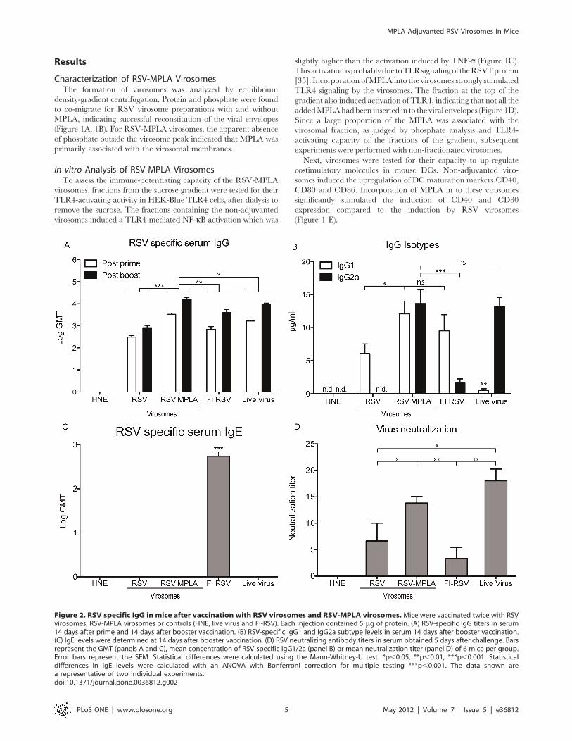
Figure 2. RSV specific IgG in mice after vaccination with RSV virosomes and RSV-MPLA virosomes. Mice were vaccinated twice with RSVvirosomes, RSV-MPLA virosomes or controls (HNE, live virus and FI-RSV). Each injection contained 5 mg of protein. (A) RSV-specific IgG titers in serum14 days after prime and 14 days after booster vaccination. (B) RSV-specific IgG1 and IgG2a subtype levels in serum 14 days after booster vaccination.(C) IgE levels were determined at 14 days after booster vaccination. (D) RSV neutralizing antibody titers in serum obtained 5 days after challenge. Barsrepresent the GMT (panels A and C), mean concentration of RSV-specific IgG1/2a (panel B) or mean neutralization titer (panel D) of 6 mice per group.Error bars represent the SEM. Statistical differences were calculated using the Mann-Whitney-U test. *p,0.05, **p,0.01, ***p,0.001. Statisticaldifferences in IgE levels were calculated with an ANOVA with Bonferroni correction for multiple testing ***p,0.001. The data shown area representative of two individual experiments.doi:10.1371/journal.pone.0036812.g002
MPLA Adjuvanted RSV Virosomes in Mice
PLoS ONE | www.plosone.org 5 May 2012 | Volume 7 | Issue 5 | e36812
In vivo ImmunogenicityTo analyze the immunogenicity of the virosomes in vivo, Balb/c
mice were vaccinated twice with RSV virosomes or RSV-MPLA
virosomes at a 2-week interval. For comparison, mice were
inoculated with live RSV (to induce a Th1-skewed immune
response) or vaccinated twice with FI-RSV (to induce a Th2-
skewed immune response). Two weeks after the first and second
vaccination, blood was drawn and serum IgG titers were
determined. After the priming immunization, RSV virosomes
induced a mean IgG titer of 2.5 Log GMT. Incorporation of
MPLA in to the virosomes resulted in significantly increased IgG
levels after both priming and booster immunizations, not only
compared to the levels induced by non-adjuvanted RSV virosomes
but also to the levels induced by FI-RSV and live virus (Figure 2A).
Next, RSV-specific IgG1 and IgG2a subtype levels were
levels of IgG2a compared to non-adjuvanted virosomes, reaching
similar levels of RSV-specific IgG2a as seen after live virus
inoculation (Figure 2B). In parallel with the increased RSV-
specific IgG2a responses, increases in RSV-specific IgG1 levels
were also noted. Non-adjuvanted RSV virosomes and FI-RSV
mainly induced IgG1, indicative of a Th2-type response. Live virus
inoculations induced low levels of IgG1 and similar levels of
IgG2a, compared to those induced by RSV-MPLA virosomes
(Figure 2B).
To further characterize the humoral immune response, we
determined IgE levels in sera and IgA levels in BAL of immunized
mice. IgE was exclusively induced by immunization with FI-RSV,
but not by immunization with virosomes or live virus (Figure 2C).
IgA in BAL was detectable in mice immunized with FI-RSV
(4.660.1 2Log GMT) and live virus (5.660.6 2Log GMT), but not
in mice immunized with virosomes. For assessment of the
functional capacity of the antibodies, we performed a microneu-
tralization assay. Non-adjuvanted RSV virosomes induced similar
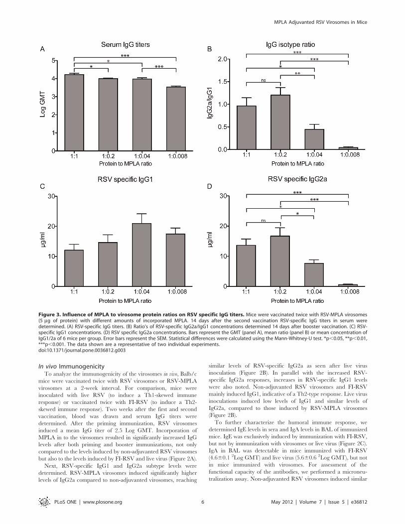
Figure 3. Influence of MPLA to virosome protein ratios on RSV specific IgG titers. Mice were vaccinated twice with RSV-MPLA virosomes(5 mg of protein) with different amounts of incorporated MPLA. 14 days after the second vaccination RSV-specific IgG titers in serum weredetermined. (A) RSV-specific IgG titers. (B) Ratio’s of RSV-specific IgG2a/IgG1 concentrations determined 14 days after booster vaccination. (C) RSV-specific IgG1 concentrations. (D) RSV specific IgG2a concentrations. Bars represent the GMT (panel A), mean ratio (panel B) or mean concentration ofIgG1/2a of 6 mice per group. Error bars represent the SEM. Statistical differences were calculated using the Mann-Whitney-U test. *p,0.05, **p,0.01,***p,0.001. The data shown are a representative of two individual experiments.doi:10.1371/journal.pone.0036812.g003
MPLA Adjuvanted RSV Virosomes in Mice
PLoS ONE | www.plosone.org 6 May 2012 | Volume 7 | Issue 5 | e36812
neutralizing antibody titers to FI-RSV. Incorporation of MPLA in
to the virosomes significantly increased the neutralizing antibody
titers to levels similar to those induced by live virus (Figure 2D).
To investigate which concentration of MPLA is needed for
optimal adjuvant activity, we added different amounts of MPLA to
the viral protein in solution before reconstitution. Apart from the
1:1 protein:MPLA ratio, we also produced virosomes with 1:0.2,
1:0.04 and 1:0.008 protein to MPLA ratios. Using a similar
immunization regimen and antigen dose as before, mice were
vaccinated, and RSV-specific serum IgG and subtype responses
were determined. The reduction in total RSV-specific serum IgG
induced by the vaccine was proportional to the decline in the
amount of MPLA in the virosomes (Figure 3A). The IgG2a/IgG1
subtype ratio remained similar when the amount of MPLA was
reduced from 1 to 0.2 mg/mg protein but decreased when the
amount of MPLA was reduced further (Figure 3B). This decrease
was primarily due to a reduction in RSV-specific IgG2a levels,
while the level of RSV-specific IgG1 did not increase significantly
with lower amounts of virosome-incorporated MPLA (Figure 3C,
3D). Because there was no significant difference between the IgG
subtypes induced by 1:1 and 1:0.2 protein to MPLA ratio
virosomes and there are other benefits to be expected from higher
MPLA concentrations (i.e. cellular immune response and re-
duction in lung pathology) we chose to perform the next
experiments with 1:1 protein:MPLA virosomes.
Cellular ImmunityTo analyze if virosome-incorporated MPLA skews the immune
response to a favorable Th1 phenotype, levels of the hallmark Th1
cytokine IFNc and Th2 cytokine IL5 were determined in
splenocyte cultures of mice, ex vivo stimulated with RSV. Super-
natants of splenocytes cultures from mice immunized with RSV-
MPLA virosomes or infected with live virus produced significantly
increased levels of IFNc compared to those from mice immunized
with RSV virosomes alone or FI-RSV (Figure 4A) Restimulated
splenocytes from non-vaccinated mice produced considerable
levels of IFNc, which may be explained by activation of innate
immunity (i.e. NK cell activation) as a result of a high viral load
occurring in infected naıve animals. Levels of IL5 were
significantly increased in splenocyte cultures from mice immunized
Figure 4. IFNc and IL5 concentrations in RSV-stimulated splenocyte cultures and lung tissue homogenates.Mice were vaccinated twicewith RSV virosomes, RSV-MPLA virosomes and control vaccines as in Figure 2, and subsequently challenged with live RSV. Four days after challenge,IFNc and IL5 responses were determined. (A) IFNc concentrations in splenocyte cultures restimulated with BPL-inactivated RSV for three days. (B) IFNcconcentrations in homogenated lung tissue, four days after challenge. (C) IL5 concentrations in splenocyte cultures, restimulated with BPL-inactivatedRSV for three days. (D) IL5 concentrations in homogenated lung tissue, four days after challenge. Bars represent the mean cytokine concentration of 6mice per group and error bars represent the SEM. Statistical differences were calculated using a Mann-Whitney-U test. *p,0.05, **p,0.01,***p,0.001. The data shown are a representative of two individual experiments.doi:10.1371/journal.pone.0036812.g004
MPLA Adjuvanted RSV Virosomes in Mice
PLoS ONE | www.plosone.org 7 May 2012 | Volume 7 | Issue 5 | e36812
with FI-RSV when compared to those from all other groups
(Figure 4B).
Next, secretion of these cytokines was measured locally, i.e. in
lung homogenates, 4 days after viral challenge. In line with the
above data, mice immunized with RSV-MPLA virosomes showed
significantly increased IFNc levels in their lungs upon live virus
challenge when compared to levels measured in the lungs of mice
immunized with non-adjuvanted virosomes, FI-RSV or live virus
immunization (Figure 4C). Also, IL5 levels were significantly
increased in the lungs of FI-RSV immunized mice when
compared to the levels measured in the lungs of mice immunized
with (adjuvanted) RSV virosomes or live virus (Figure 4D).
Virus Clearance after ChallengeTo analyze vaccination-induced virus clearance after challenge,
mice were immunized twice with HNE buffer, FI-RSV, live virus,
RSV virosomes or RSV-MPLA virosomes. Two weeks after the
second vaccination mice were challenged with 106 TCID50 live
RSV. Four days later, viral titers were determined in the lungs of
the animals. In the HNE vaccinated group, virus was recovered
from the lungs of all mice (Figure 5A). In three out of the six mice
immunized with RSV virosomes, virus could not be detected. In
the other mice, virus was detected albeit at a significant lower level
than in non-immunized mice. In contrast, in all mice immunized
with RSV-MPLA virosomes, FI-RSV and live virus, virus could
not be detected.
Lung PathologyTo further investigate ERD in the immunized mice, we
examined lung pathology upon challenge infection (Figure 6).
Mice immunized with FI-RSV showed signs of alveolitis and
infiltrates in both the peribronchial and perivascular areas
(Figure 6A). The lungs of mice immunized with live virus on the
other hand showed no signs of pathology (Figure 6B). Mice
immunized with RSV virosomes showed no signs of alveolitis but
did have perivascular infiltrates (Figure 6C) In contrast, the lungs
of the mice who received RSV-MPLA virosomes showed no signs
of lung pathology (Figure 6D) and were very similar to the lungs of
mice who received live virus or those of non-immunized mice
(Figure 6B,E). In addition to this, we assessed the presence of
eosinophils in broncho-alveolar lavages (BAL) four days after
challenge by May-Grunwald Giemsa staining of cytospotted cells.
No eosinophils were detected in BAL of mice vaccinated with
RSV or RSV-MPLA virosomes. On the other hand, in the mice
vaccinated with FI-RSV, eosinophils were clearly present
(Figure 6F).
Discussion
Despite the fact that RSV has been recognized as an important
vaccine target for more than 60 years, no vaccine is registered for
use in humans today. Various vaccine candidates have been
evaluated in clinical trials but so far none of them showed the
required safety and efficacy profiles. Generally, live attenuated
virus vaccines administered intranasally are safe and well tolerated
but it is difficult to obtain an optimal balance between
immunogenicity and attenuation [36]. Inactivated virus vaccines
appear to be hard to advance to the clinic because of the safety
concerns related to the outcome of the 1960’s FI-RSV trial.
Protein subunit vaccines are easy to produce but are generally not
very immunogenic and possibly skew towards a Th2 immune
response [36].
In this study, we evaluated the immunogenicity and protective
capacity of a virosomal RSV vaccine adjuvanted with MPLA.
Incorporation of the TLR4 ligand MPLA into the virosomal
membrane resulted in effective human TLR4 stimulation in HEK-
Blue cells in vitro and activation of mouse DC ex vivo as shown by
the upregulation of co-stimulatory molecules. Incorporation of
MPLA in virosomes resulted in increased RSV-specific serum IgG
titers, with production of RSV-specific, Th1-signature, IgG2a-
isotype antibodies similar to that induced by live virus inoculation
leading to a balanced IgG1/IgG2a profile. These antibodies
proved effective in virus neutralization. Furthermore, RSV-MPLA
virosomes skewed the cellular responses towards a Th1 profile, as
shown by enhanced IFN-c secretion, not only in ex vivo RSV-
stimulated splenocytes, but also locally in the lungs of infected
mice. Immunization with RSV-MPLA virosomes did not induce
any detectable IgE in contrast to immunization with FI-RSV. IgE
induction is a hallmark of a Th2-skewed allergy-like response,
which is implicated in RSV infections and in FI-RSV induced
enhanced disease [37–39]. MPLA-adjuvanted virosomes, similar
to FI-RSV, provided full protection against live RSV infection, but
in contrast to FI-RSV, did not lead to signs of ERD, i.e. influx of
eosinophils in the lungs or induction of lung pathology.
Importantly, previous studies in cotton rats showed that addition
of MPLA to FI-RSV reduces the induction of ERD by FI-RSV
immunization, illustrated by a reduction in lung pathology, an
increase in serum virus neutralization titers and a shift from a Th2
-skewed immune response to a balanced immune response
[31,32]. Our observations on the immune response induced by
MPLA-adjuvanted RSV virosomes in mice are in line with these
data and underline that MPLA-adjuvanted RSV virosomes hold
promise as a candidate RSV vaccine. Currently, RSV-MPLA
virosomes are being evaluated in cotton rats to optimally assess
other ERD parameters, such as alveolitis, in more detail.
Our data show that non-adjuvanted RSV virosomes stimulate
human TLR4 in HEK-Blue cells and upregulate co-stimulatory
molecules in mouse DC and that incorporated MPLA further
enhances these effects. TLR4 activation by RSV virosomes without
MPLA is likely to be caused by the RSV F protein. RSV F is a known
Figure 5. Protection against live virus challenge and infiltrationof eosinophils. Mice were vaccinated as described in figure 2 andchallenged with live virus 14 days after the booster vaccination. Fourdays after challenge, lungs were removed and the viral titer wasdetermined and expressed as TCID50. RSV TCID50 titers from the lungsof challenged animals. Statistical differences were calculated using theMann-Whitney-U test. *p,0.05. The data shown are a representative oftwo individual experiments.doi:10.1371/journal.pone.0036812.g005
MPLA Adjuvanted RSV Virosomes in Mice
PLoS ONE | www.plosone.org 8 May 2012 | Volume 7 | Issue 5 | e36812
TLR4 agonist that, for example, induces inflammatory cytokines
like IL-6 in DC [35]. Interestingly, despite this capacity to stimulate
TLR4, RSV virosomes fail to induce Th1-type responses while
MPLA, also a TLR4 agonist, effectively stimulates Th1-type
responses. This could be due to differences in the magnitude of
stimulation, which is clearly higher for MPLA (Figure 1), but could
also be caused by recruitment of different adaptor molecules
downstream of TLR4 activation. As TLR4 uses both MyD88 and
TRIF adaptor molecules, it is possible that MPLA competes with
RSV F for TLR4 activation. This competition shifts signaling from
RSV F-induced, MyD88-dependent, TLR4 signaling to MPLA-
induced, TRIF-dependent, TLR4 signaling, leading to a Th1-
skewed immune response induced by RSV-MPLA virosomes
compared to non-adjuvanted RSV virosomes.
Apart from its influence on T helper cell differentiation, TLR
signaling also has a direct effect on IgG isotype switching [40].
Antibody isotype switching is important, because different
immunoglobulin subclasses display differences in their ability
to mediate effector responses [41]. In mice, the most effective
IgG isotype protecting against viral infections is IgG2a [42]. As
stated before, MPLA signals through TLR4 to induce type I
IFNs which stimulate IgG2a production predominantly from
follicular B cells [40]. Furthermore, MPLA could also directly
activate TLR4 on B cells to facilitate isotype switching, a process
that is further augmented by IFNc and T-cell help [43].
Previously, we incorporated TLR2 ligand P3CSK4 in RSV
virosomes. P3CSK4 inclusion also skewed towards a Th1
immune response and increased IgG2a levels compared to
Figure 6. Lung pathology in mice after immunization and RSV infection.Mice were immunized and challenged as described in Figure 2 andthe lungs were harvested, sliced and stained with H&E and assessed for pathology using light microscopy. Panels represent the lungs of (A) FI-RSV, (B)live virus, (C) RSV virosomes, (D) RSV MPLA virosomes (E) buffer immunized mice. Black arrows point to alveolar infiltrates, grey arrows toperibronchial infiltrates and white arrows to perivascular infiltrates. (F) Eosinophils in BAL expressed as percentage of total BAL cells. Data pointsrepresent values from individual mice. Statistical differences were calculated using the ANOVA test with Bonferroni correction for multiple testing.***p,0.001. The data shown are a representative of two individual experiments.doi:10.1371/journal.pone.0036812.g006
MPLA Adjuvanted RSV Virosomes in Mice
PLoS ONE | www.plosone.org 9 May 2012 | Volume 7 | Issue 5 | e36812
(2009) Lack of antibody affinity maturation due to poor Toll-like receptorstimulation leads to enhanced respiratory syncytial virus disease. Nat Med 15:
34–41. doi:10.1038/nm.1894.
18. Guy B (2007) The perfect mix: recent progress in adjuvant research. Nat RevMicrobiol 5: 505–517. doi:10.1038/nrmicro1681.
19. Schnare M, Barton GM, Holt AC, Takeda K, Akira S, et al. (2001) Toll-like
receptors control activation of adaptive immune responses. Nat Immunol 2:947–950. doi:10.1038/ni712.
20. Netea MG, Van der Meer JWM, Sutmuller RP, Adema GJ, Kullberg B-J (2005)
From the Th1/Th2 Paradigm towards a Toll-Like Receptor/T-Helper Bias.Antimicrobial Agents and Chemotherapy 49: 3991. doi:10.1128/
AAC.49.10.3991–3996.2005.
21. Stegmann T, Kamphuis T, Meijerhof T, Goud E, de Haan A, et al. (2010)Lipopeptide-adjuvanted respiratory syncytial virus virosomes: A safe and
37. Welliver RC, Kaul TN, Sun M, Ogra PL (1984) Defective regulation of immune
responses in respiratory syncytial virus infection. J Immunol 133: 1925–1930.
38. Welliver RC, Wong DT, Sun M, Middleton E, Vaughan RS, et al. (1981) The
development of respiratory syncytial virus-specific IgE and the release of
histamine in nasopharyngeal secretions after infection. N Engl J Med 305:
841–846. doi:10.1056/NEJM198110083051501.39. Becker Y (2006) Respiratory syncytial virus (RSV) evades the human adaptive
immune system by skewing the Th1/Th2 cytokine balance toward increased
levels of Th2 cytokines and IgE, markers of allergy–a review. Virus Genes 33:235–252. doi:10.1007/s11262-006-0064-x.
40. Swanson CL, Wilson TJ, Strauch P, Colonna M, Pelanda R, et al. (2010) Type IIFN enhances follicular B cell contribution to the T cell-independent antibody
response. J Exp Med 207: 1485–1500. doi:10.1084/jem.20092695.
41. Nimmerjahn F, Ravetch JV (2005) Divergent immunoglobulin g subclass activitythrough selective Fc receptor binding. Science 310: 1510–1512. doi:10.1126/
science.1118948.42. Coutelier JP, van der Logt JT, Heessen FW, Warnier G, Van Snick J (1987)
IgG2a restriction of murine antibodies elicited by viral infections. J Exp Med165: 64–69.
43. Heer AK, Shamshiev A, Donda A, Uematsu S, Akira S, et al. (2007) TLR
signaling fine-tunes anti-influenza B cell responses without regulating effector Tcell responses. J Immunol 178: 2182–2191.
44. Moghaddam A, Olszewska W, Wang B, Tregoning JS, Helson R, et al. (2006) Apotential molecular mechanism for hypersensitivity caused by formalin-
inactivated vaccines. Nat Med 12: 905–907. doi:10.1038/nm1456.
45. McLellan JS, Chen M, Kim A, Yang Y, Graham BS, et al. (2010) Structuralbasis of respiratory syncytial virus neutralization by motavizumab. Nat Struct
Mol Biol 17: 248–250. doi:10.1038/nsmb.1723.
MPLA Adjuvanted RSV Virosomes in Mice
PLoS ONE | www.plosone.org 11 May 2012 | Volume 7 | Issue 5 | e36812