JOURNAL OF CLINICAL MICROBIOLOGY, June 1994, p. 1464-1469 Vol. 32, No. 6 0095-1 137/94/$04.00+0 Copyright C) 1994, American Society for Microbiology Ureaplasma urealyticum Biovar Specificity and Diversity Are Encoded in Multiple-Banded Antigen Gene LEE-JENE TENG, XIAOTIAN ZHENG, JOHN I. GLASS, HAROLD L. WATSON, JASON TSAI, AND GAIL H. CASSELL* Department of Microbiology, University of Alabama at Birmingham, Birmingham, Alabama 35294 Received 14 December 1993/Returned for modification 8 February 1994/Accepted 1 March 1994 Ureaplasma urealyticum is a commensal organism of the lower genital tract of females, but in a subpopulation of individuals, it can invade the upper genital tract. It is a significant cause of chorioamnionitis and neonatal morbidity and mortality. There are 14 recognized serovars of U. urealyticum; these can be divided into two distinct clusters or biovars. Biovar 1 is composed of serovars 1, 3, 6, and 14. Biovar 2 is composed of serovars 2, 4, 5, 7, 8, 9, 10, 11, 12, and 13. We previously identified a surface antigen, the multiple-banded (MB) antigen, which contains both serovar-specific and cross-reactive epitopes. Genotypic characterization of the C-terminal region of the MB antigen of serovar 3 indicates that serovar specificity and MB antigen size variation reside in that domain. In the present study, we used PCR analysis with primers derived from the serovar 3 MB antigen gene DNA sequence to determine if the MB antigen gene was present in the remaining 13 reference serovars as well as in invasive clinical isolates. The results indicated that not only was the MB antigen gene present in all serovars but that the genes' 5' regions were markers of biovar specificity and diversity. Further analysis of this region should reveal the phylogenetic relationship among serovars of U. urealyticum and, possibly, their invasive potential. Ureaplasma urealyticum is a common commensal organism of the urogenital tract of sexually mature females. However, ureaplasmal infection of the chorioamnion is strongly associ- ated with chorioamnionitis, premature birth, and perinatal morbidity and mortality (4, 5). U. urealyticum is the single most common microorganism isolated from the central nervous system and lower respiratory tract of newborn infants (5), particularly those born prematurely. Because of the frequency with which U. urealyticum occurs in healthy asymptomatic individuals, it has been suggested that only certain subgroups of the species are truly disease associated. However, the development of reliable typing reagents is necessary to explore this possibility. There are 14 recognized serovars of U. urealyticum; these can be divided into two distinct clusters or biovars. Biovar 1 (or parvo biovar) is composed of serovars 1, 3, 6, and 14. Biovar 2 (or T960 biovar [19]) is composed of 10 serovars, which are numbered 2, 4, 5, 7, 8, 9, 10, 11, 12, and 13. Members of the two biovars differ phenotypically in their susceptibilities to manga- nese (17). They can also be differentiated by DNA-DNA hybridization (7), restriction fragment length polymorphism (10, 15, 16), one- and two-dimensional gel electrophoresis (22), genomic sizes (18), and PCR amplification of specific genes (1, 19). We recently cloned and sequenced the multiple-banded (MB) antigen gene of the U. urealyticum serovar 3 reference strain (28). This is the predominant antigen recognized by patients infected with U. urealyticum (23). Nucleotide sequence analysis predicts that the MB antigen contains a signal peptide and acylation site in the N-terminal region, while the C- terminal region is composed of multiple six-amino-acid (en- * Corresponding author. Mailing address: BBRB, Room 276-11, Department of Microbiology, University of Alabama at Birmingham, Birmingham, AL 35294-2170. Phone: (205) 934-9339. Fax: (205) 934-9256. coded by 18 nucleotides) tandem repeats, which contain sero- var-specific epitopes. Alteration of the copy number of the repeating units results in MB antigen size variation (28). In contrast to the repeat region, nucleotide sequencing and PCR analysis of different serovar 3 MB antigen size variants sug- gested that the 5' region is conserved among serovar 3 size variants (28). Thus, we hypothesized that this conserved 5' section of the serovar 3 gene encoding the N-terminal region of the MB antigen may also have homologous counterparts in the other 13 serovars. The goal of the present study was to test this hypothesis by using PCR primers derived from the upstream region and the 5' region of the MB antigen gene of serovar 3. All 14 reference serovars plus 20 well-characterized invasive isolates were found to contain an MB antigen gene, and we found that members of the two biovars could be distinguished by the sizes of the amplification products. Although evidence presented elsewhere (28) indicates that serovar specificity is determined by the composition of the C-terminal region of MB antigens, we show here that the heterogeneity detected in the sequence of the 5' region of the MB antigen gene of the different serovars allowed us to divide the 14 serovars into several subgroups on the basis of restriction endonuclease digestion of the PCR products. MATERIALS AND METHODS Organisms. The reference strains of U. urealyticum serovars 1 through 8 used in the present study were obtained from E. A. Freundt (Institute of Medical Microbiology, University of Aarhus, Aarhus, Denmark), and serovars 9 through 14 were obtained from J. A. Robertson (Department of Medical Mi- crobiology and Infectious Diseases, University of Alberta, Edmonton, Alberta, Canada). Mycoplasma pneumoniae Eaton, Mycoplasma fermentans, Mycoplasma hominis, Mycoplasma buccale, Mycoplasma faucium, Mycoplasma orale, Mycoplasma salivarium, Mycoplasma genitalium, Mycoplasma pirum, Myco- plasma primatum, Mycoplasma arthritidis, Mycoplasma muris, 1464 on April 13, 2019 by guest http://jcm.asm.org/ Downloaded from
Transcript
JOURNAL OF CLINICAL MICROBIOLOGY, June 1994, p. 1464-1469 Vol. 32, No. 60095-1 137/94/$04.00+0Copyright C) 1994, American Society for Microbiology
Ureaplasma urealyticum Biovar Specificity and Diversity AreEncoded in Multiple-Banded Antigen Gene
LEE-JENE TENG, XIAOTIAN ZHENG, JOHN I. GLASS, HAROLD L. WATSON,JASON TSAI, AND GAIL H. CASSELL*
Department of Microbiology, University ofAlabama at Birmingham, Birmingham, Alabama 35294
Received 14 December 1993/Returned for modification 8 February 1994/Accepted 1 March 1994
Ureaplasma urealyticum is a commensal organism of the lower genital tract of females, but in a subpopulationof individuals, it can invade the upper genital tract. It is a significant cause of chorioamnionitis and neonatalmorbidity and mortality. There are 14 recognized serovars of U. urealyticum; these can be divided into twodistinct clusters or biovars. Biovar 1 is composed of serovars 1, 3, 6, and 14. Biovar 2 is composed of serovars2, 4, 5, 7, 8, 9, 10, 11, 12, and 13. We previously identified a surface antigen, the multiple-banded (MB) antigen,which contains both serovar-specific and cross-reactive epitopes. Genotypic characterization of the C-terminalregion of the MB antigen of serovar 3 indicates that serovar specificity and MB antigen size variation residein that domain. In the present study, we used PCR analysis with primers derived from the serovar 3 MBantigen gene DNA sequence to determine if the MB antigen gene was present in the remaining 13 referenceserovars as well as in invasive clinical isolates. The results indicated that not only was the MB antigen genepresent in all serovars but that the genes' 5' regions were markers of biovar specificity and diversity. Furtheranalysis of this region should reveal the phylogenetic relationship among serovars of U. urealyticum and,possibly, their invasive potential.
Ureaplasma urealyticum is a common commensal organismof the urogenital tract of sexually mature females. However,ureaplasmal infection of the chorioamnion is strongly associ-ated with chorioamnionitis, premature birth, and perinatalmorbidity and mortality (4, 5). U. urealyticum is the single mostcommon microorganism isolated from the central nervoussystem and lower respiratory tract of newborn infants (5),particularly those born prematurely. Because of the frequencywith which U. urealyticum occurs in healthy asymptomaticindividuals, it has been suggested that only certain subgroupsof the species are truly disease associated. However, thedevelopment of reliable typing reagents is necessary to explorethis possibility.There are 14 recognized serovars of U. urealyticum; these
can be divided into two distinct clusters or biovars. Biovar 1 (orparvo biovar) is composed of serovars 1, 3, 6, and 14. Biovar 2(or T960 biovar [19]) is composed of 10 serovars, which arenumbered 2, 4, 5, 7, 8, 9, 10, 11, 12, and 13. Members of the twobiovars differ phenotypically in their susceptibilities to manga-nese (17). They can also be differentiated by DNA-DNAhybridization (7), restriction fragment length polymorphism(10, 15, 16), one- and two-dimensional gel electrophoresis (22),genomic sizes (18), and PCR amplification of specific genes (1,19).We recently cloned and sequenced the multiple-banded
(MB) antigen gene of the U. urealyticum serovar 3 referencestrain (28). This is the predominant antigen recognized bypatients infected with U. urealyticum (23). Nucleotide sequenceanalysis predicts that the MB antigen contains a signal peptideand acylation site in the N-terminal region, while the C-terminal region is composed of multiple six-amino-acid (en-
* Corresponding author. Mailing address: BBRB, Room 276-11,Department of Microbiology, University of Alabama at Birmingham,Birmingham, AL 35294-2170. Phone: (205) 934-9339. Fax: (205)934-9256.
coded by 18 nucleotides) tandem repeats, which contain sero-var-specific epitopes. Alteration of the copy number of therepeating units results in MB antigen size variation (28). Incontrast to the repeat region, nucleotide sequencing and PCRanalysis of different serovar 3 MB antigen size variants sug-gested that the 5' region is conserved among serovar 3 sizevariants (28). Thus, we hypothesized that this conserved 5'section of the serovar 3 gene encoding the N-terminal region ofthe MB antigen may also have homologous counterparts in theother 13 serovars. The goal of the present study was to test thishypothesis by using PCR primers derived from the upstreamregion and the 5' region of the MB antigen gene of serovar 3.All 14 reference serovars plus 20 well-characterized invasiveisolates were found to contain an MB antigen gene, and wefound that members of the two biovars could be distinguishedby the sizes of the amplification products. Although evidencepresented elsewhere (28) indicates that serovar specificity isdetermined by the composition of the C-terminal region ofMBantigens, we show here that the heterogeneity detected in thesequence of the 5' region of the MB antigen gene of thedifferent serovars allowed us to divide the 14 serovars intoseveral subgroups on the basis of restriction endonucleasedigestion of the PCR products.
MATERIALS AND METHODS
Organisms. The reference strains of U. urealyticum serovars1 through 8 used in the present study were obtained from E. A.Freundt (Institute of Medical Microbiology, University ofAarhus, Aarhus, Denmark), and serovars 9 through 14 wereobtained from J. A. Robertson (Department of Medical Mi-crobiology and Infectious Diseases, University of Alberta,Edmonton, Alberta, Canada). Mycoplasma pneumoniae Eaton,Mycoplasma fermentans, Mycoplasma hominis, Mycoplasmabuccale, Mycoplasma faucium, Mycoplasma orale, Mycoplasmasalivarium, Mycoplasma genitalium, Mycoplasma pirum, Myco-plasma primatum, Mycoplasma arthritidis, Mycoplasma muris,
Mycoplasma neurolyticum, Mycoplasma collis, Mycoplasma pul-monis, Acholeplasma laidlawii, and Acholeplasma oculi wereobtained from J. G. Tully of the National Institute of Allergyand Infectious Diseases.Ten clinical isolates from cerebrospinal fluid of newborn
infants and 10 isolates from chorioamnions were serotyped byimmunoblotting with monoclonal antibodies (MAbs) as de-scribed previously (29). However, in the present study a newlydeveloped serovar 6-specific MAb instead of rabbit anti-serovar 6 serum was used as described previously (29). Onecerebrospinal fluid isolate (isolate C4), whose serotype wasunknown in our previous study (29), was identified as serovar6 with the new MAb.Sample preparations for PCR. Rapid sample preparation
prior to DNA amplification was done as described previously(2). Briefly, 1 ml of the cultures was centrifuged (12,000 x g,4°C, 20 min). The pellet was resuspended in 100 ,ul ofproteinase K solution (0.25 mg/ml). These cell suspensionswere incubated for 1 h at 60°C prior to incubation at 95°C for10 min. Five microliters of the deproteinized samples wasadded to the 50-,u amplification reaction mixture.PCRs. The primers used for the PCRs were derived from the
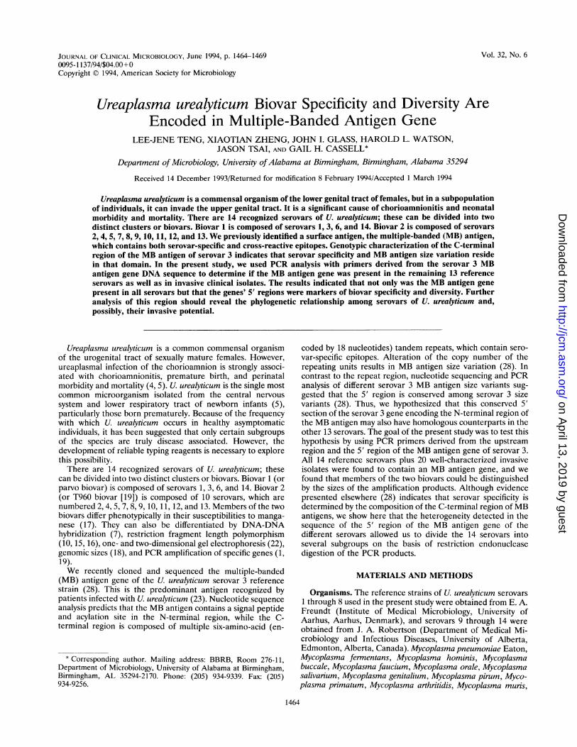
nucleotide sequence of the serovar 3 MB antigen gene (Table1) (28). The oligonucleotide nomenclature was as follows:molecules were named UMS or UMA for ureaplasma MBantigen coding sense primers or antisense primers, respec-tively. The number following UMS or UMA refers to thelocation on the MB antigen gene map corresponding to the3'-most base of the oligonucleotide. PCRs with primersUMS-125 and UMA226 were designated UM-1. Reactionswith UMS51 and UMA427 were designated UM-2. The loca-tions of the oligonucleotides used as PCR primers weremapped on the MB antigen gene as indicated in Fig. 1.The amplification reaction mixtures contained 50 ,ul of 10
MB GENE5' 3'
-150 -87f1-8Dral Pvull RsaI183 250 270
-. rSD
1 2 3
- 403 bp
B
123 bp-
c I
123bpp-
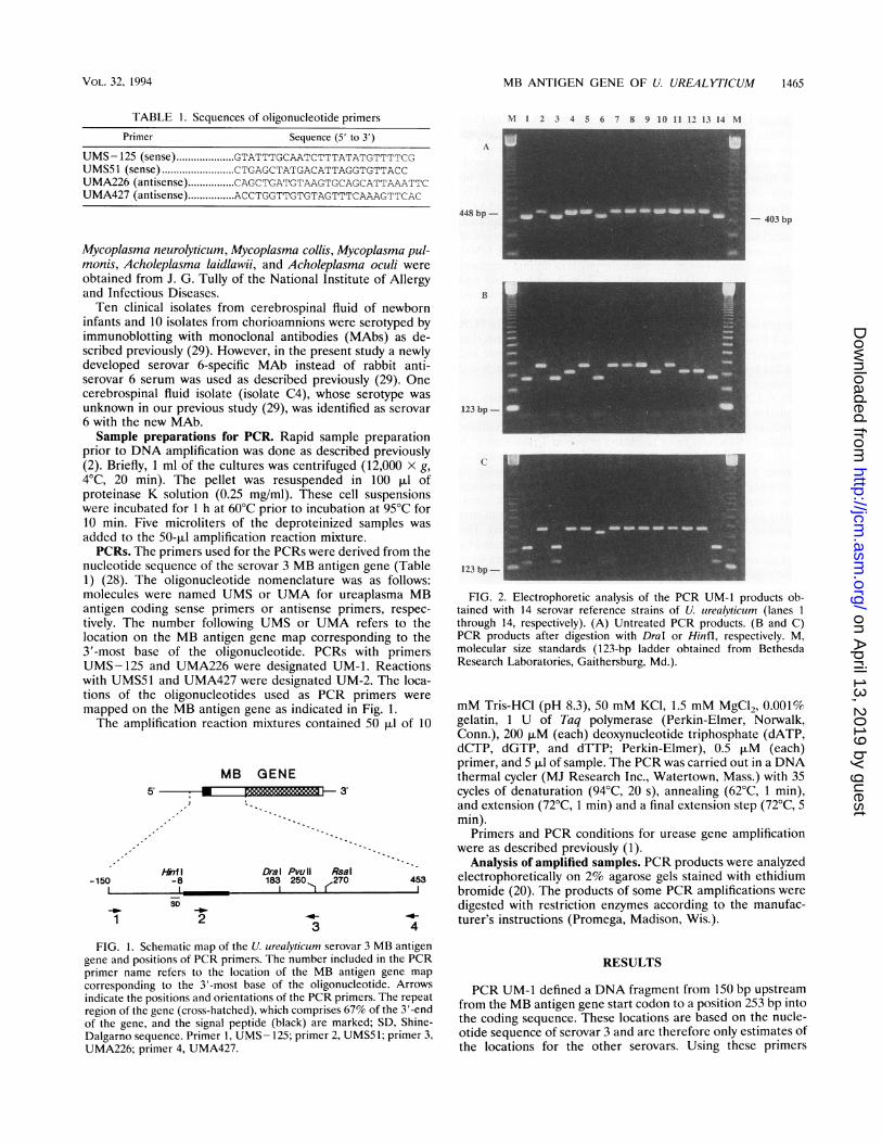
FIG. 2. Electrophoretic analysis of the PCR UM-1 products ob-tained with 14 serovar reference strains of U. urealyticum (lanes Ithrough 14, respectively). (A) Untreated PCR products. (B and C)PCR products after digestion with DraI or Hinfl, respectively. M,molecular size standards (123-bp ladder obtained from BethesdaResearch Laboratories, Gaithersburg, Md.).
mM Tris-HCl (pH 8.3), 50 mM KCl, 1.5 mM MgCl2, 0.001%gelatin, 1 U of Taq polymerase (Perkin-Elmer, Norwalk,Conn.), 200 p.M (each) deoxynucleotide triphosphate (dATP,dCTP, dGTP, and dTTP; Perkin-Elmer), 0.5 ,uM (each)primer, and 5 pul of sample. The PCR was carried out in a DNAthermal cycler (MJ Research Inc., Watertown, Mass.) with 35cycles of denaturation (94°C, 20 s), annealing (62°C, 1 min),and extension (72°C, 1 min) and a final extension step (72°C, 5min).
Primers and PCR conditions for urease gene amplificationwere as described previously (1).
Analysis of amplified samples. PCR products were analyzedelectrophoretically on 2% agarose gels stained with ethidiumbromide (20). The products of some PCR amplifications weredigested with restriction enzymes according to the manufac-turer's instructions (Promega, Madison, Wis.).
FIG. 1. Schematic map of the U. urealyticum serovar 3 MB antigengene and positions of PCR primers. The number included in the PCRprimer name refers to the location of the MB antigen gene mapcorresponding to the 3'-most base of the oligonucleotide. Arrowsindicate the positions and orientations of the PCR primers. The repeatregion of the gene (cross-hatched), which comprises 67% of the 3'-endof the gene, and the signal peptide (black) are marked; SD, Shine-Dalgarno sequence. Primer 1, UMS- 125; primer 2, UMS51; primer 3,UMA226; primer 4, UMA427.
RESULTS
PCR UM-1 defined a DNA fragment from 150 bp upstreamfrom the MB antigen gene start codon to a position 253 bp intothe coding sequence. These locations are based on the nucle-otide sequence of serovar 3 and are therefore only estimates ofthe locations for the other serovars. Using these primers
(primers UMS- 125 and UMA226), the length of the PCRproducts for serovar 3 was 403 bp. All 14 reference serovarswere amplified (Fig. 2A). The PCR products from biovar 1,which includes serovar 3, all comigrated. However, the prod-ucts from members of biovar 2 were approximately 45-bplonger (448 bp) than the 403-bp biovar 1 fragment. Theproducts of PCR UM-2 with primer pair UMS51 and UMA427were 427 bp, and serovars 1, 3, 6, and 14 were amplified.Product size calculations were based on the DNA sequence ofthe serovar 3 reference strain. Dral digestion of PCR UM-1products showed two electrophoretic patterns among membersof biovar 2 (Fig. 2B). Serovars 2, 5, 7, 8, 9, and 11 had onerestriction digestion pattern; a different pattern was observedwith serovars 4, 10, 12, and 13. Dral digests of members ofbiovar 1 were identical. Hinfl digestion of the products of PCRUM-1 from serovars 1, 3, and 14 yielded DNA fragments thatcomigrated with serovar 3 digestion products, which were 142and 261 bp in length, while serovar 6 and members of biovar 2remained undigested (Fig. 2C).PCR UM-2 used primers UMS51 and UMA427, which are
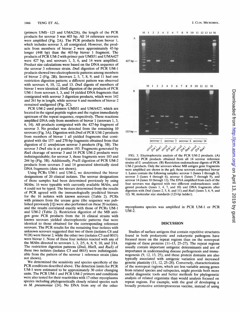
located in the signal peptide region and the region immediatelyupstream of the repeat sequence, respectively. These reactionsamplified DNA only from members of biovar 1 (serovars 1, 3,6, 14). All products comigrated with the 427-bp fragment ofserovar 3. No product was detected from the remaining 10serovars (Fig. 3A). Digestion with Dral of PCR UM-2 productsfrom members of biovar 1 all yielded fragments that comi-grated with the 157- and 270-bp fragments obtained from thatdigestion of U. urealyticum serovar 3 products (Fig. 3B). Theserovar 3 DraI site is at position 183. Fragments generated byRsaI cleavage of serovar 3 and 14 PCR UM-2 products wereindistinguishable; for serovar 3, those fragments were 183 and244 bp (Fig. 3B). Additionally, PvuII digestion of PCR UM-2products from serovars 3 and 14 generated identically sizedDNA fragments (data not shown).
Using PCRs UM-1 and UM-2, we determined the biovardesignations of 20 clinical isolates. The serovar designationsof those samples had been determined previously by usingMAbs; 16 were typeable with currently available MAbs, and4 could not be typed. The biovars determined froim the resultsof PCR agreed with the immunologically predicted biovarsfor the 16 isolates with known serovars (Table 2). PCRswith primers from the urease gene (the sequence was pub-lished previously [1]) were also performed on these 20 isolates,and the results correlated exactly with those of PCRs UM-1and UM-2 (Table 2). Restriction digestion of the MB anti-gen gene PCR products from the 16 clinical strains withknown serovars yielded electrophoretic patterns that wereidentical to those obtained for the corresponding referenceserovars. The PCR results for the remaining four isolates withunknown serovars suggested that two of them (isolates C8 and9128) were biovar 2, while the other two (isolates C3 and 0015)were biovar 1. None of these four isolates reacted with any ofthe MAbs directed to serovars 1, 3, 2/5, 6, 8, 9, 10, and 3/14.The restriction digestion patterns (Dral, Hinfl, and RsaI) ofthese two isolates (isolates C3 and 0015) were indistinguish-able from the pattern of the serovar 1 reference strain (datanot shown).We determined the sensitivity and species specificity of the
PCR conditions described above. The detection limits for PCRUM-1 were estimated to be approximately 30 color changingunits. The PCR UM-1 and PCR UM-2 primers and conditionswere also tested for their reactivities with 17 other mycoplasmaspecies including phylogenetically closely related species suchas M. pneumoniae (24). No DNA from any of the other
M 1 2 3 4 5 6 7 8 9 10 11 12 13 14 M
A
427 bp-
B
123 bp-
M 1 2 3 4 5 6 7 8 9 10 11 12 M
serovar 1 serovar 3 serovar 6 serovar 14
FIG. 3. Electrophoretic analysis of the PCR UM-2 products. (A)Untreated PCR products obtained from all 14 serovar referencestrains of U. urealyticum. (B) Restriction endonuclease digests of PCRUM-2 products. Only the serovars whose MB antigen gene fragmentswere amplified are shown in the gel; those serovars comprised biovar1. Lanes contain the following samples: serovar 1 (lanes 1 through 3),serovar 3 (lanes 4 through 6), serovar 6 (lanes 7 through 9), andserovar 14 (lanes 10 through 12). The DNA amplified from each of thefour serovars was digested with two different endonucleases: undi-gested products (lanes 1, 4, 7, and 10) and DNA fragments afterdigestion with DraI (lanes 2, 5, 8, and 11) and RsaI (lanes 3, 6, 9, and12). M, molecular size standards (123-bp ladder).
mycoplasma species was amplified in PCR UM-1 or PCRUM-2.
DISCUSSION
Studies of surface antigens that contain repetitive structuresfound in both prokaryotic and eukaryotic pathogens havefocused more on the repeat regions than on the nonrepeatregions of these proteins (11-13, 25-27). The repeat regionsusually contain important antigenic determinants and are ofimportance in understanding disease pathogenesis and immu-nogenesis (9, 12, 13, 25), and these protein domains are alsotypically associated with antigenic variation and increasedgenetic plasticity (11, 12, 25-28). Conversely, characterizationof the nonrepeat regions, which are less variable among genesfrom related species and subspecies, might provide both moreuseful diagnostic tools and better methods for phylogeneticanalysis of related organisms than would analysis focused onrepeat regions. For example, with the goal of developing abroadly protective antistreptococcus vaccine, instead of using
" Amplification of DNA is reported by a plus sign; failure to detect amplifiedDNA is reported with a minus sign.
the highly variable repeat region, a conserved region from theM-protein gene of group A streptococcus has been expressedby using a recombinant vaccinia virus. This vaccine protectsmice against challenge by several different group A streptococ-cal types (9). Conserved regions of a giardia gene have beenused as a PCR amplification target for identification anddiagnostic purposes (8).Our previous investigations indicated that the region in the
serovar 3 MB antigen gene that encoded the N-terminalnonrepeat domain of the antigen was conserved among differ-ent variants, while the lengths of the gene domains coding forthe C-terminal repeat regions were variable and were associ-ated with the size variation of this antigen (28). In the presentstudy, we used PCR to show the presence of a specificfragment of the 5' region in all 14 reference serovars as well asin each of 20 clinical isolates representing 5 different serovars.These results strongly suggest that, unlike some other serovardeterminants, which are present only among certain isolates(6), the MB antigen gene exists among all the referenceserovars and is likely to exist among all isolates of theorganism.The present results also demonstrated that biovar specificity
resides in the 5' region. With PCR UM-1, organisms ofdifferent biovars produced distinct PCR products, allowingdifferentiation between the two biovars. The differences werealso consistent for the 16 clinical isolates with known serovars.Since PCR with primers UMS51 and UMA226, which bothmap in the coding region of the MB antigen gene, resulted inamplification of DNA fragments of the same size among all 14serovars (data not shown), it is likely that the length differencesbetween the two biovars were in the 5'-upstream regions.Using the primers UMS51 and UMA427 (PCR UM-2), DNAwas amplified only from members of biovar 1. The PCR UM-2experiment as well as PCR with primers UMS- 125 andUMA427, which amplified members of biovar 1 only (data notshown), suggested that the UMA427 region contains biovar-specific sequences. Interestingly, although our results placeserovar 6 into biovar 1, the lack of sensitivity to Hinfl is a
biovar 2 characteristic. This result suggests that even beforethe nucleotide sequence is available so that the phylogeneticrelationships of the U. urealyticum serovars can be predicted,restriction enzyme digestion analysis of PCR products maypermit further subdivision of the U. urealyticum serovars.Importantly, when we eventually obtain nucleotide sequencedata for the genes of all the U. urealyticum serovars, we mayfind that restriction endonuclease digestion of the PCR prod-ucts could be an effective method for determining the serovarsof clinical isolates.These PCR results revealed that the difference in sequences
between two biovars occurred not only among reference strainsbut also among clinical isolates. In addition, the results alsosupport our previous finding that the N-terminal regions areconserved among the MB antigens of the same serovar.Importantly, in an illustration of the diagnostic potential ofPCR for U. urealyticum typing, clinical isolates that could notbe typed with currently available immunologic reagents werebiotyped. For the two isolates (isolates C8 and 9128) whichpossessed biovar 2 PCR phenotypes, the serovars could not beidentified with available serovar-specific MAbs which identifyserovars 1, 2, 3, 5, 6, 8, 9, 10, and 14. Therefore, it is likely thatthey may belong to serovar 4, 7, 11, 12, or 13. The PCRproducts from the remaining two isolates with unidentifiableserovars (isolates C3 and 0015) exhibited biovar I patterns.Although we could not confirm immunologically the serovarsof these two isolates, restriction endonuclease digestion oftheir PCR products showed that they were identical to thePCR products of the serovar I reference strain. We offerseveral explanations for this. First, although the MB antigengene exists in these two strains, the gene products may not beexpressed. Therefore, our serovar 1-specific MAb, MAb 3C4.6,was not reactive. However, because it encodes an apparentlyimportant antigen, it would be surprising if it were notexpressed in clinical pathogenic isolates. Second, and alterna-tively, the antigen recognized by MAb 3C4.6 is not the MBantigen gene product. In this scenario, these two isolatesexpressed the MB antigen; however, it was not recognized byMAb 3C4.6. Third, and perhaps the most likely explanation, isthat, although MAb 3C4.6 is serovar specific, it may recognizea nonconserved epitope. Additional serovar 1 MAbs will berequired to evaluate further these two isolates immunologi-cally.There are two published nucleotide sequences from U.
urealyticum that have been used as PCR targets for biovardifferentiation: the 16S rRNA gene sequences (19) and theurease structural genes (1). Robertson et al. (19) determinedthe 16S rRNA nucleotide sequence of the serovar 3 referencestrain, and they exploited the differences in the variable regionsof 16S rRNA genes to provide primers for biovar-specificPCRs. In their study, one set of conditions amplified DNAsfrom all biovar 1 isolates, while another set of conditions andprimers amplified gene fragments from members of biovar 2only. However, the PCR of Robertson et al. (19) for biovar 2also reacted with several phylogenetically closely related spe-cies: M. pneumoniae, M. fermentans incognitus, and animalureaplasmas. Blanchard and colleagues (1, 3) tested severalpairs of primers for PCR using urease gene sequences. One ofthose amplifications of the urease gene that used oligonucle-otides located upstream and within the 11.2-kDa unit amplifiedserovars 2, 4, 5, 7, 8, 9, and 13, and consequently, that methodclassified serovars 10 and 12 into biovar 1 (1). This resultconflicts with those of most reports, in which these twoserovars are usually classified as biovar 2 on the basis of theirsusceptibilities to manganese (17), protein analysis on poly-acrylamide gels (14), Southern blot analysis with rRNA probes
(16), and PCR based on 16S rRNA gene sequences (19).However, when Blanchard (la) performed PCR with theaforementioned urease gene primers on the 14 referencestrains obtained from our laboratory, serovars 10 and 12 wereclassified into biovar 2. In the present study, our PCR resultsclearly classified serovars 1, 3, 6, and 14 into biovar 1 and theremainder of the serovars into biovar 2. For the clinicalisolates, biovar information obtained with our primers con-curred with the results obtained by using primers based on theurease gene. Importantly, unlike the biovar determinationmethods that use PCR amplification of the 16S rRNA andurease genes, which classify samples on the basis of thepresence or absence of DNA amplification, our PCR UM-1detected all serovars and simultaneously differentiated twobiovars by the length of their amplification products.
Subdivisions within the genus Ureaplasma have been basedlargely on the host species and antigenic heterogeneity. U.urealyticum is the designation for all ureaplasmas isolated fromhumans. The two distinct biovars of the species are identifiedby various methods. It is noteworthy that the experiments withPCR analysis of different genes (including rRNA, urease, andMB antigen genes) could differentiate the two biovars, thusshowing that biovar specificity exists among the three function-ally unrelated genes. These phenomena may reflect the phylo-genetic relationship among organisms taxonomically classifiedas U. urealyticum. In fact, the question of whether the twoclusters should be separated taxonomically has been raised(19). Our results support the separation of the two biovars intoseparate subgroups.
It has been postulated that certain serovars of U. urealyticumare more likely to be associated with invasive disease. How-ever, serotyping studies of clinical isolates that sought to linkvirulence with specific serovars have yielded conflicting results(5). Furthermore, the currently available methods of serotyp-ing are both impractical and often unreliable for routinediagnosis (21). Our preliminary data obtained by using 14reference serovars and a limited number of clinical isolatesdemonstrated that biovar-specific PCR could differentiate thetwo biovars without the need for time-consuming cultivation ofU. urealyticum isolates. The PCR that we described in thisreport may provide a simple and rapid alternative method toculture for the detection and biotyping of U. urealyticumisolates.
ACKNOWLEDGMENTS
This work was supported by grant Al 28279 (to G.H.C.) from theNational Institute of Allergy and Infectious Diseases and a fellowshipfrom the National Science Council, Taiwan, Republic of China (toL.-J.T.). Support for the synthesis of oligonucleotides for PCR primersand DNA sequencing was through NCI grant CA13148 to the UABComprehensive Cancer Center.At the time of the study, L.-J.T. was on official leave from the School
of Medical Technology, College of Medicine, National Taiwan Uni-versity, Taipei, Taiwan, Republic of China.
REFERENCES1. Blanchard, A. 1990. Ureaplasma urealyticum urease genes; use of aUGA tryptophan codon. Mol. Microbiol. 4:669-676.
la.Blanchard, A. Personal communication.2. Blanchard, A., M. Gautier, and V. Mayau. 1991. Detection and
identification of mycoplasmas by amplification of rDNA. FEMSMicrobiol. Lett. 81:37-42.
3. Blanchard, A., J. Hentschel, L. Duffy, K. Baldus, and G. H.Cassell. 1993. Detection of Ureaplasma urealyticum by polymerasechain reaction in the urogenital tract of adults, in amniotic fluid,and in the respiratory tract of newborns. Clin. Infect. Dis.17(Suppl):S148-S153.
4. Cassell, G. H., K. B. Waites, and D. T. Crouse. 1991. Perinatalmycoplasmal infections. Clin. Perinatol. 18:241-262.
5. Cassell, G. H., K. B. Waites, H. L. Watson, D. T. Crouse, and R.Harasawa. 1993. Ureaplasma urealyticum intrauterine infection:role in prematurity and disease in newborns. Clin. Microbiol. Rev.6:69-87.
6. Cheng, X., A. Naessens, and S. Lauwers. 1993. Identification andcharacterization of serotype 4-specific antigens of Ureaplasmaurealyticum by use of monoclonal antibodies. Infect. Immun.61:2253-2256.
7. Christiansen, C., F. T. Black, and E. A. Freundt. 1981. Hybridi-zation experiments with deoxyribonucleic acid from Ureaplasmaurealyticum serovars I to VIII. Int. J. Syst. Bacteriol. 31:259-262.
8. Ey, P. L., R. H. Andrews, and G. Mayrhofer. 1993. Differentiationof major genotypes of Giardia intestinalis by polymerase chainreaction analysis of a gene encoding a trophozoite surface antigen.Parasitology 106:347-356.
9. Fischetti, V. A., W. M. Hodges, and D. E. Hruby. 1989. Protectionagainst streptococcal pharyngeal colonization with a vaccinia:Mprotein recombinant. Science 244:1487-1490.
10. Harasawa, R., K. Dybvig, H. L. Watson, and G. H. Cassell. 1991.Two genomic clusters among 14 serovars of Ureaplasma urealyti-cum. Syst. Appl. Microbiol. 14:393-396.
11. Hollingshead, S. K., V. A. Fischetti and J. R. Scott. 1987. Sizevariation in group A streptococcal M protein is generated byhomologous recombination between intragenic repeats. Mol. Gen.Genet. 207:196-203.
12. Kemp, D. J., R. L. Coppel, and R. F. Anders. 1987. Repetitiveproteins and genes of malaria. Annu. Rev. Microbiol. 41:181-208.
13. Michel, J. L., L. C. Madoff, K. Olson, D. E. Kling, D. L. Kasper,and F. M. Ausubel. 1992. Large, identical, tandem repeating unitsin the C protein alpha antigen gene, bca, of group B streptococci.Proc. Natl. Acad. Sci. USA 89:10060-10064.
14. Mouches, C., D. Taylor-Robinson, L. Stipkovits, and J. M. Bove.1981. Comparison of human and animal ureaplasmas by one- andtwo-dimensional protein analysis on polyacrylamide slab gel. Ann.Microbiol. (Paris) 132B:171-196.
15. Razin, S., J. G. Tully, D. L. Rose, and M. F. Barile. 1983. DNAcleavage patterns as indicators of genotypic heterogeneity amongstrains of Acholeplasma and Mycoplasma species. J. Gen. Micro-biol. 129:1935-1944.
16. Razin, S., and D. Yogev. 1986. Genetic relatedness among Urea-plasma urealyticum serotypes (serovars). Pediatr. Infect. Dis.5:S300-S304.
17. Robertson, J. A., and M. H. Chen. 1984. Effects of manganese onthe growth and morphology of Ureaplasma urealyticum. J. Clin.Microbiol. 19:857-864.
18. Robertson, J. A., L. E. Pyle, G. W. Stemke, and L. R. Finch. 1990.Human ureaplasmas show diverse genome sizes by pulsed-fieldelectrophoresis. Nucleic Acids Res. 18:1451-1455.
19. Robertson, J. A., A. Vekris, C. Bebear, and G. W. Stemke. 1993.Polymerase chain reaction using 16S rRNA gene sequences dis-tinguishes the two biovars of Ureaplasma urealyticum. J. Clin.Microbiol. 31:824-830.
20. Sambrook, J., E. F. Fritsch, and T. Maniatis (ed.). 1989. Molec-ular cloning: a laboratory manual, 2nd ed. Cold Spring HarborLaboratory Press, Cold Spring Harbor, N.Y.
21. Stemke, G. W., and J. A. Robertson. 1981. Modified colonyindirect epifluorescence test for serotyping Ureaplasma urealyti-cum and an adaption to detect common antigenic specificity. J.Clin. Microbiol. 14:582-584.
22. Swenson, C. E., J. VanHamont, and B. S. Dunbar. 1983. Specificprotein differences among strains of Ureaplasma urealyticum asdetermined by two-dimensional gel electrophoresis and a sensitivesilver stain. Int. J. Syst. Bacteriol. 33:417-421.
23. Watson, H. L., D. K. Blalock, and G. H. Cassell. 1990. Variableantigens of Ureaplasma urealyticum containing both serovar-spe-cific and serovar-cross-reactive epitopes. Infect. Immun. 58:3679-3688.
24. Weisburg, W. G., J. G. Tully, D. L. Rose, J. P. Petzel, H. Oyaizu,D. Yang, L. Mandelco, J. Sechrest, T. G. Lawrence, J. V. Etten, J.Maniloff, and C. R. Woese. 1989. A phylogenetic analysis of the
VOL. 32, 1994 MB ANTIGEN GENE OF U. UREALYTICUM 1469
mycoplasmas: basis for their classification. J. Bacteriol. 171:6455- 27. Yother, J., and D. E. Briles. 1992. Structural properties and6467. evolutionary relationships of PspA, a surface protein of Strepto-
25. Woods, J. P., S. M. Spinola, S. M. Strobel, and J. G. Cannon. 1989. coccus pneumoniae, as revealed by sequence analysis. J. Bacteriol.Conserved lipoprotein H.8 of pathogenic Neisseria consists en- 174:601-609.tirely of pentapeptide repeats. Mol. Microbiol. 3:43-48. 28. Zheng, X., L.-J. Teng, J. I. Glass, A. Blanchard, H. L. Watson, and
26. Yogev, D., R. Rosengarten, R. Watson-McKown, and K. S. Wise. G. H. Cassell. Submitted for publication.1991. Molecular basis of Mycoplasma surface antigenic variation: a 29. Zheng, X., H. L. Watson, K. B. Waites, and G. H. Cassell. 1992.novel set of divergent genes undergo spontaneous mutation of Serotype diversity and antigen variation among invasive isolates ofperiodic coding regions and 5' regulatory sequences. EMBO J. Ureaplasma urealyticum from neonates. Infect. Immun. 60:3472-10:4069-4079. 3474.