Ureaplasma urealyticum Intrauterine Infection: Role inPrematurity and Disease in Newborns
G. H. CASSELL,"* K. B. WAITES,2 H. L. WATSON,' D. T. CROUSE,3 AND R. HARASAWA4Departments ofMicrobiology, 1 Pathology, 2 and Pediatrics, 3 University ofAlabama at Birmingham,
Birmingham, Alabama 35294, and Faculty ofMedicine, The University of Tokyo, Bunkyo-ku,Tokyo 113, Japan4
INTRODUCTION....................................................................... 69EXPLANATION OF PREVIOUS CONTROVERSY ......................................................................70COLONIZATION OF THE LOWER GENITAL TRACT AND EPIDEMIOLOGICAL
CONSIDERATIONS ....................................................................... 70COLONIZATION OF THE ENDOMETRIA OF NONPREGNANT FEMALES ...................................71CHORIOAMNIONITIS ....................................................................... 71CHORIOAMNION INFECTION AND PERINATAL MORBIDI1Y AND MORTALITY.......................71INTRAUTERINE INFECTION AND PREMATURE BIRTH...........................................................72BV AS A POTENTIAL RISK FACTOR FOR U. UREALYTICUM INVASION OF
THE CHORIOAMNION ....................................................................... 73CONGENITAL AND NEONATAL PNEUMONIA....................................................................... 74CLD AND DEATH OF THE NEWBORN....................................................................... 74PNEUMONIA DURING INFANCY....................................................................... 75INFECTIONS OF THE CNS ....................................................................... 76NEONATAL SEPSIS....................................................................... 77ASSOCIATION OF U. UREALYTICUM SEROVARS WITH DISEASE .............................................77ROLE OF ANTIBODY IN DEFENSE AGAINST UREAPLASMAS ..................................................79DIAGNOSTIC AND THERAPEUTIC CONSIDERATIONS ............................................................80
Cultivation ....................................................................... 80Specimen collection ....................................................................... 80Growth and identification ....................................................................... 81Quality control of culture media ....................................................................... 81Antibiotic susceptibility testing ....................................................................... 81
Few areas of scientific investigation have been as contro-versial as that of the role of Ureaplasma urealyticum inhuman disease, particularly as it relates to pregnancy out-come (11). Over the years, ureaplasmas have been impli-cated in infertility, spontaneous abortion, stillbirth, prema-ture birth, low birth weight, and perinatal morbidity andmortality (11). While it is now clear that ureaplasmal colo-nization of the lower genital tract is not associated withadverse pregnancy outcome (9, 11), ureaplasmal infection ofthe chorioamnion is strongly associated with chorioamnio-nitis (14, 17, 33, 52, 62, 94, 95), premature birth (15, 52, 62),and perinatal morbidity and mortality (14, 33, 62, 94, 95).Case reports indicate that the infection is causal in at leastsome individuals (14, 16, 38, 96).Recent evidence indicates that U. urealyticum is the single
most common microorganism isolated from the central ner-vous system (CNS) (147, 149) and lower respiratory tract(16) of newborn infants, particularly those born prematurely.Isolation of the organism in pure culture from pleural fluid (5,16, 146, 152), lung biopsy samples (152), and lung tissue atautopsy (5, 16, 146) from infants with pneumonia and repro-
* Corresponding author.
duction of similar histologic lesions in lungs of newborn mice(112) and nonhuman primates (151) with these isolates (112)prove that this organism is a cause of respiratory disease innewborns. Prospective studies from six different centers (16,91, 113, 154, 159, 161) now indicate a significant associationbetween U. urealyticum in the lower respiratory tract anddevelopment of chronic lung disease (CLD) in low-birth-weight infants. While the occurrence of clinically significanthydrocephalus and meningitis is variable in ureaplasmalCNS infections, it is clear that in some cases it is causal (42,147, 149). Available evidence indicates that U. urealyticum-induced CNS and respiratory diseases are uncommon infull-term infants (13, 67, 68, 74, 132, 147, 149).A major need for future research is the identification of
risk factors for chorioamnion infection and risk factor forincreased susceptibility of premature infants to ureaplasmaldisease in premature infants. Current evidence suggests butdoes not prove that lack of specific antibody may be a criticaldeterminant. There is limited information concerning guide-lines or efficacy of antibiotic therapy of ureaplasmal infec-tions, particularly in neonates. Because of the frequency ofthese infections and their potential severity in some neo-nates, there is a critical need for work in the area of therapy.The present review is not meant to be inclusive; rather, its
purposes are to (i) give some insight into why there has beenso much controversy concerning the role of U. urealyticum
in adverse pregnancy outcome; (ii) summarize evidenceestablishing ureaplasmas as a cause of intrauterine infectionand premature birth; (iii) summarize evidence of a role for U.urealyticum in disease of very low-birth-weight infants; and(iv) discuss considerations for laboratory diagnosis andtherapy. Emphasis will be placed on remaining questionsand those areas of greatest need for further study.
EXPLANATION OF PREVIOUS CONTROVERSY
Most studies that have addressed the role of U. urealyti-cum in adverse pregnancy outcome have used one of threeapproaches: comparative rates of isolation from the lowergenital tract, comparative rates of isolation from placentas orinfants, or outcome of pregnancy following antibiotic ther-apy. As is often true of clinical studies with any organism,most studies of U. urealyticum have suffered from one ormore of the following defects: (i) failure to appropriatelyexclude possible involvement of other infectious agents; (ii)failure to include placebo controls; (iii) inadequate numbersof patients and controls so that statistical reliability is neverachieved; (iv) failure to match the groups for factors knownto influence ureaplasmal colonization (i.e., race, socioeco-nomic status with regard to cervical colonization, and dura-tion of membrane rupture and labor with regard to chorio-amnion or amniotic fluid infection); (v) inadequate diagnosticevaluations of patients so as to exclude obstetric and gyne-cologic factors that can confound an association with U.urealyticum; and (vi) limitation of the investigation to epide-miological studies, which by themselves can suggest a rela-tionship between ureaplasmas and disease but can neverprove a cause-and-effect relationship. Furthermore, adverseoutcome of pregnancy has many causes. Failure to incorpo-rate this concept into experimental designs coupled with thehigh prevalence of the organism almost guarantees negativeresults. Only in very few studies has the probability thatureaplasmas are likely to be related to adverse outcome onlyin certain clinicopathologic subsets been considered.As already mentioned, it is now clear that U. urealyticum
cervicovaginal colonization is not predictive of adversepregnancy outcome (9, 11). However, it is also clear that U.urealyticum can invade the upper genital tract and does soonly in a subpopulation of individuals infected with ureaplas-mas in the lower genital tract (14, 20). Herein lies the root ofmost of the controversy. Most of the earlier studies werelimited to culture of the cervix and/or vagina or to surfacecultures of infants.Most available information indicates that if any organism
causes adverse pregnancy outcome it most likely does so byinfection of the chorioamnion and/or amniotic fluid andfetus. Thus, simply comparing rates of isolation from thelower genital tracts of different patient populations is notlikely to establish whether there is an etiologic significancefor U. urealyticum or, for that matter, any other microor-ganism in pregnancy-related conditions. In fact, relying onculture of the cervix and vagina would be much like attempt-ing to determine the etiologic agent(s) of pneumonia byculturing the oro- or nasopharynx (9). Harrison and Phileloquently stated (51) the fallacy of such an attempt: "As-sume that 10% of U. urealyticum infected women are at riskfor invasion and abnormal pregnancy outcome and that this10% cannot be identified a priori. Then the other 90% wouldcarry the same risk of abnormal outcome as uninfectedwomen. Assume also that the prevalence of ureaplasmalendocervical infection in pregnancy is about 75% and thatthe overall incidence in the population of the outcome to be
investigated is about 5%. Using U. urealyticum endocervicalinfection as the risk factor, the minimum relative risk for theadverse outcome that could be detected is a p = 0.05 andpower of 0.80 is 2.1. For the overall risk in colonized womento be 2.1, the relative risk in the unknown 10% subgroup atrisk must be 12.0. This would require 60% (12 x .05) of thissubgroup to have the abnormal outcome. It is unlikely, giventhe multitude of other anatomic, physiologic and microbio-logic factors that are probably important in addition to hostsusceptibility, that the 60% level of abnormal outcome in thesubgroup at risk ever occurs."
In contrast to studies limited to isolation of U. urealyticumfrom the lower genital tract or surface cultures of the infant,studies based on infection of the placenta indicate a strongassociation between isolation of U. urealyticum from theplacenta and histologic chorioamnionitis (17, 33, 52, 62, 94,95), premature birth (15, 33, 52, 62), and perinatal morbidityand mortality (14, 16, 38, 96). While most of the earlierstudies either did not evaluate the contribution of othermicroorganisms or did not take into account membranerupture, duration of labor, or other demographic and obstet-ric confounding variables, results of more recent studies (15,52) that have taken these factors into account still show astrong association of U. urealyticum infection of the chorio-amnion with chorioamnionitis and prematurity. Individualcase reports provide compelling evidence that, in at leastsome individuals, U. urealyticum alone plays a causal role inspontaneous abortion (38), chorioamnionitis (14, 17, 38), andpremature birth (15). Recent prospective and experimentalstudies also indicate that U. urealyticum is a significantcause of morbidity and mortality in newborns, particularlythose of low birth weight (13, 16, 144).
COLONIZATION OF THE LOWER GENITAL TRACTAND EPIDEMIOLOGICAL CONSIDERATIONS
U. urealyticum can be found in the cervix or vagina of 40to 80% of sexually mature asymptomatic women (12, 18, 77,78, 80). Colonization is linked to younger age, lower socio-economic status, sexual activity with multiple partners,black ethnicity, and oral contraceptive use (77, 78, 80).Vertical transmission of U. urealyticum occurs at rates of 45to 66% in full-term infants (29, 114, 132) and 58% in preterminfants (115).
Isolation of U. urealyticum in pure culture from placenta,amniotic fluid, and internal fetal organs in the presence offunisitis and pneumonia (14) and a specific immunoglobulinM (IgM) response (96) can be taken as strong evidence thatfetal infection can occur in utero. Cassell et al. (16) foundthat up to 14% of U. urealyticum endotracheal isolatescollected within the first 12 to 24 h after birth from infantswhose birth weight was less than 2,500 g were from infantsborn by cesarean section with intact membranes, indicatingthat in utero transmission occurs rather commonly, at leastin premature infants. However, the precise rate of in uterotransmission and its impact on perinatal morbidity andmortality have not been determined. Nosocomial transmis-sion of mycoplasmas in a newborn or intensive care nurseryhas not been reported, and whether it occurs is not known.
Colonization of healthy full-term infants is relatively tran-sient, with a sharp drop in isolation rates after 3 months ofage (39, 60). Long-term follow-up studies with prematureinfants have not been conducted. In premature infants withinvasive ureaplasmal infection, persistence of the organismin the lower respiratory tract (16) and cerebrospinal fluid(CSF) (149) has been documented for weeks to months.
Fewer than 10% of older children and sexually inexperi-enced adults are colonized. Colonization after puberty in-creases with sexual activity.
COLONIZATION OF THE ENDOMETRIA OFNONPREGNANT FEMALES
U. urealyticum can be isolated from the endometria ofasymptomatic, nonpregnant females, indicating that the or-
ganism can be present at the time of implantation. Threehundred thirteen females undergoing diagnostic laparoscopyfor infertility, tubal ligation, or tubal reanastomosis were
evaluated microbiologically (20). Endometrial and uterinelavage specimens were collected by methods shown tocircumvent cervical contamination. U. urealyticum was iso-lated from the endometria of 3.1% of those individuals whowere colonized in the cervix. Bacterial growth was obtainedfrom endometrial samples in 14 of 313 (4.4%) patients butnever in association with positive ureaplasmal endometrialcultures; thus, vaginal or cervical contamination was notresponsible for ureaplasmal isolations from the endome-trium. Interestingly, neither ureaplasmal nor other bacterialcolonization of the endometrium was accompanied by mi-croscopic evidence of inflammation or clinical signs ofendometritis.
CHORIOAMNIONITIS
U. urealyticum can be isolated from amniotic fluid as earlyas 16 to 20 weeks of gestation in the absence of labor and thepresence of intact membranes (14). Prospective studiesindicate that ureaplasmas are only rarely isolated from clearamniotic fluid (14, 139) at 16 to 20 weeks of gestation but thatthey can be isolated from 4.5% of discolored amniotic fluids(14). Individual case reports (14, 38) indicate that ureaplas-mas can persist in the amniotic fluid for as long as 2 monthsin the presence of intact membranes and an intense inflam-matory response and in the absence of labor and otherdetectable microorganisms. Furthermore, in such cases,
ureaplasmas can be demonstrated directly in inflammatoryinfiltrates in the fetal membranes by immunofluorescence(17). These findings provide a convincing argument thatureaplasmas alone can actually produce chorioamnionitis.The presence of funisitis in such cases provides strongevidence that fetal infection can occur in utero (14, 16).
Studies based on isolation of U. urealyticum from theplacenta have uniformly shown a significant association withchorioamnionitis (17, 33, 52, 62, 94, 95). While some studiesdid not rigorously seek other infectious agents and did nottake into account duration of labor and/or membrane rup-
ture, recent studies in which all possible agents were soughtshowed that women whose amniotic membranes were colo-nized with ureaplasmas were more likely to have chorioam-nionitis than were women without U. urealyticum, even
after rates were adjusted for duration of labor, prematurerupture of membranes, duration of membrane rupture, andpresence of other bacteria (52). In recent studies conductedin our laboratory, U. urealyticum in the chorioamnion was
found to be significantly associated with chorioamnionitis,even in the presence of intact membranes in women whodelivered by cesarean section (15). In some of these placen-tas ureaplasmas were the only organism isolated. Thus,there is ample evidence that U. urealyticum in the placentais significantly associated with chorioamnionitis.Although colonization of the endometrium prior to con-
ception could undoubtedly result in infection of the amniotic
sac, either an ascending or a hematogenous route of infec-tion could also occur during pregnancy. U. urealyticum hasbeen isolated from maternal blood in cases of septic abortion(38), but it appears that most infections probably occur viathe ascending route and generally remain clinically silent.Unlike most other bacterial infections, ureaplasmal infectionof the amniotic fluid is not associated with fever and uterinetenderness, and thus it is not a significant cause of clinicalamnionitis (17).
CHORIOAMNION INFECTION AND PERINATALMORBIDITY AND MORTALITY
When considering the potential adverse effect of U. ure-alyticum infection on pregnancy, it is important to considerthe following facts. As already discussed, ureaplasmas cancause a clinically silent infection of the endometrium and caninvade the amniotic sac as early as 12 to 16 weeks ofgestation, even in the presence of a viable, apparentlyhealthy fetus and intact fetal membranes (14, 38).
U. urealyticum has been implicated in spontaneous abor-tion, in some cases in association with maternal septicemia(12, 38, 80). The organisms have been isolated from fetallungs, brains, hearts, and viscera (12, 14, 80, 94). U. ure-alyticum has been isolated more frequently from the prod-ucts of early abortions and midtrimester fetal losses thanfrom products of induced abortions (123, 128). Althoughrates of isolation of ureaplasmas from the lower genital tractof habitual aborters do not differ from those of normalcontrols, ureaplasmas are isolated more often from theendometria of habitual aborters (128). However, when onlythose patients with a positive cervical culture are consid-ered, no higher endometrial colonization rate is found (85).U. urealyticum is isolated more frequently from the placen-tas of aborted fetuses than from those of controls (33, 94).Antibody titers to U. urealyticum are higher in mothers witha history of fetal wastage (99). However, the results of all ofthese epidemiologic studies are difficult to interpret since thecomparability of the various groups of women is uncertainand the role of other microorganisms was often not assessed.Some studies have indicated an increase in the number ofnormal pregnancies following treatment with broad-spec-trum antibiotics or erythromycin (98), but either these trialshave been uncontrolled or the numbers of patients have beentoo few to achieve statistical reliability. However, isolationof U. urealyticum from amniotic fluid in pure culture fromwomen with preterm labor and subsequent fetal loss in thepresence of chorioamnionitis has recently been reported (14,38). Unlike previous reports, these data clearly show that thefetus was alive prior to ureaplasmal infection and that therewere no other apparent causes of abortion. This indicatesthat, in some cases, the role of U. urealyticum is causal.
Chorioamnionitis and isolation of U. urealyticum with orwithout other bacteria from the placenta are associated withstillbirth and perinatal deaths. Quinn et al. (94) studied 33perinatal deaths (28 fetuses stillborn between 20 and 42weeks of gestation and 5 neonates who died within 48 h ofbirth) and 31 random cases of normal term deliveries. Theyisolated genital mycoplasmas from placentas significantlymore often when death of the fetus could not be attributed toa known anatomic or morphologic cause (group 2) than whenthere were known congenital malformations (group 1) orwhen the group was a control. The lungs and placentas weremore likely to be inflamed in culture-positive cases fromgroup 2 than in those from group 1 or controls, and antibodytiters to genital mycoplasmas were more likely to be elevated
in sera from mothers and neonates in group 2. While thecontrol group had membranes ruptured for less than 24 h, itis not clear that duration of membrane rupture was takeninto consideration in the study groups or that considerationwas given to the time differences of processing cultures fromthe various groups.Kundsin et al. (62) cultured 801 placentas of three groups
of infants (144 subjects who died in the perinatal period, 452neonates admitted to the intensive care unit, and 205 case-matched controls). U. urealyticum was significantly associ-ated with chorioamnionitis and was isolated from 21% ofplacentas of premature and term infants who died in theperinatal period, 25% of those admitted to intensive care,and 11% of controls. While no other organism was found tobe significantly associated with chorioamnionitis, it is un-clear whether appropriate sampling methods were used.There was a 2-day delay in processing bacterial cultures.Other investigators have shown that even shorter delaysgreatly reduce the recovery of anaerobes (30).Madan et al. (72, 73) evaluated 432 placentas (201 intra-
uterine fetal deaths and 231 neonatal deaths occurring withinthe first week of life) because fetal membranes were grosslyinfected or there was clinical evidence of prolonged ruptureof membranes. U. urealyticum was found in 2.2% of placen-tal specimens, 7.4% of lung specimens, and 0.8% of liverspecimens. However, interpretation of these results is prob-lematic since the average time between death and obtainingsamples for cultures was 21.4 h (range, 1 h to 7 days), withan average of 36 h for cases in which U. urealyticum wasisolated.
INTRAUTERINE INFECTION ANDPREMATURE BIRTH
Preterm birth (gestation of <37 weeks; birth weight of<2,500 g) is the single most common risk factor for infantmorbidity and mortality, affecting 8 to 10% of all births andcontributing to more than 60% of all perinatal morbidity andmortality (53, 82). Preterm infants are 40 times more likely todie in the neonatal period than are those with normal birthweights (53, 82). Five million hospital days per year arerequired for the care of those preterm infants who survive(53, 82). Neonatal intensive care costs exceed $5 billion.A large body of evidence suggests that infection may play
a major role in premature birth (43-45, 52), and a significantproportion of that premature birth may therefore be prevent-able. The first suggestion that infection might be involved inprematurity and low birth weight resulted from studies inwhich tetracycline was administered to nonbacteriuric preg-nant women on a double-blind basis (32). Women who weretreated for 6 weeks during pregnancy gave birth to signifi-cantly fewer infants weighing <2,500 g than women given aplacebo. Although no microbiologic investigations were con-ducted, it was postulated that tetracycline-susceptible mi-croorganisms might be responsible for low birth weight, andmycoplasmas were considered among such microorganisms.
Since 1970, approximately 12 studies involving over 7,133patients have been conducted to evaluate the role of cervicalureaplasmal infection in prematurity (108). Nine of thesestudies were cohort (1, 40, 50, 81, 108-110, 130, 141), onewas a randomized clinical trial (57, 58, 79), and two werecase control studies (63, 64, 108). Almost without exception,these studies do not support an association between coloni-zation of the vagina and/or cervix and premature birth. Thiswas recently confirmed by a multicenter study involving4,934 females evaluated for vaginal colonization with U.
urealyticum at between 23 and 26 weeks of gestation (7).After adjustment for medical and sociodemographic factorsin a multivariate analysis, there was no difference in themean birth weights or proportions of low-birth-weight in-fants delivered by women who carried U. urealyticum in thecervix and/or vagina and those who did not.
Studies in high-risk populations indicate a significant as-sociation between isolation of U. urealyticum from theplacenta and low birth weight. Embree et al. (33) isolated U.urealyticum from 32% of placentas from infants of <38weeks of gestation but from only 9% of control infants of>37 weeks of gestation (P < 0.001). While the association ofU. urealyticum and low birth weight was not influenced bymaternal smoking, toxemia, or gestational diabetes, otherrisk factors, including other infectious agents, were nottaken into consideration.
In the study by Kundsin et al. (62), gestational age andbirth weight were inversely related to isolation of U. ure-alyticum from the placenta. Among infants in intensive care,the percentage of positive placental cultures declined pro-gressively, from a rate of 39% in infants weighing < 1,000 g to16% in infants weighing .2,500 g. Among placentas associ-ated with perinatal deaths, the isolation rate of ureaplasmaswas also highest in placentas of infants with the lowest birthweights. Among all subjects, recovery of ureaplasmas fromthe placenta showed a strong inverse relationship to birthweight. Similar results were obtained when data were ana-lyzed by gestational age. There was no association withintrauterine growth retardation. With respect to the strongassociation between U. urealyticum colonization and lowbirth weight, these investigators assessed possible confound-ing by maternal characteristics, including spontaneous rup-ture of membranes, preeclampsia, maternal age of less than25 years, primagravidity, marital status, race, occupation,diabetes, and smoking. Three of these factors, colonizationwith U. urealyticum, spontaneous rupture of membranes,and preeclampsia, were significantly associated with lowbirth weight. Neither of the studies by Embree et al. (33) andKundsin et al. (62) considered the duration of labor. Bacte-rial and mycoplasmal isolation from the chorioamnion in-creases after onset of labor (8, 9).
Hillier et al. (52) compared data on women in pretermlabor with those on women in labor at term. They usedmultiple logistic-regression analysis to determine the inter-relations among infection of the chorioamnion, histologicchorioamnionitis, and prematurity. One or more organismswere isolated from placentas of 61% of women with pretermlabor who delivered before 37 weeks of gestation and from21% of placentas from women without preterm labor whodelivered at term. The most frequent isolates were U.urealyticum (47%) and Gardnerella vaginalis (26%). Therecovery of any organism was strongly associated withchorioamnionitis (odds ratio, 7.2; 95% confidence interval,2.7 to 19.5). When multiple logistic regression was used tocontrol for demographic and obstetrical variables, prematuredelivery was still related to the recovery of organisms fromthe chorioamnion (odds ratio, 3.8; 95% confidence interval,1.5 to 9.9) and with chorioamnionitis (odds ratio 5.0; 95%confidence interval, 1.6 to 15.3). Thus, infection of thechorioamnion was associated with premature delivery re-gardless of the time of membrane rupture or the duration oflabor. An inverse relation was found between gestational ageand both the frequency of microorganisms in the chorioam-nion and histologic chorioamnionitis. Bacteria were recov-ered from 80% and chorioamnionitis was found in 60% of theextraplacental membranes obtained from mothers with very
low-birth-weight infants (<1,500 g), the group with thehighest rate of morbidity and mortality. The proportion ofplacentas with evidence of infection was highest amongthose infants delivered at the lowest gestational age.One of the most recent studies to evaluate the role of U.
urealyticum in prematurity was a randomized placebo-con-trolled trial based on cervical colonization by ureaplasmas(35). Erythromycin- and placebo-treated women showed nosignificant differences in infant birth weight or gestationalage at delivery, frequency of premature rupture of mem-branes, or neonatal outcome. On the basis of current evi-dence, one might have predicted failure of this trial. First, ifU. urealyticum is involved in premature birth, it probablyproduces an effect via intrauterine infection. As alreadydiscussed, if only subgroups are at risk, then it is unlikelythat a prospective study based on cervical colonization willshow an association. Another major consideration is that noinformation concerning the efficacy of erythromycin fortreating intrauterine infections is available. Erythromycin,the drug of choice (because of its lack of toxicity), does noteffectively penetrate the amniotic sac (46) or eradicateureaplasmas from the cervix and vagina (probably becauseof vaginal pH [71]). Nothing is known about its efficacy inreducing numbers of organisms or eradicating them in am-niotic fluid or the placenta.Perhaps a more important reason the treatment trial failed
is that the majority of women in this study were treatedstarting at or beyond week 29 of gestation. It is possible thattreatment earlier in pregnancy would have been more effec-tive in preventing invasion of the fetal membranes. Isolationof U. urealyticum from the chorioamnion is almost threetimes higher in infants who weigh <1,500 g at birth and areborn before 32 weeks of gestation (15, 52, 62) compared withlarger and older infants. Since only 1% of women deliverneonates weighing < 1,500 g at birth, a very large number ofwomen would have had to be treated to demonstrate ameasurable effect.
BV AS A POTENTIAL RISK FACTOR FORU. UREALYTICUM INVASION OF
THE CHORIOAMNION
Bacterial vaginosis (BV) occurs in 15 to 20% of pregnantwomen (75, 76). Symptomatic BV is characterized in part bya watery discharge with a fishy odor. Half of the patientswith this infection have no or only very mild symptoms.Patients with BV consistently have an increased prevalenceof G. vaginalis, selected anaerobic bacteria (most notablyBacteroides and Mobiluncus spp.), and Mycoplasma homi-nis and a decreased prevalence of facultative lactobacilli (34,75, 76, 124). A 1,000-fold increase in the concentration ofanaerobic microorganisms and a 100-fold increase in theconcentration of G. vaginalis have been documented (44,124). Although U. urealyticum is not independently associ-ated with BV, the prevalence of vaginal colonization by U.urealyticum is increased about 2-fold, and the intravaginalconcentration of these organisms is increased 100-fold (44).BV is associated with premature birth. However, the preciserelationship among BV, U. urealyticum, and premature birthis not known. Some have postulated that the increasedintravaginal concentrations of BV organisms may result inincreases in the synthesis of phospholipase A2 and theproduction of prostaglandins, which may lead to pretermlabor or premature rupture of membranes (34, 75). Alterna-tively (34, 75), Bacteroides spp. in the lower genital tractcould produce enough proteases to weaken the fetal mem-
brane strength, causing premature rupture of membranesand invasion by other organisms. In addition, it is possiblethat certain BV-associated microorganisms, such as U.urealyticum, may be more likely to invade the intact fetalmembranes simply because these organisms are present inlarger numbers. However, the latter possibility cannot be thetotal explanation for U. urealyticum association with prema-turity, since intravaginal concentrations ofPeptococcus spp.are also increased in patients with BV but are found infre-quently in the chorioamnion and amniotic fluid (15, 34, 52,75, 76, 124).The presence of BV is independently and significantly
associated with birth at <37 weeks of gestation when cervi-cal organisms and obstetric and demographic factors aretaken into consideration (52). However, these studies havenot determined whether BV is associated with prematuredelivery independently of chorioamnion infection (with ei-ther organisms associated with BV or those that are not, i.e.,U. urealyticum). In the study by Hillier et al. (52), multiplelogistic regression was carried out to determine the strengthof the relation between the recovery of any organism fromthe chorioamnion and BV. After adjustment for factorsrelated to both BV and the recovery of organisms from thechorioamnion, BV was associated with the isolation oforganisms from the chorioamnion (odds ratio, 3.0; 95%confidence interval, 1.1 to 6.6). The most common organ-isms recovered in their study were U. urealyticum, M.hominis, G. vaginalis, Peptostreptococcus spp., and Bacte-roides spp. Unfortunately, because of the small number ofpatients (only 38), it was not possible for Hillier and col-leagues to determine the effect of individual organisms, toaddress the question of whether BV is associated withpremature delivery independently of chorioamnion infec-tion, or, for that matter, to determine whether U. urealyti-cum chorioamnion infection can occur independently of BV.While the association between U. urealyticum chorioam-
nion infection and premature birth is strong, this associationdoes not prove a cause-and-effect relationship. If U. ure-alyticum is causal, then one should be able to prove that itprecedes onset of labor and rupture of membranes. U.urealyticum has been demonstrated in pure culture in thefetal membranes and amniotic fluid in the absence of laborand in the presence of chorioamnionitis (14, 17). A numberof published papers indicate that a large variety of microor-ganisms can be isolated from the chorioamnion of 15 to 20%of women in spontaneous labor who have intact membranes(3, 8, 45, 48, 66, 107, 143, 150). Only a few of the studieshave included specific cultures for genital mycoplasmas, butthose that have, with rare exceptions, have found U. ure-alyticum to be the most common organism isolated. How-ever, it has been hard to interpret the findings in all of thesestudies because of the large variety of organisms isolated,the failure to consider the duration of labor, and, moreimportant, the statistically insufficient number of patientsincluded in each report, which makes it impossible to fullydetermine the etiologic significance of single organisms or tostudy interactions among organisms. Recent studies in ourlaboratory indicate that U. urealyticum is the single mostcommon microorganism isolated from the chorioamnion ofwomen with spontaneous labor delivering by cesarean sec-tion who have intact membranes (15). Furthermore, logisticregression analyses of demographic and obstetric variablesindicate that the presence of U. urealyticum alone or withother bacteria in the chorioamnion is independently associ-ated with birth at <37 weeks of gestation regardless of theduration of labor (15). As in other studies, isolation of U.
urealyticum from the chorioamnion was inversely related toboth gestational age and birth weight. In our study, almost50% of the ureaplasmal chorioamnion isolates were in pureculture. Although M. hominis and G. vaginalis were the nextmost common organisms isolated, they, unlike U. urealyti-cum, were not independently associated with birth at <37weeks of gestation. Thus, the relationship between BV andprematurity with or without U. urealyticum infection re-mains unclear. Current evidence indicates a distinct possi-bility that both BV and U. urealyticum are of etiologicsignificance independently of each other yet may have anadditive effect when present simultaneously. In this regard,it is interesting that in our study the effect of U. urealyticumplus other bacteria was additive. U. urealyticum, like otherbacteria implicated in prematurity, is known to producephospholipase A2, a precursor of prostaglandin synthesisthat is thought to lead to uterine contractions (26, 27).
CONGENITAL AND NEONATAL PNEUMONIA
Congenital pneumonia or pneumonia acquired during birthis almost always accompanied by chorioamnionitis (31, 86).Since it appears that U. urealyticum can cause chorioamnio-nitis, it is reasonable to suspect that this organism is also acause of pneumonia in newborns. In fact, retrospectivestudies indicate an association of ureaplasmas with congen-ital pneumonia (133). U. urealyticum has been isolated fromaffected lungs in the absence of bacteria, fungi, viruses, andchlamydiae and in the presence of chorioamnionitis andfunisitis (14, 38). It has also been demonstrated within fetalmembranes by immunofluorescence (17) and in lung lesionsby electron and immunofluorescence microscopy (96). Aspecific IgM response has been demonstrated in individualpatients with pneumonia, further documenting in utero in-fection (96). U. urealyticum can induce ciliostasis and mu-cosal lesions in human fetal tracheal organ cultures (96).Furthermore, ureaplasmas isolated from the lungs of humaninfants with congenital pneumonia produce pneumonia innewborn animals (24, 112, 151).We have previously described (146) a series of newborn
infants in whom U. urealyticum was isolated from the lowerrespiratory tract and pneumonia and persistent pulmonaryhypertension were present. A severely asphyxiated, 2,200-gmale infant born after 33 weeks of gestation had multiple U.urealyticum-positive cultures from blood, pleural fluid, andtracheal aspirates in the absence of other microorganismsprior to his death on postnatal day 6. At autopsy, severebilateral pneumonia with a mixed cellular intraalveolar in-flammatory exudate and early fibrotic changes of broncho-pulmonary dysplasia were confirmed. U. urealyticum wasisolated in pure culture from nasopharynx, lung, and brainpostmortem. The occurrence of histologically proven pneu-monia in an infant from whom U. urealyticum was the onlyorganism isolated from multiple sites before and after death,including the lungs at autopsy, proves unequivocally thatthis organism can produce pneumonia in a newborn infant (5,146).The association of ureaplasmal infection with persistent
pulmonary hypertension of the newborn is of particularinterest in view of another study implicating group B strep-tococci with this condition and experimental studies inwhich streptococci have been shown to induce pulmonaryhypertension when infused into the pulmonary circulation ofanimals (117). A proposed mechanism relates to thrombox-ane formation as a result of arachidonic acid activity induced
by phospholipases (135). The production of phospholipasesby ureaplasmas has been described elsewhere (26, 27).
CLD AND DEATH OF THE NEWBORN
In addition to the acute respiratory distress commonlyseen in preterm infants, the survival of a greater number ofvery low-birth-weight infants than ever before has led to therecognition of the clinical entity bronchopulmonary dyspla-sia, also known as CLD of prematurity. The pathophysiol-ogy and known risk factors for CLD have been reviewed indetail elsewhere (87). Despite multiple intervention strate-gies, there has been little progress in reducing the mortalityof CLD, which affects 15 to 38% of survivors of neonatalpulmonary disease with birth weights of < 1,500 g, since thefirst description of this condition over 20 years ago. This lackof progress has led to consideration of other pathologicmediators of the condition, one of which is U. urealyticum.Four recent independent studies (16, 91, 113, 154) link U.
urealyticum with CLD. The four studies, performed atdifferent institutions, indicate a significant association be-tween ureaplasmal colonization of the respiratory tract doc-umented within 24 to 72 h of birth and development of CLDin very low-birth-weight infants. The populations studied,entrance criteria, and study designs differed, yet the relativerisks of developing CLD in infants colonized with U. ure-alyticum were remarkably similar. Infected infants did notdiffer from uninfected infants demographically or with re-spect to other risk factors for development of CLD. In ameta analysis (153) of these four cohort studies (16, 91, 113,154), relative risk was examined for birth weight groups ofinfants weighing <750, 750 to 999, 1,000 to 1,249, 1,250 to1,499, 1,500 to 1,749, and 1,750 to 2,000 g. A significantlyincreased risk of CLD in ureaplasma-infected newborns wasobserved in all infants weighing <1,250 g. This risk was nolonger observed in infants weighing .1,250 g. The magni-tude of the relative risk varied from 2.04 to 2.78 times ininfected versus uninfected infants.Sanchez and Regan (113) studied 111 infants in a newborn
intensive care unit and found a 30% incidence of broncho-pulmonary dysplasia among infants weighing <2,000 g whowere colonized with U. urealyticum in the throat in compar-ison to an 8% incidence of the disease in those who were notcolonized (P < 0.05). They concluded that duration ofventilation and oxygen therapy could not account for thehigher incidence of CLD in infected infants. Other potentialinfectious agents were not sought, and no attempt to culturethe actual infected site, i.e., the lung, was made.Wang et al. (154) reported CLD in 51% of 107 infants
weighing <1,250 g who were colonized with U. urealyticumin gastric, nasopharyngeal, or tracheal specimens but in only16% of those not colonized. They concluded that U. ure-
alyticum contributed to development of CLD independentlyof the effects of ventilation, gestational age, and severity ofinitial respiratory disease.Payne et al. (91) examined the association between U.
urealyticum colonization and the development of CLD in 93premature infants who were treated with a surfactant andwho had birth weights of <1,251 g. Nasopharyngeal andtracheal cultures for U. urealyticum were obtained at 2 + 1and 14 + 1 days after birth and were positive for 17 of 93(18%) patients. Infants born vaginally were 4.5 times as
likely to be colonized as were those born by cesarean
section. Colonization with U. urealyticum was associatedwith 1.66 (95% confidence intervals, 1.24 to 2.20; P = 0.024)times the risk of developing CLD and with a greater inci-
dence of .2+ polymorphonuclear leukocytes in the trachealaspirate at 2 ± 1 days of age compared with uncolonizedinfants (P = 0.025). They concluded that U. urealyticumcolonization is associated with CLD even after surfactanttreatment and with inflammatory cells in the tracheal aspi-rate.
Cassell et al. (16) isolated U. urealyticum from 17% ofendotracheal aspirates obtained within 12 to 24 h of birth(85% in pure culture) from 200 infants with respiratorydisease who weighed <2,500 g. CLD occurred in 82% ofinfants weighing <1,000 g whose cultures were positive forU. urealyticum but in only 41% of those with negativecultures (P < 0.02); no association of U. urealyticum andCLD was found among infants with birth weights of >1,000g. Very low-birth-weight, infected infants did not differ fromuninfected infants with respect to demographics or otherpotential risk factors for development of CLD. Fourteenpercent of the ureaplasma isolates from endotracheal aspi-rates were from infants born by cesarean section with intactmembranes, indicating in utero infection. We also (16)evaluated death and found that infants weighing <1,000 gwho were infected were not only more likely to developCLD but also twice as likely to die as those who wereuninfected or whose birth weights were >1,000 g. Thesefindings reinforce the hypothesis that only a select group ofinfants, i.e., those with very low birth weights, is verysusceptible to disease due to U. urealyticum. This fact mayaccount for the seeming disparities in conclusions concern-ing the role of U. urealyticum in neonatal respiratory diseasereached in earlier prospective studies (111, 137) which failedto distinguish this high-risk subpopulation from the whole.The etiologic significance of U. urealyticum in develop-
ment of CLD is strengthened by finding the organisms inpure culture in numbers exceeding 103/ml of endotrachealaspirate in 85% of infants, concomitant recovery of theorganism from blood cultures in 26% of infants, repeatedisolations from endotracheal secretions from the same in-fants for several weeks (16), and elevated leukocyte countsin infected versus uninfected infants (88, 91). Recentlyreported studies by Wesenberg et al. (159) and Witman et al.(161) at two additional centers also indicate a role for U.urealyticum in pneumonia and CLD in very low-birth-weightinfants. Walsh et al. (152) have reported the isolation of U.urealyticum directly from lung biopsy samples from infantswith CLD and have found that biopsy samples can bepositive even when tracheal aspirates are negative. U.urealyticum has also been shown to produce pneumonia andin some cases to invade the bloodstream of prematurebaboons (151).
Current knowledge of the pathophysiology of CLD ofprematurity would suggest that U. urealyticum is not aprimary cause but that U. urealyticum produces pneumoniathat goes undetected and untreated and results in an in-creased requirement for oxygen and subsequent develop-ment of CLD as a result of oxygen toxicity (13, 24, 91, 151).The occurrence of histologically proven pneumonia in in-fants from whom U. urealyticum was the only organismisolated from endotracheal aspirate, pleural fluid, and lungtissue (5, 14, 16, 146) suggests that this organism canproduce pneumonia in newborn infants. Furthermore, U.urealyticum isolated from the endotracheal aspirate of aninfant in the study of CLD by Cassell et al. (16) and anotherisolated from the lungs of an infant with proven congenitalpneumonia (14) produced pneumonia in two different strainsof newborn mice proven to be free of other known pathogens(112). Age was a critical determinant of development and
severity of disease. Newborn mice were susceptible,whereas 14-day-old mice were resistant. Furthermore, innewborn mice, infection with U. urealyticum and exposureto 80% oxygen resulted in more severe lung lesions, organ-ism persistence, and death than occurred in unexposedinfected mice or oxygen-exposed uninfected mice (24).These results suggest that increased oxygen requirements ofvery low-birth-weight infants might predispose them tolower respiratory tract infection or, alternatively, that U.urealyticum infection potentiates oxygen-induced injury.Exposure to oxidants is known to enhance respiratorydisease and death due to other mycoplasmal respiratorydiseases (90).That U. urealyticum is a cause of pneumonia in newborns
can no longer be questioned. Likewise, its true associationwith CLD is convincing. The available data provide verystrong evidence that U. urealyticum can actually be aprimary cause or a contributing cofactor in development ofCLD in humans, but the data are not definitive. Cohortstudies allow follow-up of exposed individuals and thusreduce bias, but the designs of these studies cannot rule outthe possibility that a third factor associated with U. urealyti-cum is not the true cause of CLD. A randomized trial ofexposure to infection in humans is not ethical or practical.While a randomized trial of antibiotic treatment could pro-vide critical information related to patient management, itwould still not bring us closer to proving causality. Even iftreatment is found to be efficacious, conclusions aboutcausation will be limited by the fact that the third factormight also be susceptible to the antibiotic chosen. Neverthe-less, a treatment trial is urgently needed to determinewhether appropriate therapy can reduce the incidence ofmorbidity and mortality associated with CLD.
PNEUMONIA DURING INFANCY
The preterm neonate constitutes a different host from theolder, otherwise healthy infant who may be subject todevelopment of pneumonitis. The fact that the majority ofinfants who present for medical care with respiratory illnessnever have a precise microbiologic diagnosis has led to asearch for other fastidious organisms in addition to the usualbacterial and viral pathogens. Genital mycoplasmas repre-sent only one group of organisms falling into this category.Stagno et al. (125) performed a microbiologic study of 125
infants aged 2 to 12 weeks who were hospitalized withrespiratory syndromes. Infants with CLD or acute pneumo-nia were excluded. The rate of isolation of U. urealyticumfrom nasopharyngeal aspirates of these infants was com-pared with that of hospitalized, age-matched controls with-out respiratory disease. Although the cervicovaginal isola-tion rate did not differ between mothers of the subjects andthose of the controls, U. urealyticum was isolated signifi-cantly more often from nasopharyngeal aspirates of infantswith pneumonitis than from those of controls, while M.hominis was isolated from comparable numbers of infants ineach group. The majority of ureaplasmal isolates wereassociated with other organisms, which makes the role of U.urealyticum, if any, in the clinical pneumonitis in thispopulation unclear. Moreover, mere isolation from the upperrespiratory tract may not accurately reflect the flora of thelower respiratory tract.
Syrogiannopoulos et al. (132) studied 108 full-term infantswho were colonized with U. urealyticum at birth. They weremonitored during the first 3 months of life. These researcherswere unable to demonstrate an increased risk of lower
respiratory illness during this period of early infancy inureaplasma-colonized infants compared with infants whowere without pharyngeal ureaplasmal colonization.
Considering that there have been no prospective studiesaddressing the role of ureaplasmas in lower respiratory tractinfections of infants outside the neonatal period that utilizeddirect cultures from the affected site (i.e., tracheal aspirates,lung biopsy samples, or autopsy material), no compellingevidence suggests that ureaplasmas are significant pathogensin lower respiratory tract infections in this population.Because of the well-documented difference in susceptibilityof very low-birth-weight (i.e., extremely premature) infantsversus older infants, we do not think that these organismsare likely to be a major cause of respiratory disease inotherwise healthy infants after the first month of life.
INFECTIONS OF THE CNS
The incidence of bacterial meningitis is greater in theneonatal period than in any other period in life (61), yet evenin this select group epidemiological surveys place the attackrate at less than 1% (61). The importance of bacterialinvasion of the CNS and the need for its prompt investiga-tion when suspected or when found are underscored becauseof the significant death rate and the incidence of neurologichandicaps in survivors. Only within the past decade has anappreciation of ureaplasmas as pathogens in the newborngained sufficient acceptance that investigations of theirrole(s) as pathogens in the CNS are beginning to be under-taken.We previously (149) studied 100 predominantly preterm
infants undergoing lumbar puncture for suspected sepsisand/or meningitis or for treatment of posthemorrhagic hy-drocephalus. Infants were derived from a high-risk, univer-sity-based obstetric population. U. urealyticum was isolatedfrom eight infants and M. hominis was isolated from fiveinfants. Only one other CSF infection, in an infant withEscherichia coli meningitis, was identified in this group of100 infants, making ureaplasmas the most common organ-isms isolated. U. urealyticum was isolated from six infantswith severe intraventricular hemorrhage and from threeinfants with hydrocephalus. U. urealyticum was isolatedfrom the respiratory tracts of four of eight infants with CSFinfections. One infant had clinical pneumonia with pleuraleffusions, from which the organism was also isolated. Fourinfants in whom multiple isolations of U. urealyticum weremade over several weeks had each sustained an intraventric-ular hemorrhage at or shortly following birth and had largeintraventricular blood clots, which may have sequesteredorganisms over long periods (149). Four ureaplasma-infectedinfants died. The most striking features of M. hominis-induced CNS infection occurred in a full-term infant inwhom the clinical features of congenital infection resembledthose seen with viral or toxoplasmal infections and in whommajor neurological impairment was noted.A fundamental question that arose as a result of our
former study (149) was whether mycoplasmal CSF infectionsoccur with the same frequency in patients of higher socio-economic levels, i.e., those in private hospitals. We culturedCSF from an additional 318 infants delivered in suburbancommunity hospitals in Birmingham, Ala. (147). M. hominiswas isolated from nine of the infants and U. urealyticum wascultured from five infants. With only three other verifiedCSF bacterial isolations in this population, mycoplasmaswere again the most common microorganisms recovered,
although the isolation rates were lower than in the originalstudy.
Particular care was taken to ensure that the microbiologicresults were valid. Lumbar skin cultures were taken from 80newborn infants after the skin had been washed. No myco-plasmas were recovered from any infant. Multiple isolationsfrom the CSF of the same infant over several weeks and thenumber of organisms (up to 105/ml) recovered also make itunlikely that the isolation of ureaplasmas reflects skin orlaboratory contamination. The possibility that traumaticlumbar puncture with blood in the CSF specimen may haveaccounted for some positive cultures cannot be completelydiscounted. However, a number of isolations occurred ininfants with few or no erythrocytes in the CSF and in somewith no evidence of intraventricular hemorrhage.
In other prospective studies, Likitnukul et al. (67, 68) andMardh (74) failed to recover mycoplasmas from CSF ofinfants. A possible explanation for these negative findingscould be that the infants they studied were not really thepopulation in whom mycoplasmal infection is most likely.The study by Likitnukul et al. (67) involved primarily olderterm infants, all of whom had been previously dischargedfrom the hospital and had returned because of suspectedsepsis or meningitis. Mardh did not specify the ages andbirth weights of infants in his study (74). Shaw et al. (118)performed a prospective study of 135 preterm infants under-going lumbar puncture and found only one isolate of U.urealyticum. The reason for lumbar puncture was not stated.In some hospitals it has been common practice to evaluateCSF of all infants weighing <2,500 g regardless of clinicalevidence of sepsis or meningitis. Despite conclusions byShaw et al. (118) that in their hospital the single isolation didnot justify routine investigation of infants for mycoplasmalinfection, a similar or even lower bacterial isolation ratefrom CSF does not justify withholding diagnostic proceduresto identify bacterial infections. As mentioned earlier, theattack rate of bacterial meningitis is greater in the neonatalperiod than in any other period in life, yet even in this selectgroup the attack rate is less than 1% (61). Not enough isknown about the long-term effects of perinatal mycoplasmalinfections to ignore their presence. It is obvious from theserecent studies that the incidence in various populationsdiffers and, at least in preterm infants, ureaplasmas are themicroorganisms most commonly isolated from the CSF.
U. urealyticum may produce CSF pleocytosis, with eitherpolymorphonuclear or mononuclear cells predominating, or
the inflammatory reaction in CSF may be minimal or absent(147, 149). Lack of inflammation, when the presence of theorganism has been verified on multiple occasions, maylogically lead to some skepticism about the significance ofmycoplasmal CSF infection. It should be noted that, early inthe course of infection with a number of other provenbacterial pathogens that may infect the meninges, inflamma-tory reactions may be scant or absent (28, 47). The severelyill infant with meningitis may in fact represent only a fractionof the total number of ureaplasma-infected infants, with themajority experiencing only a mild, often subclinical infectionthat may resolve spontaneously.That ureaplasmas can invade the CNS of newborn human
infants is plausible when one considers that naturally occur-
ring mycoplasmal CNS infections in animals have long beenappreciated (144). Furthermore, a wide range of CNS dys-functions associated with Mycoplasma pneumoniae infec-tion has been reported in humans. These include asepticmeningitis, meningoencephalitis, transverse myelitis, cranialnerve palsies, myeloradiculopathy, cerebellar ataxia, cere-
bral infarction, Guillain-Barre syndrome, and acute psycho-sis (144). The organism has actually been isolated directlyfrom the CSF and brain tissue in some reported cases (144).The recent production of meningitis and hydrocephalus in
newborn mice and beagles by using pure cultures of U.urealyticum isolated from CSF of human infants (21a) indi-cates that these organisms do have the potential to produceCNS disease.
NEONATAL SEPSIS
The factor associated most significantly with sepsis due toany microorganism in the neonate is low birth weight (122).Other factors include prolonged ruptured membranes, trau-matic delivery, maternal infection, chorioamnionitis, andfetal hypoxia. In the case of U. urealyticum, as with anyother bacterium, infection can occur at the time of birthwhen the infant is inoculated with the organism as ittraverses the birth canal. Alternatively, the fetus may ac-quire the organism in utero through infected amniotic fluid orplacenta. Placental infection with involvement of the umbil-ical vessels could lead to disseminated infection in the fetus,or aspiration of the organisms into the lungs could lead topneumonitis and septicemia from a respiratory tract focus.
U. urealyticum has been isolated from cord blood (127).However, the possibility of contamination by vaginal organ-isms or maternal blood cannot be discounted. Waites et al.(149) performed blood cultures for mycoplasmas in 43 new-born infants as part of a study of CSF infections. Two infantswere positive for M. hominis and two were positive for U.urealyticum. Cassell et al. (16) found that 26% of preterminfants with positive endotracheal aspirates had positiveureaplasmal blood cultures. These results suggest that sep-ticemia with ureaplasmas can be rather common in preterminfants. Ureaplasmal septicemia may accompany severeneonatal pneumonia (5, 146), a fact that is not at all surpris-ing when the typical natural histories of better-known bac-terial infections such as group B streptococcal infection areconsidered.
In contrast to findings in neonates, Likitnukul et al. (67,68) failed to isolate ureaplasmas from the blood in a prospec-tive study of 191 older infants readmitted to the hospital forsuspected sepsis. It appears unlikely that ureaplasmas are asignificant cause of sepsis outside of the perinatal period inotherwise healthy infants. However, under special circum-stances their presence still should be considered.
ASSOCIATION OF U. UREALYTICUM SEROVARSWITH DISEASE
Development of disease in only a subpopulation of in-fected individuals led very early to the hypothesis that onlycertain strains of U. urealyticum may be pathogenic. Thus,subspecies of U. urealyticum have been determined by anumber of serological methods (105). Eight serovars havebeen identified by modified metabolic inhibition, growthinhibition, and indirect hemagglutination tests. Fourteenserovars have been identified by immunofluorescence (105).There is a preliminary report that two additional serovarshave been found by the mycoplasmacidal assay (69, 70).However, these two serovars have not undergone rigorouscomparison with the 14 reference strains and thus have notyet been accepted by the International Research Program ofComparative Mycoplasmology (129). In fact, the investiga-tors who suggested expanding the serotyping scheme to 14have proposed deleting serovars 11 and 13 (126). In serotyp-
ing clinical isolates, they found that repeated tests of serovar11 showed poor agreement, while serovar 13 not onlyshowed poor agreement but also occurred too frequently toaid in strain differentiation.The percentages of clinical isolates typeable by using
antisera to serovars 1 to 8, 1 to 10, or 1 to 14 are conflicting.It is disconcerting that up to 96% of isolates are typeablewith antisera only to serovars 1 to 10 (84) and that up to 94%are typeable with antisera to serovars 1 to 8 (56), while otherstudies using antisera to all 14 serovars found that serovars11 through 14 can occur in 20 to 50% of patients (104). Theseresults may be explained by the tendency of clinical isolatesto express multiple specificities (54, 84, 126, 157). This is inpart because of cross-reactivity alone but is also because ofactual common expression of epitopes (54, 126, 157). Admit-tedly, part of the problem may be lack of standardization ofreagents and methods between laboratories.
Unfortunately, little is known concerning the nature ofeither type-specific or group-specific antigens. Preliminarydata suggest that complement-fixing antigens are membranelipids, whereas metabolism-inhibiting antibodies are directedmainly against membrane protein antigens (106). Type-specific antigens appear to be sensitive to mild protease andTriton X-114 treatment, which suggests that they may besurface-exposed proteins (126).Two studies involve analysis of U. urealyticum proteins
by two-dimensional gel electrophoresis. One involved types1 to 9, and the other involved types 1 to 9, 11, 12, and 14 (83,131). Although the serovars could be divided into the twogenetic clusters (see below), only one serovar-unique proteinwas found and this was in serovar 9 (131). All other poly-peptides were common to all serovars, limited to one of thetwo genetic clusters, or found in two or more serovars whichwere not always of the same group. It appeared that aboutone-third of the polypeptides were common to all serovarsand that most of the remainder were group specific. How-ever, it is dangerous to draw conclusions concerning differ-ences in antigenicity without the use of antibody, especiallywithout the use of monospecific or monoclonal reagents.One study using immunoblotting reported detection of sero-group-specific antigens, but only 5 of the 14 serovars wereevaluated with a limited number of sera (65).
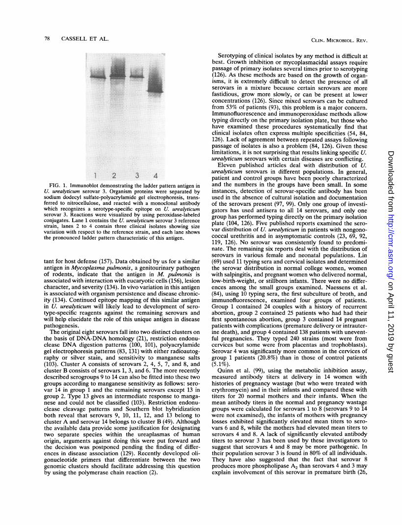
Previously (19, 54, 157), we have shown that immunoblot-ting with rabbit antisera reveals an intensely staining, com-plex "ladder" pattern consisting of a series of proteins withmolecular weight heterogeneity when some serovars, sero-vars 1, 2, 3, 5, and 6, are reacted with homologous antisera.The same ladder pattern also indicates a certain degree ofrelatedness between some members of the two biotypes ofureaplasmas (between serovars 2 and 5 and between sero-vars 3 and 14). Using monoclonal antibodies, we havedemonstrated the occurrence of a ladder antigen also onserotypes 8 and 10 (Fig. 1). Thus, the ladder antigen has nowbeen identified on 8 of the 14 serovars. Monoclonal antibod-ies to these ladder antigens have been produced. Detailedanalysis of this antigen in serovars 3, 8, and 10 indicates thatthis antigen (i) is species specific; (ii) contains both serotype-specific and a cross-reactive epitope(s); (iii) is produced notonly in vitro but also in vivo; (iv) undergoes a high rate ofstructural variation in vitro; (v) is present on invasiveureaplasma isolates, i.e., those from placenta, lung, andCSF; and (vi) is one of the most predominant proteinantigens recognized during infections in humans (157). Fur-thermore, we have shown that monoclonal antibodies to thisantigen can inhibit the growth of these organisms in vitro,which suggests that antibody to this antigen may be impor-
1 2 3 4FIG. 1. Immunoblot demonstrating the ladder pattern antigen in
U. urealyticum serovar 3. Organism proteins were separated bysodium dodecyl sulfate-polyacrylamide gel electrophoresis, trans-ferred to nitrocellulose, and reacted with a monoclonal antibodywhich recognizes a serotype-specific epitope on U. urealyticumserovar 3. Reactions were visualized by using peroxidase-labeledconjugates. Lane 1 contains the U. urealyticum serovar 3 referencestrain, lanes 2 to 4 contain three clinical isolates showing sizevariation with respect to the reference strain, and each lane showsthe pronounced ladder pattern characteristic of this antigen.
tant for host defense (157). Data obtained by us for a similarantigen in Mycoplasma pulmonis, a genitourinary pathogenof rodents, indicate that the antigen in M. pulmonis isassociated with interaction with eucaryotic cells (156), lesioncharacter, and severity (134). In vivo variation in this antigenis associated with organism persistence and disease chronic-ity (134). Continued epitope mapping of this similar antigenin U. urealyticum will likely lead to development of sero-
type-specific reagents against the remaining serovars andwill help elucidate the role of this unique antigen in diseasepathogenesis.The original eight serovars fall into two distinct clusters on
the basis of DNA-DNA homology (21), restriction endonu-clease DNA digestion patterns (100, 101), polyacrylamidegel electrophoresis patterns (83, 131) with either radioautog-raphy or silver stain, and sensitivity to manganese salts(103). Cluster A consists of serovars 2, 4, 5, 7, and 8, andcluster B consists of serovars 1, 3, and 6. The more recentlydescribed serogroups 9 to 14 can also be fitted into these twogroups according to manganese sensitivity as follows: sero-var 14 in group 1 and the remaining serovars except 13 ingroup 2. Type 13 gives an intermediate response to manga-
nese and could not be classified (103). Restriction endonu-clease cleavage patterns and Southern blot hybridizationboth reveal that serovars 9, 10, 11, 12, and 13 belong tocluster A and serovar 14 belongs to cluster B (49). Althoughthe available data provide some justification for designatingtwo separate species within the ureaplasmas of humanorigin, arguments against doing this were put forward andthe decision was postponed pending the finding of differ-ences in disease association (129). Recently developed oli-gonucleotide primers that differentiate between the twogenomic clusters should facilitate addressing this questionby using the polymerase chain reaction (2).
Serotyping of clinical isolates by any method is difficult atbest. Growth inhibition or mycoplasmacidal assays requirepassage of primary isolates several times prior to serotyping(126). As these methods are based on the growth of organ-isms, it is extremely difficult to detect the presence of allserovars in a mixture because certain serovars are morefastidious, grow more slowly, or can be present at lowerconcentrations (126). Since mixed serovars can be culturedfrom 53% of patients (93), this problem is a major concern.Immunofluorescence and immunoperoxidase methods allowtyping directly on the primary isolation plate, but those whohave examined these procedures systematically find thatclinical isolates often express multiple specificities (54, 84,126). Lack of agreement between repeated assays followingpassage of isolates is also a problem (84, 126). Given theselimitations, it is not surprising that results linking specific U.urealyticum serovars with certain diseases are conflicting.
Eleven published articles deal with distribution of U.urealyticum serovars in different populations. In general,patient and control groups have been poorly characterizedand the numbers in the groups have been small. In someinstances, detection of serovar-specific antibody has beenused in the absence of cultural isolation and documentationof the serovars present (97, 99). Only one group of investi-gators has used antisera to all 14 serovars, and only onegroup has performed typing directly on the primary isolationplate (104, 126). Five published reports examined the sero-var distribution of U. urealyticum in patients with nongono-coccal urethritis and in asymptomatic controls (23, 69, 92,119, 126). No serovar was consistently found to predomi-nate. The remaining six reports deal with the distribution ofserovars in various female and neonatal populations. Lin(69) used 11 typing sera and cervical isolates and determinedthe serovar distribution in normal college women, womenwith salpingitis, and pregnant women who delivered normal,low-birth-weight, or stillborn infants. There were no differ-ences among the small groups examined. Naessens et al.(84), using 10 typing sera, the first subculture of broth, andimmunofluorescence, examined four groups of patients.Group 1 contained 24 couples with a history of recurrentabortion, group 2 contained 25 patients who had had theirfirst spontaneous abortion, group 3 contained 14 pregnantpatients with complications (premature delivery or intrauter-ine death), and group 4 contained 138 patients with unevent-ful pregnancies. They typed 240 strains (most were fromcervices but some were from placentas and trophoblasts).Serovar 4 was significantly more common in the cervices ofgroup 1 patients (20.8%) than in those of control patients(5.1%).Quinn et al. (99), using the metabolic inhibition assay,
measured antibody titers at delivery in 14 women withhistories of pregnancy wastage (but who were treated witherythromycin) and in their infants and compared these withtiters for 20 normal mothers and their infants. When themean antibody titers in the normal and pregnancy wastagegroups were calculated for serovars 1 to 8 (serovars 9 to 14were not examined), the infants of mothers with pregnancylosses exhibited significantly elevated mean titers to sero-vars 6 and 8, while the mothers had elevated mean titers toserovars 4 and 8. A lack of significantly elevated antibodytiters to serovar 3 has been used by these investigators tosuggest that serovars 4 and 8 may be more pathogenic. Intheir population serovar 3 is found in 80% of all individuals.They have also suggested that the fact that serovar 8produces more phospholipase A2 than serovars 4 and 3 mayexplain involvement of this serovar in premature birth (26,
27). However, in the study by Naessens et al. (84), therewere no isolations of serovar 8 from cervices or placentas ofwomen with intrauterine fetal death or premature delivery.Even after adjusting for age, mean titers of antibody to U.
urealyticum are significantly lower in women who are expe-riencing their first pregnancy than in those who have beenpregnant before (70). The study by Quinn et al. (99) isdifficult to interpret because no details concerning the parityof the control mothers are given. No information is givenconcerning the duration of membrane rupture in the twogroups (i.e., ureaplasmal amniotic fluid infection resultingfrom membrane rupture could explain differences in anti-body levels). Furthermore, since the U. urealyticum isolatesfrom the two groups were not serotyped, the apparentselected increase in antibodies to serovars 4 and 8 cannot beevaluated. Other investigators found that approximately40% of women demonstrated a significant rise in antibodytiter to one or more serovars of U. urealyticum during anapparently normal pregnancy (70).
In a somewhat similar study, Quinn et al. (97) evaluatedthe serologic responses (but not the culture status) of pre-term infants with respiratory distress and compared themwith the responses and status of normal term infants. Neo-nates with respiratory disease had significantly elevatedmean titers to serovars 4, 7, and 8 compared with mean titersof normal neonates and slight but not significant elevationsof titers to serovars 3 and 6. When the respiratory diseasecases were assessed according to whether the infant sur-vived or died, the mean titer to serovar 3 was slightlyelevated in all groups. With serovars 4 and 8, the mean titerswere significantly higher among neonates who died thanamong the survivors. For serovar 5, a significant elevationoccurred only among the survivors. The difficulty in inter-preting these results is that sera were collected from infantswith respiratory disease from 0 to 20 days after birth andfrom the control term infants at delivery only. Also, thefrequent transfusions received by premature infants werenot taken into account. This can affect immunoglobulinlevels (25).Even though there are weaknesses in both studies, it is
interesting that Quinn et al. (97, 99) by serology only andNaessens et al. (84) by culture only obtained data thatsuggest that serovar 4 may be more often isolated fromwomen with recurrent abortion. It is also of interest thatQuinn et al. (93, 97, 99) found significantly elevated antibodyto serovars 4 and 8 more often in infants with respiratorydisease and in infants who died than in those who survived.Some investigators have found serovar 4 more commonly inmales with urethritis (23, 119) and in the cervices of femaleswith pelvic inflammatory disease (22). Of eight serovarsexamined for their abilities to reduce the penetration ofzona-free hamster eggs by human spermatozoa, serovar 4was the only one to produce a significant effect (6). It is notclear whether all serovars were compared within a singleexperiment, to what degree the assay was reproducible, howmany times the experiments were repeated, or how the datawere analyzed. Regarding the other studies linking serovar 4to urethritis and spontaneous abortion, other investigatorswho used antisera to all 14 serovars, identified isolatesdirectly on the primary isolation plate, and studied largernumbers of patients (104, 126) have not confirmed theseresults.From all serotyping studies to date, there appears to be
only one consistent finding. Serovar 3 is the most commonserovar isolated from females regardless of the patientpopulation and geographic location. The data were obtained
by six different investigators from four different countriesand were derived by using at least three different typingmethods (23, 56, 69, 97, 99, 104, 119). The data implicatingserovars 4 and 8 in perinatal morbidity and mortality are byno means conclusive, and further studies are warranted,considering that serovars 4 and 8 are two of the serovarsleast commonly recovered by culture in all normal femaleand infant populations examined to date (23, 56, 69, 97, 99,104, 119). However, these studies should by no means beundertaken until serovar-specific reagents and improvedmethods of antibody detection are available. Using mono-clonal antibodies to the ladder antigen discussed above, wehave developed immunoblotting methods for serotyping ofclinical isolates (162). We previously isolated U. urealyticumfrom the CSF of 13 of 418 newborn infants; additionalbloodstream isolates were obtained from the same popula-tion (147, 149). Ten of the 13 CSF isolates and 3 bloodstreamisolates were available for serotyping. By the use of sero-type-specific reagents, including monoclonal antibodies,70% of the CSF isolates were identifiable as serotype 1, 3, 6,8, or 10; i.e., they represented 5 of the 14 establishedserotypes and both presently defined genomic clusters. Oneof the bloodstream isolates was identified as serotype 3.These data support the hypothesis that the property ofinvasiveness for ureaplasmas is likely not limited to one or afew particular serotypes among the 14 established serovars.Additionally, this study showed that, even in isolates of thesame serotype from different patients and in isolates fromdifferent body sites within the same patient, there can be sizevariation in the serotype antigens expressed (Fig. 1). There-fore, it appears that many serotypes are invasive and thatperhaps antigen variability and host factors may be moreimportant determinants for ureaplasma infections than dif-ferent serotypes per se.
ROLE OF ANTIBODY IN DEFENSEAGAINST UREAPLASMAS
Available evidence indicates that identification of specificantibody to U. urealyticum may be useful in detecting anactive infectious process. Specific antibody to U. urealyti-cum can be detected by complement fixation, growth inhi-bition, metabolic inhibition, and mycoplasmacidal assays(121). However, these methods are limited by technicaldifficulties and lack the ability to distinguish the responsesamong immunoglobulin classes. The latter capability may beof utmost importance in ultimately determining the etiologicsignificance of U. urealyticum. We have developed anenzyme-linked immunosorbent assay (ELISA) based on awhole organism lysate as the antigen (4). The assay agreesexceptionally well with results obtained by metabolic inhi-bition assay (overall agreements of 82 and 95% for acute- andconvalescent-phase sera, respectively). Using this assay, wecharacterized the antibody response in a well-defined popu-lation of males with nongonococcal urethritis for whomquantitative cultural results were available over time. Serumantibody levels in nongonococcal urethritis patients weresignificantly higher than the normal serum standard for theIgG, IgM, and IgA classes. Additionally, the magnitude ofchange between acute- and convalescent-phase sera wasgreater for nongonococcal urethritis patients than for normalasymptomatic ureaplasma-positive male controls. A signifi-cant change in antibody levels of one or more antibodyclasses was detected by ELISA in 12 of 18 nongonococcalurethritis patients. Ten of the 12 individuals had a change inthe IgM class, which suggests an active infectious process.
We have used the ELISA to analyze sera from variousfemale patient populations. Whereas the mere presence orabsence of antibody, especially IgA, agrees well with cervi-covaginal isolation of U. urealyticum (4), changes in anti-body titer do not necessarily agree with cultural isolation ofthe organism from invasive sites (17). This may be becauseour ELISA failed to detect serovar-specific antibody in-creases (54). Two papers purport to demonstrate ureaplasmaserovar-specific ELISAs (140, 160). Both assays were testedonly with rabbit antisera, and both showed considerablecross-reactions which could be diluted out. However, care-ful examination of the data suggests that neither assay wouldprove clinically useful even if human serum reacted in thesame fashion as rabbit antiserum. Considerable cross-reac-tions remained, despite serum dilutions of up to 1:1,000.Furthermore, a serovar-specific antibody response could notbe demonstrated with sera from humans presumably ex-posed to only a single serovar (54).The unique susceptibility of hypogammaglobulinemic pa-
tients to chronic arthritis due to U. urealyticum (138) suggeststhat antibody is important for protection against invasivedisease caused by this organism. Increased susceptibility ofinfants of <30 weeks of gestational age to U. urealyticum-induced respiratory disease may be related to their hypogam-maglobulinemia (13, 16).Study of a hypogammaglobulinemic patient with arthritis
provides evidence that serovar-specific antibody may berequired for protection against invasive disease. A non-serovar-specific ELISA revealed high levels of ureaplasma-specific antibodies in the serum of the patient as well as intwo serum donors who served as sources of immunoglobulinreplacement during the period of joint infection. However,by the serovar-specific metabolic inhibition assay, no anti-bodies to serovar 4, which was isolated from the joint andsubcutaneous abscesses, were detected (142). In anotherhypogammaglobulinemic patient with chronic arthritis dueto an antibiotic-resistant strain of U. urealyticum, treatmentwith commercial immunoglobulin (Sandoz) was not effectivebut arthritis and abscesses resolved when the patient wasgiven infusions of goat hyperimmune ureaplasma serum(138, 158). Thus far, the serovars isolated from joints ofhypogammaglobulinemic patients include 2, 4, 5, 7, and 8and an untypeable strain. Generally, these are not the morecommon serovars isolated from the patient and controlpopulations that have been surveyed. Perhaps commercialimmunoglobulin preparations contain antibodies to the morecommon serovars such as 3 and 6.That detection of serovar-specific U. urealyticum re-
sponses is clinically significant has also been suggested byKass et al. (57). They showed that women who experienceda fourfold or greater rise in antibody titer to any single strainof U. urealyticum had a 30% rate of low-birth-weight infantswhereas women who did not experience such an increase inantibody titer had a 39% rate. Correspondingly, the birthweights in the two groups differed by 230 g. In mostinstances of significant increases in antibody titer, the titerincrease was specific for a serologic type other than those forwhich the women had preexistent antibody and the type-specific titer increased from undetectable levels. The datareported by Quinn et al. (96, 97, 99), discussed above, alsosuggest that selective antibody to certain serovars increasesin women with pregnancy wastage and in infants withrespiratory disease compared with control patients. Studiesby Gallo et al. (41) suggest that the presence of U. urealyti-cum-specific IgM antibody in infants is predictive of disease.Taken together, the available evidence suggests but does
not prove that immunity to U. urealyticum invasive infectionis type specific. The mechanism of protection afforded byantibody seems to be mediated by metabolic inhibition of theorganism and not by opsonization (138, 158). U. urealyticumbinds spontaneously to neutrophils and also directly acti-vates the first component of complement.
DIAGNOSTIC AND THERAPEUTIC CONSIDERATIONS
There are insufficient data to make specific recommenda-tions related to indications for diagnosis and treattnent of U.urealyticum infection of the chorioamnion. However, it isobvious from available data that routine culture of the cervixand/or vagina is not indicated. Culture of amniotic fluid alone(15) is unreliable for detection of chorioamnion infection.Only half of those with ureaplasma-positive cultures of thechorioamnion will also have positive culture of the amnioticfluid (15). The fact that analysis by the more sensitivetechnique of polymerase chain reaction also indicates amuch lower frequency of infection in amniotic fluid than inthe chorioamnion confirms that reliance on analysis ofamniotic fluid will lead to underdiagnosis of chorioamnioninfection and hence chorioamnionitis (15). There is no infor-mation upon which to base a rational therapy of chorioam-nion infection.
Recognition that U. urealyticum indeed causes severedisease in newborn infants, including septicemia, meningitis,and pneumonia, is sufficient justification for treatment inmost instances when the presence of ureaplasmas is docu-mented in normally sterile sites such as blood, CSF, pleuralfluid, or endotracheal aspirates in infants who weigh <1,250g. Techniques for specimen collection, cultivation, identifi-cation, and drug susceptibility testing and therapeutic con-
siderations have recently been reviewed elsewhere (10, 18,120, 121, 145, 148) so only the more important points will bediscussed here.
Cultivation
Specimen collection. Liquid specimens, including blood,amniotic fluid, CSF, urine, pleural fluid, and tracheobron-chial secretions, are acceptable for ureaplasmal culture. Inaddition, chorioamnion or any tissue collected from a biopsyor an autopsy can be submitted for culture if there is reasonto suspect the presence of ureaplasmas in newborn infants.Ureaplasmas are extremely susceptible to adverse envi-
ronmental conditions, especially drying, osmotic changes,and toxic metabolites. Particular care must be taken toensure that specimens are not subjected to extreme environ-mental fluctuations. Whenever possible, specific ureaplas-mal transport medium such as Shepard's 10B broth (119,120) should be provided for direct inoculation of clinicalspecimens at the time they are collected. The medium shouldthen be sent directly to the laboratory and processed imme-diately. If specimens are allowed to sit at room temperatureand are not inoculated into appropriate media, significantloss of mycoplasmal viability or overgrowth of endogenousbacteria is to be expected.Body fluids should be inoculated in an approximately 1:10
ratio into the transport medium (usually 0.1 ml of fluid per0.9 ml of broth), but it is also desirable that some uninocu-lated material be sent to the laboratory. Swabs should alsobe inoculated into suitable transport medium. Only calciumalginate-, dacron-, or polyester-tipped swabs with plastic or
wire shafts should be utilized for sampling of mucosalsurfaces, and the swab must always be extracted from the
specimen. Blood should be collected free of anticoagulantsand immediately inoculated into transport medium in a 1:5 to1:10 ratio. Inoculate as much blood as possible, preferably 5to 10 ml from adults, although smaller volumes from infantsare acceptable. Specimens should be kept refrigerated at 4°Cand protected from drying in a sealed container until theycan be transported to the laboratory. If specimens arecollected in a facility that does not have immediate access toureaplasmal broth for transport, a satisfactory alternative is2SP medium (10% heat-inactivated fetal calf serum; 0.2 Msucrose in 0.02 M phosphate buffer, pH 7.2). If transport isnot possible within 6 to 12 h after collection, the specimen intransport medium should be stored at -70°C and shippedfrozen. Ureaplasmas are stable for long periods when keptfrozen at -70°C in a protein-containing support mediumsuch as lOB. Storage at -20°C is less reliable and subject tosignificant loss in titer in a relatively short time.The original specimen from all swab cultures should be
thoroughly mixed on a Vortex mixer. Following mixing,specimens for ureaplasmal culture should always be seriallydiluted in broth to at least iO-', preferably 10-', and analiquot of the original sample and of each dilution should beinoculated onto A8 agar (120). All dilutions (10-1 to 10-5) ofthe broth should be incubated. The remainder of the originalspecimen should be frozen (-70°C) for future confirmation.Dilution is necessary to overcome potential inhibitory sub-stances or metabolites, including antibiotics, which may bepresent in the body fluid or tissue (119, 136) and to facilitatequantitative estimation of the numbers of organisms present.Greater isolation sensitivity may be obtained by centrifugingbody fluids such as urine (600 x g) to deposit epithelial andother cells and by performing serial dilutions on an aliquotfrom the sediment. Tissues are preferably minced ratherthan ground for cultivation to circumvent potential growthinhibitors that are more likely to be released with grinding(136).Growth and identification. Ureaplasmas are fastidious and
demanding in their requirements for special media. For thisreason alone, many laboratories choose not to culture my-coplasmas. Although commercially prepared media are nowon the market, they have not been thoroughly evaluated in aclinical setting. Growth media are usually enriched withanimal serum (fetal calf or horse), peptones, yeast extract,and metabolic substrates, including urea. Shepard's lOBbroth and A8 agar are the most widely used media (119, 120),although others are also satisfactory (119, 120).The relatively rapid growth rate of U. urealyticum makes
identification of most positive cultures possible within 1 to 5days. Broth cultures should be incubated at 37°C underatmospheric conditions, and agar plates should be incubatedunder 5% CO2. Ureaplasmas are susceptible to a rapid, steepdeath phase in culture which is likely because of a combina-tion of urea depletion and elevated pH due to urease activity.Ureaplasma broth cultures should therefore be monitoredvery closely (two to three times daily). The presence ofgrowth in lOB medium is suggested by an alkaline shift dueto urease activity of ureaplasmas or arginine hydrolysis byM. hominis, causing the phenol red indicator to turn fromyellow to pink. Ureaplasmas produce no or minimal turbid-ity in broth. Broth cultures demonstrating color changeshould be subcultured to broth and agar just as the pHindicator begins to turn. This combination of broth to agarinoculation technique is the most sensitive for recovery of allmycoplasmas. Positive broth cultures should be frozen(-70°C) immediately after being subcultured for future ref-erence, e.g., for identification and antibiotic susceptibility
testing. Plates should be evaluated microscopically forgrowth every 1 to 3 days and incubated for 7 to 10 days priorto being called negative. Positive agar cultures can beexpanded by transfer of an agar plug to broth.
Quality control of culture media. Ureaplasmas are verysensitive to inhibitors present in some lots of horse serum,yeast extract, and even mycoplasma medium base. It is notuncommon for the standard media in a laboratory to betemporarily insufficient for cultivation of ureaplasmas. Rig-orous quality control should be performed on media ob-tained commercially or prepared in the laboratory. Organ-isms to be used for quality control should be type strain (all14 serotypes) and low-passage clinical isolates, grown inlarge volumes, and stored frozen in small aliquots at -70°C.Serial dilutions of the thawed stock strains should be made,and the numbers of organisms growing on the test batch ofmedium should be quantitated. If the number of CFU onsolid medium is less than 90% of that on the medium fromthe previous batch, the test should be repeated. If it is stillless than 90%, the new medium should be consideredsubstandard and discarded. For broth medium, a differencegreater than 10 to 100 color-changing units per ml (that is, 1to 2 10-fold dilutions) between the control and test liquidmedium should be considered unacceptable. Each batch ofcomplete medium, both agar and broth, should be tested forsterility by incubation of an aliquot of broth and an agar plateat 37°C for at least 24 h. Finally, uninoculated tubes of thecomplete medium should be incubated with each series ofpatient specimens. Observation of mycoplasma colonies anddetermination of colonial morphology require the aid of astereomicroscope. U. urealyticum colonies appear brown orblack on A8 agar because of urease production in thepresence of the CaCl2 indicator.
Antibiotic susceptibility testing. Either the microdilution orthe agar dilution technique can be employed for testingantibiotic susceptibilities (59, 116, 148). The broth methodinvolves serially diluting antibiotics in liquid media in a96-well microtiter plate, adding known concentrations oforganisms, incubating, and observing for growth. The lowestdilution of antibiotic in which there is no evidence oforganism growth is designated the MIC. The agar dilutiontechnique involves incorporating multiple concentrations ofantibiotics directly into agar media on which organisms areinoculated. Plates are examined following incubation for thepresence of colonies, and a MIC is determined in the samemanner as for broth dilution. Each method has relativeadvantages and disadvantages that have been reviewed byWaites et al. (148) in a comparative study utilizing bothmethods for testing susceptibility of U. urealyticum toerythromycin and tetracycline. Agar dilution typically yields1- to 2-dilution-higher MICs than broth dilution methods.Unfortunately, there are no universally applied standards forsusceptibility testing; therefore, results from different labo-ratories may not be truly comparable. This lack of standard-ization has led to confusion about the susceptibility patterns,prevalence, and degree of drug resistance among clinicalisolates, particularly among ureaplasmas. Regardless of thetechnique employed, it is extremely important to have aconsistent time point for reading results, since the endpointmay shift over time, and to inoculate the proper number oforganisms into the test system. The importance of utilizingstandardized inocula has been well documented, particularlywith ureaplasmas (55).The broth dilution method is the most widely used. Pure
cultures to be tested should be passaged in SP-4 or 10Bbroth, aliquoted, and frozen (-70°C), and the color-changing
units or CFU for the frozen stock should be determinedsubsequently. On the day the assay is to be performed, thespecimen should be thawed, diluted in fresh medium to yield1,000 to 10,000 color-changing units or CFU/0.2 ml, andincubated (37°C) for 2 h prior to assay to allow the organismto begin active growth. A final color-changing unit countshould be determined at this time. If results indicate that thenumber of organisms used in the assay falls outside theacceptable range of 1,000 to 10,000 per microtiter well, theassay must be considered invalid.Mycoplasma reference strains for which the MICs of the
antibiotics being tested are known should be routinelyincluded for evaluation of the suitability of the antibioticpreparations being tested and to help control for any extra-neous effects of pH or medium components. When a newdrug is to be tested for antimycoplasmal activity, Staphylo-coccus aureus (ATCC 25923) and Enterococcus faecalis(ATCC 29212) or other appropriate bacterial referencestrains should be tested according to guidelines from theNational Committee for Clinical Laboratory Standards, us-ing SP-4 and 10B broth, and results should be compared withresults obtained using Mueller-Hinton broth to determinepotential drug-mycoplasma medium interactions.Each drug prepared according to guidelines from the
National Committee for Clinical Laboratory Standardsshould be tested over a range of dilutions from 0.008 to 256,ug/ml. A drug control, positive organism control, and nega-tive broth control should be included with each organismtested.
After inoculation, the microtiter plates should be sealedwith acetate tape to prevent the release of ammonia contam-ination from a positive well to an adjacent negative well andalso to prevent evaporation during long-term incubation.Plates should be incubated at 37°C, and results should bedetermined as soon as the well containing the control organ-ism is positive; for U. urealyticum, this could be as early as16 h. The initial MIC is defined as the lowest dilution ofantibiotic in which metabolism of the organism is inhibited asevidenced by lack of color change in the medium at the timethe control organism well first shows color change. The finalMIC is defined as the lowest dilution of antibiotic in whichno further color change occurs at readings on two consecu-tive days after prolonged incubation.To ensure reliable results, susceptibility testing should be
performed in duplicate. Ideally, tests on those isolates foundto be resistant should be repeated.
Therapy
Routine drug susceptibility testing of clinical isolatesremains largely the province of a relatively small number ofreference laboratories because of cost, time, and laborfactors. This lack of diagnostic facilities in many areascoupled with the potentially serious nature of some of thesystemic ureaplasmal infections in neonates makes empirictreatment necessary in many instances until proper microbi-ologic diagnosis and antimicrobial susceptibility informationcan be obtained.
Since ureaplasmas lack peptidoglycan, they are not af-fected by beta-lactam antibiotics. They are not susceptible tosulfonamides or trimethoprim because they do not synthe-size folic acid. However, they are generally susceptible tocertain antibiotics that interfere with protein synthesis, suchas tetracyclines and macrolides, but usually not to lincosa-mides except in high concentrations. Some isolates may besusceptible to streptomycin or other aminoglycosides but
not predictably so. Some of the newer investigational qui-nolones are active in vitro and in vivo against U. urealyticum(145), but their roles as therapeutic agents have not beenestablished in pediatric infections because of potential ef-fects on cartilage development.Aminoglycosides, which are commonly used against
gram-negative infections in neonates, do not appear to beeffective against U. urealyticum. Infants at the University ofAlabama Hospital who have previously received >3 days oftreatment with intravenous gentamicin for suspected sepsishave frequently had ureaplasmas subsequently isolated fromthe CSF, the bloodstream, or the lower respiratory tract(145a).Chloramphenicol has potential bone marrow toxicity. Its
clinical use has been relegated primarily to that of a second-line treatment for enteric bacillus infections unresponsive toaminoglycosides. Neither the clinical nor the microbiologicefficacy of chloramphenicol against ureaplasmal infectionshas been assessed in any population, although its in vitroactivity against U. urealyticum was favorable in a recentstudy (145). Data from the studies of Waites et al. (145)suggest that clindamycin should not be used empiricallyagainst U. urealyticum, but neither should it be used rou-tinely as a selective agent to isolate U. urealyticum frommixed cultures, since some strains are apparently suscepti-ble at relatively low concentrations.
In 1974, Ford and Smith (37) published the first report oftetracycline resistance to U. urealyticum. In 1978, Evansand Taylor-Robinson (36) reported the incidence of tetracy-cline resistance among genitourinary ureaplasmal isolates tobe approximately 10%. Since that time, a number of addi-tional studies have reported variable resistance rates as highas 42%. Roberts and Kenny (102) increased the understand-ing of tetracycline resistance in U. urealyticum by demon-strating that resistant strains contain DNA sequences homol-ogous to the streptococcal determinant TetM whichmediates resistance at the level of ribosomal protein synthe-SiS.
Macrolide resistance in U. urealyticum has not beenstudied nearly as much as tetracycline resistance. High-levelerythromycin resistance (>100 ,ug/ml) has been known tooccur but is apparently relatively uncommon when appro-priate quantitative techniques to determine MICs are em-ployed (89). Palu et al. (89) speculated that reduced bindingto ribosomes may be the most common means by whichmacrolide resistance arises in U. urealyticum since macro-
lide uptake into the cell depends on passive diffusion.While it is now clear that antimicrobial resistance in U.
urealyticum is a definite laboratory as well as clinical phe-nomenon, the true incidence of resistance to drugs such as
tetracyclines and erythromycin is really unknown. This lackof knowledge is due to several factors, such as small samplesizes in many studies, samples often collected in a nonran-
dom manner without regard for prior exposure to antibiotics,differences in nonstandardized methods, interpretation ofendpoints, samples collected from different populations fromwidely varying geographic and social strata, and the fact thatthe actual resistance patterns may change over time.
In a recent survey by Waites et al. (145), 43 isolates of U.urealyticum obtained from the lower respiratory tracts ofneonates were tested against chloramphenicol, ciprofloxa-cin, clindamycin, erythromycin, doxycycline, and gentami-cin by a broth microdilution technique in lOB broth. In vitroresistance was observed in one or more strains for each ofthe drugs tested except erythromycin (MIC range = 0.125 to4,ug/ml; MIC for 90% of strains = 2,ug/ml). MICs of the
remaining five antibiotics were 2 ,ug/ml for doxycycline, 8,ug/ml for chloramphenicol, 8 ,g/ml for ciprofloxacin, 16,g/ml for clindamycin, and 32 ,ug/ml for gentamicin.Among the limited options, for 90% of strains erythromy-
cin appears to be the most promising currently availableantimicrobial agent for use in neonatal ureaplasmal infec-tions. Because of its toxicity, doxycycline, though activeagainst most strains of U. urealyticum, is not a realistictherapeutic alternative except for ill infants from whom U.urealyticum is isolated from the CSF. Finding only 7%doxycycline resistance in neonatal ureaplasmal isolates,despite the small sample size, is encouraging (145).Making specific recommendations for treating ureaplasmal
infections is particularly difficult in view of the fact that thespectrum of ureaplasmal disease in neonates has not beenfully described. Therefore, not only is choice among thelimited drug options for treating ureaplasmal infections con-troversial, but also the actual indications concerning condi-tions under which treatment should be offered are debatable.Ureaplasmas can frequently be isolated from the upperrespiratory tracts of neonates, but there is no evidence thatcultures should be obtained routinely in the absence ofsymptomatic disease. Neonates clinically ill with pneumoni-tis or showing signs of CNS disease, particularly progressivehydrocephalus with or without CSF pleocytosis, for whombacterial cultures are negative or in whom there is noimprovement with antibiotic therapy warrant cultures for U.urealyticum and treatment if cultures are positive. Thehigher levels of erythromycin in serum achievable by intra-venous administration, with good penetration into tissuesincluding lung tissue, make it likely that the MICs for moststrains may actually be exceeded in vivo when the drug isgiven parenterally (147, 149, 155).Empiric treatment should be initiated in an unstable infant
with a positive culture for U. urealyticum from CSF, tra-chea, pleural fluid, or blood because of the time (severaldays) required to obtain antimicrobial susceptibility data.Although CSF is normally considered a sterile body fluid inwhich any bacterial infection can be potentially life-threat-ening, some ureaplasma-caused CSF infections in termneonates may be asymptomatic, without a CSF inflamma-tory response, and self-limited (147, 149). However, thismay not be the case in low-birth-weight, preterm neonates,who without agressive intervention typically have difficultyin localizing and combatting systemic bacterial infections.Because of poor CSF penetration by erythromycin, tetracy-clines are probably the most effective drugs available forureaplasmal infections of the CSF. It may be prudent toobserve stable, asymptomatic infants and to document per-sistent infection through follow-up CSF cultures beforetreatment with these potentially toxic antibiotics is initiated.
Several mechanisms may be operative in the developmentof CLD in neonates with U. urealyticum present in the lowerrespiratory tract. This condition is quite distinct from themore acute entity of pneumonia which may also be causedby U. urealyticum in an undetermined number of cases.These organisms may be contributory causes of CLD, initi-ating an inflammatory response, promoting the productionand release of toxic oxygen species, interfering with pulmo-nary defenses, or promoting injury by other organisms.Therefore, the appropriate treatment may involve eradica-tion of the organisms by antibiotics, modulation of theimmune response by steroids, administration of antioxi-dants, or even surfactant therapy. Until the mechanism ofdisease production is known, therapy will need to be basedon the judgment of each individual clinician.
Since clinically useful antibiotics are only mycoplasma-static, not mycoplasmacidal, the immune status of the pre-mature infant might well be a crucial component in anysuccessful drug treatment. Experience in treatment ofchronic ureaplasmal and mycoplasmal arthritis in hypogam-maglobulinemic patients clearly suggests that this is the case(138, 142, 158). Though there are no established guidelines,limited clinical experience with neonates and other personswith systemic infections due to U. urealyticum suggests thata minimum of 10 to 14 days of treatment is best. Whether theintravenous or oral route with erythromycin, doxycycline,or alternatives is employed depends on the overall conditionof the patient and the nature of the infection being treated.Optimally, isolates from infants treated with antibiotics forureaplasmal infection should be tested for drug susceptibilityand the infants should be monitored for clinical response tothe chosen antibiotic. Follow-up cultures of the infected siteto document microbiologic efficacy of the drug are suggestedwhen clinical improvement does not occur.
Future studies are needed to assess the utility of antibiotics,especially erythromycin, in treating neonatal ureaplasmalinfections and should include pharmacokinetic investigationsaimed at determining peak levels, half-life, tolerability, safety,and microbiologic efficacy of intravenous preparations. Moreinformation is needed concerning the immune status of thepremature infant and the relationship to successful therapy.Such studies are necessary before any definitive recommen-dations regarding treatment of systemic neonatal ureaplasmalinfections are made and before any controlled trials aimed atclarifying the etiologic significance of this organism in patho-logic conditions in this population, particularly the importantquestion of its association with CLD of prematurity, are done.
ACKNOWLEDGMENTS
Work summarized in this review was supported by grants HD20929 and Al 28279 from the National Institutes of Health to G.H.C.and by a grant from the American Lung Association to K.B.W. andG.H.C. D.T.C. was supported by training grant HL 07553 (toG.H.C.) and Physician-Scientist Award Kll HL 02109 from theNational Institutes of Health.
REFERENCES1. Berman, S. M., H. R. Harrison, W. T. Boyce, J. J. Haffner, M.
Lewis, and J. B. Arthur. 1987. Low birth weight, prematurity,and postpartum endometritis. JAMA 257:1189-1194.
2. Blanchard, A. 1990. Ureaplasma urealyticum urease genes;use of a UGA tryptophan codon. Mol. Microbiol. 4:669-676.
3. Bobitt, J. R., C. C. Hayslip, and J. D. Damato. 1981. Amnioticfluid infection as determined by transabdominal amniocentesisin patients with intact membranes in premature labor. Am. J.Obstet. Gynecol. 140:947-952.
4. Brown, M. B., D. Taylor-Robinson, M. C. Shepard, and G. H.Cassell. 1983. Measurement of antibody to Ureaplasma ure-alyticum by an enzyme-linked immunosorbent assay and de-tection of antibody response in patients with nongonococcalurethritis. J. Clin. Microbiol. 17:288-295.
5. Brus, F., W. M. Van Waande, C. Schoots, and S. B. Oetomo.1991. Fetal ureaplasmal pneumonia and sepsis in a newborninfant. Eur. J. Pediatr. 150:782-783.
6. Busolo, F., and R. Zanchetta. 1985. The effect of Mycoplasmahominis and Ureaplasma urealyticum on hamster egg in vitropenetration by human spermatozoa. Fertil. Steril. 43:110-114.
7. Carey, J. C., W. C. Blackwelder, R. P. Nugent, M. A. Matte-son, A. V. Rao, D. A. Eschenbach, M. L. F. Lee, P. J. Rettig,J. A. Regan, K. L. Feromanos, D. H. Maritn, J. G. Pastorek,R. S. Gibbs, K. A. Lipscomb, and the Vaginal Infections andPrematurity Group. 1991. Antepartum cultures for Ure-aplasma urealyticum are not useful in predicting pregnancyoutcome. Am. J. Obstet. Gynecol. 164:728-733.
8. Caspi, E., F. Solomon, R. Langer, and D. Sompolinsky. 1976.Isolation of mycoplasma from the placenta after cesareansection. Obstet. Gynecol. 48:682-684.
9. Cassell, G. H. 1986. Future considerations: maternal andneonatal aspects. Pediatr. Infect. Dis. J. 5:S341-S344.
10. Cassell, G. H., A. Blanchard, K. B. Waites, L. Duffy, and D.Crabb. Mycoplasmas. In B. J. Howard, J. Klaas II, S. J.Rubin, A. S. Weissfeld, and R. C. Tilton (ed.), Clinical andpathogenic microbiology, in press.
11. Cassell, G. H., W. A. Clyde, G. E. Kenny, W. M. McCormack,and D. Taylor-Robinson. 1986. Ureaplasmas of humans: withemphasis upon maternal and neonatal infections. Pediatr.Infect. Dis. J. 5:S221-S354.
12. Cassell, G. H., and B. C. Cole. 1981. Mycoplasmas as agents ofhuman disease. N. Engl. J. Med. 304:80-89.
13. Cassell, G. H., D. T. Crouse, K. B. Waites, P. T. Rudd, andJ. K. Davis. 1988. Does Ureaplasma urealyticum cause respi-ratory disease in the newborn? Pediatr. Infect. Dis. J. 7:535-541.
14. Cassell, G. H., R. 0. Davis, K. B. Waites, M. B. Brown, P. A.Marriott, S. Stagno, and J. K. Davis. 1983. Isolation of Myco-plasma hominis and Ureaplasma urealyticum from amnioticfluid at 16-20 weeks of gestation: potential effect on outcome ofpregnancy. Sex. Transm. Dis. 10:294-302.
15. Cassell, G. H., J. Hauth, G. Cutter, K. Waites, M. Mazur, W.Hamrick, K. Baldus, R. Sinsky, and B. Andrews. Ureaplasmaurealyticum chorioamnion infection and prematurity. Clin.Infect. Dis., in press.
16. Cassell, G. H., K. B. Waites, D. T. Crouse, P. T. Rudd, K. C.Canupp, S. Stagno, and G. R. Cutter. 1988. Association ofUreaplasma urealyticum infection of the lower respiratorytract with chronic lung disease and death in very low birthweight infants. Lancet ii:240-244.
17. Cassell, G. H., K. B. Waites, R. S. Gibbs, and J. K. Davis. 1986.Role of Ureaplasma urealyticum in amnionitis. Pediatr. Infect.Dis. J. 5:S247-S252.
18. Cassell, G. H., K. B. Waites, and D. Taylor-Robinson. 1990.Genital mycoplasmas, p. 7.2-7.15. In S. A. Morse, A. A.Moreland, and S. E. Thompson (ed.), Atlas of sexually trans-mitted diseases. Gower Medical Publishers, New York.
19. Cassell, G. H., H. L. Watson, D. K. Blalock, S. A. Horowitz,and L. B. Duffy. 1988. Protein antigens of the genital myco-plasmas. Rev. Infect. Dis. 10:S391-S398.
20. Cassell, G. H., J. B. Younger, M. Brown, R. E. Blackwell, J. K.Davis, B. S. Marriott, and S. Stagno. 1983. Microbiologic studyof infertile women at the time of diagnostic laparoscopy. N.Engl. J. Med. 308:502-505.
21. Christiansen, C., F. T. Black, and E. A. Freundt. 1981. Hy-bridization experiments with deoxyribonucleic acid from Ure-aplasma urealyticum serovars I to VIII. Int. J. Syst. Bacteriol.31:259-262.
21a.Cox, N. R., D. F. Kohn, and G. H. Cassell. Unpublished data.22. Cracea, E., D. Boetz, S. Constaninescu, and M. Georgescu-
23. Cracea, E., S. Constantinescu, and A. Lazar. 1985. Serotypesof Ureaplasma urealyticum isolated from patients with non-gonococcal urethritis and gonorrhoea and from asymptomaticcarriers. Sex. Transm. Dis. 12:219-223.
24. Crouse, D. T., G. H. Cassell, K. B. Waites, J. M. Foster, and G.Cassady. 1990. Hyperoxia potentiates Ureaplasma urealyti-cum pneumonia in newborn mice. Infect. Immun. 58:3487-3493.
25. Decsi, T., and M. Fekete. 1990. Early postnatal changes ofimmunoglobulin levels in very low birth weight (<1500 g)preterm infants. Acta Paediatr. Hung. 30:225-232.
26. Desilva, N. S., and P. A. Quinn. 1986. Endogenous activity ofphospholipases A and C in Ureaplasma urealyticum. J. Clin.Microbiol. 23:354-359.
27. Desilva, N. S., and P. A. Quinn. 1991. Localization of endoger-ous activity of phospholipases A and C in Ureaplasma ure-alyticum. J. Clin. Microbiol. 29:1498-1503.
28. Dillon, H. C., and B. M. Gary. 1987. Infection caused by groupB streptococci in perinatal, neonatal and early infancy, p. 1-12.In V. C. Kelly (ed.), Practice of pediatrics, vol. 23. Harper &Row, New York.
29. Dinsmoor, M. J., R. S. Ramamurthy, and R. S. Gibbs. 1989.Transmission of genital mycoplasmas from mother to neonatein women with prolonged membrane rupture. Pediatr. Infect.Dis. J. 8:483-487.
30. Dong, Y., P. J. St. Clair, I. Ramzy, K. S. Kagan-Hallet, andR. S. Gibbs. 1987. A microbiologic and clinical study ofplacental inflammation at term. Obstet. Gynecol. 70:175-182.
31. Driscoll, S. G. 1986. Chorioamnionitis: perinatal morbidity andmortality. Pediatr. Infect. Dis. 5:S273-S275.
32. Elder, H. A., B. A. G. Santa Maria, and S. Smithietal. 1971.The natural history of asymptomatic bacteruria during preg-nancy: the effect of tetracycline on the clinical course and theoutcome of pregnancy. Am. J. Obstet. Gynecol. 3:441-462.
33. Embree, J. E., V. W. Kraus, J. A. Embil, and S. MacDonald.1980. Placental infection with Mycoplasma hominis and Ure-aplasma urealyticum: clinical correlation. Obstet. Gynecol.56:475-481.
34. Eschenbach, D. A. 1989. Bacterial vaginosis. Obstet. Gynecol.Clin. North Am. 16:593.
35. Eschenbach, D. A., R. P. Nugent, A. V. Rao, M. F. Cotch, R. S.Gibbs, K. A. Lipscomb, D. H. Martin, J. G. Pastorek, P. J.Rettig, J. C. Carey, J. A. Regan, K. L. Geromanos, M. L. F.Lee, W. K. Poole, R. Edelman, and Vaginal Infections andPrematurity Study Group. 1991. A randomized placebo-con-trolled trial of erythromycin for the treatment of Ureaplasmaurealyticum to prevent premature delivery. Am. J. Obstet.Gynecol. 164:734-742.
36. Evans, R. T., and D. Taylor-Robinson. 1978. The incidence oftetracycline-resistant strains of Ureaplasma urealyticum. J.Antimicrob. Chemother. 4:57-63.
37. Ford, D. K., and J. R. Smith. 1974. Nonspecific urethritisassociated with a tetracycline-resistant T-mycoplasma. Br. J.Vener. Dis. 50:373-374.
38. Foulon, W., A. Naessens, M. Dewaele, S. Lauwers, and J. J.Amy. 1986. Chronic Ureaplasma urealyticum amnionitis asso-ciated with abruptio placentae. Obstet. Gynecol. 68:280.
39. Foy, H. M., G. E. Kenny, E. M. Levinsohn, and J. T. Grayston.1970. Acquisition of mycoplasmata and T-strains during in-fancy. J. Infect. Dis. 121:579-587.
40. Foy, H. M., G. E. Kenny, B. B. Wentworth, W. L. Johnson, andJ. T. Garson. 1970. Isolation of mycoplasma hominis,T-strains, and cytomegalovirus from the cervix of pregnantwomen. Am. J. Obstet. Gynecol. 106:635-643.
41. Gallo, D., K. W. Dupuis, N. J. Schmidt, and G. E. Kenny. 1983.Broadly reactive immunofluorescence test for measurement ofimmunoglobulin M and G antibodies to Ureaplasma urealyti-cum in infant and adult sera. J. Clin. Microbiol. 17:614-618.
42. Garland, S. M., and L. J. Murton. 1987. Neonatal meningitiscaused by Ureaplasma urealyticum. Pediatr. Infect. Dis. J.6:868-869.
43. Gravett, M. G. 1984. Causes of preterm delivery. Semin.Perinatol. 8:246-257.
44. Gravett, M. G., and D. A. Eschenbach. 1986. Possible role ofUreaplasma urealyticum in preterm premature rupture of fetalmembranes. Pediatr. Infect. Dis. 5:S253-S257.
45. Gravett, M. G., D. Hummel, D. A. Eschenbach, and K. K.Holmes. 1986. Preterm labor associated with subclinical amni-otic fluid infection and with bacterial vaginosis. Obstet. Gyne-col. 67:229-237.
46. Gribble, M. J., and A. W. Chow. 1982. Erythromycin. Med.Clin. North Am. 66:79-89.
47. Gruskay, J., M. C. Harris, A. T. Constarino, and S. Baumgart.1989. Neonatal staphylococcal epidermidis meningitis withunremarkable CSF examination results. Am. J. Dis. Child.143:580-582.
48. Hameed, C., N. Tejani, U. L. Verma, and F. Archbald. 1984.Silent chorioamnionitis as a cause of preterm labor refractoryto tocolytic therapy. Am. J. Obstet. Gynecol. 149:726-730.
49. Harasawa, R., K. Dybvig, H. Watson, and G. H. Cassell. 1991.
Two genomic clusters among 14 serovars of ureaplasma ure-alyticum. Syst. Appl. Microbiol. 14:393-396.
50. Harrison, H. R., E. R. Alexander, L. Weinstein, M. Lewis, M.Nash, and D. A. Sim. 1983. Cervical chlamydia trachomatis andmycoplasmal infections in pregnancy: epidemiology and out-comes. JAMA 250:1721-1727.
51. Harrison, H. R., and D. Phil. 1986. Cervical colonization withUreaplasma urealyticum and pregnancy outcome: prospectivestudies. Pediatr. Infect. Dis. J. 5:S266-S269.
52. Hillier, S. L., J. Martius, M. Krohn, N. Kiviat, K. K. Holmes,and D. A. Eschenbach. 1988. A case-control study of chorio-amnionic infection and histologic chorioamnionitis in prematu-rity. N. Engl. J. Med. 319:972-978.
53. Hogue, C. J. R., L. T. Strauss, J. W. Buehler, and J. C. Smith.1990. Overview of the neonatal infant mortality surveillance(NIMS) project. 39:148-151.
54. Horowitz, S. A., L. B. Duffy, B. Garrett, J. Stephens, J. K.Davis, and G. H. Cassell. 1986. Can group- and serovar-specificproteins be detected in Ureaplasma urealyticum? Pediatr.Infect. Dis. J. 5:S325-S331.
55. Jalil, N., C. Gilchrist, and D. Taylor-Robinson. 1989. Factorsinfluencing the in vitro sensitivity of Ureaplasma urealyticumto tetracyclines. J. Antimicrob. Chemother. 23:341.
56. Kanamoto, Y. 1985. Studies on genital mycoplasmas. III.Serological characterization of ureaplasmas isolated from preg-nant women and neonates. Hiroshima Univ. Med. J. 33:1129-1134.
57. Kass, E. H., J. S. Lin, and W. M. McCormack. 1986. Low birthweight and maternal colonization with genital mycoplasmas.Pediatr. Infect. Dis. J. 5:S279-S281.
58. Kass, E. H., W. M. McCormack, J. S. Lin, B. Rosner, and A.Munoz. 1981. Genital mycoplasmas as a cause of excesspremature delivery. Trans. Assoc. Am. Physicians 94:261-266.
59. Kenny, G. E., F. D. Cartwright, and M. C. Roberts. 1986. Agardilution method for determination of antibiotic susceptibility ofUreaplasma urealyticum. Pediatr. Infect. Dis. J. 5:S332-S334.
60. Klein, J. O., D. 0. Buckland, and M. Finland. 1969. Coloniza-tion of newborn infants by mycoplasmas. N. Engl. J. Med.280:1025-1030.
61. Klein, J. O., and S. M. Marcy. 1983. Bacterial sepsis andmeningitis, p. 679-735. In J. S. Remington and J. 0. Klein(ed.), Infectious diseases of the fetus and newborn infant. TheW. B. Saunders Co., Philadelphia.
62. Kundsin, R. B., S. G. Driscoll, R. R. Monson, C. Yeh, S. A.Biano, and W. D. Cochran. 1984. Association of Ureaplasmaurealyticum in the placenta with perinatal morbidity and mor-tality. N. Engl. J. Med. 310:941-945.
63. Lamont, R. F., D. Taylor-Robinson, M. Newman, J. Wiggles-worth, and M. G. Elder. 1986. Spontaneous early pretermlabour associated with abnormal genital bacterial colonization.Br. J. Obstet. Gynaecol. 93:804-810.
64. Lamont, R. F., D. Taylor-Robinson, J. S. Wigglesworth, P. M.Furr, R. T. Evans, and M. G. Elder. 1987. The role ofmycoplasmas, ureaplasmas, and chlamydiae in the genital tractof women presenting in spontaneous early preterm labor. J.Med. Microbiol. 24:253-257.
65. Lee, G. Y., and G. E. Kenny. 1987. Humoral immune responseto polypeptides of Ureaplasma urealyticum in women withpostpartum fever. J. Clin. Microbiol. 25:1841-1844.
66. Leigh, J., and T. J. Garite. 1986. Amniocentesis and themanagement of premature labor. Obstet. Gynecol. 67:500-506.
67. Likitnukul, S., H. Kusmiesz, J. D. Nelson, and G. H. Mc-Cracken. 1986. Role of genital mycoplasmas in young infantswith suspected sepsis. J. Pediatr. 109:971-974.
68. Likitnukul, S., J. D. Nelson, G. H. McCracken, and H.Kusmiesz. 1987. Rarity of genital mycoplasm infection in younginfants with aseptic meningitis. J. Pediatr. 110:998.
69. Lin, J. S. 1977. Ureaplasma urealyticum: serotyping and thehuman humoral response, p. 370-375. In D. Hobson and K. K.Holmes (ed.), Nongonococcal urethritis and related infections.American Society for Microbiology, Washington, D.C.
70. Lin, J. S., K. Radney, M. I. Kendrick, B. Rosner, and E. H.Kass. 1978. Serologic studies of human genital mycoplasmas:
distribution of titers of mycoplasmacidal antibody to Ure-aplasma urealyticum and Mycoplasma hominis in pregnantwomen. J. Infect. Dis. 137:266-273.
71. Lorian, V., and L. D. Sabath. 1970. Effect of pH on the activityof erythromycin against 500 isolates of gram-negative bacilli.Appl. Microbiol. 20:754-756.
72. Madan, E., M. P. Meyer, and A. J. Amortegui. 1988. Isolationof genital mycoplasmas and Chlamydia trachomatis in stillbornand neonatal autopsy material. Arch. Pathol. Lab. Med. 112:749-751.
73. Madan, E., M. P. Meyer, and A. J. Amortegui. 1989. Histologicmanifestations of perinatal genital mycoplasmal infection.Arch. Pathol. Lab. Med. 113:465-469.
74. Mardh, P. A. 1983. Mycoplasma hominis infections of thecentral nervous system in newborn infants. Sex. Transm. Dis.10:331-334.
75. Martius, J., and D. A. Eschenbach. 1990. The role of bacterialvaginosis as a cause of amniotic fluid infection, chorioamnio-nitis and prematurity-a review. Arch. Gynecol. Obstet. 247:1-13.
76. Martius, J., M. A. Krohn, S. L. Hillier, W. E. Stamm, K. K.Holmes, and D. A. Eschenbach. 1988. Relationships of vaginalLactobacillus species, cervical Chlamydia trachomatis, andbacterial vaginosis to preterm birth. Obstet. Gynecol. 71:89-95.
77. McCormack, W. M. 1986. Ureaplasma urealyticum: ecologicalniche and epidemiological considerations. Pediatr. Infect. Dis.J. 5:S232-S233.
78. McCormack, W. M., B. Rosner, S. Alpert, J. R. Evrard, V. A.Crockett, and S. H. Zinner. 1986. Vaginal colonization withMycoplasma hominis and Ureaplasma urealyticum. Sex.Transm. Dis. 134:67-70.
79. McCormack, W. M., B. Rosner, L. Yhu-Hsiung, A. Munoz, D.Charles, and E. H. Kass. 1987. Effect on birth weight oferythromycin treatment of pregnant women. Obstet. Gynecol.69:202-207.
80. McCormack, W. M., and D. Taylor-Robinson. 1984. The gen-ital mycoplasmas, p. 408-419. In K. K. Holmes, P. A. Mardh,P. F. Sparling, and P. J. Wiesner (ed.), Sexually transmitteddiseases. McGraw-Hill Book Co., New York.
81. Minkoff, H., A. N. Grunebaum, R. H. Schwarz, J. Feldman, M.Cummings, W. Cromblehome, L. Clark, G. Pringle, and W. M.McCormack. 1984. Risk factors for prematurity and prematurerupture of membranes: a prospective study of the vaginal florain pregnancy. Am. J. Obstet. Gynecol. 150:965-972.
82. Morrison, J. C. 1990. Preterm birth: a puzzle worth solving.Obstet. Gynecol. 76:5S.
83. Mouches, C., D. Taylor-Robinson, L. Spikovits, and J. M. Bove.1981. Comparison of human and animal ureaplasmas by one-and two-dimensional protein analysis on polyacrylamide slabgel. Ann. Microbiol. (Paris) 132B:171-196.
84. Naessens, A., W. Foulon, J. Breynaert, and S. Lauwers. 1988.Serotypes of Ureaplasma urealyticum isolated from normalpregnant women and patients with pregnancy complications. J.Clin. Microbiol. 26:319-322.
85. Naessens, A., W. Foulon, H. Cammu, A. Goossens, and S.Lauwers. 1987. Epidemiology and pathogenesis of Ureaplasmaurealyticum in spontaneous abortion and early preterm labor.Acta Obstet. Gynecol. Scand. 66:513-516.
86. Nayeye, R. L., W. S. Dellinger, and W. A. Banc. 1971. Fetaland maternal features of antenatal bacterial infections. J.Pediatr. 79:733.
87. O'Brodovich, H. M., and B. Mellins. 1985. Bronchopulmonarydysplasia. Am. Rev. Respir. Dis. 132:694-704.
88. Ohlsson, A., and E. Wang. Leucocyte response to Ureaplasmaurealyticum colonization in preterm neonates. Clin. Infect.Dis., in press.
89. Palu, G., S. Valisena, M. F. Barile, and G. A. Meloni. 1989.Mechanisms of macrolide resistance in Ureaplasma urealyti-cum: a study on collection and clinical strains. Eur. J. Epide-miol. 5:146-153.
90. Parker, R. F., J. K. Davis, G. H. Cassell, H. White, D. Dziedzic,D. K. Blalock, R. B. Thorp, and J. W. Simecka. 1989. Short-
term exposure to nitrogen dioxide enhances susceptibility tomurine respiratory mycoplasmosis and decreases intrapulmo-nary killing of Mycoplasma pulmonis. Am. Rev. Respir. Dis.140:502-512.
91. Payne, N. R., S. Steinberg, P. Ackerman, B. A. Chrenka, S.Sane, and K. T. Anderson. New prospective studies of theassociation of Ureaplasma urealyticum colonization andchronic lung disease. Clin. Infect. Dis., in press.
92. Piot, P. 1976. Distribution of eight serotypes of Ureaplasmaurealyticum in cases of nongonococcal urethritis and of gon-orrhoea and in healthy persons. Br. J. Vener. Dis. 52:266.
93. Quinn, P. A. 1986. Evidence of an immune response toUreaplasma urealyticum in perinatal morbidity and mortality.Pediatr. Infect. Dis. J. 5:S282-S287.
94. Quinn, P. A., J. Butany, M. Chipman, J. Taylor, and W.Hannah. 1985. A prospective study of microbial infection instillbirths and early neonatal death. Am. J. Obstet. Gynecol.151:238-249.
95. Quinn, P. A., J. Butany, J. Taylor, and W. Hannah. 1987.Chorioamnionitis: its association with pregnancy outcome andmicrobial infection. Am. J. Obstet. Gynecol. 156:379-387.
96. Quinn, P. A., J. E. Gillian, T. Markestad, M. A. St. John, A.Daneman, K. I. Lie, H. C. S. Li, RT., E. Czegledy-Nagy, andA. M. Klein. 1985. Intrauterine infection with Ureaplasmaurealyticum as a cause of fatal neonatal pneumonia. Pediatr.Infect. Dis. J. 4:538-543.
97. Quinn, P. A., S. Rubin, H. C. S. Li, D. M. Nocilla, S. E. Read,and M. Chipman. 1983. Serological evidence of Ureaplasmaurealyticum infection in neonatal respiratory disease. Yale J.Biol. Med. 56:565-572.
98. Quinn, P. A., A. B. Shewchuk, J. Shuber, K. I. Lie, E. Ryan,M. L. Chipman, and D. M. Nocilla. 1983. Efficacy of antibiotictherapy in preventing spontaneous pregnancy loss amongcouples colonized with genital mycoplasmas. Am. J. Obstet.Gynecol. 145:239.
99. Quinn, P. A., A. B. Shewchuk, J. Shuber, K. I. Lie, E. Ryan,M. Sheu, and M. L. Chipman. 1983. Serologic evidence ofUreaplasma urealyticum infection in women with spontaneouspregnancy loss. Am. J. Obstet. Gynecol. 145:245-250.
100. Razin, S., R. Harasawa, and M. F. Barile. 1983. Cleavagepatterns of the mycoplasma chromosome, obtained by usingrestriction endonucleases, as indicators of genetic relatednessamong strains. Int. J. Syst. Bacteriol. 33:201-206.
101. Razin, S., and D. Yogev. 1986. Genetic relatedness amongUreaplasma urealyticum serotypes (serovars). Pediatr. Infect.Dis. J. 5:S300-S304.
102. Roberts, M. C., and G. E. Kenny. 1986. Dissemination of thetetM tetracycline resistance determinant to Ureaplasma ure-alyticum. Antimicrob. Agents Chemother. 29:350-352.
103. Robertson, J. A., and M. H. Chen. 1984. Effects of manganeseon the growth and morphology of Ureaplasma urealyticum. J.Clin. Microbiol. 19:857-869.
104. Robertson, J. A., L. H. Honore, and G. W. Stemke. 1986.Serotypes of Ureaplasma urealyticum in spontaneous abor-tion. Pediatr. Infect. Dis. J. 5:S270-S272.
105. Robertson, J. A., and G. W. Stemke. 1982. Expanded serotyp-ing scheme for Ureaplasma urealyticum strains isolated fromhumans. J. Clin. Microbiol. 15:873-878.
106. Romano, N., R. La Licata, and G. Scarlata. 1977. Immunolog-ical analysis of plasma membranes of a T-strain of mycoplasma(Ureaplasma urealyticum). Infect. Immun. 16:734-737.
107. Romero, R., C. Avila, U. Santhanam, and P. B. Sehgal. 1990.Amniotic fluid interleukin 6 in preterm labor. J. Clin. Invest.85:1392-1400.
108. Romero, R., M. Mazor, E. Oyarzun, M. Sitori, Y. K. Wu, andJ. C. Hobbins. 1989. Is genital colonization with Mycoplasmahominis or Ureaplasma urealyticum associated with prematu-rity/low birth weight? Obstet. Gynecol. 73:532.
109. Romero, R., M. Sirtori, E. Oyarzun, C. Avila, M. Mazor, R.Callahan, V. Sabo, A. P. Athanassiadis, and J. C. Hobbins.1989. Infection and labor. V. Prevalence, microbiology, andclinical significance of intraamniotic infection in women withpreterm labor and intact membranes. Am. J. Obstet. Gynecol.
161:817-824.110. Ross, J. M., P. M. Furr, D. Taylor-Robinson, D. C. Altman,
and C. R. Coid. 1981. The effect of genital mycoplasmas onhuman fetal growth. Br. J. Obstet. Gynaecol. 88:749-755.
111. Rudd, P. T., and D. A. Carrington. 1985. A prospective studyof chlamydial, mycoplasmal and viral infections in a neonatalintensive care unit. Arch. Dis. Child. 59:120-125.
112. Rudd, P. T., G. H. Cassell, K. B. Waites, J. K. Davis, and L. B.Duffy. 1989. Experimental production of Ureaplasma urealyti-cum pneumonia and demonstration of age-related susceptibil-ity. Infect. Immun. 57:918-925.
113. Sanchez, P. J., and J. A. Regan. 1988. Ureaplasma urealyticumcolonization and chronic lung disease in low birth weightinfants. Pediatr. Infect. Dis. J. 7:542-546.
114. Sanchez, P. J., and J. A. Regan. 1988. Vertical transmission ofUreaplasma urealyticum in full term infants. Pediatr. Infect.Dis. J. 6:825-828.
115. Sanchez, P. J., and J. A. Regan. 1990. Vertical transmission ofUreaplasma urealyticum from mothers to preterm infants.Pediatr. Infect. Dis. J. 9:398-401.
116. Senterfit, L. B. 1983. Antibiotic susceptibility testing of myco-plasmas, p. 397-401. In J. G. Tully and S. Razin (ed.), Methodsin mycoplasmology, vol. 2. Academic Press, Inc., New York.
117. Shankaran, S., Z. Q. Farooki, and R. Desal. 1982. P-Hemolyticstreptococcal infection appearing as persistent fetal circula-tion. Am. J. Dis. Child. 136:725-727.
118. Shaw, N. J., B. C. Pratt, and A. M. Weindling. 1989. Ure-aplasma and mycoplasma infections of central nervous sys-tems in preterm infants. Lancet ii:1530-1531.
119. Shepard, M. C. 1978. Serological typing of Ureaplasma ure-alyticum isolates from urethritis patients using an agar growthinhibition method. J. Clin. Microbiol. 8:566-574.
120. Shepard, M. C. 1983. Culture media for ureaplasmas, p.137-146. In S. Razin and J. G. Tully (ed.), Methods inmycoplasmology, vol. 1. Academic Press, Inc., New York.
121. Shepard, M. C., and G. K. Masover. 1979. Special features ofureaplasmas, p. 452-494. In M. F. Barile and S. Razin (ed.),The mycoplasmas, vol. 1. Cell biology. Academic Press, Inc.,New York.
122. Soman, M., B. Green, and J. Daling. 1985. Risk factors forearly neonatal sepsis. Am. J. Epidemiol. 121:712-719.
123. Sompolinsky, D., F. Solomon, L. Elkina, Z. Weintraub, I.Bukovasky, and E. Caspi. 1975. Infections with mycoplasmaand bacteria in induced mid trimester abortion and fetal loss.Am. J. Obstet. Gynecol. 121:610.
124. Spiegal, C. A., R. Amsel, D. A. Eschenbach, F. Schoenknecht,and K. K. Holmes. 1980. Anaerobic bacteria in nonspecificvaginitis. N. Engl. J. Med. 303:601-607.
125. Stagno, S., D. M. Brasfield, M. B. Brown, G. H. Cassell, L. L.Pifer, R. J. Whitley, and R. E. Tiller. 1981. Infant pneumonitisassociated with cytomegalovirus, Chlamydia, Pneumocystis,and Ureaplasma: a prospective study. Pediatrics 68:322-327.
126. Stemke, G. W., and J. A. Robertson. 1985. Problems associatedwith serotyping Ureaplasma urealyticum. Diagn. Microbiol.Infect. Dis. 3:311-320.
127. Steytler, J. G. 1970. Statistical studies on mycoplasma-positivehuman umbilical cord blood cultures. S. Afr. J. Obstet. Gyne-col. 8:10-13.
128. Stray-Pederson, B., J. Engard, and T. M. Reikvam. 1978.Uterine T-mycoplasma colonization in reproductive failure.Am. J. Obstet. Gynecol. 130:307.
129. Subcommittee on the Taxonomy of MoUicutes. 1984. Minutes ofthe interim meeting, 30 August and 6 September 1982, Tokyo,Japan. Int. J. Syst. Bacteriol. 34:361-365.
130. Sweet, R. L., D. V. Landers, C. Walker, and J. Schachter. 1987.Chlamydia trachomatis infection and pregnancy outcome. Am.J. Obstet. Gynecol. 156:824-833.
131. Swenson, C. E., J. VanHamont, and B. S. Dunbar. 1983.Specific protein differences among strains of Ureaplasmaurealyticum as determined by two-dimensional gel electro-phoresis and a sensitive silver stain. Int. J. Syst. Bacteriol.33:417-421.
132. Syrogiannopoulos, G. A., K. Kapatais-Zoumbos, G. 0. Decav-
alas, C. G. Markantes, V. A. Katsarou, and N. G. Beratis. 1990.Ureaplasma urealyticum colonization of full term infants:perinatal acquisition and persistence during early infancy.Pediatr. Infect. Dis. J. 9:236-240.
133. Tafari, N., S. Ross, and R. L. Naeye. 1976. Mycoplasma Tstrains and perinatal death. Lancet i:108-109.
134. Talkington, D. F., M. T. Fallon, H. L. Watson, R. K. Thorp,and G. H. Cassell. 1989. Mycoplasma pulmonis V-1 surfaceprotein variation: occurrence in vivo and association with lunglesions. Microb. Pathog. 7:429-436.
135. Tarpey, M. N., G. B. Graybar, R. K. Lyrene, G. Godoy, J.Oliver, B. M. Gray, and J. B. Philips. 1987. Thromboxanesynthesis inhibition reverses group B streptococcus-inducedpulmonary hypertension. Crit. Care Med. 15:644-647.
136. Taylor-Robinson, D., and T. A. Chen. 1983. Growth inhibitoryfactors in animal and plant tissues, p. 109-114. In S. Razin andJ. G. Tully (ed.), Methods in mycoplasmology, vol. 1. Aca-demic Press, Inc., New York.
137. Taylor-Robinson, D., P. M. Furr, and M. M. Liberman. 1984.The occurrence of genital mycoplasmas in babies with andwithout respiratory diseases. Acta Paediatr. Scand. 73:383-386.
138. Taylor-Robinson, D., P. M. Furr, and A. D. B. Webster. 1986.Ureaplasma urealyticum in the immunocompromised host.Pediatr. Infect. Dis. J. 5:S236-S238.
139. Thomsen, A. C., D. Taylor-Robinson, K. B. Hanson, P. M.Furr, J. M. Ross, P. J. Milton, and M. J. Hare. 1984. Theinfrequent occurrence of mycoplasmas in amniotic fluid fromwomen with intact fetal membranes. Acta Obstet. Gynecol.Scand. 63:425-429.
140. Turunen, H., P. Leinikki, and E. Jansson. 1982. Serologicalcharacterization of Ureaplasma urealyticum strains by en-zyme-linked immunosorbent assay. J. Clin. Microbiol. 35:439-443.
141. Upadhyaya, M., B. M. Hibbard, and S. M. Walker. 1983. Therole of mycoplasmas in reproduction. Fertil. Steril. 39:814-818.
142. Volger, L. B., K. B. Waites, P. F. Wright, and G. H. Cassell.1985. Ureaplasma urealyticum polyarthritis in agammaglobu-linemia. Pediatr. Infect. Dis. J. 4:687-691.
143. Wahbeh, C. J., G. B. Hill, R. D. Eden, and S. A. Gall. 1984.Intra-amniotic bacterial colonization in premature labor. Am.J. Obstet. Gynecol. 148:739-742.
144. Waites, K. B., N. R. Cox, D. T. Crouse, C. McIntosh, and G. H.Cassell. 1990. Mycoplasmal infection of the central nervoussystem in humans and animals. Int. J. Med. Microbiol. Suppl.20:379-386.
145. Waites, K. B., D. T. Crouse, and G. H. Cassell. 1992. Antibioticsusceptibilities and therapeutic options for Ureaplasma ure-alyticum infections in neonates. Pediatr. Infect. Dis. J. 11:23-29.
145a.Waites, K. B., D. T. Crouse, and G. H. Cassell. Unpublisheddata.
146. Waites, K. B., D. T. Crouse, J. B. Philips, K. C. Canupp, andG. H. Cassell. 1991. Ureaplasma pneumonia and sepsis asso-ciated with persistent pulmonary hypertension of the newborn.Pediatrics 83:84-89.
147. Waites, K. B., L. B. Duffy, D. T. Crouse, M. E. Dworsky, M. J.
Strange, K. G. Nelson, and G. H. Cassell. 1990. Mycoplasmalinfections of cerebrospinal fluid in newborn infants from acommunity hospital population. Pediatr. Infect. Dis. J. 9:241-245.
148. Waites, K. B., T. A. Figarola, T. Schmid, D. M. Crabb, L. B.Duffy, and J. W. Simecka. 1991. Comparison of broth versusagar dilution for determining antibiotic susceptibilities of Ure-aplasma urealyticum. Diagn. Microbiol. Infect. Dis. 14:26-32.
149. Waites, K. B., P. T. Rudd, D. T. Crouse, K. C. Canupp, K. G.Nelson, C. Ramsey, and G. H. Cassell. 1988. Chronic Ure-aplasma urealyticum and Mycoplasma hominis infections ofthe central nervous system in preterm infants. Lancet ii:17-22.
150. Wallace, R. L., and C. N. Herricl 1981. Amniocentesis in theevaluation of premature labor. Obstet. Gynecol. 57:483-486.
151. Walsh, W. F., J. Butler, J. Coalson, D. Hensley, G. Cassell, andR. A. deLemos. A primate model of Ureaplasma urealyticuminfection in the premature infant with hyaline membrane dis-ease. Clin. Infect. Dis., in press.
152. Walsh, W. F., S. Stanky, K. P. Lally, R. E. Stribley, D. P.Treece, F. McCleskey, and D. M. Null. 1991. Ureaplasmaurealyticum demonstrated by open lung biopsy in newbornswith chronic lung disease. Pediatr. Infect. Dis. J. 10:823-827.
153. Wang, E., G. H. Cassell, P. Sanchez, J. Regan, and P. Liu.Ureaplasma urealyticum and chronic lung disease of prematu-rity. Clin. Infect. Dis., in press.
154. Wang, E. E., H. Frayha, J. Watts, 0. Hamberg, M. A.Chernesky, J. B. Mahoney, and G. H. Cassell. 1988. The role ofUreaplasma urealyticum and other pathogens in the develop-ment of chronic lung disease of prematurity. Pediatr. Infect.Dis. J. 7:547-551.
155. Washington, J. A., and W. R. Wilson. 1985. Erythromycin: amicrobial and clinical perspective after 30 years of clinical use(first of two parts). Mayo Clin. Proc. 60:189-203.
156. Watson, H. L., D. K. Blalock, and G. H. Cassell. 1990.Relationship of hydrophobicity of the V-1 antigen of Myco-plasma pulmonis. Zentralbl. Bakteriol. Mikrobiol. Hyg. 20(Suppl.):650-654.
157. Watson, H. L., D. K. Blalock, and G. H. Cassell. 1990. Variableantigens of Ureaplasma urealyticum containing both serovar-specific and serovar-cross-reactive epitopes. Infect. Immun.58:3679-3688.
158. Webster, A. D. B., P. M. Furr, N. C. Hughes-Jones, B. D.Gorick, and D. Taylor-Robinson. 1988. Critical dependence onantibody for defense against mycoplasmas. Clin. Exp. Immu-nol. 71:383-387.
159. Wesenberg, R. L., D. N. Pillers, P. Gilhooly, D. C. Buffkin, andJ. W. Reynolds. 1991. Ureaplasma pneumonia in neonates:radiographic changes. Pediatr. Res. 29:239A.
160. Wiley, C. A., and P. A. Quinn. 1984. Enzyme-linked immu-nosorbent assay for detection of antibodies to Ureaplasmaurealyticum serotypes. J. Clin. Microbiol. 19:421-426.
161. Witman, M. N., G. M. Johnson, M. Holman, and W. E. Scorza.1991. Pulmonary effects of the genital mycoplasmas in ex-tremely low birth weight infants. Pediatr. Res. 29:336A.
162. Zheng, X., H. L. Watson, K. B. Waites, and G. H. Cassell.1992. Serotype diversity and antigen variation among invasiveisolates of Ureaplasma urealyticum from neonates. Infect.Immun. 60:3472-3474.