82
Urine Cytology Diagnostic Categories and Atypia Tarik M. Elsheikh, MD Professor and Medical Director Anatomic Pathology Cleveland Clinic

Urine Cytology
Diagnostic Categories and Atypia
Tarik M. Elsheikh, MD
Professor and Medical Director
Anatomic Pathology
Cleveland Clinic
Outline
• Indications and diagnostic accuracy of urine cytology
• Atypia in urine cytology
• Diagnostic categories
• The Paris system classification 2015
Introduction
• Majority of UT malignancies are UC
– Urothelial carcinoma, 80-90%
– Mixed Carcinoma- UC (5%)
– Squamous cell carcinoma (5%)
– Adenocarcinoma (2%)
– Small Cell Carcinoma (1%)
• The main function of urine cytology is to diagnose urothelial carcinoma (UC)

Indications
1. Establish Dx in symptomatic patients-hematuria
– Most common, low yield (5-10% malignancy)
2. Screen high risk patients (exposure to industrial chemicals, metals, etc.)
3. Follow-up patients with Hx of UC
4. Complementary to cystoscopy and biopsy: detect small and hidden lesions (diverticuli, ureters, renal pelvis)
• Urine cytology is the most reliable method for detecting urothelial CIS (> biopsies)
Diagnostic Accuracy of Urine Cytology
• Number of Specimens
-Voided urine on 3 consecutive days
- 50% accuracy (1 specimen)
- 75-90% accuracy (3 specimens)
• Patient Population
- High risk and history of CA
• Tumor Grade
• HGUC: > 90 %
• LGUC: <50 %
Atypia in Urine Cytology

Diagnostic Categories
• JH created a template
similar to Gyn TBS:
1. Negative
2. AUC-US
3. AUC-H
4. LG neoplasm
5. HG neoplasm
6. Non-diagnostic
Rosenthal, cancer cytopath 2013

Diagnostic Categories
• JH created a template
similar to Gyn TBS:
1. Negative
2. AUC-US (26%)
3. AUC-H (5%)
4. LG neoplasm
5. HG neoplasm
6. Non-diagnostic
Rosenthal, cancer cytopath 2013
• Cleveland Clinic
1. Negative
2. Atypical (14%)
3. Suspicious for HG UC
(2%)
5. Positive
Diagnostic Categories
Preferred by Urologists
1. Negative for HGUC
2. Suspicious for HGUC
3. Positive for HGUC

Should We Eliminate the
“Atypical” Category?
• Approx 10-20% of urines classified as
“atypical”
• Considerable inter-observer variability among
pathologists as to what constitutes atypia
• Currently, most urologists interpret “atypia” as
negative or unhelpful
Arguments for Not Eliminating “Atypia”
• Significant proportion of malignant cases
would be missed if “atypia” was eliminated
– Malignant rate on FU: 23-68%
• Ancillary studies such as FISH can be helpful
in those cases

Variations in Atypical rate
• Inter-institutional:
– Reported wide variation: 2-31%
• Intra-departmental:
• ≈ 23,000 urine cases signed out by 12
cytopathologists at CC, over 3 yr period
- All were cytopathology board certified
• Variable experience ranging from 2-26 yrs
Reynolds, Elsheikh, 2015
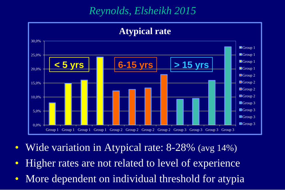
Reynolds, Elsheikh 2015
• Wide variation in Atypical rate: 8-28% (avg 14%)
• Higher rates are not related to level of experience
• More dependent on individual threshold for atypia
0,0%
5,0%
10,0%
15,0%
20,0%
25,0%
30,0%
Group 1 Group 1 Group 1 Group 1 Group 2 Group 2 Group 2 Group 2 Group 3 Group 3 Group 3 Group 3
Atypical rate
Group 1
Group 1
Group 1
Group 1
Group 2
Group 2
Group 2
Group 2
Group 3
Group 3
Group 3
Group 3
< 5 yrs 6-15 yrs > 15 yrs

Urine Dx’s Categorized by Pathologist BMH, Indiana 2009
In Need of Standardization!
• Standard classification and terminology system
• Well defined and reproducible diagnostic criteria
• Uniform inter- and intra-departmental
communications
• Consistent prognostic and management
information leading to optimal patient care

The Paris System for Reporting
Urinary Tract Cytology


TPS Diagnostic Categories
• Negative for HGUC
• Atypical Urothelial Cells
• Suspicious for HGUC
• High Grade Urothelial Carcinoma
• Low Grade Urothelial Neoplasm
• Other malignancies, both primary and
secondary
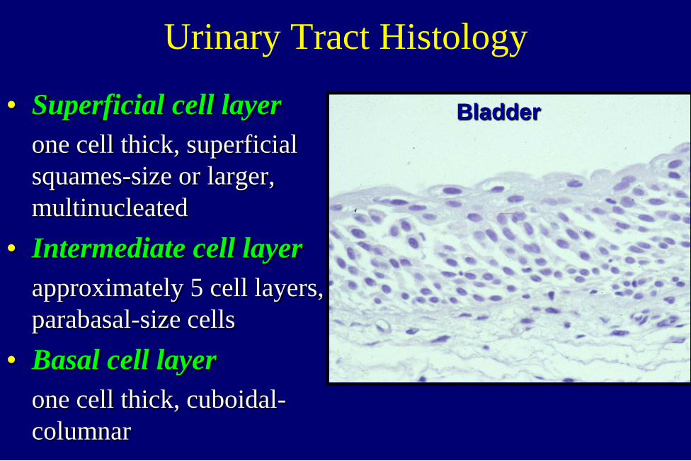
Urinary Tract Histology
• Superficial cell layer
one cell thick, superficial
squames-size or larger,
multinucleated
• Intermediate cell layer
approximately 5 cell layers,
parabasal-size cells
• Basal cell layer
one cell thick, cuboidal-
columnar
Bladder
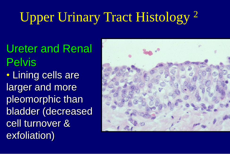
Ureter and Renal
Pelvis • Lining cells are
larger and more
pleomorphic than
bladder (decreased
cell turnover &
exfoliation)
Upper Urinary Tract Histology 2
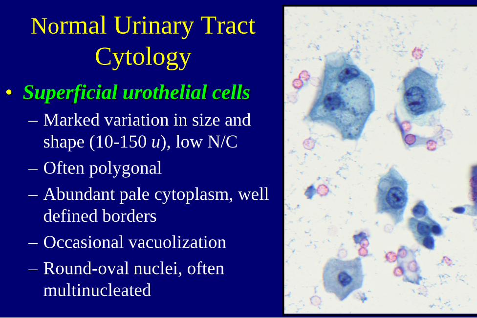
Normal Urinary Tract
Cytology
• Superficial urothelial cells
– Marked variation in size and
shape (10-150 u), low N/C
– Often polygonal
– Abundant pale cytoplasm, well
defined borders
– Occasional vacuolization
– Round-oval nuclei, often
multinucleated

Normal Urinary Tract Cytology 2
• Deep urothelial cells
– Uniform in shape and
size (10-20 u)
– Scant to moderate dense
cytoplasm, distinct
borders, fine
vacuolization
– Central nuclei, finely
granular chromatin,
small nucleoli
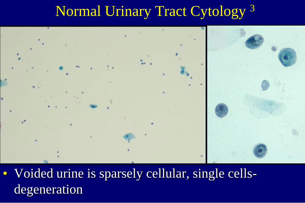
Normal Urinary Tract Cytology 3
• Voided urine is sparsely cellular, single cells-
degeneration
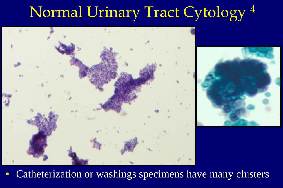
• Catheterization or washings specimens have many clusters
Normal Urinary Tract Cytology 4
TPS Diagnostic Categories
• Negative for HGUC
• Atypical Urothelial Cells
• Suspicious for HGUC
• High Grade Urothelial Carcinoma
• Low Grade Urothelial Neoplasm
• Other malignancies, both primary and
secondary

High Grade Urothelial CA
Often invasive, 70% mortality
90% of pts dying of disease
present initially with HGUC
• Cytology cannot reliably
separate CIS from invasive CA
• High diagnostic accuracy of
cytology
– Sensitivity 80-90 %
– Specificity > 95%
HGUC- TPS Criteria
Non-superficial and non-degenerated (viable) urothelial cells
• High N/C ratio > 0.5-0.7 (required)
• Hyperchromasia, moderate-severe (required)
–and one of the following:
• Irregular clumpy chromatin
• Irregular nuclear membranes
At least 5-10 abnormal cells
– Based on pathologist’s level of comfort
– Voided vs. upper tract instrumented specimen

• Single cells and/or disorganized
clusters
• Irregular, hyperchromatic nuclei
HGUC
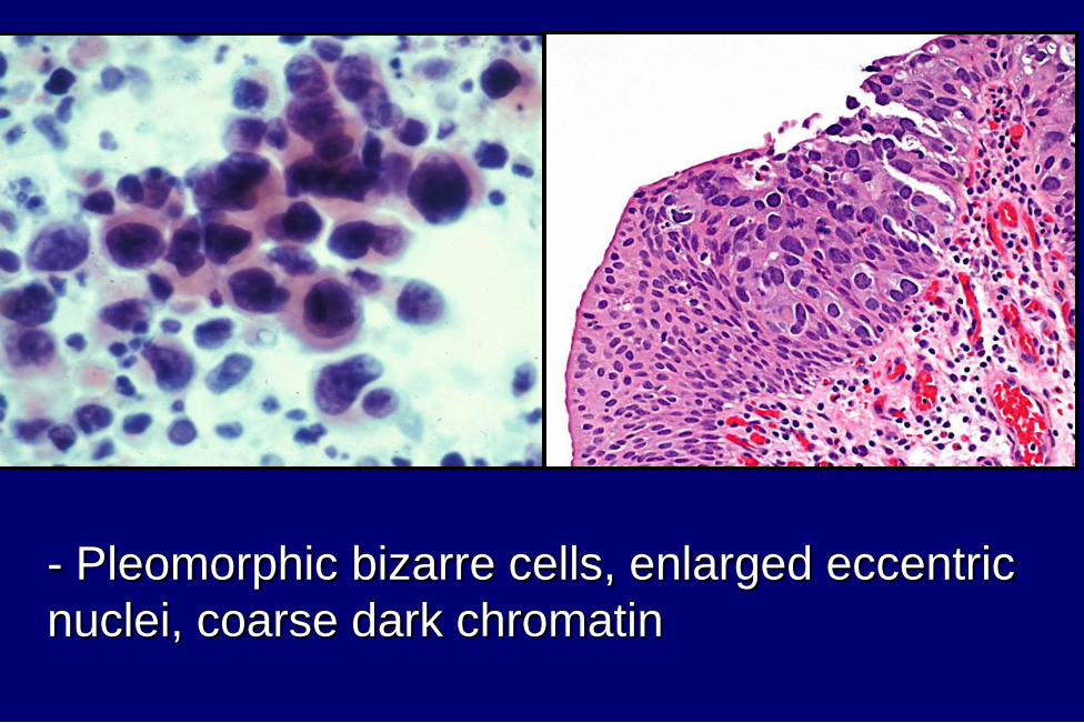
- Pleomorphic bizarre cells, enlarged eccentric
nuclei, coarse dark chromatin

TPS Diagnostic Categories
• Negative for HGUC
• Atypical Urothelial Cells
• Suspicious for HGUC
• High Grade Urothelial Carcinoma
• Low Grade Urothelial Neoplasm
• Other malignancies, both primary and
secondary

Suspicious for HGUC- TPS Criteria
• Same criteria as HGUC:
- Viable deep urothelial cells
- High N/C ratio > 0.5-0.7
- Marked Hyperchromasia
- Irregular clumpy chromatin
- Irregular nuclear membranes
• Less than 5-10 abnormal cells
– Based on pathologist’s level of comfort
– Voided vs. upper tract instrumented specimen

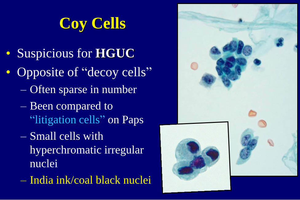
Coy Cells
• Suspicious for HGUC
• Opposite of “decoy cells”
– Often sparse in number
– Been compared to
“litigation cells” on Paps
– Small cells with
hyperchromatic irregular
nuclei
– India ink/coal black nuclei

Differential Diagnosis of HGUC
• Human polyoma viral infection
• Therapy effect
• Stones and reactive changes
• Other malignancies
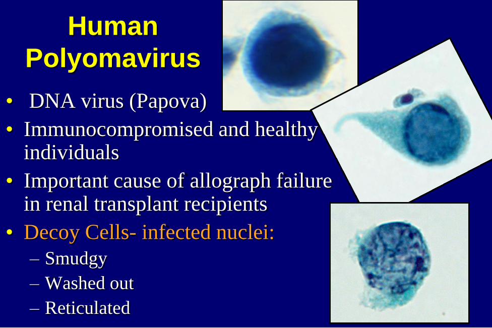
• DNA virus (Papova)
• Immunocompromised and healthy individuals
• Important cause of allograph failure in renal transplant recipients
• Decoy Cells- infected nuclei:
– Smudgy
– Washed out
– Reticulated
Human
Polyomavirus
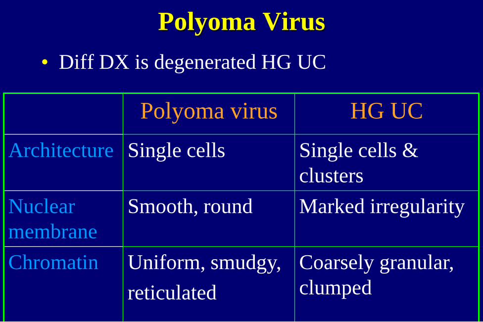
Polyoma Virus
• Diff DX is degenerated HG UC
Polyoma virus HG UC
Architecture Single cells Single cells &
clusters
Nuclear
membrane
Smooth, round Marked irregularity
Chromatin Uniform, smudgy,
reticulated
Coarsely granular,
clumped

HGUC Polyoma
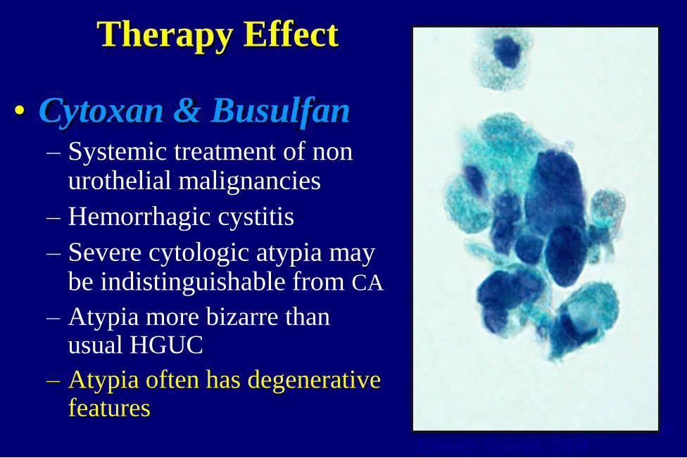
Therapy Effect
• Cytoxan & Busulfan – Systemic treatment of non
urothelial malignancies
– Hemorrhagic cystitis
– Severe cytologic atypia may be indistinguishable from CA
– Atypia more bizarre than usual HGUC
– Atypia often has degenerative features
Photo from Modern Cytopathology.
Elsevier Science, 2004

Therapy Effect 2
Thiotepa & Mitomycin C
Intravesical Rx of sup UC
Repair-like changes
BCG Vaccine
Treatment of CIS
Granulomas, mild atypia
Radiation Change
Extreme cytomegaly,
multinucleation, but low N/C ratio
Photo from Murphy WM. Urinary
Cytopatholgy. ASCP Press,2000

Lithiasis
• Papillary clusters common
• Smooth bordered clusters
• Centrally placed nuclei,
smooth nuclear membranes,
finely granular chromatin
• Hyperchromatic smudgy
nuclei (degenerative
changes)

Lithiasis 2
• Occasionally marked cytologic atypia, including
nuclear pleomorphism, coarsely granular chromatin,
mitotic figures false-positive diagnosis of HGUC
• Inflammation & debris
in background may be
misinterpreted as tumor
diathesis
• May be impossible
to distinguish from
LGUC

Lithiasis 3
• Important source of false positive Dx for
LGUC and HGUC
• Clinical history not reliable: filling defect in
upper UT stone vs. neoplasm
• Persistent atypical features (weeks)
aggressively worked up for neoplasia

TPS Diagnostic Categories
• Negative for HGUC
• Atypical Urothelial Cells
• Suspicious for HGUC
• High Grade Urothelial Carcinoma
• Low Grade Urothelial Neoplasm
• Other malignancies, both primary and
secondary

Mild Nuclear Atypia
• Single cells with enlarged and irregular nuclei
(no significant hyperchromasia)
• Most common and most frustrating
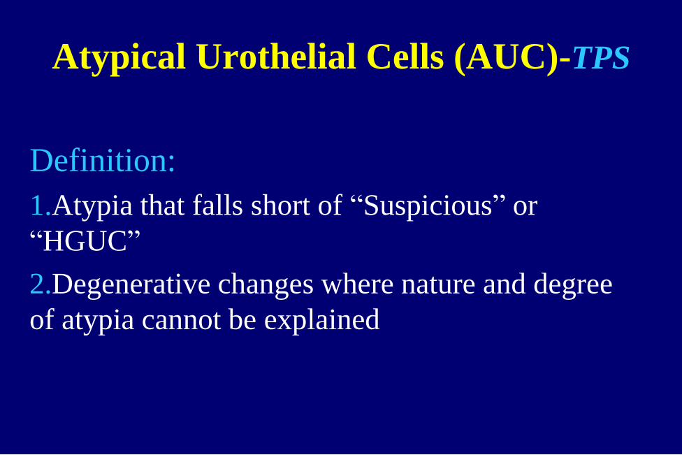
Atypical Urothelial Cells (AUC)-TPS
Definition:
1.Atypia that falls short of “Suspicious” or
“HGUC”
2.Degenerative changes where nature and degree
of atypia cannot be explained

AUC- TPS Criteria
• High N/C ratio > 0.5 (required)
and one of the following:
• Hyperchromasia, mild-moderate
- Compared to benign urothelial or
squamous cell nuclei
• Nuclear Irregularity, significant
• Irregular clumpy chromatin, mild
1. Non-degenerated, Non-superficial urothelial cells

• Nuclear irregularity, high NC ratio, but no significant
hyperchromasia, compared to benign urothelial cells
AUC
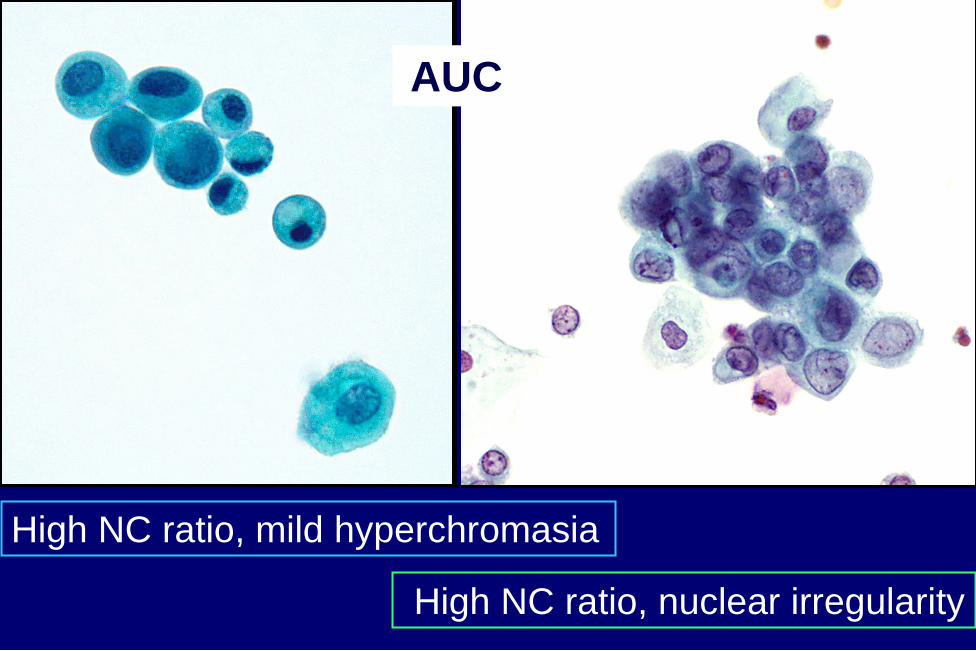
High NC ratio, mild hyperchromasia
AUC
High NC ratio, nuclear irregularity

AUC- TPS Criteria
• High N/C ratio and
hyperchromasia
• Extensive degeneration of nuclei
and/or incomplete cytoplasm
2. Degenerated non-superficial urothelial cells
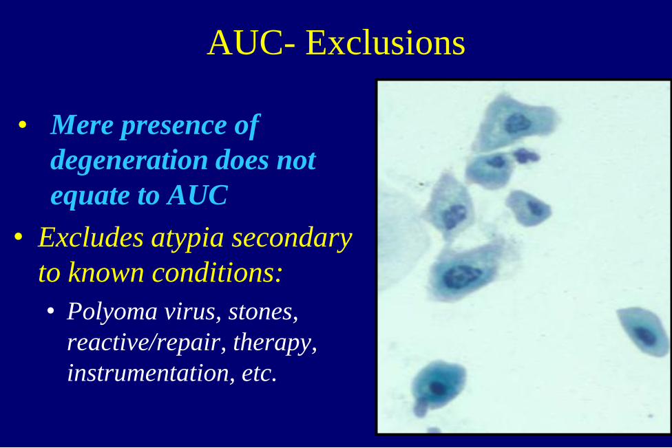
AUC- Exclusions
• Mere presence of
degeneration does not
equate to AUC
• Excludes atypia secondary
to known conditions:
• Polyoma virus, stones,
reactive/repair, therapy,
instrumentation, etc.

AUC Negative
Lost nuclear membrane Intact nuclear membrane,
hyperchromatic

TPS Diagnostic Categories
• Negative for HGUC
• Low Grade Urothelial Neoplasm
• Other malignancies, both primary and
secondary
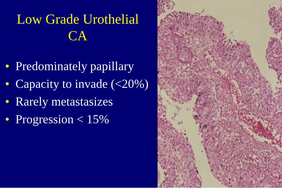
Low Grade Urothelial
CA
• Predominately papillary
• Capacity to invade (<20%)
• Rarely metastasizes
• Progression < 15%
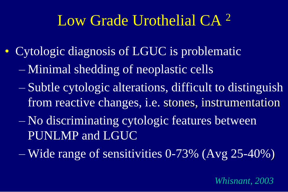
Low Grade Urothelial CA 2
• Cytologic diagnosis of LGUC is problematic
– Minimal shedding of neoplastic cells
– Subtle cytologic alterations, difficult to distinguish
from reactive changes, i.e. stones, instrumentation
– No discriminating cytologic features between
PUNLMP and LGUC
– Wide range of sensitivities 0-73% (Avg 25-40%)
Whisnant, 2003

Mcroskey 2015
• Compared biopsy proven LGUC cytologies (98
cases) to negative cytologies (53 cases)
• Instrumented urine specimens
• Evaluated 17 published cytologic features
• All cases were examined blinded to histology
• No single cytologic feature was found to be
helpful in DDX, except for papillary clusters
with fibrovascular cores (2/98 cases)

LGUC Benign
Few cells with enlarged slightly irregular nuclei.

Clusters in voided urine
• Papillary clusters (without fibrovascular core are not associated with increased risk of neoplasia
• Should place less reliance on presence or shape of clusters
• More emphasis on nuclear features
(Deshpande & Mckee, Cancer Cytopathol, 2005)
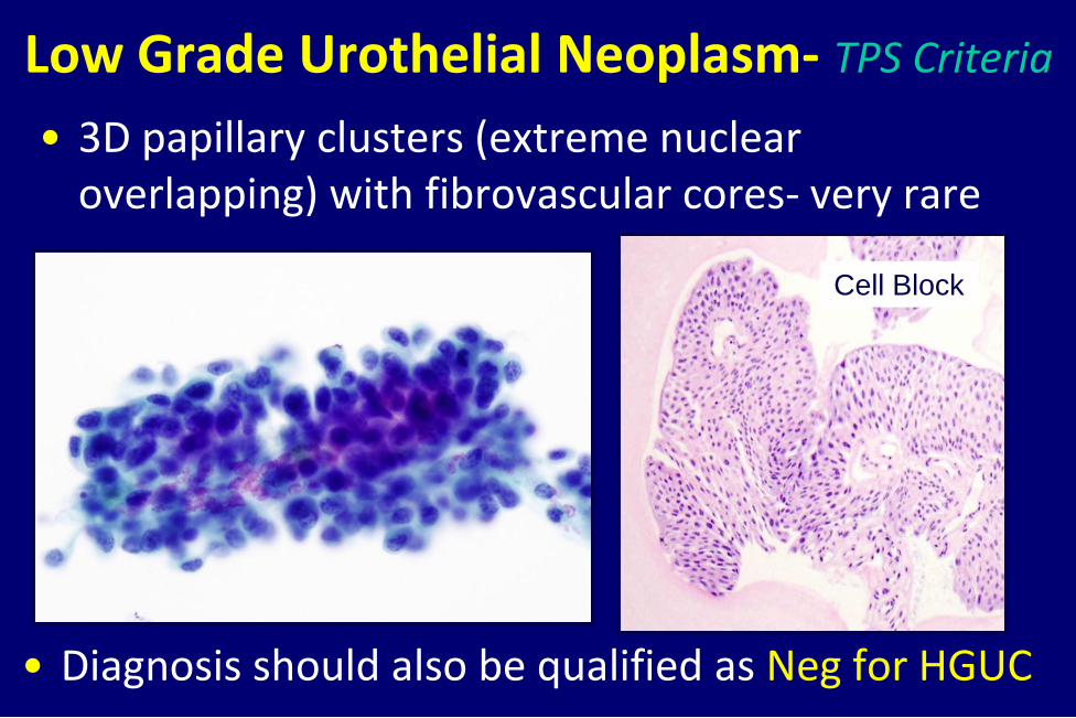
Low Grade Urothelial Neoplasm- TPS Criteria
• 3D papillary clusters (extreme nuclear overlapping) with fibrovascular cores- very rare
Cell Block
• Diagnosis should also be qualified as Neg for HGUC
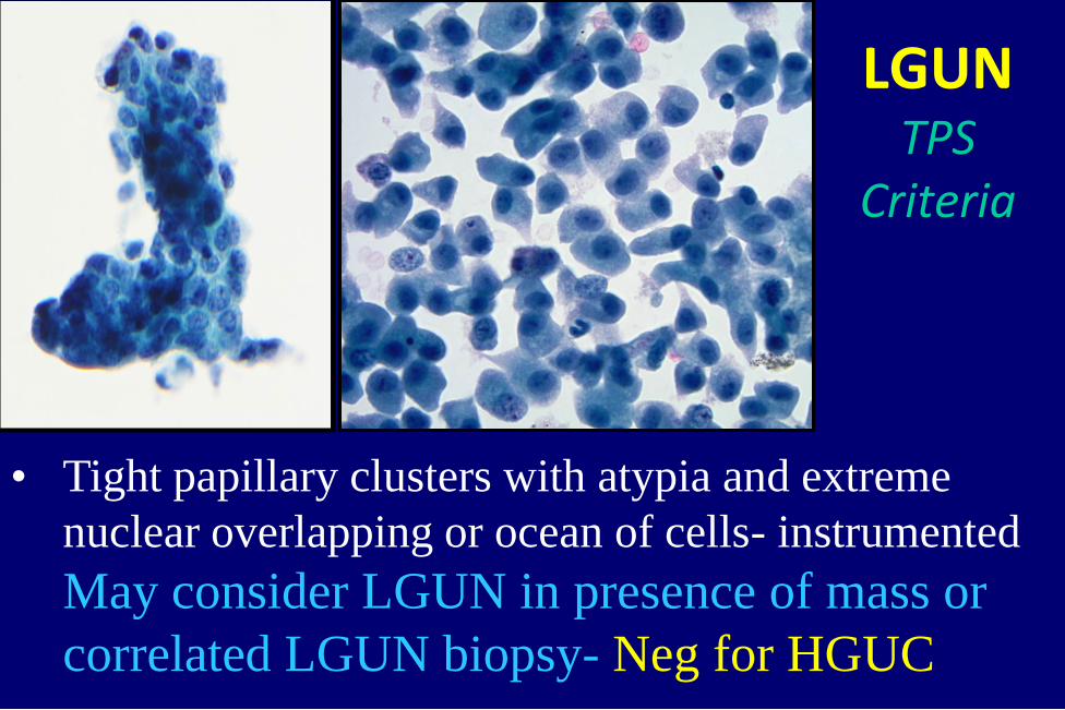
• Tight papillary clusters with atypia and extreme
nuclear overlapping or ocean of cells- instrumented
May consider LGUN in presence of mass or
correlated LGUN biopsy- Neg for HGUC
LGUN TPS
Criteria

Differential Diagnosis of
LG Urothelial CA
• Reactive/reparative changes
• Upper urinary tract sampling
• Instrumentation effect
• Lithiasis
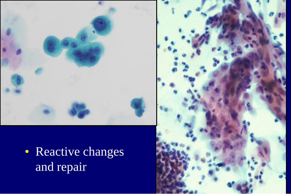
• Reactive changes
and repair

Upper Urinary Tract specimens
• Direct sampling of upper UT is effective in
detecting HGUC, but poor for low grade lesions
• Sensitivity: LGUC 37% vs. HGUC 80% Barkan 2015
• Normal upper UT epithelium shows more atypia
than lower UT and occasionally more than LGUC
N/C ratio, nuclear irregularities, papillary clusters
• Almost impossible to distinguish low grade UC
from upper tract benign changes
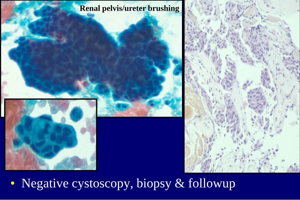
• Negative cystoscopy, biopsy & followup
Renal pelvis/ureter brushing
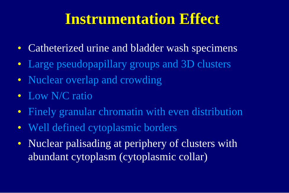
Instrumentation Effect
• Catheterized urine and bladder wash specimens
• Large pseudopapillary groups and 3D clusters
• Nuclear overlap and crowding
• Low N/C ratio
• Finely granular chromatin with even distribution
• Well defined cytoplasmic borders
• Nuclear palisading at periphery of clusters with
abundant cytoplasm (cytoplasmic collar)

Instrumentation effect
ThinPrep
How Long is Cytology Abnormal
after Cystoscopy?
• Evaluated 48 patients
• Examined urine before, immediately after, 1,
2, 7, 14 and 28 days
• Instrumentation effect was transient, mostly
disappearing within 1 day after cystoscopy
McVey et al. BJU INT, 2004

TPS Diagnostic Categories
• Negative for HGUC
• Atypical Urothelial Cells
• Suspicious for HGUC
• High Grade Urothelial Carcinoma
• Low Grade Urothelial Neoplasm
• Other malignancies, both primary and
secondary
Negative for HGUC-TPS Criteria
• If there is a known cause for “atypia”- it’s
Negative
– Reactive urothelial cells
– Instrumentation effect
– Upper urinary tract specimens
– Changes associated with lithiasis
– Polyoma viral cytopathic effect
– Post-therapy effects
– Clusters without fibrovascular cores or atypia

NEGATIVE
Instrumentation
Reactive Repair
Therapy
Polyoma
Upper tract
Stones
Clusters
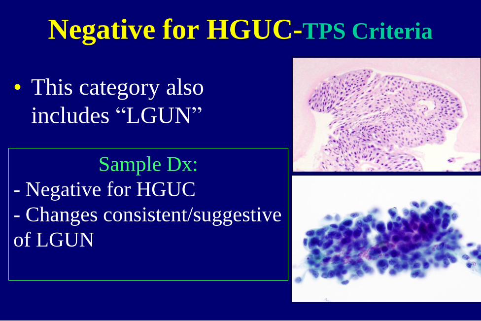
Negative for HGUC-TPS Criteria
• This category also
includes “LGUN”
Sample Dx:
- Negative for HGUC
- Changes consistent/suggestive
of LGUN

Nu
cle
ar
/ cyto
log
ic a
typ
ia
Probability of high grade UC
low moderate/high certain
AUC-Suspicious
8%-30%
HGUC NFM
Slide courtesy of
D. Rosenthal, MD
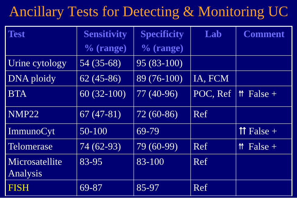
Ancillary Tests for Detecting & Monitoring UC
Test Sensitivity
% (range)
Specificity
% (range)
Lab Comment
Urine cytology 54 (35-68) 95 (83-100)
DNA ploidy 62 (45-86) 89 (76-100) IA, FCM
BTA 60 (32-100) 77 (40-96) POC, Ref ⇈ False +
NMP22 67 (47-81) 72 (60-86) Ref
ImmunoCyt 50-100 69-79 ⇈ False +
Telomerase 74 (62-93) 79 (60-99) Ref ⇈ False +
Microsatellite
Analysis
83-95 83-100 Ref
FISH 69-87 85-97 Ref
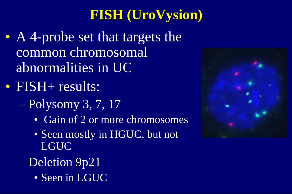
FISH (UroVysion)
• A 4-probe set that targets the common chromosomal abnormalities in UC
• FISH+ results:
– Polysomy 3, 7, 17
• Gain of 2 or more chromosomes
• Seen mostly in HGUC, but not LGUC
– Deletion 9p21
• Seen in LGUC

FISH 2
• FDA approved for surveillance of patients with
hematuria and history of UC
• Recommended in hematuria pts with other risk
factors such as smoking hx and age > 45
• High sensitivity 69-93%, esp. for
HGUC, lower for LG UC
• ? FISH positive AUC treated as Susp/ HGUC

FISH 3
Impressive Sensitivity results:
• Surveillance UC patients: – FISH +/ cystoscopy-/ cytology- 65%
recurrent CA (within 29 months)
– FISH- 13% recurrent CA
• Hematuria surveillance: – FISH+/cytology- (30%) 60% UC
• Post BCG therapy – FISH+ approx 10 times more likely to develop
invasive cancer
False-Positive FISH
• Be careful about significance of FISH+ in upper
tract cytlogy
– Limited value for upper tract tumor surveillance
– High false + (Johannes, J Urol. 2010)
• Polyoma virus can cause false + FISH (approx
15%)
– Usually in pts with high viral titers (renal transplant)
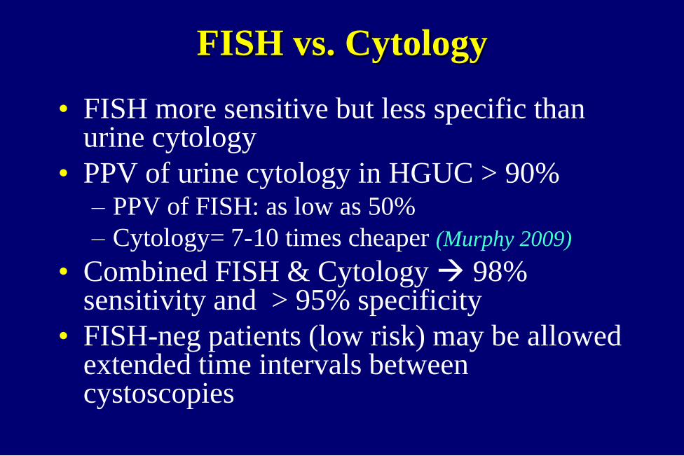
FISH vs. Cytology
• FISH more sensitive but less specific than urine cytology
• PPV of urine cytology in HGUC > 90% – PPV of FISH: as low as 50%
– Cytology= 7-10 times cheaper (Murphy 2009)
• Combined FISH & Cytology 98% sensitivity and > 95% specificity
• FISH-neg patients (low risk) may be allowed extended time intervals between cystoscopies

Summary
• Urine cytology is best applied to HGUC
• Cytology less helpful for detecting and monitoring LG neoplasms
– Not major limitation
– LG neoplasms rarely aggressive and can be readily detected by cystoscopy

Dogs Sniff Out Cancer
• Willis et al, British Medical
Journal 2004:
– Dogs correctly identified urine
from cancer patients: 41%
success rate vs. 14% chance
alone (Pathologist sensitivity for Dx
of LGUC 25-40%)
– Suggested that tumor-related
volatile compounds are present
in urine imparting a
characteristic odor

Summary 2
• “Atypical” diagnoses should not be used for reactive/reparative changes Negative for HGUC
• TPS provides strict cytologic criteria and aims to establish a standardized practical approach to urinary cytology classification

Is there a pathologist-or dog- in the house?

Thank You!


![7 Catheter-associated Urinary Tract Infection (CAUTI) · UTI Urinary Tract Infection (Catheter-Associated Urinary Tract Infection [CAUTI] and Non-Catheter-Associated Urinary Tract](https://static.documents.pub/doc/80x56/5c40b88393f3c338af353b7f/7-catheter-associated-urinary-tract-infection-cauti-uti-urinary-tract-infection.jpg)


