Urinary Tract Pathology: Urinary Bladder, Renal Pelvis & Urethra John F. Madden, M.D., Ph.D. Spring 2010
Transcript
Urinary Tract
Pathology:
Urinary Bladder,
Renal Pelvis &
Urethra John F. Madden, M.D., Ph.D.
Spring 2010
apm15
Stamp
apm15
Callout
Bladder time.
hulet001
Approved
Cystitis
apm15
Text Box
First set of benign conditions to discuss.
•“Ascending” infection due to enteric bacteria
• >95% of cases due to E. coli
• Klebsiella, Proteus, etc. in predisposed pts
• Yeast, viruses (CMV, polyoma, adenovirus)
with immunosuppression
•Favored by obstruction
•Prostatism, congenital anomalies, stones
Infectious cystitis
apm15
Highlight
apm15
Highlight
apm15
Highlight
apm15
Highlight
apm15
Highlight
apm15
Highlight
apm15
Text Box
Most conditions of ureteritis and pyelonephritis are also ascending infections
apm15
Text Box
Fungal cystitis is unusual except in chronic catherization and patients on multiple antibiotics. Usually develop yeast (candida) infection.
apm15
Callout
important for patients on immunosuppression (transplant patients, neutropenic patients)
apm15
Line
apm15
Text Box
We keep bacteria out of urinary tract by peeing. Therefore obstruct the urinary flow = infection. Female anatomy (shorter urethra) puts them at greater risk. Risk for males is obstruction of the prostate
apm15
Text Box
Older men due to BPH are at risk of obstruction and cystitis
apm15
Text Box
Stones favors infectious cystitis.
apm15
Highlight
Urethral
colonization
Asymptomati
c bacteriuria
(<104/ml)
“Urethral
syndrome”
(104–105/ml)
Cystitis
(≥105/ml)
Pyelonephritis
apm15
Text Box
Pathogenetic sequence is reflective in the diagnostic sequence. On wards diagnosing cystitis is done by a urine culture and quantitatively determine the diagnosis.
apm15
Callout
Grey zone often associated with burning symptoms. So, often urethritis ("urethral syndrome") precedes cystitis.
apm15
Callout
Numerical criteria to diagnose cystitis. This number is of a single species.
apm15
Text Box
UTI is a spectrum of degrees across which ascending infection has assembled itself across various areas of the urinary tract
apm15
Callout
Occurs for various reasons, but does not warrant treatment
apm15
Text Box
Case of bacterial cystitis. This patient had a catheter. Hallmarks of severe acute infection: - yellowish grey pus on bladder - erythema / hemorrhage due to infection
apm15
Text Box
Microscopically: - reactive hyperplasia of bladder epithelium -hallmarks of infection (pure PMN or PMN w/ mixed chronic inflammatory cells depending on stage of inflammation) - Whenever bladder gets ulcerated and urine enters stroma beneath epithelium, the urine attracts eosinophils
• Idiopathic (? autoimmune, mast cell
dysfunction) cystitis
• Typically, women in later adulthood
• Hematuria, pain
• Extensive ulceration, often transmural,
with fibrosis
• dDx: infection, cancer
Interstitial (“Hunner’s”) cystitis
apm15
Text Box
There are a couple of other non-infectious kinds of cystitis. Interstitial cystitis is one of them. Frustrating diagnosis / unknown etiology
apm15
Highlight
apm15
Text Box
Chronic, recurrent, mild to severe w/possible transmural ulceratoin. Supposedly an autoimmune process
apm15
Text Box
Many mast cells found in infiltrate.
apm15
Text Box
AKA "Bladder Pain Syndrome"
apm15
Highlight
apm15
Highlight
apm15
Highlight
apm15
Callout
Superficial to transmural ulceration
apm15
Callout
No epithelium and plenty of ulceration.
apm15
Text Box
Not high powered, therefore can't see mast cells.
apm15
Text Box
Difficult to treat due to unknown etiology.
apm15
Text Box
Ulcerating, no PMN, mast cells, chronic inflammation.
apm15
Text Box
Difficult to know how to treat these patients. Sometimes steroids are given.
• Complication of chemo-therapy
or therapeutic pelvic irradiation
• Cyclophosphamide, others
• Can cause severe hemorrhage
Hemorrhagic cystitis
apm15
Text Box
Another kind of cystitis. Inpatient and outpatient chemotherapy patients are the prime target.
apm15
Callout
Kind of cystitis associated with cytotoxic chemotherapy agents / RT. Blood found in urine.
apm15
Highlight
apm15
Highlight
apm15
Highlight
apm15
Highlight
apm15
Highlight
apm15
Text Box
Can be PO therapy (such as cyclophosphamide) or intravenous. Both can cause hemorrhagic cystitis.
apm15
Callout
Often require a cystectomy to control the bleeding
apm15
Text Box
Severe hemorrhagic cystitis. Surgical case where the patient was losing lots of blood and a cystectomy was necessary.
apm15
Text Box
Histology shows a lot of reactive, proliferation and granulation tissue. Lots of nuclear atypia, which you may mistake for cancer, but it is due to the chemo / RT.
•Chronic bacterial infection with
ineffective clearance of organisms
• Proteus often involved
•“Pseudotumor”
•Sheets of histiocytes packed lysosomes
•Malakoplakia has Michaelis-Gutmann
bodies
Malakoplakia &
Xanthogranulomatous pyelonephritis
apm15
Text Box
Xanthogranulomatous pyelo is similar to Malakoplakia of the urinary bladder. Both are entities that result from chronic bacterial infection and ineffective clearance of bacteria. Occurs often when you have stones in the renal pelvis or patients who are paraplegic w/o bladder control who constantly develop cystitis.
apm15
Highlight
apm15
Highlight
apm15
Highlight
apm15
Highlight
apm15
Highlight
apm15
Highlight
apm15
Highlight
apm15
Callout
Lysosomes have shreds of partially digested bacteria
apm15
Callout
Difference between the two is that Malakoplakia have calcified / fossilized bacteria in the lysosomes creating these bodies.
apm15
Highlight
apm15
Highlight
apm15
Text Box
Case of xanthogranulomatous pyelonephritis presenting as a renal tumor. This patient had the kidney removed. The physician thought this was clear cell RCC, but it is simply a mass of histocytes mimicking a tumor. Entirely reasonable to excise this kidney, although a partial nephrectomy would be more advisable.
apm15
Text Box
These people usually have large renal calculi
apm15
Text Box
Picture of the histocytes. This is a case of Malakoplakia. You can see the histiocytes and under EM it would be packed with lysosomes.
apm15
Callout
Reddish smudge are the Michalis-Gutmann bodies
apm15
Callout
another Michalis-Gutmann body.
apm15
Text Box
Malakoplakia can present in bladder or kidney. In each case it would raise the suspicion of cancer.
Urothelial metaplasia
apm15
Text Box
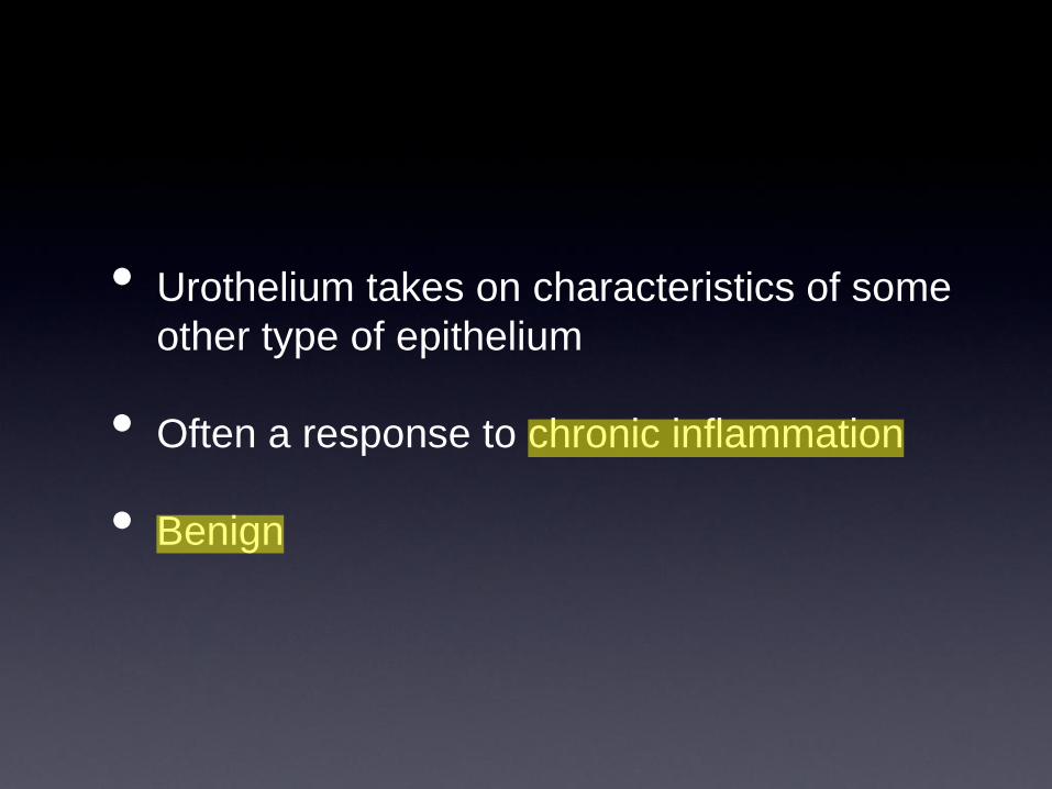
When a normal cell type undergoes differentiation to another cell type = metaplasia. It does so due to insults. At times these areas undergo biopsy and report states "squamous cell metaplasia". It is a common benign change and you don't want to mistake it for a carcinoma. Metaplasia is not neoplasia. It is not cancerous and does not necessarily precede cancer.
• Urothelium takes on characteristics of some
other type of epithelium
• Often a response to chronic inflammation
• Benign
apm15
Highlight
apm15
Highlight
apm15
Text Box
Urothelium has incredible ability to undergo metaplasia.
Normal urothelium
apm15
Text Box
Transitional epithelium. Usually ~7 cell layers thick, umbrella cell on top.
apm15
Callout
basal cells
apm15
Callout
umbrella cells
Cystitis cystica Normal submucosal nests of urothelium (“von
Brunn’s nests”) develop central cystic change
apm15
Text Box
Sort of metaplasia that is common in the bladder and appears as a domed mass on the bladder and is often biopsied in fear of cancer. Odd name since we frequently have no cystitis, but do have a cystic change.
apm15
Callout
Normal invagination of the urothelium underneath submucosa that undergoes central cystic change, inflates, and causes a mass.
apm15
Highlight
apm15
Highlight
apm15
Text Box
Benign metaplastic change.
apm15
Line
Cystitis glandularis Transitional cells convert to mucinous
columnar type
apm15
Text Box
Cystitis cystica can undergo secondary metaplasis to look like colon. Causing cystitis glandularis. Negative for malignancy. May be spontaneous or associated w/inflammation.
apm15
Text Box
Most bladder cancers are those of urothelium. We may see adenocarcinoma arising due to this type of metaplasia.
Squamous metaplasia Transitional cells convert to squamous
cells under chronic irritation
apm15
Text Box
Common in bladder, especially w/ patients who have schistosomiasis. Theory is that the squamous epithelium is more protective than the typical urothelium, hence the metaplasia during chronic irritation.
apm15
Text Box
Again, rarely we see squamous carcinoma of the bladder due to underlying squamous metaplasia
Urothelial hyperplasia
apm15
Text Box
Not metaplasia. It is thickened hyperplastic urothelium due to irritation.
“Nephrogenic adenoma”
apm15
Text Box
Disease where you get a mass / lump / tumor in bladder / urethra / ureter, that looks just like kidney epithelium. Called adenoma since some ppl. consider it a tumor (misnomer), but other ppl. consider it metaplasia. Ppl. with chronic irritation get this condition at a higher rate. Theory (in at least the transplant population) is that this represents bits of kidney that break off, float, and re-implant.
Urothelial
(transitional
cell)carcinoma
apm15
Text Box
Bladder carcinoma. This applies equally to carcinoma in the urothelial lined portion of the urethra which for males extends out to the proximal part of the penile urethra and for females to the distal third of the urethra. After that point squamous epithelium takes over. The ureters and renal pelvis are also lined with urothelium.
• Most common carcinoma of urinary
bladder (85%)
• Y > X, white > black
• Known risk factors
• Smoking → ~50% of U.S. cases
• Aromatic amines
• Some occupations
• Schistosomiasis (squamous>TCC)
apm15
Highlight
apm15
Highlight
apm15
Highlight
apm15
Text Box
More common in males. More common in the white race.
apm15
Highlight
apm15
Highlight
apm15
Highlight
apm15
Callout
Hair dye (in the past), no longer permitted.
apm15
Callout
Nickel industry.
apm15
Callout
Most of the cancer is squamous in these patients, but some are urothelial
apm15
Callout
Single most important risk factor for bladder cancer
apm15
Text Box
Various exposures to environmental carcinogens is typically the cause. Unlike RCC, which seems to just occur.
•Tends to occur multifocally
•Tends to recur
apm15
Highlight
apm15
Highlight
apm15
Text Box
So, most bladder cancer are urothelial carcinoma (90-95%), the remaining are squamous, adeno. (due to the metaplasia as explained previously
apm15
Text Box
Because it is so closely related to chemical exposure, the chemical gets concentrated in the urine and is stirred around in the bladder = multifocal. In addition, it is typically triggered by numerous genetic hits = high reoccurrence
apm15
Text Box
Bladder cancer is described by the term "polychronotropism" (historically) due to the following factors:
•Molecular alterations in multiple regulatory
pathways are seen (Ras-MAPK, p53, Rb)
•Abnormalities of chromosome 9 (mostly del 9)
are a consistent, early finding
• p16 (CDKN2A) underexpression (9p21-)
(Rb pathway) especially common
•One FDA-approved ancillary test
(UroVysion™ Abbott) detects aneuploidy 3, 7,
17, and loss of the 9p21 via fluorescence in
situ hybridization (FISH) in urine
apm15
Text Box
There is no one knockout genetic change / gene involved in bladder cancer.
apm15
Highlight
apm15
Highlight
apm15
Callout
very common
apm15
Highlight
apm15
Callout
Also common
apm15
Highlight
apm15
Text Box
UroVysion is used as a screening test for bladder cancer.
Molecular Pathways in Invasive
Bladder Cancer:
New Insights Into Mechanisms,
Progression, and
Target Identification
Anirban P. Mitra, Ram H. Datar, and Richard J. Cote From the Departments of Pathology
JOURNAL OF CLINICAL ONCOLOGY R E V I E W A
R T I C L E
V O L U M E 2 4 N U M B E R 3 5 D E C E M B E R 1 0 2 0 0 6
Text
del 9
apm15
Text Box
Not all that important. For those interested it shows an early view of where some of these genetic changes occur. Early cancers at top and more invasive cancers at bottom
• Symptoms
• Episodic painless hematuria (80%)
• Diagnostic evaluation
• Urinary cytology
• Sensitivity modest, detects mainly high
grade lesions
• Okay for following patients with
established Dx
• Molecular tests
• Cystoscopy with biopsy
• Most useful
Bladder cancer: clinical
apm15
Callout
In 80% of patients Bladder cancer presents to medical attention with painless hematuria (text obscured by slide title)
apm15
Text Box
If you have cystitis there is blood in the urine with pain. Unlike bladder cancer which causes blood and no pain
apm15
Highlight
apm15
Text Box
Urine cytology is not good for early / low grade cancer
apm15
Text Box
You can perform the molecular test as mention on previous slide (UroVysion)
apm15
Text Box
Gold Standard is cystoscopy with biopsy
apm15
Callout
Least invasive way to start workup is a urine sample.
• Superficial
• Non-invasive or Invasive into lamina propria
only
• Traditionally, treated by transurethral resection
• Muscle-invasive
• Invasion into or through muscularis propria
• Treated by cystectomy and/or radiation
Bladder cancer in two broad
categories
by extent of invasion
apm15
Text Box
Several ways to subcategorize bladder cancer. One important way is based on how deeply invasive it is. Two groups: 1. Superficial 2. Muscle Invasive
apm15
Highlight
apm15
Highlight
apm15
Highlight
apm15
Highlight
apm15
Callout
Much worse prognosis
• Papillary
• Majority of urothelial cancers
• Exophytic, cystoscopic resection often
possible
• On average, lower grade
• Non-papillary
• 10-40% of urothelial cancers
• Cystoscopically occult
• Usually higher grade, multifocal at
presentation
Superficial urothelial neoplasia:
two histologic types
apm15
Text Box
Two main histo subtypes: 1. Papillary: Cauliflower mass (lower grade risk) 2. Non-papillary: analogous to dysplasia in the cervix, flat lesion (higher grade risk)
apm15
Highlight
apm15
Highlight
apm15
Highlight
apm15
Text Box
More aggressive, high grade
apm15
Callout
Superficial is lower grade, less aggressive
apm15
Callout
usually low grade / lower risk of invasion
apm15
Callout
flat carcinoma are higher grade / high risk of becoming invasive
apm15
Text Box
Episodic twisting off papillary tumor can lead to random hematuria.
apm15
Text Box
Roughly 25% of pts belong to the bottom two "flat" kind. These are more aggressive
Superficial papillary
urothelial neoplasia
apm15
Callout
bladder with lots of papillary carcinomas
apm15
Callout
couple of smaller papillary carcinomas
apm15
Text Box
Both of these papillary carcinoma examples are fairly advanced and invasive.
apm15
Callout
non-invasive papillary carcinoma in the renal pelvis
• By convention, papillary neoplasms of
urothelium are always called
“carcinoma” even if non-invasive
• Why call this “carcinoma”?
• Comparison with colonic adenoma
Warning!
apm15
Text Box
Bladder cancer exception (for historic reasons): Whether invasive or pre-invasive, lesions of the bladder are called cancer. Pre- or non-invasive "cancer" have very good prognosis and rarely progress to invasive disease.
apm15
Highlight
apm15
Text Box
Superficial non-invasive papillary "carcinoma" of the bladder, low grade, excised cystoscopically.
apm15
Text Box
Microscopic view of a pre-invasive bladder carcinoma
apm15
Text Box
urothelium on these papillae are seen as fingers w/fibrovascular cords lined with urothelium that is slightly thickened
apm15
Callout
Atypical enlarged cells.
apm15
Callout
Apoptosis occurring around here
•Papilloma
•(Low malignant potential)
•Low grade UC
•High grade UC
Papillary urothelial neoplasia:
grading
apm15
Text Box
When these papillary urothelial neoplasms are pre-invasive can be divided into low grade and high grade. The majority of the papillary are low grade and don't progress.
apm15
Text Box
Example of a low grade one
apm15
Text Box
Another example of a low grade one
apm15
Text Box
Example of a high grade one
• Frequent recurrence
• Infrequent
progression or
invasion
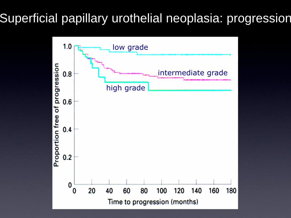
Superficial papillary urothelial neoplasia:
natural history
apm15
Text Box
Since papillary neoplasia is usually low grade and doesn't progress, they typically present as episodic hematuria, urologist will perform a cystoscopy, snips the cauliflower lesion, pathologist labels it as low grade, and it may recur. None of these tumors develop an invasive component. So this patient must keep coming back to have these papillae snipped out every six months.
Progression (development of invasive component) is uncommon in these patients
apm15
Callout
Low grade (out to 15 years) well over half have recurred, but 5% have progressed
• Cystoscopic resection
• Periodic (lifelong) follow-up
• Urine cytology
• Cystoscopy
• Intravesical therapy
• Partial cystectomy for high-grade
tumors
Therapy for superficial
papillary urothelial neoplasia
apm15
Text Box
Alluded to on previous slide. Keep snipping the papillae out.
apm15
Callout
Routine for urologist to give single dose of mitomycin (intravesically) following cystoscopic resection of a papillary urothelium neoplasm. This pushes out the time to recurrence.
apm15
Highlight
apm15
Highlight
apm15
Highlight
apm15
Highlight
apm15
Highlight
apm15
Callout
Bladder-sparring surgery is not really done
Superficial “flat”
urothelial neoplasia
apm15
Text Box
So ... we have two histological types. The papillary ones that we just discussed, and here we have the "flat" ones. These are rather aggressive.
Atypia Dysplasia CIS
Cheng et al. Cancer
apm15
Text Box
Carcinoma in stiu was term used in cervical lectures and is used to describe these flat neoplasms
apm15
Text Box
Does not form characteristic papillary fronds, but instead flat lesion
apm15
Text Box
Carcinoma in situ of the bladder. It does not form papillae, but has nasty looking cytologically atypical cells, nuclear enlargement, and nuclear pleomorphism.
• Over 70% have diffuse disease at
diagnosis
• Over 30% of CIS have undiagnosed
invasive disease at cystectomy
• Over 5% dead of (metastatic)
disease in 5 years after cystectomy
for CIS
Non-papillary (“Flat”) urothelial neoplasia
(urothelial carcinoma-in-situ): natural
history
apm15
Text Box
For "flat" urothelial neoplasia pre-invasive or in situ, the situation is very different than that for papillary neoplasm. Read the slide.
apm15
Highlight
apm15
Highlight
apm15
Highlight
apm15
Highlight
apm15
Highlight
apm15
Highlight
apm15
Highlight
apm15
Highlight
apm15
Highlight
apm15
Highlight
apm15
Highlight
apm15
Highlight
apm15
Highlight
apm15
Highlight
apm15
Highlight
•BCG
• >70% durable response in CIS
•Intravesical chemotherapy
• Thiotepa/doxorubicin/mitomycin
•Interferon
•Cystectomy
“Flat” urothelial neoplasia
(urothelial carcinoma-in-situ): therapy
apm15
Text Box
What do we do if we catch it early? We can biopsy, but can't resect b/c it's multifocal. Therefore use intravesical chemotherapy / immunotherapeutic agent or cystectomy.
apm15
Callout
cytotoxic
apm15
Callout
immunotherapeutic agent. Attenuated form of mycobacterium TB.
apm15
Highlight
apm15
Highlight
apm15
Text Box
BCG works not only for flat urothelium neoplasia, but also papillary type.
Alvaro Morales
Guerin & Calmette
apm15
Callout
Veterinarian
apm15
Text Box
Dudes on the left created BCG. Noted early on that patients w/ TB developed cancer at lower rates. Therefore, ppl realized that BCG might have some anti-cancer effects as a vaccine. Finally, in the 1970's Alvaro Morales instilled BCG directly into the bladder with in-situ carcinoma causing regression of carcinoma and durable responses. It works great in high proportion of pts. Often need to repeat treatment in six months.
apm15
Callout
Spanish urologist from Canada.
Muscle-invasive
urothelial carcinoma
apm15
Text Box
We discussed the lower grade papillary type and higher grade flat type. Either of these two types can evolve into muscle-invasive urothelial carcinoma (the flat kind at a higher rate). Once muscle involvement occurs it is very hard to distinguish papillary versus flat type.
apm15
Callout
Muscle invasive at higher rate
apm15
Text Box
Visual flow chart of what we discussed and the potential treatments. You can see that for muscle invasive carcinoma the gold treatment is cystectomy.
apm15
Text Box
Here is a muscle invasive carcinoma presenting as an ulcer. This is a cystectomy specimen.
apm15
Text Box
Here is an invasive carcinoma of the bladder that probably started as a papillary carcinoma and evolved into a large carcinoma which invades muscle.
apm15
Text Box
This is what invasive carcinoma looks like. Very high grade, malignant appearing cells.
apm15
Callout
These are muscle fibers and it infiltrates through through the muscularis propria. The depth of invasion determines staging.
Year
Death
s p
er
100
,00
0
Bladder cancer
mortality
apm15
Text Box
Over the years the mortality has been decreasing due to better chemical hygiene and better diagnosis.
apm15
Callout
Bladder cancer occurs predominantly in men, possibly due to previous smoking statistics
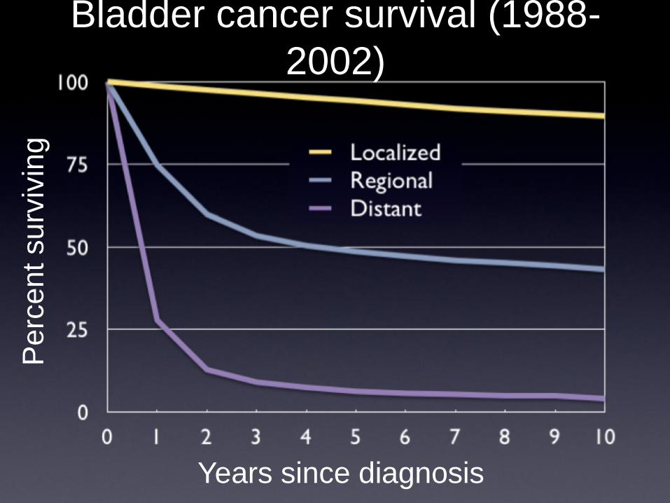
Bladder cancer survival (1988-
2002)
Years since diagnosis
Pe
rce
nt su
rviv
ing
apm15
Text Box
Survival is great for low grade papillary disease and dismal for patients with distant disease at diagnosis.
Bladder cancer stage distribution (1988-
2002)
Percent of cases
apm15
Text Box
Fortunately most are diagnosed at time when it is localized. Good alarm is the hematuria.
Therapy for invasive
urothelial carcinoma
•Radical cystectomy
•Partial cystectomy
• Transurethral resection
•Chemotherapy
• MVAC (methotrexate +
vinblastine + adriamycin +
cisplatin)
apm15
Text Box
Therapy for invasive urothelial carcinoma (gold standard) is radical cystectomy.
apm15
Text Box
Should be called bladder sparring. They resect as much tumor as possible via the transurethral approach and then the person gets systemic and intravesical therapy.
apm15
Text Box
Deleted on bottom of slide: MVAC (methotrexate + vinblastine + adriamycin + cysplatin)