36
UROLOGY IMAGING UROLOGY IMAGING Dr Mukosai S Dr Mukosai S Urology, Dept of Surgery Urology, Dept of Surgery 13 13 th th March 2015 March 2015

UROLOGY IMAGINGUROLOGY IMAGING
Dr Mukosai S Dr Mukosai S
Urology, Dept of SurgeryUrology, Dept of Surgery
1313thth March 2015 March 2015

IntroductionIntroduction
Urological Imaging use of Urological Imaging use of radiological picture to diagnose radiological picture to diagnose disease of the genitourinary disease of the genitourinary system.system.
These picture may be static (X These picture may be static (X ray) or ray) or dynamic(Fluoroscopic,Image dynamic(Fluoroscopic,Image intensifier)intensifier)

UROLOGICAL IMAGINGUROLOGICAL IMAGING
PLAIN X RAYPLAIN X RAY SPECIAL X RAYS(CONTRAST SPECIAL X RAYS(CONTRAST
ENHENCED)ENHENCED) ULTRASONOGRAPHYULTRASONOGRAPHY COMPUTERISED AXIAL COMPUTERISED AXIAL
TOMOGRAPHYTOMOGRAPHY NUCLEAR MEDICAL IMAGINGNUCLEAR MEDICAL IMAGING

PLAIN X RAYPLAIN X RAY
Use in UrologyUse in Urology Diagnosis of stone disease most Diagnosis of stone disease most
stones 80% radiopaquestones 80% radiopaque Dystrophc calcification eg Dystrophc calcification eg
curvilinear calcification of the curvilinear calcification of the bladder in schistomiasis, TB bladder in schistomiasis, TB calification Kidney,seminal calification Kidney,seminal vesiclesvesicles

Bladder stone
This patient had a h/o of STI and presented with dysuria and hematuria. Plain x ray revealed a trifolate bladder stone.


INTRAVENOUS INTRAVENOUS UROGRAPHY (IVU)UROGRAPHY (IVU)
DEFN: Special Contrast enhance x ray DEFN: Special Contrast enhance x ray to evaluate the function urinary to evaluate the function urinary production and outflow.production and outflow.
Technique: Intravenous injection of 50-Technique: Intravenous injection of 50-100ml of radiopaque contrast eg 100ml of radiopaque contrast eg Omnipaque is given.Omnipaque is given.
Serial X ray are taken beginning with a Serial X ray are taken beginning with a control (KUB before contrast control (KUB before contrast Injection). This followed by serial x Injection). This followed by serial x rays at timed intervals of 5mins, rays at timed intervals of 5mins, 10mins, 30mins up to 24hrs)10mins, 30mins up to 24hrs)

Nephrogram phase of IVUNephrogram phase of IVU
Opacification of NephrogramPyelogramContrast in ureter

Post voiding IVUPost voiding IVUUsed to assess outflow Used to assess outflow
obstruction Ureterovesical reflux.obstruction Ureterovesical reflux.
Cystogram phase

Delay orPersistentNephrogram
Cystogram
Tourtous dilated ureter

IVU demonstrating IVU demonstrating distorted calyceal pattern in distorted calyceal pattern in polycystic kidneypolycystic kidney

Retrograde UrethrogramRetrograde Urethrogram
Urethral stricture represents 30-40% Urethral stricture represents 30-40% of all urology cases seen at of all urology cases seen at UTH.These occur secondary to UTH.These occur secondary to urethritis, trauma , post catherisation urethritis, trauma , post catherisation or congential urethral diseases.or congential urethral diseases.
The assessment of urethral stricture The assessment of urethral stricture disease is by ascending disease is by ascending (retrograge)or descending (retrograge)or descending urethrography(antegrade)urethrography(antegrade)
TechniqueTechnique
A small foleys cathether is insert A small foleys cathether is insert up to the navicular fossa and 10 up to the navicular fossa and 10 mls of contrast(urograffin) is mls of contrast(urograffin) is inserted.inserted.
The contrast outlines the urethra The contrast outlines the urethra and will show the site,number, and will show the site,number, width and length of the stricture.width and length of the stricture.
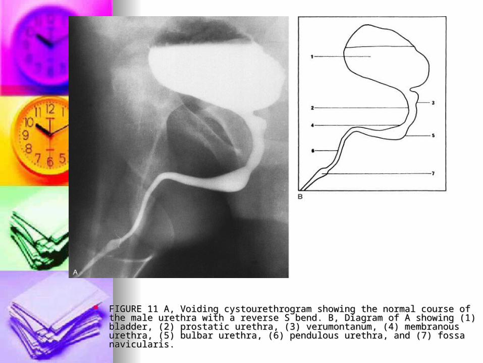
FIGURE 11 A, Voiding cystourethrogram showing the normal course of the FIGURE 11 A, Voiding cystourethrogram showing the normal course of the male urethra with a reverse S bend. B, Diagram of A showing (1) bladder, (2) male urethra with a reverse S bend. B, Diagram of A showing (1) bladder, (2) prostatic urethra, (3) verumontanum, (4) membranous urethra, (5) bulbar prostatic urethra, (3) verumontanum, (4) membranous urethra, (5) bulbar urethra, (6) pendulous urethra, and (7) fossa navicularis. urethra, (6) pendulous urethra, and (7) fossa navicularis.
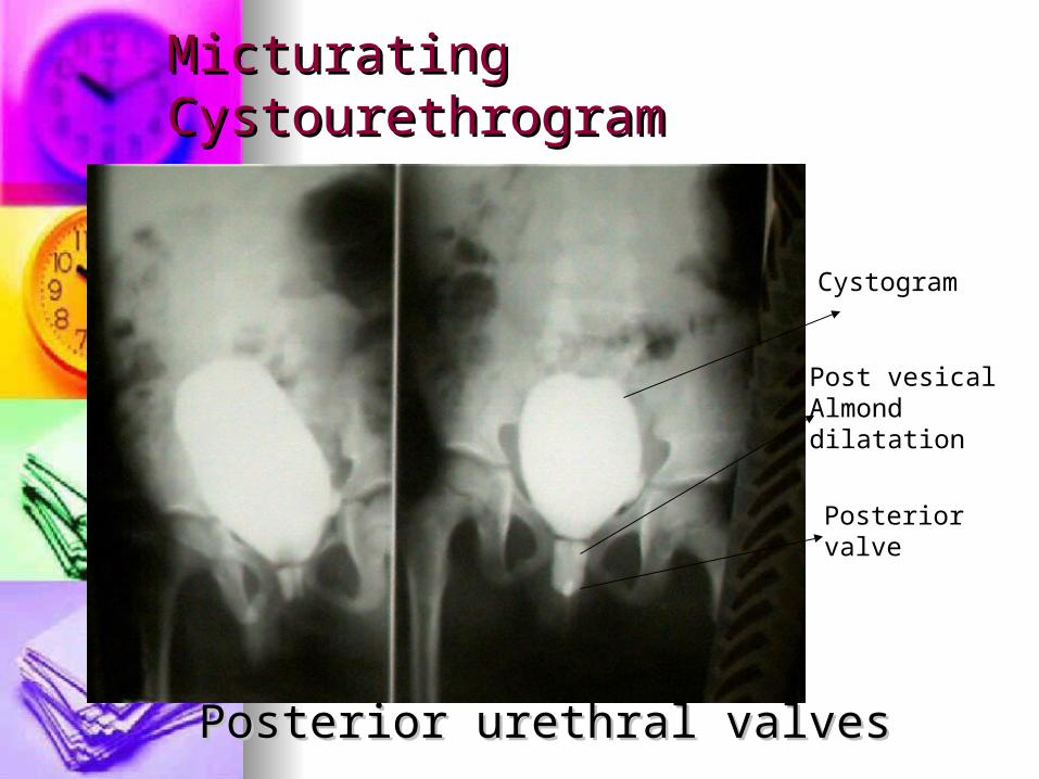
Micturating CystourethrogramMicturating Cystourethrogram
Posterior urethral valves Posterior urethral valves
Cystogram
Posteriorvalve
Post vesicalAlmonddilatation

April 19, 2023April 19, 2023 1616
CystographyCystography
A-intraperitoneal A-intraperitoneal perforationperforationthe peritoneum the peritoneum overlying the overlying the bladder, has been bladder, has been breached along with breached along with the wall of the the wall of the bladder, allowing bladder, allowing urine to escape into urine to escape into the peritoneal cavity.the peritoneal cavity.
Traumatic emergency

April 19, 2023April 19, 2023 1717
CystographyCystographyB- extraperitoneal B- extraperitoneal perforationperforation
the peritoneum is intactthe peritoneum is intact
and urine escapes into and urine escapes into the space around the the space around the bladder, but not intobladder, but not into
the peritoneal cavity.the peritoneal cavity.
Traumatic emergency

Normal Retrograde urethrogramNormal Retrograde urethrogram
Smooth narrowing in Smooth narrowing in the posterior & the posterior & prostatic urethral is prostatic urethral is normalnormal

Retrograde urethrogramRetrograde urethrogram
Multiple strictures in Multiple strictures in the anterior urethralthe anterior urethral

UltrasoundUltrasound
This uses high frequency sound This uses high frequency sound waves to diagnose diseases in waves to diagnose diseases in solid organs(particularly). The solid organs(particularly). The machine has a transducer which machine has a transducer which transforms the sound into an transforms the sound into an image. The probe has a image. The probe has a transmitter with a frequency of transmitter with a frequency of 3.5-5 MHertz.3.5-5 MHertz.

UltrasoundUltrasound
In the kidney it helps to diagnose In the kidney it helps to diagnose cyst from solid lesions, cyst from solid lesions, hydronephrosis, polycystic kidney, hydronephrosis, polycystic kidney, congenital abnormalities of the congenital abnormalities of the kidney eg horse shoe kidneykidney eg horse shoe kidney
In the bladder stones, cancer, In the bladder stones, cancer, residual urine etcresidual urine etc
It is used with colour for the It is used with colour for the diagnosis of testicular torsion as diagnosis of testicular torsion as doppler U/Sdoppler U/S

ultrasonography of the normal kidney. Longitudinal (A) and transverse (B) ultrasound views outline the contour of the right kidney (R), the
parenchyma of which is hypoechoic relative to the liver (L). The renal sinus fat (S) appears echogenic.

Normal testis and epididymal head. A, Longitudinal view of a scrotal ultrasonogram reveals the homogeneous texture of the right testis. The mediastinum of the testis (not shown) appears as an elongated echogenic structure extending craniocaudally on the posterolateral side of the testis. B, Longitudinal view demonstrates a normal head of the epididymis (arrow).
CAT scanCAT scan
Discovered by Godfrey Discovered by Godfrey Housfield in the 1980s from Housfield in the 1980s from University of Reading in the UKUniversity of Reading in the UK
This is a series of multiple x This is a series of multiple x rays from a tube(gantry) that are rays from a tube(gantry) that are summated by computer to summated by computer to produce crossectional body produce crossectional body images.images.
UsesUses
Trauma eg kidney Trauma eg kidney Congential abnormalitiesCongential abnormalities Functional disease eg Functional disease eg
hydronephrosis with contrasthydronephrosis with contrast Cancers used particularly for Cancers used particularly for
staging.staging.
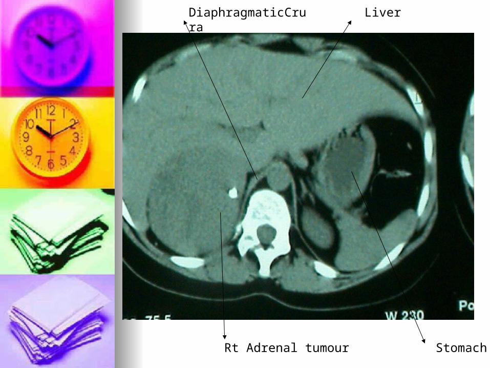
Rt Adrenal tumour Stomach
LiverDiaphragmaticCrura

Hyperpigmented striaehirsutism
Light skin colour

Cortisol producing Adrenal Tumour

Magnetic Resonance ImagingMagnetic Resonance Imaging
This uses magnetic waves to create This uses magnetic waves to create a change in alignment of hydrogen a change in alignment of hydrogen ions in the body. This change can be ions in the body. This change can be captured on computer as an image.captured on computer as an image.
Artificial electronically operated Artificial electronically operated medical devices are a medical devices are a contraindication to it’s use such as contraindication to it’s use such as Pacemakers.Pacemakers.
Indications include staging of cancer Indications include staging of cancer kidney, bladder and prostate.kidney, bladder and prostate.

Normal kidneys. A, Coronal T1-weighted (time of repetition [TR] of 500 msec and time of echo [TE] of 14 msec) spin-echo image demonstrates bilateral normal kidneys. The renal cortex (c) is higher in signal intensity than the medulla (m) and isointense to the liver (L). Note the hyperintensity of the perinephric fat relative to the renal cortex, resulting in excellent delineation of the cortex from surrounding perinephric fat. The renal sinus fat (s) appears hyperintense. B, Coronal T2-weighted fast spin-echo image (TR of 7500 msec, TE of 135 msec, and echo train [ET] of 16) obtained at the same level. The cortex (c) intermediate in signal intensity and hypointense relative to the liver (L) and medulla (m). (A and B, From Kramer LA: Magnetic resonance imaging of renal masses.

MRI of the normal prostate and seminal vesicles. A, On axial T1-weighted images, the prostate (solid arrow) appears as a homogeneous low–signal intensity structure in contrast to the periprostatic fat. The puborectalis is indicated with open arrows. B, urinary bladder; R, rectum. B, In a view cephalad to A, the paired seminal vesicles (arrows) appear as a characteristic bow-tie–shaped soft tissue structure and are well outlined by the surrounding fat plane. The lumens of the seminal vesicles appear markedly high in signal intensity on T2-weighted images (not shown). B, urinary bladder; R, rectum. C, A T2-weighted spin-echo image corresponding to A reveals the normal zonal anatomy of the prostate with a hyperintense peripheral zone (p) and intermediate–signal intensity central gland (c). The puborectalis is indicated with arrows. B, urinary bladder; R, rectum.

Nuclear Medicine TechniquesNuclear Medicine Techniques
This uses radiolabelled chemicals to image This uses radiolabelled chemicals to image the urinary systemthe urinary system
It has uses in determining the renal It has uses in determining the renal physiology, morphology and cortical statusphysiology, morphology and cortical status
Radioactive chemicals are Radioactive chemicals are visualised(iodine, technetium 99, gallium visualised(iodine, technetium 99, gallium etc) using gamma camera and presented etc) using gamma camera and presented visually on a computer screen.visually on a computer screen.

Picture of Technetium scan showing normal uptake of isotopeThis represents a normal study with no focal lesion seen

Summary questions Summary questions
80% of urinary stones are 80% of urinary stones are radiopaque Tor Fradiopaque Tor F
Differentials for filling defect in Differentials for filling defect in the renal pelvis ? 4 pointsthe renal pelvis ? 4 points
Schistosomiasis presents as Schistosomiasis presents as curvilinear calcification of the curvilinear calcification of the bladder on X ray T or Fbladder on X ray T or F
Differentials for nonfuntional Differentials for nonfuntional kidney on IVU?kidney on IVU?
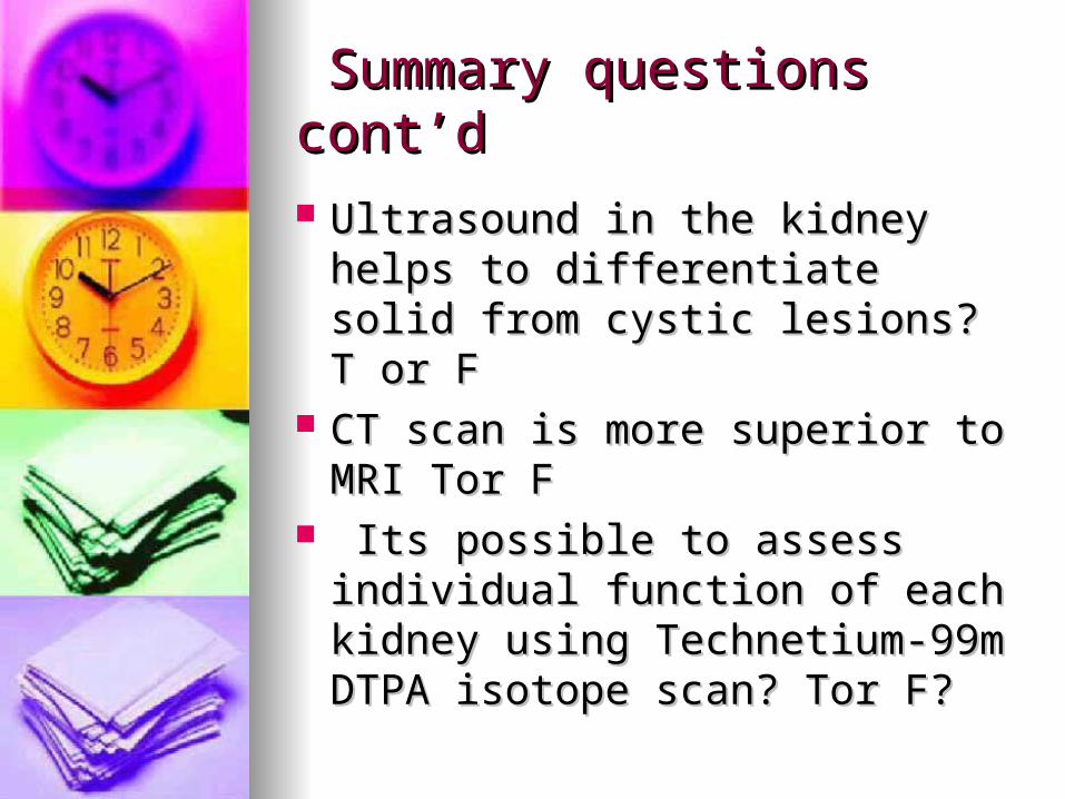
Summary questions cont’dSummary questions cont’d
Ultrasound in the kidney helps Ultrasound in the kidney helps to differentiate solid from cystic to differentiate solid from cystic lesions? T or Flesions? T or F
CT scan is more superior to MRI CT scan is more superior to MRI Tor FTor F
Its possible to assess individual Its possible to assess individual function of each kidney using function of each kidney using Technetium-99m DTPA isotope Technetium-99m DTPA isotope scan? Tor F?scan? Tor F?

April 19, 2023April 19, 2023 3636
ReferencesReferences
1.1. Misra R.R ,Uthappa M.C, Misra R.R ,Uthappa M.C, Datta P.K, 2002 Datta P.K, 2002 Radiology Radiology for Surgeonsfor Surgeons..
2.2. Patel P.R, 1998, Patel P.R, 1998, Lecture Lecture notes on Radiologynotes on Radiology


