US Environmental Protection Agency Office of Pesticide Programs Office of Pesticide Programs Microbiology Laboratory Environmental Science Center, Ft. Meade, MD Standard Operating Procedure for AOAC Use Dilution Method for Testing Disinfectants SOP Number: MB-05-16 Date Revised: 01-17-20
Transcript
US Environmental Protection Agency Office of Pesticide Programs
Office of Pesticide Programs Microbiology Laboratory Environmental Science Center, Ft. Meade, MD
Standard Operating Procedure for AOAC Use Dilution Method for Testing Disinfectants
SOP Number: MB-05-16
Date Revised: 01-17-20
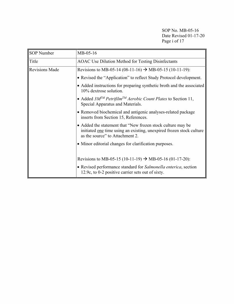
SOP No. MB-05-16 Date Revised 01-17-20 Page i of 17 SOP Number MB-05-16
Title AOAC Use Dilution Method for Testing Disinfectants
Revisions Made Revisions to MB-05-14 (08-11-16) MB-05-15 (10-11-19):
• Revised the “Application” to reflect Study Protocol development.
• Added instructions for preparing synthetic broth and the associated 10% dextrose solution.
• Added 3MTM PetrifilmTM Aerobic Count Plates to Section 11, Special Apparatus and Materials.
• Removed biochemical and antigenic analyses-related package inserts from Section 15, References.
• Added the statement that “New frozen stock culture may be initiated one time using an existing, unexpired frozen stock culture as the source” to Attachment 2.
• Minor editorial changes for clarification purposes.
Revisions to MB-05-15 (10-11-19) MB-05-16 (01-17-20):
• Revised performance standard for Salmonella enterica, section 12.9c, to 0-2 positive carrier sets out of sixty.
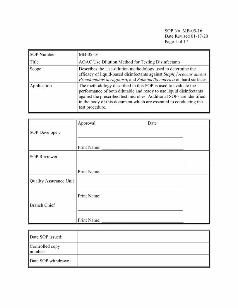
SOP No. MB-05-16 Date Revised 01-17-20 Page 1 of 17 SOP Number MB-05-16 Title AOAC Use Dilution Method for Testing Disinfectants Scope Describes the Use-dilution methodology used to determine the
efficacy of liquid-based disinfectants against Staphylococcus aureus, Pseudomonas aeruginosa, and Salmonella enterica on hard surfaces.
Application The methodology described in this SOP is used to evaluate the performance of both dilutable and ready to use liquid disinfectants against the prescribed test microbes. Additional SOPs are identified in the body of this document which are essential to conducting the test procedure.
SOP No. MB-05-16 Date Revised 01-17-20 Page 2 of 17
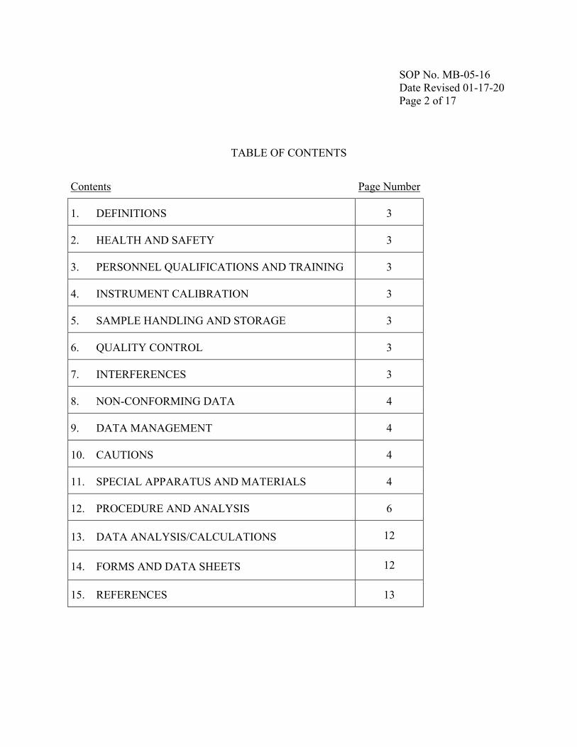
TABLE OF CONTENTS
Contents Page Number
1. DEFINITIONS 3
2. HEALTH AND SAFETY 3
3. PERSONNEL QUALIFICATIONS AND TRAINING 3
4. INSTRUMENT CALIBRATION 3
5. SAMPLE HANDLING AND STORAGE 3
6. QUALITY CONTROL 3
7. INTERFERENCES 3
8. NON-CONFORMING DATA 4
9. DATA MANAGEMENT 4
10. CAUTIONS 4
11. SPECIAL APPARATUS AND MATERIALS 4
12. PROCEDURE AND ANALYSIS 6
13. DATA ANALYSIS/CALCULATIONS 12
14. FORMS AND DATA SHEETS 12
15. REFERENCES 13
SOP No. MB-05-16 Date Revised 01-17-20 Page 3 of 17 1. Definitions 1. Refer to Series 810 – Product Performance Test Guidelines
(https://www.epa.gov/test-guidelines-pesticides-and-toxic-substances/series-810-product-performance-test-guidelines) for testing requirements of antimicrobials under FIFRA.
2. Mean Log Density (LD) = Mean log10 converted carrier count data per test day.
3. A “carrier set” is a test with both primary and secondary subculture tubes for each carrier.
4. Additional abbreviations/definitions are provided in the text.
2. Health and Safety
Follow appropriate biosafety practices specified in SOP MB-01, Laboratory Biosafety. Consult the Safety Data Sheet for specific hazards associated with antimicrobial products.
3. Personnel Qualifications and Training
Refer to SOP ADM-04, OPP Microbiology Laboratory Training.
4. Instrument Calibration
Refer to SOPs EQ-01 (pH meters), EQ-02 (thermometers), EQ-03 (weigh balances), EQ-04 (spectrophotometers) and EQ-05 (timers) for details on method and frequency of calibration.
5. Sample Handling and Storage
Refer to SOP MB-22, Disinfectant Sample Preparation, and SOP COC-01, Chain of Custody Procedures, as necessary.
6. Quality Control For quality control purposes, document the required information on the appropriate form(s) (see section 14).
7. Interferences 1. Any disruption of the Pseudomonas aeruginosa pellicle resulting in the disrupting or breaking of the pellicle in culture before or during its removal renders that culture unusable.
2. Transferring the inoculated carriers into the disinfectant is a critical, technique-sensitive step. False positives can result from transfer of test microbe to sides of tubes due to contact or aerosol formation.
3. Viscous test chemicals may result in a substantial amount of product remaining on treated carriers following the contact time, which upon transfer to the primary subculture medium (neutralizer) produces cloudiness in the medium. This cloudiness may negatively impact the recording of results.
4. For neutralizers/subculture media that do not result in turbidity as the outcome of growth, such as Dey/Engley broth, assess the interpretation of a positive tube in advance of the test (see section 12.7.c.).
SOP No. MB-05-16 Date Revised 01-17-20 Page 4 of 17 8. Non-
conforming Data
1. Sterility and/or viability controls do not yield expected results. 2. The mean LD for control carriers falls outside the specified range.
a. The mean LD for carriers inoculated with S. aureus and P. aeruginosa must be at least 6.0 (corresponding to a geometric mean density of 1.0 × 10⁶) and not above 7.0 (corresponding to a geometric mean density of 1.0 × 10⁷).
b. The mean LD for carriers inoculated with S. enterica must be at least 5.0 (corresponding to a geometric mean density of 1.0 × 10⁵) and not above 6.0 (corresponding to a geometric mean density of 1.0 × 10⁶).
c. Refer to Series 810 for guidance on retesting scenarios. 3. Contamination in subculture tubes deems the test invalid. 4. Manage non-conforming data as specified in the study protocol;
procedures are consistent with SOP ADM-07, Non-Conformance Reports.
9. Data Management
Data will be archived consistent with SOP ADM-03, Records and Archives.
10. Cautions 1. There are time sensitive steps in this procedure including the use periods of the inoculated carriers and the test chemical.
2. Verify the volume of dilution blanks, neutralizer tubes, and subculture tubes in advance and adjust accordingly.
3. When transferring inoculated carriers to disinfectant tubes during testing (see section 12.5.b), avoid intense swirling and agitation of the carriers.
11. Special Apparatus and Materials
1. Subculture/neutralizer media (e.g., letheen broth, fluid thioglycollate medium). Note: Commercial media made to conform to the recipes provided in AOAC Methods 955.15, 964.02, and 955.14 may be substituted.
2. Test organisms. Pseudomonas aeruginosa (ATCC No. 15442), Staphylococcus aureus (ATCC No. 6538) and Salmonella enterica (ATCC No. 10708) obtained directly from ATCC.
3. Culture media. Note: Commercial media (e.g., HiMedia synthetic broth) made to conform to the recipes provided in AOAC Methods 955.15, 964.02, and 955.14 may be substituted. a. Synthetic broth (SB; 10 mL tubes) for daily transfers and final test
cultures. Commercial media (HIMEDIA, Synthetic Broth, AOAC, #M334-500G). Suspend 16.9 g in 1000 mL DI water. Heat if necessary to dissolve the medium completely. Final pH at 25°C should be 7.1±02. Medium may be dispensed in 10 mL amounts in
SOP No. MB-05-16 Date Revised 01-17-20 Page 5 of 17
20×150 mm culture tubes or alternatively in 500 mL volumes in a 1 L bottle; sterilize by autoclaving at 15 lbs pressure (121°C) for 15 minutes. Refrigerate prepared SB (without dextrose added). Just before use, bring SB to room temperature and aseptically add 0.1 mL of 10% sterile dextrose solution per 10 mL SB. i. Alternatively, SB made in-house per the recipe provided in
AOAC Methods 955.15, 964.02, and 955.14 may be substituted.
b. 10% dextrose solution. Add 5.0 g dextrose to 50 mL de-ionized water and mix by stirring. Filter sterilize the solution using a 0.2 µm filter. Refrigerate the sterile solution for up to 30 days.
4. Other media. For example, Tryptic Soy Broth (TSB) and Nutrient Broth (NB) for rehydrating the lyophilized cultures.
5. Trypticase soy agar (TSA). For use in propagation of the test organism to generate frozen cultures and as a plating medium for carrier enumeration. Alternately, TSA with 5% sheep blood (BAP) may be used.
6. Sterile water. Use reagent-grade water free of substances that interfere with analytical methods. Any method of preparation of reagent-grade water is acceptable provided that the requisite quality can be met. See Standard Methods for the Examination of Water and Wastewater and SOP QC-01, Quality Assurance of Purified Water for details on reagent-grade water.
7. Carriers. Polished stainless steel cylinders, 8 ± 1 mm outer diameter, 6 ± 1 mm inner diameter, 10 ± 1 mm length; type 304 stainless steel, SS 18-8 (S & L Aerospace Metals, Maspeth, NY or Fisher Scientific catalog number 07-907-5Q as of September 2019). a. For physical screening, cleaning, and storage of carriers, refer to SOP
MB-03, Screening of Stainless Steel Cylinders, Porcelain Cylinders and Glass Slide Carriers Used in Disinfectant Efficacy Testing.
b. Use only carriers that pass bioscreening. Bioscreen carriers according to SOP MB-03.
8. Specialized glassware. For disinfectant, use autoclavable 25 × 100 mm tubes (Bellco Glass Inc., Vineland, NJ). For glassware used to prepare test chemical, refer to SOP MB-22.
9. Recirculating chiller unit. For maintaining specified temperature of the test chemical.
10. Transfer loops. For performing culture transfers. Make 4 mm inner diameter single loop at end of 50–75 mm (2–3 in.) Pt or Pt alloy wire No.
SOP No. MB-05-16 Date Revised 01-17-20 Page 6 of 17
23 B&S gage or 4 mm loop fused on 75 mm (3 in.) shaft (available from Johnson Matthey, West Chester, PA 19380, USA). Fit other end in suitable holder. Bend loop at 30° angle with stem.
11. Micropipettes. For performing culture transfers and serial dilutions, and adding 10% dextrose solution to SB.
12. Wire Hook. For carrier transfer. Make 3 mm right angle bend at end of 50–75 mm nichrome wire No. 18 B&S gage. Place other end in suitable holder.
13. Timer. For managing timed activities, any certified timer that can display time in seconds.
14. Sonicator (ultrasonic cleaner). For conducting control carrier counts. 15. 3MTM PetrifilmTM Aerobic Count Plates. 3M Food Safety, St. Paul, MN,
USA, Cat. No. 6400. 16. Vitek 2 Compact. For microbe identification.
12. Procedure and Analysis
Prior to testing, perform the neutralization confirmation assay to confirm the use of the appropriate neutralizer and to determine if secondary subculture tubes are necessary (refer to SOP MB-17, Neutralization Confirmation).
12.1 Test Culture Preparation
Refer to SOP MB-02 for the test microbe culture transfer notation. Refer to Attachment 2 for culture initiation and generation of frozen stock cultures.
a. Defrost a cryovial at room temperature and briefly vortex to mix. Add 10 µL of the thawed frozen stock (single use) to a tube containing 10 mL of synthetic broth, vortex, and incubate at 36 ± 1°C for 24 ± 2 h. One daily transfer is required prior to the inoculation of a final test culture. Daily cultures may be subcultured for up to 5 days; each daily culture may be used to generate a test culture. For S. aureus and S. enterica only, briefly vortex1 the 24 h cultures prior to transfer.
b. To generate test cultures, inoculate a sufficient number of 20 × 150 mm tubes containing 10 mL synthetic broth with 10 µL per tube of the 24 h culture then vortex2 to mix. Incubate 48-54 h at 36 ± 1°C. Do not shake the 48-54 h test culture. Record all culture transfers on the Organism Culture Tracking Form (see section 14).
12.2 Carrier Inoculation
Inoculate approximately 80 carriers; 60 carriers are required for testing, 6 for control carrier counts, and 1 for the viability control.
1 Step not contained in the AOAC standard methods 955.14 and 955.15. 2Step not contained in the AOAC standard methods 955.14, 955.15, and 964.02.
SOP No. MB-05-16 Date Revised 01-17-20 Page 7 of 17
a. For P. aeruginosa, remove the pellicle from the 48-54 h test culture either by decanting the liquid aseptically into a sterile tube, by gently aspirating the broth away from the pellicle using a pipette, or by vacuum removal. Avoid harvesting pellicle from the bottom of the tube. Transfer test culture after pellicle removal into sterile 25 × 150 mm test tubes (up to approximately 20 mL per tube) and visually inspect for pellicle fragments. Presence of pellicle in the final culture makes it unusable for testing. Proceed as below in 12.2b.
b. For S. aureus, S. enterica, and P. aeruginosa (from 12.2.a), using a vortex-style mixer, mix 48-54 h test cultures 3-4 s and let stand 10 min at room temperature before continuing. Remove the upper portion of each culture (e.g., upper ¾), leaving behind any debris or clumps, and transfer to a sterile flask; pool cultures in the flask and swirl to mix. Measure and record the OD at 650 nm. Use sterile broth medium to calibrate the spectrophotometer. Use the test culture for carrier inoculation within 30 minutes. Note: To achieve mean carrier counts within the appropriate range (see section 8), the final test culture may be diluted (e.g., one part culture plus one part sterile broth) prior to the addition of the organic soil to the inoculum using the sterile culture medium used to generate the final test culture (e.g., synthetic broth). Use the diluted test culture for carrier inoculation within 30 min. Note: Concentration of the final test culture may be used in the event the bacterial titer in the final test cultures is too low (OD ≤ 0.2). Concentration may be achieved using centrifugation (e.g., 5000 g for 20 min) and resuspending the pellet in the appropriate volume of the sterile final test culture medium necessary to meet the carrier count range. Use the concentrated test culture for carrier inoculation within 30 min.
c. Add appropriate amount of organic soil if required. Swirl to mix. d. Aliquot 20 mL portions into sterile 25 x 150 mm test tubes. e. Drain the water from the carriers. Aseptically transfer 20 carriers
into each of the tubes containing the test culture. The test culture must completely cover the carriers; reposition carriers as necessary to ensure coverage. Alternatively, siphon off the water from the carriers and add 20 mL test culture directly to the carriers without transferring.
f. Allow carriers to remain in the inoculum for 15 ± 2 min.
SOP No. MB-05-16 Date Revised 01-17-20 Page 8 of 17
g. Following the carrier exposure period, remove carriers individually from the inoculum using a flamed nichrome wire hook, briefly tap each carrier against the side of the tube to remove excess culture, and place on end in vertical position in sterile Petri dish matted with 2 layers of Whatman No. 2 (or equivalent) sterile filter paper. Do not remove inoculum from the tube in advance of removing carriers.3 Ensure that carriers do not touch or fall over in the Petri dish. Place no more than 12 carriers in a Petri dish. Place lid on Petri dish.
h. Dry carriers in incubator at 36 ± 1°C for 40 ± 2 min. Record the timed carrier inoculation activities on the AOAC Use-Dilution Method Processing Sheet (see section 14). Expose all carriers to disinfectant within two hours of drying.
12.3 Enumeration of viable bacteria from carriers (control carrier counts)
a. Select one carrier from each of 6 Petri dishes, assay dried carriers in 2 sets of three carriers, one set immediately prior to conducting the efficacy test and one set immediately following the test.
b. Place each inoculated dried carrier into a tube containing 10 mL of letheen broth and sonicate in an ultrasonic cleaner for 1 min ± 5 s. Record the time of sonication on the AOAC Use-Dilution Method Processing Sheet (see section 14).
c. For sonication, place tubes into an appropriately sized glass beaker with tap water to the level of the letheen broth in the tubes. Place the beaker in an ultrasonic cleaner so that the water level in the beaker is even with the water level fill-line on the tank. Fill the tank with tap water to the water level fill-line. Hold the beaker so that it does not touch the bottom of the tank and all 3 liquid levels (inside the test tubes, inside the beaker, and inside the tank) are approximately the same.
d. After sonication, briefly mix and make serial ten-fold dilutions in 9 mL dilution blanks of PBDW. Briefly vortex and plate 0.1 mL aliquots of appropriate dilutions in duplicate on TSA or BAP using spread plating. Plate appropriate dilutions to achieve colony counts in the range of 30-300 colony forming units (CFU) per plate. Spread inoculum evenly over the surface of the agar. Plates must be dry prior to incubation. If the serial dilutions are not made and plated immediately, keep the sonicated tubes at 2-5°C until this step can be done. Complete the dilutions and plating within 2 h after sonication. Alternatively, pool the letheen broth from the tubes with the carriers
3Note: Draining of inoculum with a pipette after contact time is currently provided in the AOAC standard methods 955.14, 955.15, and 964.02.
SOP No. MB-05-16 Date Revised 01-17-20 Page 9 of 17
and briefly vortex for each set of three carriers. Serially dilute and plate 0.1 mL aliquots of the pooled media (30 mL).
e. Incubate plates (inverted) at 36 ± 1°C for up to 48 ± 2 h. f. Count colonies. Plates that have colony counts over 300 will be
reported as TNTC. Record counts on the AOAC Use-Dilution Method Carrier Counts Form and calculate the mean counts (see sections 13 and 14).
g. Alternatively, Petrifilm may be used for enumeration of bacterial organisms. Follow manufacturer’s instructions for preparation and incubation of Petrifilm cards. Note: At a minimum, conduct a culture purity check (isolation streak) using suspension from one dilution tube of one carrier or pooled set.
12.4 Disinfectant Sample Preparation
a. Prepare disinfectant sample per SOP MB-22, Disinfectant Product Preparation.
b. Equilibrate the water bath and allow it to come to 20 ± 1°C or the temperature specified (±1°C). Prepare the disinfectant dilutions within 3 hours of performing the assay unless test parameters specify otherwise. Record the time of disinfectant preparation on the AOAC Use-Dilution Method Processing Sheet (see section 14).
c. Dispense 10 mL aliquots of the disinfectant into 25 × 100 mm test tubes, one tube per carrier. Place tubes in the equilibrated water bath for approximately 10 min to allow disinfectant to come to specified temperature. Record the temperature of the water bath and recirculating chiller before and after testing on the AOAC Use-Dilution Method Information Sheet (see section 14).
12.5 Test Procedure a. Sequentially transfer the carriers from the Petri dish to the test tubes containing the disinfectant at appropriate intervals (e.g., 30 second intervals).
b. Add one carrier per tube and swirl the tube using 2-3 gentle rotations before placing it back in the water bath. Add carrier within ±5 seconds of the specified time for a contact time of 1-10 minutes or within ±3 seconds for contact times <1 minute.
c. Using alternating hooks, flame-sterilize the hook and allow it to cool after each carrier transfer. When lowering the carriers into the disinfectant tubes, neither the carrier itself nor the tip of the wire hook can touch the interior sides of the tube. If the interior sides of the tube are touched, repeat the carrier.
SOP No. MB-05-16 Date Revised 01-17-20 Page 10 of 17
d. Following the exposure time, sequentially transfer the carriers into subculture/neutralizer media. Remove the carrier from the disinfectant with a sterile hook, tap it against the interior sides of the tube to remove the excess disinfectant, and transfer it into the subculture tube within ±5 s. Avoid tapping the carrier against the upper third of the tube. Avoid contact of the carrier to the interior sides of the subculture tube during transfer.
e. Recap the subculture tube and shake thoroughly. Incubate at 36 ± 1°C for 48 ± 2 h.
f. If a secondary subculture tube is deemed necessary to achieve neutralization, then transfer the carrier from the primary tube to a secondary tube of sterile medium after a minimum of 30 ± 5 min at room temperature from the end of the initial transfer. Within 25-60 min of the initial transfer, transfer the carriers using a sterile wire hook to a second subculture tube. Move the carriers in order but the movements do not have to be timed. Thoroughly shake the subculture tubes after all of the carriers have been transferred. Incubate both the primary and secondary subculture tubes 48 ± 2 h at 36 ± 1°C.
g. Record timed events on the AOAC Use-Dilution Method Time Recording Sheet for Carrier Transfers (see section 14).
12.6 Sterility and viability controls
a. Viability controls. Place 1 (or 2) dried inoculated untreated carrier(s) into separate tubes of the neutralizing subculture broth (if primary and secondary media are different). Incubate tubes with the efficacy test. Report results as + (growth) or 0 (no growth) as determined by presence or absence of turbidity. Growth should occur in both tubes. Record results on AOAC Use-Dilution Method Results Sheet (see section 14).
b. Sterility controls. Place one sterile, uninoculated carrier into a tube of neutralizing subculture broth. Incubate tube with the efficacy test. Report results as + (growth), or 0 (no growth) as determined by presence or absence of turbidity. Growth should not occur in the tube. Record results on AOAC Use-Dilution Method Results Sheet (see section 14).
12.7 Results a. Gently shake each tube prior to recording results. Record results as + (growth) or 0 (no growth) as determined by presence or absence of turbidity, on the AOAC Use-Dilution Method Results Sheet (see section 14).
b. For a test with secondary subculture tubes, a positive result in either
SOP No. MB-05-16 Date Revised 01-17-20 Page 11 of 17
the primary or secondary subculture tube is considered a positive result for a carrier set.
c. Specialized neutralizer/subculture medium such as Dey/Engley broth will not show turbidity; rather the presence of pellicle at the surface of the medium (for P. aeruginosa) or a color change to the medium (yellow for growth of S. aureus or S. enterica) must be used to assess the results as a positive or negative outcome. i. Use viability controls for comparative determination of a
positive tube. ii. If the product passes the performance standard, a minimum of
20% of the remaining negative tubes will be assayed for the presence of the test microbe using isolations streaks on TSA or BAP. Record preliminary results and conduct isolation streaks at 48 ± 2 h, however, continue to incubate negative tubes for up to an additional 24 hours to confirm the results.4
12.8 Confirmatory Steps for Test Microbes5
a. For S. aureus, confirm the presence of the test microbe in a minimum of four positive carriers, if present, per test.
b. For P. aeruginosa confirm a minimum of seven positive carriers, if present, per test.
c. For S. enterica, confirm a minimum of three positive carrier sets, if present, per test.
d. For tests with fewer positives than indicated above for each microbe, confirm each positive carrier.
e. For any test with ≥20 positive carriers, confirm a minimum of 50% of the positives.
f. If secondary subculture tubes are used and both primary and secondary subculture tubes are positive in a carrier set, select only the secondary subculture tube for microbe confirmation.
g. To confirm the presence of the test microbe, use Gram staining, solid media, and biochemical evaluation (i.e., Vitek 2 Compact) i. Streak isolate growth from a positive subculture medium tube
onto TSA or TSA with 5% sheep blood, and appropriate selective medium. Incubate media plates 24±2 h at 36 ± 1°C and record the results. Examine colonies on plates for morphology and characteristics of the test organism
4Step not contained in the AOAC standard methods 955.14, 955.15, and 964.02. 5Step not contained in the AOAC standard methods 955.14, 955.15, and 964.02.
SOP No. MB-05-16 Date Revised 01-17-20 Page 12 of 17
(conforming to the morphology in Bergey's Manual). h. See Attachment 1 for typical diagnostic characteristics of the test
microbes (Gram stain reactions, cell morphology, and colony characteristics on solid media).
i. If confirmatory testing determines that the identity of the unknown was not the test organism, annotate the positive entry (+) on the results sheet to indicate a contaminant was present.
12.9 Performance Standard
a. The performance standard for S. aureus is 0-3 positive carriers out of sixty.
b. The performance standard for P. aeruginosa is 0-6 positive carriers out of sixty.
c. The performance standard for S. enterica is 0-2 positive carriers out of sixty.
d. If replicated testing is required for any microbe, conduct testing with that microbe on independent test days.
12.10 Re-use of Stainless Steel Carriers
a. After use, autoclave all carriers. Carriers for which test results were negative may be reused after cleaning. Carriers that are positive are re-cleaned and screened biologically (see SOP MB-03, Screening Carriers) before re-use. These carriers may be reused if the biological screening test results in no growth. The extra inoculated carriers, positive control, and those used for carrier counts may be autoclaved, re-cleaned, and used again.6
13. Data Analysis/ Calculations
Calculate control counts using a Microsoft Excel spreadsheet (see section 14). Both electronic and hard copies of the spreadsheet will be retained.
14. Forms and Data Sheets
1. Attachment 1: Typical Growth Characteristics of strains of P. aeruginosa, S. aureus, and S. enterica
2. Attachment 2: Culture Initiation Flow Chart for S. aureus, P. aeruginosa, and S. enterica, and Preparation of Frozen Stocks
3. Test Sheets. Test sheets are stored separately from the SOP under the following file names: Organism Culture Tracking Form MB-05-16_F1.docx Test Microbe Confirmation Sheet (Quality Control)
MB-05-16_F2.docx
AOAC Use-Dilution Method Time Recording MB-05-16_F3.docx
6Step not contained in the AOAC standard methods 955.14, 955.15, and 964.02.
SOP No. MB-05-16 Date Revised 01-17-20 Page 13 of 17
AOAC Use-Dilution Method Carrier Counts Form (Pooled Carriers)
MB-05-16_F11.docx
15. References 1. Official Methods of Analysis. Method 955.14 – Salmonella enterica. Posted March 2013. AOAC INTERNATIONAL, Gaithersburg, MD.
2. Official Methods of Analysis. Methods 955.15 – Staphylococcus aureus. Posted September 2013. AOAC INTERNATIONAL, Gaithersburg, MD.
3. Official Methods of Analysis. Method 964.02 – Pseudomonas aeruginosa. Posted September 2013. AOAC INTERNATIONAL, Gaithersburg, MD.
4. Krieg, Noel R. and Holt, John G. 1984. Bergey’s Manual of Systematic Bacteriology Volume 1. Williams & Wilkins, Baltimore, MD. P. aeruginosa p. 164, S. enterica p. 447.
5. Sneath, P., Mair, N., Sharpe, M.E., and Holt, J. eds. 1986. Bergey’s Manual of Systematic Bacteriology Volume 2. Williams & Wilkins, Baltimore, MD. S. aureus p. 1015.
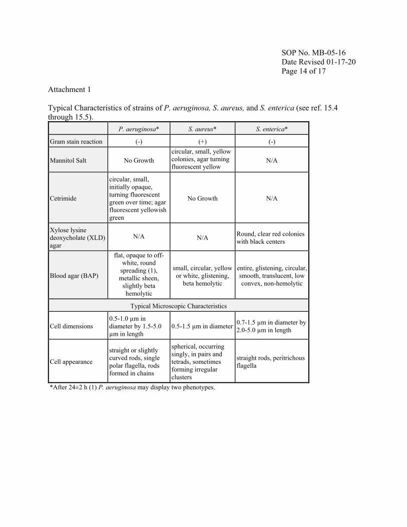
SOP No. MB-05-16 Date Revised 01-17-20 Page 14 of 17 Attachment 1 Typical Characteristics of strains of P. aeruginosa, S. aureus, and S. enterica (see ref. 15.4 through 15.5). P. aeruginosa* S. aureus* S. enterica*
Gram stain reaction (-) (+) (-)
Mannitol Salt No Growth circular, small, yellow colonies, agar turning fluorescent yellow
N/A
Cetrimide
circular, small, initially opaque, turning fluorescent green over time; agar fluorescent yellowish green
No Growth N/A
Xylose lysine deoxycholate (XLD) agar
N/A N/A Round, clear red colonies with black centers
Cell dimensions 0.5-1.0 µm in diameter by 1.5-5.0 µm in length
0.5-1.5 µm in diameter 0.7-1.5 µm in diameter by 2.0-5.0 µm in length
Cell appearance
straight or slightly curved rods, single polar flagella, rods formed in chains
spherical, occurring singly, in pairs and tetrads, sometimes forming irregular clusters
straight rods, peritrichous flagella
*After 24±2 h (1) P. aeruginosa may display two phenotypes.
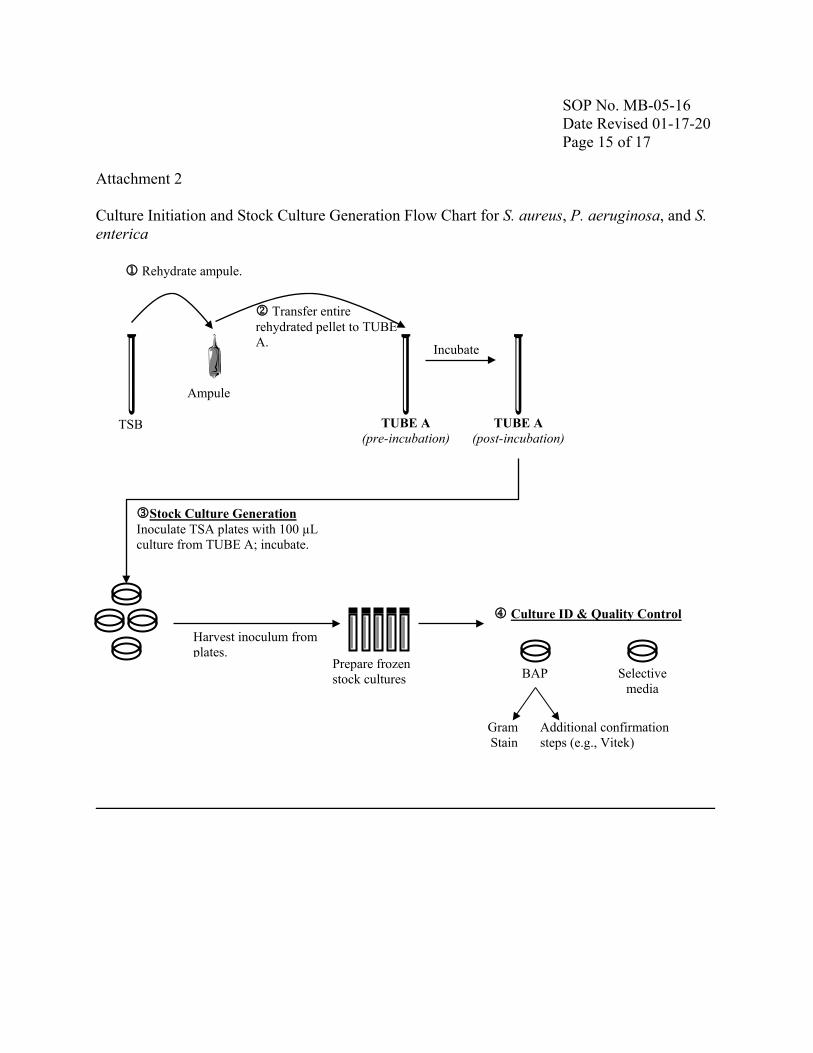
SOP No. MB-05-16 Date Revised 01-17-20 Page 15 of 17 Attachment 2 Culture Initiation and Stock Culture Generation Flow Chart for S. aureus, P. aeruginosa, and S. enterica
Ampule
TUBE A (pre-incubation)
TUBE A (post-incubation)
TSB
Rehydrate ampule.
Transfer entire rehydrated pellet to TUBE A. Incubate
Stock Culture Generation Inoculate TSA plates with 100 µL culture from TUBE A; incubate.
Prepare frozen stock cultures
Harvest inoculum from plates.
Culture ID & Quality Control
Gram Stain
BAP Selective media
Additional confirmation steps (e.g., Vitek)
SOP No. MB-05-16 Date Revised 01-17-20 Page 16 of 17
Attachment 2 continued. Preparation of Frozen Stock Cultures. Refer to SOP MB-02 for establishment of the organism control number.
a. Initiate new stock cultures from lyophilized cultures of Pseudomonas aeruginosa (ATCC 15442), Staphylococcus aureus (ATCC 6538), and Salmonella enterica (ATCC 10708) from ATCC within 18 months. i. New frozen stock culture may be initiated one time using an existing, unexpired
frozen stock culture as the source. Begin process at step C below, by streaking a loopful of the frozen stock culture onto 2 TSA plates.
b. Open ampule of freeze dried organism as indicated by ATCC. Using a tube containing 5-6 mL of TSB for P. aeruginosa and S. aureus and 5-6 mL of NB for S. enterica, aseptically withdraw 0.5 to 1.0 mL and rehydrate the lyophilized culture. Aseptically transfer the entire rehydrated pellet back into the original tube of broth designated as “TUBE A.” Mix well. i. Incubate broth culture (TUBE A) at 36 ± 1ºC for 24 ± 2 h. Record all
manipulations on the Organism Culture Tracking Form (see section 14). c. Following incubation, use a sterile spreader to inoculate a sufficient number of TSA
plates (e.g., 5 to 10 plates per organism) with 100 µL each of the 24 ± 2 h culture. Incubate plates at 36 ± 1°C for 24 ± 2 h. i. For QC purposes, perform a streak isolation of the 24 ± 2 h broth culture, or
frozen stock culture (a.i.), on a BAP. In addition, for S. aureus and P. aeruginosa, streak a loopful onto both selective media (MSA and Cetrimide); for S. enterica, streak a loopful onto XLD. Incubate all plates at 36 ± 1°C for 24 ± 2 h.
d. Following incubation, add 5 mL cryoprotectant solution (TSB with 15% v/v glycerol for S. aureus and P. aeruginosa and NB with 15% v/v glycerol for S. enterica) to the surface of each agar plate. Re-suspend the cells in this solution using a sterile spreader or a sterile swab and aspirate the cell suspension from the surface of the agar. Transfer the suspension into a sterile vessel. Repeat by adding another 5 mL of cryoprotectant to the agar plates, re-suspend the cells, aspirate the suspension and pool with the initial cell suspension. i. For QC purposes, use the pooled suspension to perform a streak isolation on a
BAP. In addition, for S. aureus and P. aeruginosa, streak a loopful onto both selective media (MSA and Cetrimide); for S. enterica, streak a loopful onto XLD. Incubate all plates at 36 ± 1°C for 24 ± 2 h. Continue QC steps as per sections g through i.
e. Mix the pooled contents of the vessel thoroughly. Immediately after mixing, dispense approximately 0.5 to 1.0 mL aliquots into cryovials (e.g., 1.5 mL cryovials).
f. Place and store the cryovials at -70°C or below; these are the frozen stock cultures. Stock
SOP No. MB-05-16 Date Revised 01-17-20 Page 17 of 17
cultures may be used up to 18 months; reinitiate using a new lyophilized culture.7 These cultures are single-use only.
g. Following the incubation period (see d.i), record the colony morphology as observed on the BAPs and selective media plates (including the absence of growth). See Attachment 1 for details on cell and colony morphology, colony characteristics on selective media, and stain reactions.
h. For each organism, perform a Gram stain and Vitek from growth taken from the BAPs according to the manufacturer’s instructions. Observe the Gram reaction by using brightfield microscopy at 1000X magnification (oil immersion).
i. Record all confirmation results on the Test Microbe Confirmation Sheet (Quality Control) (see section 14).
7 Step not contained in the AOAC standard methods 955.14, 955.15, and 964.02.