UvA-DARE is a service provided by the library of the University of Amsterdam (http://dare.uva.nl) UvA-DARE (Digital Academic Repository) Basolateral and apical ABC transporters in liver and intestine de Waart, D.R. Link to publication Citation for published version (APA): de Waart, D. R. (2011). Basolateral and apical ABC transporters in liver and intestine. General rights It is not permitted to download or to forward/distribute the text or part of it without the consent of the author(s) and/or copyright holder(s), other than for strictly personal, individual use, unless the work is under an open content license (like Creative Commons). Disclaimer/Complaints regulations If you believe that digital publication of certain material infringes any of your rights or (privacy) interests, please let the Library know, stating your reasons. In case of a legitimate complaint, the Library will make the material inaccessible and/or remove it from the website. Please Ask the Library: https://uba.uva.nl/en/contact, or a letter to: Library of the University of Amsterdam, Secretariat, Singel 425, 1012 WP Amsterdam, The Netherlands. You will be contacted as soon as possible. Download date: 03 Jan 2020
Transcript
UvA-DARE is a service provided by the library of the University of Amsterdam (http://dare.uva.nl)
UvA-DARE (Digital Academic Repository)
Basolateral and apical ABC transporters in liver and intestine
de Waart, D.R.
Link to publication
Citation for published version (APA):de Waart, D. R. (2011). Basolateral and apical ABC transporters in liver and intestine.
General rightsIt is not permitted to download or to forward/distribute the text or part of it without the consent of the author(s) and/or copyright holder(s),other than for strictly personal, individual use, unless the work is under an open content license (like Creative Commons).
Disclaimer/Complaints regulationsIf you believe that digital publication of certain material infringes any of your rights or (privacy) interests, please let the Library know, statingyour reasons. In case of a legitimate complaint, the Library will make the material inaccessible and/or remove it from the website. Please Askthe Library: https://uba.uva.nl/en/contact, or a letter to: Library of the University of Amsterdam, Secretariat, Singel 425, 1012 WP Amsterdam,The Netherlands. You will be contacted as soon as possible.
acetonitrile/ 90% ammonium formate (20 mM), pH 3.5, followed by a linear gradient to 30%
acetonitrile in the same buffer in 20 min. Cefadroxil had a retention time of 14 min. Detection
of cefadroxil was done at 260 nm. Quantification of cefadroxil was done by using a
calibration curve of cefadroxil.
In vivo experiment: Jejunal administration of cefadroxil and portal blood collection
Mice were anesthetized with a combination of Hypnorm (VetaPharma, Leeds, UK; 11.8
mg/kg fluanisone and 0.37 mg/kg fentanyl citrate) and Valium (Centrafarm, Etten-Leur, The
Netherlands; 5.9 mg/kg Diazepam). Body temperature was maintained between 35°C and
37°C by keeping the mice on thermostatted heating pads. After induction of anaesthesia, the
vena porta was cannulated, followed by ligation of 10 cm of the middle part of the jejunum.
500 µl cefadroxil (5 µM; 2µCi) was subsequently injected into the ligated jejunum and portal
blood samples were collected after indicated time points. At the end of the experiment a
peripheral blood sample was taken by cardiac puncture. Mice were subsequently
anaesthetized and liver, the ligated jejunum, kidneys and gallbladder were collected. After
118
119
the addition of H2O2 (30%) to blood, jejunum, kidney, liver and jejunum radioactivity was
measured by liquid scintillation counting.
Statistical analyses
Statistical differences were determined by an unpaired Student’s t-test. All data were
expressed as means ± standard deviation.
Table 1: Cephalosporins used in the inhibition studies.
molecular weight oral available
Cephalexin 347.4 yes
Cephradine 349.4 yes
Cefadroxil 363.4 yes
Cefaclor 367.8 yes
Cefuroxime 424.4 yes
Cefoxitin 427.4 no
Cefamandole 462.5 no
Cefmetazole 471.5 no
Ceftriaxon 554.6 no
Cefoperazone 645.7 no
119
120
Figure 1: Inhibition of estradiol-17ß-glucuronide transport by cephalosporins. Cephalosporin inhibition of E217ßG (0.05 µCi; 1.0 µM) transport into, ABCC2, ABCC3 and ABCC4 containing Sf21 membrane vesicles. Shown are the measurements of the IC50 values for E217ßG (0.05 µCi; 1.0 µM) transport mediated by (A) ABCC2 (solid line), (B) ABCC3 (dashed line) and (C) ABCC4 (dotted line), inhibited by different concentrations of cefadroxil. (D) IC50 values for inhibition of E217ßG transport by cephalosporins. Transport mediated by ABCC2, ABCC3 and ABCC4 versus molecular weight of these cephalosporins.
120
121
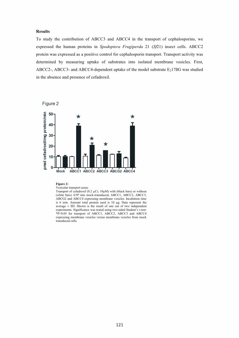
Results
To study the contribution of ABCC3 and ABCC4 in the transport of cephalosporins, we
expressed the human proteins in Spodoptera Frugiperda 21 (Sf21) insect cells. ABCC2
protein was expressed as a positive control for cephalosporin transport. Transport activity was
determined by measuring uptake of substrates into isolated membrane vesicles. First,
ABCC2-, ABCC3- and ABCC4-dependent uptake of the model substrate E217ßG was studied
in the absence and presence of cefadroxil.
Figure 2: Vesicular transport assay. Transport of cefadroxil (0.2 µCi; 10µM) with (black bars) or without (white bars) ATP into mock-transduced, ABCC1, ABCC2, ABCC3, ABCG2 and ABCC4 expressing membrane vesicles. Incubation time is 6 min. Amount total protein used is 10 µg. Data represent the average ± SD. Shown is the result of one out of two independent experiments. Significance was tested using two-sided Student’s t-test: *P<0.05 for transport of ABCC1, ABCC2, ABCC3 and ABCC4 expressing membrane vesicles versus membrane vesicles from mock transduced cells.
121
122
Figure 3: Time- and concentration-dependent transport. Time-dependent transport of cefadroxil (0.2 µCi; 10 µM) into plasma membrane vesicles from Sf21 cells expressing ABCC1 (A), ABCC3 (B) and ABCC4 (C). Open squares (with dotted line) and open diamonds represent incubations with or without ATP, respectively. Filled triangles (with solid line) represent ATP dependent transport. Concentration-dependent transport of cefadroxil (0.2 µCi; 10 µM) into plasma membrane vesicles (open diamonds with solid line) from Sf21 cells expressing ABCC1 (D), ABCC3 (E) and ABCC4 (F). Shown are the average ± SD of ATP-dependent transport of a representative experiment with triplicate incubations performed 3-4 times.
122
123
Figure 4: Transport of cephadroxil across jejunal explants in Ussing chambers. Appearance of cefadroxil on the basolateral side of jejunal explants from Abcc3-/-, Abcc4-/- and Abcc3-/-/Abcc4-/- and wild type mice. Cefadroxil was applied to the apical compartment of Ussing chambers at a final concentration of 5 µM. At indicated time points samples were taken from the basolateral side and analyzed by HPLC. Shown is the average ± standard deviation of 3-4 experiments with triplicate incubations. ‡, p < 0.05 comparing appearance of cefadroxil using tissue from Abcc4-/- versus wild type mice for indicated time points. #, p < 0.05 comparing appearance of cefadroxil using tissue from Abcc3-/-/Abcc4-/- versus wild type mice for indicated time points.
A concentration-dependent inhibition of E217ßG uptake by cefadroxil was observed in
with very different IC50’s. The same inhibition studies were performed for nine other
cephalosporins (table 1). For all cephalosporins the concentration at which E217ßG uptake
was inhibited by 50% (IC50) was determined and plotted as a function of the molecular weight
of the cephalosporins (Fig. 1D). For all three ABC transporters a near linear, inverse
correlation between IC50-values and molecular weight of the cephalosporins was observed.
The orally-available cephalosporins (see Table 1) have the lowest molecular weight,
indicating that molecular weight/bulkiness of the substrate is an important determinant for
oral availability. Moreover, the orally available cephalosporins show the largest difference
with regard to IC50-values of cephalosporins towards transport of E217ßG mediated by
ABCC4 versus ABCC2 and ABCC3, respectively. Competitive inhibition is an indication that
the inhibitor may be a transported substrate. Cefadroxil is an orally prescribed cephalosporin
that is commercially available in a radiolabelled form allowing its use in direct transport
experiments. We, therefore, used cefadroxil as a model substrate for orally prescribed
cephalosporins, in vesicular transport experiments. ABCG2 did not transport cefadroxil.
However, membrane vesicles containing ABCC1, ABCC2, ABCC3 and ABCC4 all showed
ATP-dependent uptake of cefadroxil (Fig. 2). In line with the inhibition experiments,
transport of cefadroxil mediated by ABCC4 was three- to six-fold higher compared to
transport by ABCC2 and ABCC3 (Fig. 2). Furthermore, cefadroxil transport was time
dependent (Fig. 3A-C) and saturable (Fig. 3D-E) for ABCC1, ABCC3 and ABCC4 with Km
values of 3.9±0.6, 2.5±0.7 and 0.25±0.07 mM, respectively.
We subsequently tested whether there could be a role for Abcc3 and/or Abcc4 in vivo in the
transport of cefadroxil across the basolateral membrane of the enterocyte in Ussing chamber
experiments. No difference was found in transport of cefadroxil from the apical to the
123
124
basolateral compartment using jejunal tissue from wild type and Abcc3-/- mice. Transport of
cefadroxil, (5 µM), was reduced to about 50% of normal levels, when jejunal tissue of Abcc4-
/- and Abcc3-/-/Abcc4-/- mice were used (Fig. 4). Similar results were found with 50 µM
cefadroxil (data not shown). Finally, in intestinal loop experiments we found that injection of
cefadroxil in the ligated jejunum resulted in the time-dependent appearance of cefadroxil in
portal blood of wild type mice (Fig. 5). The appearance of cefadroxil in portal blood of Abcc3-
/- and Abcc4-/- mice was however not different from wild type mice, the latter result being in
contrast to the Ussing chamber data. Interestingly, the appearance of cefadroxil in portal
blood of Abcc3-/-/Abcc4-/- mice was reduced. Similar results were found for peripheral blood:
A significantly lower concentration of cefadroxil was found in Abcc3-/-/Abcc4-/- mice in
comparison with wild type mice. There was no difference in the cefadroxil tissue content of
intestine, kidney and liver nor in its urinary excretion between the mice strains tested.
However, an increased amount of cefadroxil was noticed in the very low (less than 0.4% of
dose) biliary content in Abcc3-/-/Abcc4-/- versus wild type mice (Fig. 5D).
Figure 5: In vivo uptake of cefadroxil by the jejunum. Appearance of cefadroxil in portal (closed symbols) and peripheral blood (open symbols) of Abcc3-/- versus wild type mice (A), Abcc4-/- versus wild type mice (B) and Abcc3-/-/Abcc4-/- versus wild type mice (C) after injection of 500 µl cefadroxil (5 µM; 2µCi) into ligated jejunum. Portal blood was collected after indicated time points, peripheral blood after the last portal blood sample was drawn. Appearance of cefadroxil in bile of Abcc3-/-/Abcc4-/- versus wild type mice (D) after injection of 500 µl cefadroxil (5 µM; 2µCi) into ligated jejunum. Data represent average ± SD of at least 5 mice. Significance was tested using two-sided Student’s t-test: *P<0.05 for Abcc3-/-/Abcc4-/- versus wild type mice.
124
125
Discussion
Orally prescribed cephalosporins are efficiently taken up in the gut. At the luminal
side of the small intestine the dipeptide transporter PepT1 is involved in the import of
cephalosporin. At present the transporter(s) involved in the translocation of
cephalosporins from the enterocytes into blood is (are) still unknown. ABCC2
transports several cephalosporins like ceftriaxone, cefaperazone and cephalexin (302,
471). Because of its apical expression, ABCC2 can not be responsible for the
transport of oral cephalosporins into blood. As ABCC3 and ABCC4 are related
transporters we tested their ability to mediate transport of one of the orally prescribed
cephalosporins, namely cefadroxil. In this article we show that both ABCC3 and
ABCC4 mediate transport of cefadroxil albeit with different kinetics: of the ABC
transporters under study ABCC4 mediates transport with the highest affinity (Fig. 2
and 3). This is in line with an earlier report showing that the non-oral available
cephalosporins ceftizoxime, cefazolin, cefotaxime and cefmetazole are substrates of
ABCC4 (484). In this paper the Km values of 18 and 81 µM were stated for MRP4-
mediated transport of respectively ceftizoxime and cefazolin (484). Since cefadroxil is
a smaller compound than both these cephalosporins the Km value of 246 µM is higher
as expected. This is based on our findings that an inverse correlation exists between
molecular weights of cephalosporins and IC50 values (Fig. 1D) and a relation between
Km and IC50 values. Our in vivo experiments with wild type, Abcc3-/- and Abcc4-/-
single knockout mice showed no difference in transport of cefadroxil from luminal to
serosal side (Fig 5). We have two explanations for this result: one explanation is that
murine Abcc3 and Abcc4, unlike their human orthologues used in the vesicular
transport experiments, are unable to mediate transport of cefadroxil (Fig. 2 and 3).
This explanation is unlikely as we observed in Ussing chamber experiments that
transport was reduced in intestinal explants from Abcc4-/- mice compared to wild type
mice. Another explanation is that these transporters can compensate for the loss of
each other. Therefore, we extended the experiments with Abcc3-/-/Abcc4-/- mice. The
Abcc3-/-/Abcc4-/- mice had reduced levels of cefadroxil in their portal blood (Fig. 5).
As similar results were found in Ussing chamber experiments using jejunal explants
from Abcc3-/-/Abcc4-/- and wild type mice, conclude that both Abcc3 and Abcc4
transport cefadroxil and when one transporter is lacking the other can fully
compensate. Human ABCC4 transports cefadroxil at higher rates than ABCC3 in
vesicular transport experiments. Expression of murine Abcc3 is higher than murine
125
126
Abcc4 in the gut. This might be the reason that although Abcc3 transports cefadroxil
only at moderate rates it can still compensate for the loss of Abcc4. As the difference
in expression level between Abcc3/ABCC3 and Abcc4/ABCC4 in jejunum is similar
in mouse and man we speculate that also in the human situation ABCC3 and ABCC4
are involved in transport of cefadroxil from enterocyte to blood (485, 486).
The potential involvement of Abcc4 in the basolateral transport of cefadroxil is
important since literature data are not consistent about the cellular localization of
Abcc4. In the human colonic cell line HT29-CL19A, Li et al. found Abcc4 in both the
apical and basolateral membrane, with a higher expression apically (477). In Caco2
cells Ming and Thakker detected Abcc4 mainly in basolateral membrane (478). Our
data that intestinal Abcc4 influences cefadroxil uptake supports the notion that Abcc4
is present at the basolateral mebrane of the enterocytes. Our functional data suggest
that Abcc4 is present at the basolateral membrane of enterocytes and confirm the data
obtained in Caco2 cells by Ming and Thakker (478).
Abcc3-/-/Abcc4-/- mice still show a considerable amount of transport of cefadroxil over
the basolateral membrane of enterocytes. Theoretically, Abcc1 could be responsible
for the residual transport of cefadroxil as we found that human ABCC1 is able to
mediate transport of cefadroxil (Fig 2 and 3). However, Abcc1 is expressed in the
small intestine, mainly in the crypts (86, 189) which does not colocalize with PepT1,
which is abundantly present in the villus tip with decreasing levels towards the villus
base. The localization of Abcc1 therefore argues against a role of Abcc1 in the
basolateral efflux of cefadroxil in enterocytes. We can, however, not fully exclude
that there is a role of Abcc1 in the efflux of cefadroxil as this transporter protects
against the intestinal toxicity evoked by methotrexate (86). This means that Abcc1 is
pumping methotrexate out of the cell at a physiological relevant speed. The same
might be the case for Abcc1 mediated transport of cefadroxil into blood from the
enterocyte, especially in the absence of Abcc3 and Abcc4 in the used murine model:
Abcc3-/-/Abcc4-/- mice.
Oral administration is a preferable route of administration of drugs. In general, the
oral available cephalosporins are low molecular weight molecules. Kato et al. showed
a correlation between size and biliary excretion for several cephalosporins (471). This
can be partially caused by a size-dependent affinity for the transporter mainly
responsible for the biliary excretion, Abcc2/ABCC2 (471). In line with data of Kato et
al., we found a size dependent inhibition by cephalosporins of ABCC2-mediated
126
127
transport of E217ßG (Fig 1D). Also, a size-dependent inhibition by cephalosporins
was found by us for ABCC3- and ABCC4-mediated transport of E217ßG (Fig 1D),
but the slope for ABCC2 is steeper than for ABCC4 and similar for ABCC3. This
could indicate that the influence of size is relatively big for ABCC2 (which prevents
net uptake) and relatively small for ABCC4 (which stimulates net uptake).
Interestingly, an inverse selectivity of PepT1 was shown to be dependent on
molecular weight as well, with small cephalosporins being preferentially transported.
Hence, the combined selectivity of the uptake transporter Pept1 and the efflux
transporter ABCC2 may determine the typical pharmacokinetic behaviour of
cephalosporins.
In conclusion, the data presented in this paper demonstrate that murine intestinal
uptake depends partly on Abcc3 and Abcc4. We therefore speculate that in the human
situation oral availability involves, at least partly, uptake via ABCC3 and ABCC4.
127
128
Reference List
Assem M, Schuetz EG, Leggas M, Sun D, Yasuda K, Reid G, Zelcer N, Adachi M, Strom S, Evans RM, Moore DD, Borst P and Schuetz JD (2004) Interactions between hepatic Mrp4 and Sult2a as revealed by the constitutive androstane receptor and Mrp4 knockout mice. J Biol Chem 279:22250-22257. Bakos E, Evers R, Sinko E, Varadi A, Borst P and Sarkadi B (2000) Interactions of the human multidrug resistance proteins MRP1 and MRP2 with organic anions. Mol Pharmacol 57:760-768. Borst P, de WC and van de WK (2007) Multidrug resistance-associated proteins 3, 4, and 5. Pflugers Arch 453:661-673. Breedveld P, Zelcer N, Pluim D, Sonmezer O, Tibben MM, Beijnen JH, Schinkel AH, van TO, Borst P and Schellens JH (2004) Mechanism of the pharmacokinetic interaction between methotrexate and benzimidazoles: potential role for breast cancer resistance protein in clinical drug-drug interactions. Cancer Res 64:5804-5811. Ci L, Kusuhara H, Adachi M, Schuetz JD, Takeuchi K and Sugiyama Y (2007) Involvement of MRP4 (ABCC4) in the luminal efflux of ceftizoxime and cefazolin in the kidney. Mol Pharmacol 71:1591-1597.
de Waart DR, Paulusma CC, Kunne C and Oude Elferink RP (2006) Multidrug resistance associated protein 2 mediates transport of prostaglandin E2. Liver Int 26:362-368.
Heijn M, Oude Elferink RP and Jansen PL (1992) ATP-dependent multispecific organic anion transport system in rat erythrocyte membrane vesicles. Am J Physiol 262:C104-C110.
Kato S, Ito K, Kato Y, Wakayama T, Kubo Y, Iseki S and Tsuji A (2009) Involvement of multidrug resistance-associated protein 1 in intestinal toxicity of methotrexate. Pharm Res 26:1467-1476. Kato Y, Takahara S, Kato S, Kubo Y, Sai Y, Tamai I, Yabuuchi H and Tsuji A (2008) Involvement of multidrug resistance-associated protein 2 (Abcc2) in molecular weight-dependent biliary excretion of beta-lactam antibiotics. Drug Metab Dispos 36:1088-1096. Leggas M, Adachi M, Scheffer GL, Sun D, Wielinga P, Du G, Mercer KE, Zhuang Y, Panetta JC, Johnston B, Scheper RJ, Stewart CF and Schuetz JD (2004) Mrp4 confers resistance to topotecan and protects the brain from chemotherapy. Mol Cell Biol 24:7612-7621. Li C, Krishnamurthy PC, Penmatsa H, Marrs KL, Wang XQ, Zaccolo M, Jalink K, Li M, Nelson DJ, Schuetz JD and Naren AP (2007) Spatiotemporal coupling of cAMP transporter to CFTR chloride channel function in the gut epithelia. Cell 131:940-951. Livermore DM (2009) Has the era of untreatable infections arrived? J Antimicrob Chemother 64 Suppl 1:i29-i36. Maher JM, Slitt AL, Cherrington NJ, Cheng X and Klaassen CD (2005) Tissue distribution and hepatic and renal ontogeny of the multidrug resistance-associated protein (Mrp) family in mice. Drug Metab Dispos 33:947-955. Maliepaard M, Scheffer GL, Faneyte IF, van Gastelen MA, Pijnenborg AC, Schinkel AH, van d, V, Scheper RJ and Schellens JH (2001) Subcellular localization and distribution of the breast cancer resistance protein transporter in normal human tissues. Cancer Res 61:3458-3464. Ming X and Thakker DR (2010) Role of basolateral efflux transporter MRP4 in the intestinal absorption of the antiviral drug adefovir dipivoxil. Biochem Pharmacol 79:455-462. Mottino AD, Hoffman T, Jennes L and Vore M (2000) Expression and localization of multidrug resistant protein mrp2 in rat small intestine. J Pharmacol Exp Ther 293:717-723.
128
129
Nakashima E, Tsuji A, Mizuo H and Yamana T (1984) Kinetics and mechanism of in vitro uptake of amino-beta-lactam antibiotics by rat small intestine and relation to the intact-peptide transport system. Biochem Pharmacol 33:3345-3352. Oude Elferink RP and de WR (2007) Transporters in the intestine limiting drug and toxin absorption. J Physiol Biochem 63:75-81. Oude Elferink RP and Jansen PL (1994) The role of the canalicular multispecific organic anion transporter in the disposal of endo- and xenobiotics. Pharmacol Ther 64:77-97. Paulusma CC, Kool M, Bosma PJ, Scheffer GL, ter Borg F, Scheper RJ, Tytgat GN, Borst P, Baas F and Oude Elferink RP (1997) A mutation in the human canalicular multispecific organic anion transporter gene causes the Dubin-Johnson syndrome. Hepatology 25:1539-1542.
Peng KC, Cluzeaud F, Bens M, Van Huyen JP, Wioland MA, Lacave R and Vandewalle A (1999) Tissue and cell distribution of the multidrug resistance-associated protein (MRP) in mouse intestine and kidney. J Histochem Cytochem 47:757-768. Rius M, Hummel-Eisenbeiss J and Keppler D (2008) ATP-dependent transport of leukotrienes B4 and C4 by the multidrug resistance protein ABCC4 (MRP4). J Pharmacol Exp Ther 324:86-94. Russel FG, Koenderink JB and Masereeuw R (2008) Multidrug resistance protein 4 (MRP4/ABCC4): a versatile efflux transporter for drugs and signalling molecules. Trends Pharmacol Sci 29:200-207. Scheffer GL, Kool M, de Haas M, de Vree JM, Pijnenborg AC, Bosman DK, Elferink RP, van d, V, Borst P and Scheper RJ (2002) Tissue distribution and induction of human multidrug resistant protein 3. Lab Invest 82:193-201.
Scheffer GL, Kool M, Heijn M, De HM, Pijnenborg AC, Wijnholds J, van HA, de Jong MC, Hooijberg JH, Mol CA, van der LM, de Vree JM, van d, V, Elferink RP, Borst P and Scheper RJ (2000) Specific detection of multidrug resistance proteins MRP1, MRP2, MRP3, MRP5, and MDR3 P-glycoprotein with a panel of monoclonal antibodies. Cancer Res 60:5269-5277. Taipalensuu J, Tornblom H, Lindberg G, Einarsson C, Sjoqvist F, Melhus H, Garberg P, Sjostrom B, Lundgren B and Artursson P (2001) Correlation of gene expression of ten drug efflux proteins of the ATP-binding cassette transporter family in normal human jejunum and in human intestinal epithelial Caco-2 cell monolayers. J Pharmacol Exp Ther 299:164-170. Tsuji A, Nakashima E, Kagami I and Yamana T (1981) Intestinal absorption mechanism of amphoteric beta-lactam antibiotics I: Comparative absorption and evidence for saturable transport of amino-beta-lactam antibiotics by in situ rat small intestine. J Pharm Sci 70:768-772. van Aubel RA, Smeets PH, Peters JG, Bindels RJ and Russel FG (2002) The MRP4/ABCC4 gene encodes a novel apical organic anion transporter in human kidney proximal tubules: putative efflux pump for urinary cAMP and cGMP. J Am Soc Nephrol 13:595-603. Zelcer N, Saeki T, Reid G, Beijnen JH and Borst P (2001) Characterization of drug transport by the human multidrug resistance protein 3 (ABCC3). J Biol Chem 276:46400-46407. Zelcer N, van de WK, de WR, Scheffer GL, Marschall HU, Wielinga PR, Kuil A, Kunne C, Smith A, van d, V, Wijnholds J, Elferink RO and Borst P (2006) Mice lacking Mrp3 (Abcc3) have normal bile salt transport, but altered hepatic transport of endogenous glucuronides. J Hepatol 44:768-775.