Page 1
AIP Conference Proceedings 2157, 020010 (2019); https://doi.org/10.1063/1.5126545 2157, 020010
© 2019 Author(s).
Vibrational frequencies and electronicstructures of 2-amino-1, 9-dihydro-9-[(2-hydroxuethoxy) methyl]-6H-purin-6-oneusing density functional theory methodCite as: AIP Conference Proceedings 2157, 020010 (2019); https://doi.org/10.1063/1.5126545Published Online: 18 September 2019
Suh-Miin Wang, Lee Muei Chng, Montha Meepripruk, and Pek-Lan Toh
Page 2
Vibrational Frequencies and Electronic Structures of 2-
amino-1, 9-dihydro-9-[(2-hydroxuethoxy) methyl]-6H-
purin-6-one using Density Functional Theory Method
Suh-Miin Wang1, Lee Muei Chng1, Montha Meepripruk2, Pek-Lan Toh1*
1Faculty of Engineering and Green Technology, Universiti Tunku Abdul Rahman, Malaysia
2Institute of Science and Technology, Kampheang Phet Rajabhat University, Thailand
*Corrsponding author: [email protected]
Abstract. In this study, Density Functional Theory (DFT) calculations were conducted on Acyclovir (ACV) hydrate molecular
system with the chemical formula of 3[C8H11N5O3]·2[H2O]. Geometry optimization calculations were performed to find the
equilibrium structure of 3[C8H11N5O3]·2[H2O]. The optimized geometries were used to find the electronic structures of title
molecule, i.e. total energies, frontier molecular orbital energies, atomic charges, and others. The results show that all structural
parameters obtained are close to that of experiment. The total energies of 3[C8H11N5O3]·2[H2O] are -70355.92 eV and -
70373.84 eV, respectively for DFT/B3LYP/6-31G** and DFT/B3LYP/6-311G** levels of calculations. Using the scheme of
Mulliken population analysis (MPA), the highest positive charge was localised on the atom of C12, while for natural atomic
charge distribution, C9 atom possesses the highest positive charge. Moreover, the results of vibrational frequencies computed
with two different basis sets are close to each other. The computed vibrational frequencies are overall corresponded with the
region in the literature studies.
INTRODUCTION
In recent years, the subject of drug polymorphism has become an essential part of drug development.
Numerous experiments have been conducted on the polymorph drugs [1-10]. In 2015, Censi and Martino studied
the stability and bioavailability on the anhydrous and solvated forms of polymorph drugs [8]. The bioavailability
of several polymorph drugs were summarised and discussed in the studied. Nicoud et al, in 2018 reported the
solubility results of three metastable polymorphs, such as paracetamol, chloramphenicol palmitate, and
sulfathiazole using three types of experiments [10]. It was known that the different molecular configurations can
be affected the physical and chemical properties, such as colour, solubility, stability, and others. For example, a
monoclinic form of ACV was studied by Birnbaum et al. [1], and a triclinic form of ACV was presented by
Suwinska et al. [2]. Birnbaum and his co-workers, in 1984 found that the monoclinic form of ACV contains three
molecules of ACV and two water molecules. The lattice parameters obtained are a = 25.459Å, b = 11.282Å, c
= 10.768Å, and β = 95.16°, respectively. While for Suwinska et al. studies, ACV also contains ACV and water
molecules. In 2013, Montha and Kenneth also re-investigated the triclinic form of tricyclic ACV [6]. The findings
are close to Suwinska et al. studies. ACV is one of the anti-viral drugs [1-7, 9]. It can help to treat Herpes simplex
virus (i.e. HSV-1 and HSV-2) infections, i.e. cold sore, chickenpox, and shingles by inhibiting the replication of
viral DNA. The polymorph drugs can change the physicochemical properties, i.e. stability, solubility,
bioavailability, and dissolution rate [1-10]. However, previously literature studies for the electronic structures of
3[C8H11N5O3]·2[H2O] have several limitations. To improve the better understanding of polymorphism features
for ACV under the drug development and industrial pharmacy, a DFT method was employed to study the
structural molecules and electronic properties of 3[C8H11N5O3]·2[H2O] in this work.
COMPUTATIONAL METHODOLOGY
All DFT calculations were carried out using personal computer with the help Gaussian 09 program
package [11]. A molecular system of 3[C8H11N5O3]·2[H2O] was used as the host environment in this work. Figure
International Symposium on Green and Sustainable Technology (ISGST2019)AIP Conf. Proc. 2157, 020010-1–020010-9; https://doi.org/10.1063/1.5126545
Published by AIP Publishing. 978-0-7354-1902-5/$30.00
020010-1
Page 3
1 shows the molecular structure view of 3[C8H11N5O3].2[H2O]. For the geometry optimization calculations,
Becke’s Three Lee-Yang-Parr (B3LYP) functional with 6-31G** and 6-311G** basis sets were chosen. The
optimized geometry structures and electronic properties were then obtained. In addition, the surface plots of
HOMO, LUMO, and molecular electrostatic potential (MEP) were also illustrated using GaussView 5.0 and
Multiwfn software package [11-12].
Figure 1. Molecular structure view of 3[C8H11N5O3]·2[H2O]
RESULT AND DISCUSSION
Table 1 shows the results of geometrical structures of 3[C8H11N5O3]·2[H2O] using the hybrid B3LYP
functional and two basis sets (i.e. 6-31G** and 6-311G**). Since our computational results are focused on
isolated molecule, the comparison geometries between the computational data and experiment are presented in
this work. From the results reported in the table, the results obtained from both DFT/B3LYP/6-31G** and
DFT/B3LYP/6-311G** levels of calculations are almost identical. All computed geometries are very close with
experimental values [9]. For example, the percentage difference between the overall computed C-O and
experimental bond values are less than 2.9% in the molecular system. While for C-N bonds in
[C8H11N5O3]·2[H2O], the percentage difference between the calculated data and experiment are less than 3.9%. In
the case of bond angle, C-C-O possesses the highest percentage difference between the computational and
experimental values, which is less than 6% in the molecular system. On the other hand, it was noted that all
computed dihedral angles obtained from B3LYP/6-31G** and B3LYP/6-311G** technique are significantly
closer to measurement data as shown in the table. From the optimised structure of 3[C8H11N5O3]·2[H2O], the
findings notice that there is no significantly change in the molecular system. Therefore, there is no further study
on the geometry structures.
Table 2 gathers the computed total and Frontier molecular orbital energies of 3[C8H11N5O3]·2[H2O]. Both
of the results obtained from DFT/B3LYP/6-31G** and DFT/B3LYP/6-311G** levels of calculations show good
agreement with each other. The total energy of 3[C8H11N5O3]·2[H2O] obtained from B3LYP/6-31G** method is -
70355.92 eV, whereas the value of -70373.84 eV is found for DFT/B3LYP/6-311G** level of calculation. To
understand the details of chemical reactivity, electron transport mechanism, and stability of 3[C8H11N5O3]·2[H2O],
the values of HOMO-LUMO gaps in Table 2 and Figure 2 are determined to be about 5.01 eV and 4.91 eV,
respectively for B3LYP functional with employing 6-31G** and 6-311G** basis sets. Moreover, the dipole
020010-2
Page 4
moments of 14.82 debye is obtained from DFT/B3LYP/6-31G** level of theory, whereas for B3LYP/6-311G**
method, the dipole moments of 3[C8H11N5O3]·2[H2O] is determined to be about 14.60 debye. The details of
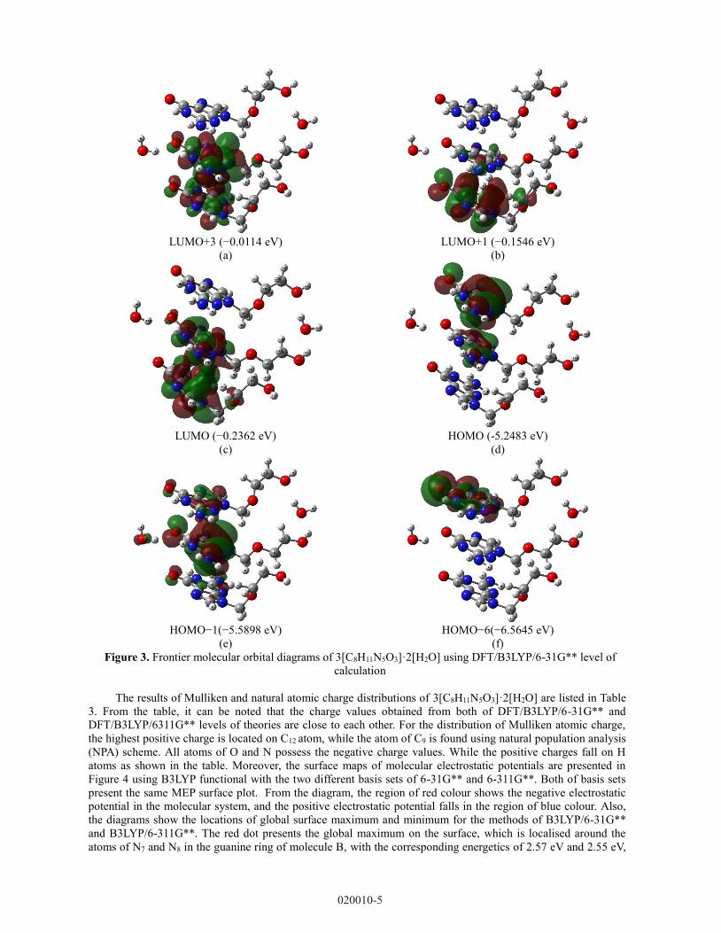
Frontier molecular orbitals (FMOs) are studied in this work. It was found that the diagrams obtained from the
functional of B3LYP with two basis sets of 6-31G** and 6-311G** are close to each other. In Figure 3, the
diagrams of Frontier molecular orbitals are illustrated using B3LYP/6-31G** method. As can be seen from the
diagrams, the electron density distribution of Lowest Unoccupied Molecular Orbital (LUMO) is mainly localised
on the guanine group of molecules B and C. For Highest Occupied Molecular Orbital (HOMO), the distribution
of electron density is focused on the guanine group of molecule A.
Table 1. Computed molecular geometries of 3[C8H11N5O3]·2[H2O]
Molec
ule
A
B3LY
P/
6-
31G*
*
B3LY
P/
6-
311G*
*
Exp
[9]
Molecul
e
B
B3LY
P/
6-
31G*
*
B3LY
P/
6-
311G*
*
Exp
[9]
Molecul
e
C
B3LY
P/
6-
31G*
*
B3LY
P/
6-
311G*
*
Exp
[9]
Bond Distance (Å)
O1-C1 1.216 1.209 1.23
9 O4-C9 1.222 1.216
1.24
1 O7-C17 1.217 1.210
1.24
5
C1-N1 1.441 1.442 1.39
1 C9-N7 1.419 1.419
1.38
3 C17-N12 1.442 1.443
1.38
8
N2-C4 1.372 1.370 1.37
5 N7-C12 1.375 1.373
1.37
2 N12-C20 1.366 1.364
1.36
8
N1-C3 1.361 1.359 1.35
3 N6-C11 1.357 1.355
1.33
9 N11-C19 1.356 1.353
1.34
8
C3-C2 1.393 1.390 1.37
5 C11-C10 1.391 1.389
1.39
2 C19-C18 1.394 1.392
1.37
5
C4-N3 1.369 1.369 1.33
5 C12-N8 1.361 1.361
1.33
6 C20-N13 1.362 1.362
1.33
6
C2-N4 1.380 1.378 1.38
7 C10-N9 1.381 1.379
1.38
5 C18-N14 1.383 1.382
1.38
5
C3-N5 1.378 1.377 1.36
7 C11-N10 1.379 1.378
1.37
9 C19-N15 1.378 1.377
1.37
6
N5-C6 1.454 1.454 1.46
9 N10-C14 1.443 1.443
1.44
4 N15-C22 1.459 1.459
1.46
2
C6-O2 1.417 1.417 1.39
1 C14-O5 1.407 1.406
1.39
5 C22-O8 1.400 1.399
1.40
8
O2-C7 1.420 1.421 1.42
8 O5-C15 1.429 1.430
1.42
4 O8-C23 1.429 1.429
1.43
5
C7-C8 1.508 1.505 1.49
0 C15-C16 1.510 1.507
1.46
0 C23-C24 1.502 1.500
1.49
9
C8-O3 1.426 1.428 1.41
9 C16-O6 1.410 1.411
1.39
1 C24-O9 1.426 1.427
1.42
1
Bond Angle (°)
O1-C1-
N2
119.5
3 119.66
120.
02 O4-C9-
N7
122.2
4 122.12
120.
03 O7-C17-
N12
117.9
7 118.23
120.
10
O1-C1-
C2
130.9
6 130.90
128.
39 O4-C9-
C10
127.2
0 127.46
127.
73 O7-C17-
C18
132.7
1 132.54
128.
23
N3-C4-
N1
118.9
6 119.09
119.
36 N8-C12-
N6
119.3
9 119.51
120.
66 N13-C20-
N11
119.0
1 119.08
119.
67
C4-N1-
C3
112.6
9 112.94
112.
23 C12-N6-
C11
113.0
2 113.20
112.
32 C20-N11-
C19
112.5
1 112.78
112.
29
C3-C2-
N4 111.18 111.11
110.
40 C11-C10-
N9 111.55 111.43
110.
74 C19-C18-
N14
110.7
7 110.68
110.
90
C3-N5-
C5
105.4
7 105.48
105.
94 C11-N10-
C13
105.7
6 105.77
106.
72 C19-N15-
C21
105.5
6 105.56
106.
17
C3-N5-
C6
127.7
8 127.64
126.
28 C11-N10-
C14
129.8
1 129.81
126.
87 C19-N15-
C22
125.2
4 125.33
125.
62
020010-3
Page 5
Table 1 continued
N5-C6-O2 112.
55
112.
47
112.
21 N10-C14-O5
107.0
4
107.1
5
109.2
6 N15-C22-O8
113.2
4
113.4
5
111.5
2
O2-C7-C8 110.
85
110.
97
109.
86 O5-C15-C16
104.2
5
104.4
7
110.5
5 O8-C23-C24
115.7
9
115.8
9
109.4
4
C7-C8-O3 111.
10
111.
34
109.
38 C15-C16-O6
110.4
5
110.4
3
109.8
9 C23-C24-O9
111.6
9
111.7
3
113.2
2
Dihedral Angle (°)
O1-C1-
C2-C3
179.
88
179.
88
179.
88 O4-C9-C10-
C11
-
178.0
6
-
178.0
6
-
178.0
6
O7-C17-
C18-C19
-
178.3
6
-
178.3
6
-
178.3
6
O1-C1-
N2-C4
179.
39
179.
39
179.
4 O4-C9-N7-
C12
-
179.0
2
-
179.0
2
-
179.0
3
O7-C17-
N12-C20
178.5
2
178.5
2
178.4
9
N3-C4-
N1-C3
179.
59
179.
59
179.
59 N8-C12-N6-
C11
-
178.3
7
-
178.3
7
-
178.3
7
N13-C20-
N11-C19
177.5
1
177.5
1
177.5
1
N1-C3-
N5-C6
-
1.87
-
1.87
-
1.87 N6-C11-
N10-C14 3.44 3.44 3.38
N11-C19-
N15-C22 4.11 4.11 4.1
C3-C2-
N4-C5
-
0.24
-
0.24
-
0.24 C11-C10-
N9-C13 -0.05 -0.05 -0.06
C19-C18-
N14-C21 -0.52 -0.51 -0.52
N5-C6-
O2-C7
-
66.2
9
-
66.2
9
-
66.2
9
N10-C14-
O5-C15
-
173.3
4
-
173.3
4
-
173.3
4
N15-C22-
O8-C23 76.85 76.85 76.85
O2-C7-
C8-O3
73.5
1
73.5
1
73.5
1 O5-C15-
C16-O6
-
174.3
4
-
174.3
4
-
174.3
4
O8-C23-
C24-O9
-
60.56
-
60.56
-
60.56
Table 2. Total energies, Frontier molecular orbital energies, and dipole moments of 3[C8H11N5O3]·2[H2O]
B3LYP/6-31G** B3LYP/6-311G**
Total energy -70355.92 eV -70373.84 eV
HOMO -5.25 eV -5.48 eV
LUMO -0.24 eV -0.57 eV
HOMO-LUMO Gap 5.01 eV 4.91 eV
Dipole Moment 14.82 debye 14.60 debye
DFT/B3LYP/6-31G**
(a)
DFT/B3LYP/6-311G**
(b)
Figure 2: HOMO-LUMO energy gaps of 3[C8H11N5O3]·2[H2O].
HOMO-LUMO gap = 5.01 eV HOMO-LUMO gap = 4.91 eV
020010-4
Page 6
LUMO+3 (−0.0114 eV)
(a)
LUMO+1 (−0.1546 eV)
(b)
LUMO (−0.2362 eV)
(c)
HOMO (-5.2483 eV)
(d)
HOMO−1(−5.5898 eV)
(e)
HOMO−6(−6.5645 eV)
(f)
Figure 3. Frontier molecular orbital diagrams of 3[C8H11N5O3]·2[H2O] using DFT/B3LYP/6-31G** level of
calculation
The results of Mulliken and natural atomic charge distributions of 3[C8H11N5O3]·2[H2O] are listed in Table
3. From the table, it can be noted that the charge values obtained from both of DFT/B3LYP/6-31G** and
DFT/B3LYP/6311G** levels of theories are close to each other. For the distribution of Mulliken atomic charge,
the highest positive charge is located on C12 atom, while the atom of C9 is found using natural population analysis
(NPA) scheme. All atoms of O and N possess the negative charge values. While the positive charges fall on H
atoms as shown in the table. Moreover, the surface maps of molecular electrostatic potentials are presented in
Figure 4 using B3LYP functional with the two different basis sets of 6-31G** and 6-311G**. Both of basis sets
present the same MEP surface plot. From the diagram, the region of red colour shows the negative electrostatic
potential in the molecular system, and the positive electrostatic potential falls in the region of blue colour. Also,
the diagrams show the locations of global surface maximum and minimum for the methods of B3LYP/6-31G**
and B3LYP/6-311G**. The red dot presents the global maximum on the surface, which is localised around the
atoms of N7 and N8 in the guanine ring of molecule B, with the corresponding energetics of 2.57 eV and 2.55 eV,
020010-5
Page 7
respectively obtained from DFT/B3LYP/6-31G** and DFT/B3LYP/6311G** levels of calculations. While the
location of global minimum (blue dot) is closest to the atom of O10 in the water I molecule. with the same energy
value of -3.06 eV for 6-31G** and 6-311G** basis sets, respectively. The computed MEP plots can be used to
understand the atomic charge distribution of 3[C8H11N5O3]·2[H2O], and also the charge transfer within the
molecular system in this study. Furthermore, the results of MEP plots obtained from two different basis sets show
good agreement with the Mulliken and natural population analyses.
Table 3. Mulliken and natural atomic charge distributions of 3[C8H11N5O3] ·2[H2O]
Mole
cule
A
B3LYP/6-
31G**
B3LYP/6-
311G** Mole
cule
B
B3LYP/6-
31G**
B3LYP/6-
311G** Mole
cule
C
B3LYP/6-
31G**
B3LYP/6-
311G**
MP
A
NP
A
MP
A
NP
A
MP
A
NP
A
MP
A
NP
A
MP
A
NP
A
MP
A
NP
A
O1
-
0.50
4
-
0.58
8
-
0.33
1
-
0.58
9 O4
-
0.54
2
-
0.62
4
-
0.38
9
-
0.63
1 O7
-
0.49
6
-
0.57
8
-
0.31
9
-
0.57
9
C1 0.58
2
0.66
6
0.42
7
0.67
1 C9
0.62
6
0.67
1
0.48
5
0.67
3 C17
0.57
2
0.65
6
0.41
9
0.66
1
C2 0.14
6
-
0.04
0
-
0.10
1
-
0.04
1 C10
0.12
6
-
0.04
7
-
0.14
5
-
0.04
7 C18
0.13
2
-
0.05
1
-
0.12
9
-
0.05
1
C3 0.50
2
0.35
0
0.33
2
0.36
3 C11
0.53
2
0.36
1
0.37
6
0.37
5 C19
0.53
7
0.36
6
0.39
0
0.37
9
N1
-
0.59
4
-
0.58
6
-
0.41
3
-
0.59
9 N6
-
0.61
3
-
0.58
8
-
0.43
7
-
0.60
4 N11
-
0.61
5
-
0.60
2
-
0.43
2
-
0.61
5
C4 0.73
4
0.62
8
0.57
7
0.64
1 C12
0.79
7
0.65
4
0.63
8
0.66
6 C20
0.75
5
0.64
0
0.59
2
0.65
2
N2
-
0.62
5
-
0.66
8
-
0.48
5
-
0.63
8 N7
-
0.62
4
-
0.64
5
-
0.47
0
-
0.61
4 N12
-
0.61
4
-
0.65
9
-
0.47
4
-
0.62
9
H1 0.26
5
0.43
5
0.22
3
0.39
9 H12
0.27
0
0.43
9
0.22
6
0.40
4 H23
0.28
4
0.44
9
0.24
2
0.41
3
N3
-
0.67
7
-
0.86
7
-
0.52
0
-
0.80
0 N8
-
0.66
7
-
0.84
3
-
0.50
0
-
0.77
6 N13
-
0.66
5
-
0.85
3
-
0.50
2
-
0.78
4
H2 0.29
1
0.43
7
0.24
4
0.40
2 H13
0.28
3
0.42
8
0.23
3
0.39
3 H24
0.29
4
0.43
8
0.24
8
0.40
3
H3 0.27
1
0.41
8
0.22
8
0.38
3 H14
0.27
3
0.42
0
0.22
4
0.38
5 H25
0.28
4
0.42
7
0.23
8
0.39
1
N4
-
0.48
7
-
0.43
5
-
0.28
8
-
0.44
1 N9
-
0.49
2
-
0.44
4
-
0.28
2
-
0.45
1 N14
-
0.49
6
-
0.44
7
-
0.29
6
-
0.45
3
C5 0.27
7
0.19
4
0.16
9
0.23
4 C13
0.25
4
0.18
1
0.13
2
0.21
9 C21
0.24
8
0.17
2
0.12
7
0.20
9
N5
-
0.52
0
-
0.43
7
-
0.40
5
-
0.43
7 N10
-
0.52
5
-
0.41
9
-
0.38
6
-
0.41
7 N15
-
0.51
4
-
0.43
7
-
0.40
2
-
0.43
7
H4 0.10
9
0.22
5
0.10
0
0.18
7 H15
0.10
5
0.22
4
0.09
5
0.18
7 H26
0.10
5
0.22
0
0.10
6
0.18
3
C6 0.18
3
0.07
3
0.12
5
0.16
3 C14
0.21
4
0.08
3
0.14
5
0.16
4 C22
0.16
4
0.07
0
0.11
5
0.16
6
H5 0.11
4
0.21
9
0.12
1
0.17
3 H16
0.08
2
0.20
4
0.08
4
0.15
8 H27
0.10
9
0.21
2
0.11
7
0.16
5
H6 0.15
2
0.26
1
0.15
4
0.21
2 H17
0.15
0
0.24
4
0.16
2
0.20
3 H28
0.14
7
0.25
7
0.14
2
0.20
5
O2
-
0.47
9
-
0.57
6
-
0.36
4
-
0.57
4 O5
-
0.51
1
-
0.60
5
-
0.39
6
-
0.60
3 O8
-
0.45
1
-
0.55
8
-
0.33
3
-
0.56
3
020010-6
Page 8
Table 3 continued
C7 0.06
0
-
0.14
6
-
0.02
8
-
0.05
4
C1
5
0.04
0
-
0.13
0
-
0.03
5
-
0.03
9
C2
3
0.05
7
-
0.23
1
-
0.03
3
-
0.14
0
H7 0.111 0.22
3
0.12
3
0.17
6 H1
8
0.12
7
0.22
2
0.12
9
0.17
7 H2
9
0.05
3
0.22
8
0.06
5
0.18
4
H8 0.09
4
0.20
9
0.09
2
0.16
3 H1
9
0.08
4
0.19
8
0.09
4
0.15
1 H3
0
0.09
9
0.25
3
0.08
6
0.21
9
C8 0.03
0
-
0.12
3
-
0.00
7
-
0.02
1
C1
6
0.08
2
-
0.11
4
0.01
3
-
0.01
4
C2
4
0.02
4
-
0.13
8
-
0.03
1
-
0.04
6
H9 0.09
6
0.21
3
0.09
8
0.16
3 H2
0
0.06
7
0.18
9
0.07
1
0.14
3 H3
1
0.08
0
0.20
8
0.09
2
0.16
1
H1
0
0.11
5
0.21
9
0.11
5
0.16
9 H2
1
0.10
3
0.21
3
0.10
0
0.16
7 H3
2
0.15
1
0.23
9
0.15
7
0.19
0
O3
-
0.53
8
-
0.77
5
-
0.42
4
-
0.75
3 O6
-
0.57
1
-
0.77
4
-
0.42
4
-
0.74
4 O9
-
0.51
8
-
0.75
0
-
0.37
8
-
0.72
1
H1
1
0.31
9
0.49
3
0.25
4
0.46
0 H2
2
0.31
5
0.49
5
0.23
4
0.46
9 H3
3
0.30
5
0.47
8
0.23
3
0.44
8
DFT/B3LYP/6-31G**
(a)
DFT/B3LYP/6-311G**
(b)
Figure 4. Computed MEP surface plots of 3[C8H11N5O3]·2[H2O] molecular system
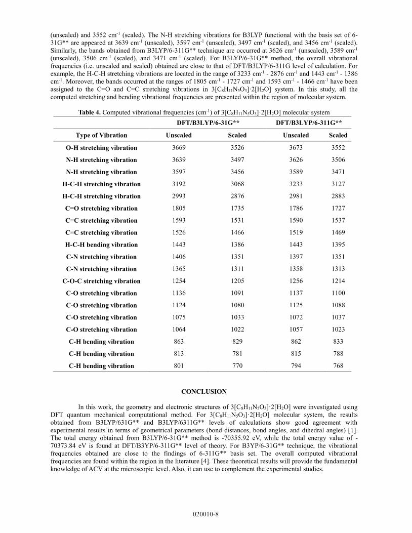
There are no reported experimental findings on the different vibration modes of 3[C8H11N5O3]·2[H2O]
molecular system. To study the infrared spectroscopic signature of title molecule, the DFT computational
calculations are conducted on the vibrational frequency analysis. The results in Table 4 presents the vibrational
frequencies (i.e. unscaled and scaled) of 3[C8H11N5O3]·2[H2O] molecular system using B3LYP method with two
different basis sets (i.e. 6-31G** and 6-311G**). The scale factors used are 0.961 and 0.967, respectively for
B3LYP/6-31G** and B3LYP/6-311G** techniques. The molecular structure of 3[C8H11N5O3]·2[H2O] has the
space group of P21/n, consists 87 atoms in the system. Therefore, it has 19 out of 255 normal vibrational modes,
which can be distributed as 10 types of vibrations. The calculated vibrational frequencies either unscaled or
scaled obtained from DFT/B3LYP/6-31G** and DFT/B3LYP/6-311G** levels of theories show good agreement
with the recorded spectral in the literature data [4]. For 3[C8H11N5O3]·2[H2O] molecular system, the O-H
stretching vibrations are found at 3669 cm-1 (unscaled) and 3526 cm-1 (scaled) in the molecular system using
DFT/B3LYP/6-31G** level of theory. Similar to B3LYP/6-311G** results, the peaks are observed at 3673 cm-1
2.6797 a.u.
2.57 eV
-3.06 eV
2.55 eV
-3.06 eV
020010-7
Page 9
(unscaled) and 3552 cm-1 (scaled). The N-H stretching vibrations for B3LYP functional with the basis set of 6-
31G** are appeared at 3639 cm-1 (unscaled), 3597 cm-1 (unscaled), 3497 cm-1 (scaled), and 3456 cm-1 (scaled).
Similarly, the bands obtained from B3LYP/6-311G** technique are occurred at 3626 cm-1 (unscaled), 3589 cm-1
(unscaled), 3506 cm-1 (scaled), and 3471 cm-1 (scaled). For B3LYP/6-31G** method, the overall vibrational
frequencies (i.e. unscaled and scaled) obtained are close to that of DFT/B3LYP/6-311G level of calculation. For
example, the H-C-H stretching vibrations are located in the range of 3233 cm-1 - 2876 cm-1 and 1443 cm-1 - 1386
cm-1. Moreover, the bands occurred at the ranges of 1805 cm-1 - 1727 cm-1 and 1593 cm-1 - 1466 cm-1 have been
assigned to the C=O and C=C stretching vibrations in 3[C8H11N5O3]·2[H2O] system. In this study, all the
computed stretching and bending vibrational frequencies are presented within the region of molecular system.
Table 4. Computed vibrational frequencies (cm-1) of 3[C8H11N5O3]·2[H2O] molecular system
DFT/B3LYP/6-31G** DFT/B3LYP/6-311G**
Type of Vibration Unscaled Scaled Unscaled Scaled
O-H stretching vibration 3669 3526 3673 3552
N-H stretching vibration 3639 3497 3626 3506
N-H stretching vibration 3597 3456 3589 3471
H-C-H stretching vibration 3192 3068 3233 3127
H-C-H stretching vibration 2993 2876 2981 2883
C=O stretching vibration 1805 1735 1786 1727
C=C stretching vibration 1593 1531 1590 1537
C=C stretching vibration 1526 1466 1519 1469
H-C-H bending vibration 1443 1386 1443 1395
C-N stretching vibration 1406 1351 1397 1351
C-N stretching vibration 1365 1311 1358 1313
C-O-C stretching vibration 1254 1205 1256 1214
C-O stretching vibration 1136 1091 1137 1100
C-O stretching vibration 1124 1080 1125 1088
C-O stretching vibration 1075 1033 1072 1037
C-O stretching vibration 1064 1022 1057 1023
C-H bending vibration 863 829 862 833
C-H bending vibration 813 781 815 788
C-H bending vibration 801 770 794 768
CONCLUSION
In this work, the geometry and electronic structures of 3[C8H11N5O3]·2[H2O] were investigated using
DFT quantum mechanical computational method. For 3[C8H11N5O3]·2[H2O] molecular system, the results
obtained from B3LYP/631G** and B3LYP/6311G** levels of calculations show good agreement with
experimental results in terms of geometrical parameters (bond distances, bond angles, and dihedral angles) [1].
The total energy obtained from B3LYP/6-31G** method is -70355.92 eV, while the total energy value of -
70373.84 eV is found at DFT/B3YP/6-311G** level of theory. For B3YP/6-31G** technique, the vibrational
frequencies obtained are close to the findings of 6-311G** basis set. The overall computed vibrational
frequencies are found within the region in the literature [4]. These theoretical results will provide the fundamental
knowledge of ACV at the microscopic level. Also, it can use to complement the experimental studies.
020010-8
Page 10
ACKNOWLEDGMENTS
The authors would like to thank Universiti Tunku Abdul Rahman and UTARRF grant.
REFERENCES
1. G. I. Birnbaum, M. Cygler and D. Shugar, Canadian journal of chemistry 62(12), pp. 2646-2652
(1984).
2. K. Suwińska, B. Golankiewicz and W. Zielenkiewicz, Acta Crystallographica Section C: Crystal
Structure Communications 57(6), pp. 767-769 (2001).
3. Y. T. Sohn, and S. H. Kim, Archives of Pharmacal Research 31(2), pp. 231-234 (2008).
4. K. M. Lutker, R. Quinones, J. Xu, A. Ramamoorthy and A. J. Matzger, Journal of Pharmaceutical
Sciences 100(3), pp. 949-963 (2011).
5. T. Masuda, Y. Yoshihashi, E. Yonemochi, K. Fujii, H. Uekusa and K. Terada, International Journal of
Pharmaceutics 422(1-2), pp. 160-169 (2012).
6. M. Montha and J. H. Kenneth, Acta Crystallographica Section C: Crystal Structure Communications
69(9), pp. 1077-1080 (2013).
7. A. Sarkar and S. Rohani, Journal of Pharmaceutical Sciences 104(1), pp. 98-105(2015).
8. R. Censi and P. D. Martino, Molecules, 20, pp. 18759-18776 (2015).
9. Q. Cai, J. Xue, Q. Wang and Y. Du, Spectrochimica Acta Part A: Molecular and Biomolecular
Spectroscopy 186, pp. 29-36 (2017).
10. L. Nicoud, F. Licordari and A. S. Myerson, Crystal Growth & Design 18(11), pp. 7228-7237 (2018).
11. M. J. Frisch, G. W. Trucks, H. B. Schlegel, G. E. Scuseria, M. A. Robb and J. R. Cheeseman,
GAUSSIAN09, Revision E. 01. Gaussian Inc., Wallingford, CT, USA. (2009)
12. T. Lu and F. Chen, Journal of Computational Chemistry 33(5), pp. 580-592 (2012).
020010-9