54
Vision “El ojo que ves no es ojo porque tu lo veas, es ojo porque te ve” Antonio Machado “The eye you see is not an eye due to you seeing it, It’s an eye because it sees you”
| Date post: | 28-Dec-2015 |
| Category: |
Documents |
| Upload: | marlene-moore |
| View: | 221 times |
| Download: | 0 times |

Vision
“El ojo que ves no es
ojo porque tu lo veas,
es ojo porque te ve”
Antonio Machado
“The eye you see
is not an eye due to you seeing it,
It’s an eye because it sees you”

Vision: Outline
• Light
• Eye
• Visual Path
• Visual Cortex

Perceptual Dimensions of Light
Wave amplitude
Purity of the wave
Wave frequency
UV rays

Vision: Outline
EYE
• Anatomy
• Retina
• Receptive Field
• Edge Detection
• Color vision

Eye Anatomy
We study this in the lab

transparent medium air(cornea, aqueous humor, pupil,lens, vitreous humor)
lens lensiris diaphragmretina film a focal point a focal point
Similarity btw eye & camera known
since 1600’s
Eye anatomy: Functions

Near-sightedness (Myopia ):
image falls too short of retina (eyeball too long)
newborns
Far-sightedness:
focal point of light falls beyond retina
(Eyeball is too short)
Lasik Changes the shape of the cornea(Laser-Assisted In Situ Keratomileusis)
Eye Anatomy: Abnormalities

Near-Sightedness
nearby things are on focus

Cataracts
• Reduced illumination, acuity, and color saturation• Deposits in the lens
• Common in older adults

Eye Anatomy: Retina
• fovea: center of the retina, high concentration of cones• optic disk (blindspot) & direct view of arteries (clinical importance)• photorreceptors: cones (color vision) and rods
Red eye in photos due to dilated pupils
Retina of diabetic patient

Concentration ofCones & Rods
in Retina
Visual Acuity
Eye Anatomy: Retina

Eye Anatomy: Retina

One Cones --> one ganglion cell high acuity (fovea) Many Rods --> ganglion cells. High sensitivity (periphery)
(e.g, night vision)

Lateral visual field
Medial Retina
The eye is a device 'designed' to see, but the ‘blindspot’ reveals it is not perfect
Eye Anatomy: Optic disc (blindspot)

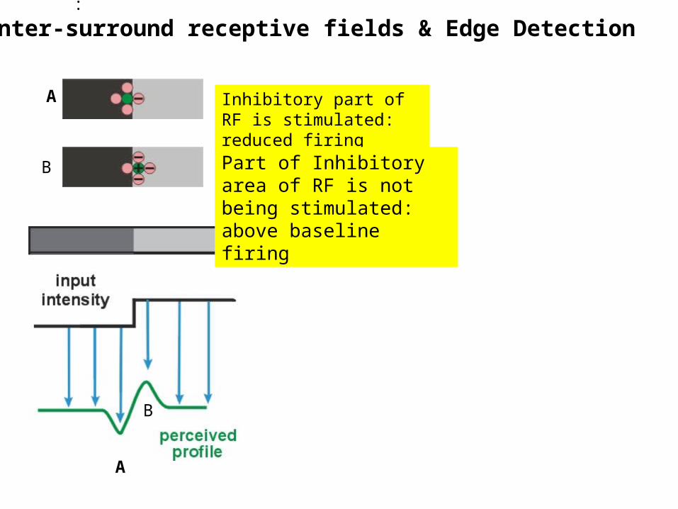
Receptive field (RF)• is that portion of the visual field (outside world) in which
the presentation of visual stimuli will produce an alteration in the firing rate of a particular neuron

• Each strip is uniform,
• Strips look lighter on the left
Edge detection

:
Center-surround receptive fields & Edge Detection Neither excitatory nor inhibitory parts of RF are stimulated
Both excitatory & inhibitory parts of RF are stimulated, canceling each other
:
Center-surround receptive fields & Edge Detection
Inhibitory part of RF is stimulated: reduced firing
A
A
Part of Inhibitory area of RF is not being stimulated: above baseline firing
B
B

Edge detection

Hermann Grid
+- -- -
- -
- -
- -- -
+- -- -
- -
- -
- -- -
Inhibition (-)
Excitation (+)+
- -- -
- -
- -
- -- -

Tri-chromatic theory – Blue, red, & green “color”
receptors– But some colors don’t mix!
Peak sensitivities of the three cones
Opponent process theory Red vs. Green; Blue vs. Yellow- Negative afterimage- But there is no ‘yellow’ receptor!
COLOR VISION
(-)
BLUE
(-)
GREEN
(+)
RED
(+)
YELLOW


Test for Deuteranopia: Name number: (‘5’ or ‘2’)If you see a 2: Red/Green Color blindness (male)
Most people who are color blind can see colors
No ‘green’ cones

Vision: Outline
• Light
• Eye
• Visual Path & its deficits
• Visual cortex
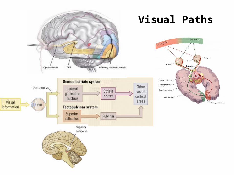
Visual Paths

LGN thalamic organization
• Magnocellular – M ganglion cells– large receptive fields– motion detection– locating stimulus in space – dorsal cortical stream– parietal lobe
• Parvocellular– P ganglion cells – small receptive fields– Object identity– Color recognition– ventral cortical stream– temporal lobe
Concentric receptive fields (center surround)

VISUAL PATHWAY
Retinal Field: representation of visual field in the retina (reversed: right/left, up/down)
Visual Field: outside world you see

Right homonymous most commonCan also get upper and lower deficits and scotoma
Visual Path: Lesions & Deficits
QuadrantanopiaHomonymousHemianopia
Bitemporal

Scotoma: A small blindspot in the visual field caused by a small lesion, usually in the occipital lobe
Hemianopia – objects are bisected with ½ obscured experiencing the obscured part as “blank” or “void”

Vision: Outline
• Light
• Eye
• Visual Path & its deficits
• Visual cortex• V1: Orientation sensitive
– Ventral Pathway – Dorsal Pathway
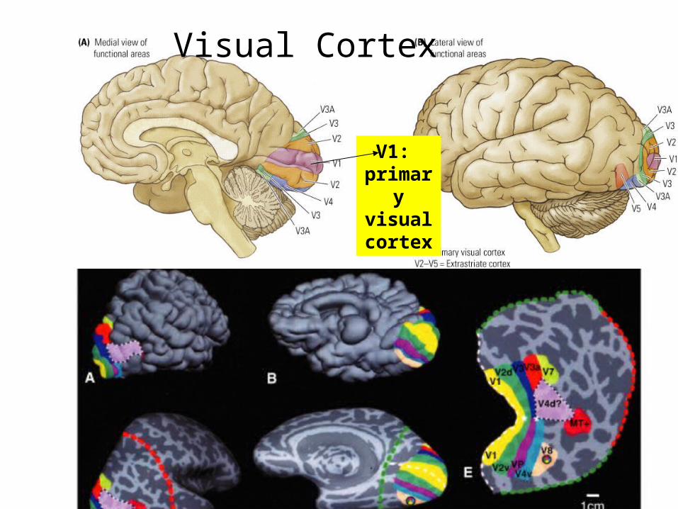
Visual Cortex
V1: primaryvisual cortex

Primary visual cortex (V1)
• V1 cells respond to lines – of particular orientations – of particular widths.

Primary visual cortex (V1)
• Orientation selective cells are organized in a topographic map in V1

How does orientation selectivity in V1 emerges?
• LGN cells with concentric receptive field provide input to simple cells in V1

Vision: Outline
• Light
• Eye
• Visual Path & its deficits
• Visual cortex• Orientation sensitive
– Ventral Pathway• Area MT (motion), Object Recognition, Area V4 (color)• synesthesia
– Dorsal Pathway • Spatial Attention• Hemispatial Neglect

• Complex & with multiple connections
• Over-simplified version: dorsal & ventral paths
Cortical Connections of Visual areas

Ventral & Dorsal Paths
¼ of the brain is involved in visual processing, more than for all other senses

Ventral & Dorsal Paths
& how

Ventral & Dorsal Paths

Ventral Path: Object recognition
Lesion of ventral pathwayAgnosia
fMRI: Object recognition

R.V. has bilateral parietal lobe damage
ventral lesion (patient DF):
- Agnosia - Normal grip
dorsal lesion (patient RV):
- Normal recognition - poor grip
Independence of Dorsal and Ventral paths: Neurpsychological evidence

Cerebral Achromatopsia: bilateral damage to V4
Color is more important of ‘what’ than for ‘where’ Synesthesia
Ventral Pathway (V4): Color perception

Ventral Path: Objects vs. Faces
Are faces very difficult objects or special ones (i.e., specific process)

Neuroimaging of face, bird and car experts
“Face Experts”
FusiformGyrus
FusiformGyrus
CarExperts
BirdExperts
FusiformGyrus
Gauthier et al., 2000
Cars-Objects Birds-Objects

ControlGroup
AutismGroup
Hypoactivation of fusiform face area Schultz, et al. 2000
Faces
FusiformGyrus
FusiformGyrus
Children with autism as face “novices”

Area MT: motion perception


• Different parts of the visual cortex are specialized in the processing of specific features
• For example,• movement, • color.• Objects• Faces• Location
Binding problem: If the brain processes features separately, how does it bind those features into a single conscious representation:
Answer: Attention (next week)

Spare slides
• Spatial frequency

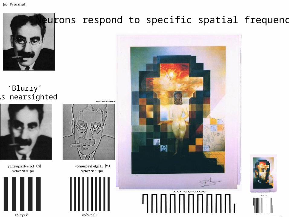
Neurons respond to specific spatial frequencies
‘Blurry’ As nearsighted


3-D vision
• Retinal disparity• Stereograms

http://www.ritsumei.ac.jp/%7Eakitaoka/index-e.html


