Vitamin D manipulates miR-181c, miR-20b and miR-15a in human umbilical vein endothelial cells exposed to a diabetic-like environment Zitman-Gal et al. C ARDI O V ASCULAR D IABETOLOG Y Zitman-Gal et al. Cardiovascular Diabetology 2014, 13:8 http://www.cardiab.com/content/13/1/8
Transcript
Vitamin D manipulates miR-181c, miR-20b andmiR-15a in human umbilical vein endothelial cellsexposed to a diabetic-like environmentZitman-Gal et al.
C ARDI OVASCULAR D IABETOLOG Y
Zitman-Gal et al. Cardiovascular Diabetology 2014, 13:8http://www.cardiab.com/content/13/1/8
ORIGINAL INVESTIGATION Open Access
Vitamin D manipulates miR-181c, miR-20b andmiR-15a in human umbilical vein endothelial cellsexposed to a diabetic-like environmentTali Zitman-Gal1*, Janice Green1, Metsada Pasmanik-Chor2, Eliezer Golan1,3, Jacques Bernheim1,3
and Sydney Benchetrit1,3
Abstract
Background: High blood and tissue concentrations of glucose and advanced glycation end-products are believedto play an important role in the development of vascular complications in patients with diabetes mellitus (DM) andchronic kidney disease. MicroRNAs (miRNA) are non-coding RNAs that regulate gene expression in a sequencespecific manner. MiRNA are involved in various biological processes and become novel biomarkers, modulators andtherapeutic targets for diseases such as cancer, atherosclerosis, and DM. Calcitriol (the active form of vitamin D) mayinhibit endothelial proliferation, blunt angiogenesis, and be a cardioprotective agent. Calcitriol deficiency is a riskfactor for DM and hypertension. The aim of this project was to study the miRNA microarray expression changes inhuman umbilical vein endothelial cells (HUVEC) treated in a diabetic-like environment with the addition of calcitriol.
Methods: HUVEC were treated for 24 h with 200 μg/ml human serum albumin (HSA) and 100 mg/dl glucose(control group) or 200 μg/ml AGE-HSA, and 250 mg/dl glucose (diabetic-like environment), and physiologicalconcentrations (10-10 mol/l) of calcitriol. miRNA microarray analysis and real time PCR to validate the miRNAexpression profile and mRNA target gene expression were carried out.
Results: Compared to control, 31 mature human miRNA were differentially expressed in the presence of adiabetic-like environment. Addition of physiological concentrations of calcitriol revealed 39 differentially expressedmature human miRNA. MiR-181c, miR-15a, miR-20b, miR-411, miR-659, miR-126 and miR-510 were selected forfurther analysis because they are known to be modified in DM and in other biological disorders. The predictedtargets of these miRNA (such as KLF6, KLF9, KLF10, TXNIP and IL8) correspond to molecular and biological processessuch as immune and defense responses, signal transduction and regulation of RNA.
Conclusion: This study identified novel miRNA in the field of diabetic vasculopathy and might provide newinformation about the effect of vitamin D on gene regulation induced by a diabetic-like environment. New genetargets that are part of the molecular mechanism and the therapeutic treatment in diabetic vasculopathy arehighlighted.
* Correspondence: [email protected] Physiology Laboratory, Department of Nephrology and Hypertension,Meir Medical Center, Kfar Saba 44281, IsraelFull list of author information is available at the end of the article
IntroductionMicroRNAs (miRNA) are non-coding RNA species ofapproximately 22 nucleotides that regulate gene expres-sion in a sequence-specific manner. miRNA join to par-tial complementary sequences in the 3'UTRs of targetmRNA of protein coding genes to specify translationalrepression and/or mRNA cleavage [1,2]. miRNA are in-volved in various biological processes and have becomenovel biomarkers, modulators and therapeutic targetsfor diseases such as cancer, heart disease, and diabetes[1,2]. Several studies investigated the effect of miRNAon the development of diabetes and its complications,including endothelial and vascular smooth muscle celldysfunction, diabetic cardiomyopathy and diabetic ne-phropathy, and demonstrated involvement of specifictypes of miRNA, which regulate a broad range of in-flammatory genes [3-5].
Diabetes mellitus (DM) is associated with endothelialdysfunction including changes in barrier function andhomeostasis, reduced vasodilator response, inflammatoryactivation, increased plasma levels of endothelial prod-ucts, and angiogenesis, all of which are associated with agreater incidence and severity of cardiovascular diseases[6-10]. Hyperglycemia is thought to affect endothelialfunction, increasing stiffness of the peripheral arteriesand arterioles partly due to reduced nitric oxide produc-tion [8,10]. Elevated concentrations of environmentalglucose initiate events that stimulate extracellular andintracellular formation of advanced glycation end prod-ucts (AGEs) [10-12]. The increased formation of AGEsplays a relevant role in the development of vascular andrenal-related complications seen in DM, chronic kidneydisease (CKD), and aging [11,12]. Previous studies per-formed in our laboratory have demonstrated that AGEsstimulate the endothelial expression of AGE receptor(RAGE) and interleukin 6 (IL6), and depress the eNOSmRNA expression and eNOS enzymatic activity [13,14],while calcitriol blunted the deleterious effect of AGEs inthis in vitro model [14].
Our previous studies in endothelial and vascular smoothmuscle cells using a high throughput microarray approach[15-17], showed that diabetic-like conditions (250 mg/dlglucose and AGEs at a concentration similar to that foundin the blood of diabetic patients) induced significantly ele-vated expressions of cellular and metabolic processes, in-cluding endothelial inflammatory-related genes throughthe NFκB pathway and stimulated the production of thior-edoxin interacting protein (TXNIP). Calcitriol (1,25 dihy-droxycholecalciferol), the active form of vitamin D thatcontrols calcium homeostasis, hormonal secretions, andcell proliferation and differentiation, was found to have aprotective effect on the development of renal or cardiovas-cular disorders in stimulated endothelial cells in vitro andin vivo [18,19]. Vitamin D deficiency is a risk factor for
DM and hypertension [20]. Our previous studies showedthat adding calcitriol to endothelial and vascular smoothmuscle cells significantly down-regulated the inflamma-tory response of gene and protein expression involved inthe NFκB signal transduction pathway [16,17]. In thisstudy we evaluated the expression of miRNA and theirpredicted gene targets in human umbilical vein cord endo-thelial cells (HUVEC) exposed to a diabetic like environ-ment and the effect of calcitriol on their expression.
Materials and methodsCell culture and incubationHUVEC were isolated from umbilical cords obtained fromthe maternity unit at Meir Medical Center, Kfar Saba,Israel [13]. The Ethical Review Committee approved thestudy and the parturient provided written informed con-sent. HUVEC were identified by their typical cobblestonemorphology and by immunostaining for von Willebrandfactor. HUVEC were grown in M-199 medium supple-mented with 20% FCS, 100 U/ml penicillin, 100 μ/mlstreptomycin (Biological Industries, Bet Haemek, Israel),5 U/ml heparin, and 25 μ/ml endothelial mitogen(Biomedical Technologies Inc., Stoughton, MA, USA).Confluent cultures of HUVEC were used for experimentsat passages 3–4. The cells were stimulated in media con-taining 200 μg/μl HSA and 100 mg/dl glucose (controlgroup) or 200 μg/μl AGE-HSA and 250 mg/dl glucose(diabetic-like environment) for 24 h. Calcitriol 10-10 mol/l(PH&T SpA, Milan, Italy), corresponding to physiologicalblood concentrations, [16,17] was added to cells 1 h afterhaving started the stimulation with AGE-HSA and glucosefor an additional 23 h.
miRNA microarray analysismiRNA were extracted using the mirVANA™ RT-PCRmiRNA isolation and detection kit (Ambion, Austin, Texas,USA) according to manufacturer’s instructions. RNA quan-tity and quality were determined using Nanodrop (ThermoFisher Scientific, Inc, Wilmington, DE, USA). AffymetrixGeneChip® miRNA 2.0 arrays were used for genome-widemiRNA expression analysis (15,644 probe sets for 131 or-ganisms, including 1,105 human miRNA, 1,105 humanpre-miRNA and 2,302 human small nucleolar RNAs) ac-cording to the instruction manual, as described in URL 1(Affymetrix, Santa Clara, CA, USA). Three biological re-peats were used for each treatment.
Bioinformatics analysisMicroarray analysis was performed on CEL files usingPartek® Genomics Suite™ (Partek GS, Partek Inc., MO,USA; Quantile normalization was performed by the ro-bust multi-average method (RMA). Batch effect removalwas applied for the different samples to remove individ-ual variations, followed by one-way analysis of variance
Zitman-Gal et al. Cardiovascular Diabetology 2014, 13:8 Page 2 of 10http://www.cardiab.com/content/13/1/8
(ANOVA). Human miRNA that were differentially ex-pressed (p < 0.05; fold-change cutoff 1.5) were obtained.Each relevant miRNA was analyzed using TargetScan,which uses a computational algorithm to predict biologicalgene targets of miRNA. David and WebGestalt databases(http://david.abcc.ncifcrf.gov and http://bioinfo.vanderbilt.edu/webgestalt, respectively) were used for functional an-notations of the target gene lists. The String database(http://string-db.org) was used to predict the miR-target-interactions including direct (physical) and indirect (func-tional) associations. Enrichment of GO biological pro-cesses with FDR correction was used.
miRNA – real time PCRmiRNA real time PCR was performed on 2 additionalbiological repeats (total of 5 repeats) using TaqMan spe-cific Small RNA primer and probe sets for miR-181c,miR-411, miR-659, miR-510, miR-126, miR-15a and miR-20b (Applied Biosystems, Inc., Foster City, CA, USA).miRNA expression is represented relative to the expres-sion of the internal control U6-snRNA. Data were ana-lyzed using the 2-ΔΔCt method.
RNA extraction and real-time PCRTotal RNA was extracted from HUVEC using mir-VANA™ RT-PCR isolation and detection kit (Ambion,Austin, Texas, USA) and RNeasy Mini Kit, includingDNase digestion with RNase-free DNase set (Qiagen,Valencia, CA, USA) according to manufacturer's in-structions. RNA integrity was assessed using NanoDrop(Thermo Fisher Scientific, Inc, Wilmington, DE, USA).RNA (1 μg) was then reverse transcribed into single-strand DNA using the High Capacity cDNA ReverseTranscription Kit (Applied Biosystems, Inc, Foster City,CA, USA), according to manufacturer's instructions.TaqMan real-time PCR amplification was used withgene specific primer for Kruppel-like Factor 6 (KLF6),KLF9, KLF10 using glucuronidase, beta (GUSB) as con-trol genes. Data were analyzed using the 2-ΔΔCt method.
Statistical analysisAll data are expressed as mean ± standard deviation (SD).Friedman and repeated measures with Bonferroni multiplecomparisons were used to evaluate the effect of the differ-ent treatments on the expression of selected miRNA. Stu-dent t-test and Mann–Whitney non-parametric test wereused to evaluate the different treatments on the expres-sion of selected target mRNA genes. P-values of 0.05 orless were considered significant.
ResultsDifferentially expressed miRNAMiRNA expression in cultured HUVEC exposed to adiabetic-like environment revealed 31 mature human
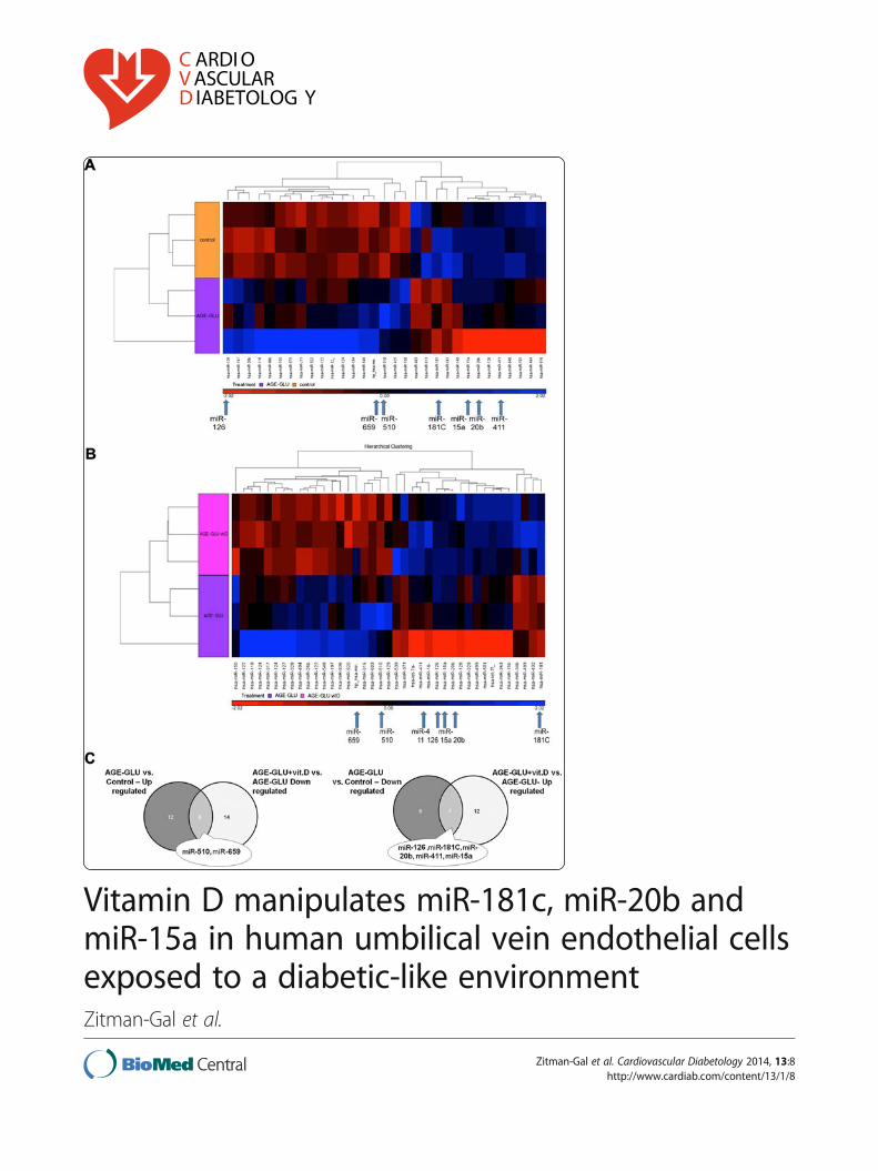
miRNA that were significantly changed (p < 0.05, foldchange cut-off 1.5), of which 18 were up-regulated and13 were down-regulated (Figure 1A).
The addition of calcitriol (10-10 mol/l) to culturedHUVEC exposed to a diabetic-like environment revealed39 mature human miRNA that were significantly chan-ged (p < 0.05 and fold-change cut-off 1.5), of which 19miRNA were up-regulated and 20 were down-regulated(Figure 1B). Table 1 presents a partial list of selectedmiRNA (from the differential mature human miRNA)that were chosen according to their p-value, their foldchanges or their physiological expression. From themiRNA list presented in Table 1 and from the corre-sponding Venn diagram (Figure 1C), we validated severalmiRNA (marked in bold in Table 1) that are known tobe modified in a diabetic environment (miR-510, miR-15a, miR-20b, miR-126, and miR-181C). miR-659 andmiR-411 were validated because in the microarray ana-lysis they were significantly changed after the addition ofcalcitriol. Each miRNA was normalized to that of theU6-snRNA, which was used as a reference. A total of 5biological repeats (5 different umbilical cords) were usedfor the validation to ensure that the variations observedwere biological. The miRNA expression patterns (Figure 2,A-G) were consistent between the microarray (insets) andreal-time PCR validations.
Gene target and pathway analysis of miR-181C, miR-15aand miR-20bMiRNA are known to regulate many target genes andconsequently can modulate different signaling pathways.We focused on three differentially expressed miRNA:miR-181C, miR-15a and miR-20b, which were found tobe down-regulated in a diabetic-like environment andup-regulated after the addition of calcitriol. They werechosen for further investigation because their gene tar-gets play a key role in endothelial cell function, which isrelevant to our research (Table 2). We found that genestargets from the Kruppel-like family, which are tran-scription factors that play key regulatory roles in cellulargrowth, differentiation, proliferation, apoptosis and angio-genesis [20,21], take part as putative targets for miR-181Cand miR-20b. TXNIP, a pro-apoptotic protein, which isknown to regulate endothelial cell metabolism, growth,and inflammation [22,23] and IL8, an inflammatory-related protein [24] are putative targets of miR-20b andmiR-15a. Based on these target genes, using the Stringdatabase, we predicted the miRNA-target protein interac-tions including direct (physical) and indirect (functional)associations. Based on these results, we found that thepredicted gene targets correspond to biological processessuch as immune and defense responses, signal transduc-tion and regulation of RNA and primary metabolic pro-cesses (Figure 3). We decided to further analyze KLF6,
Zitman-Gal et al. Cardiovascular Diabetology 2014, 13:8 Page 3 of 10http://www.cardiab.com/content/13/1/8
Zitman-Gal et al. Cardiovascular Diabetology 2014, 13:8 Page 4 of 10http://www.cardiab.com/content/13/1/8
KLF9 and KLF10 and their expression patterns. Using realtime PCR analysis, we found that the expression level ofKLF6 was significantly up-regulated in a diabetic-likeenvironment (Figure 4). KLF9 and KLF10 were also up-regulated, but not significantly. The addition of calcitriolsignificantly down-regulated KLF6, KLF9 and KLF10mRNA expression (Figure 4). TXNIP and IL8 expres-sion patterns, as previously described, were significantlyup-regulated in a diabetic-like environment, whereasthe addition of calcitriol significantly down-regulatedIL8 [14,15].
DiscussionEndothelial cell, diabetic environment and vitamin DThe regulation of miRNA that are released in the endo-thelium in response to a diabetic-like environment andvitamin D might provide important insights to the eventsthat play a major role in the development of vascularcomplications. Our previous studies showed that diabetic-like conditions characterized by mildly elevated glucoseconcentrations (250 mg/dl) and AGEs in an HUVEC cul-ture model, were found to stimulate endothelial TXNIPsystem activity, as well as expression of the inflammatoryrelated proteins IL6, IL8, RAGE, and NFκB [15]. Addingphysiological concentrations of calcitriol (10-10 mol/l) tothis in vitro model had a beneficial effect on the endothe-lial expression of pro-inflammatory parameters, probablythrough the NFκB signal transduction pathway [16].Vitamin D was found to play an important role in renal,endothelial, and cardiovascular protection and to have an-titumor activity [18-20]. In this study, we used a miRNAmicroarray to obtain expression profiles of miRNA in cul-tured HUVEC that were exposed to a diabetic-like envir-onment in order to mimic a clinical model of DM in vitro.We demonstrated that stimulation of vitamin D in thistype of environment has a beneficial effect on the endo-thelial expression of miRNA profile and their target genes.
Differentially expressed miRNAWe identified a list of miRNA that were differentiallyexpressed following the various manipulations. As men-tioned previously, we focused on selected miRNAs thatare known to be modified in a diabetic-like environmentor that were highly changed after the addition of calci-triol. MiR-510 and miR-659 were over-expressed underdiabetic-like conditions and decreased after calcitriolwas added. MiR-126, miR-411, miR-20b, miR-15a and
miR-181c were down-regulated under diabetic condi-tions and over-expressed after calcitriol was added.
MiR-510 was first identified in association with irrit-able bowel syndrome; its co-expression was involved inthe regulation of 5-HT3 receptors in colonic enterocytes[23]. Recently, Hezova et al. [24] demonstrated thatmiR-510 expression was elevated in regulatory T cells ofdiabetic patients. To the best of our knowledge, ourfindings are the first demonstration of miR-510 involve-ment in HUVEC exposed to a diabetic-like environmentwith significant changes induced by calcitriol. Manipula-tion of miR-510 may represent a beneficial effect of cal-citriol if we consider the possible deleterious effect ofincreased miR-510 in DM. MiR-659 is not described inthe database as being connected to a diabetic-like envir-onment and appears to be novel in genomic control inthe presence of diabetic-like conditions affected by vita-min D. Further investigation of the effect of the miRNAon their predicted target genes is warranted.
MiR-126 is highly enriched in endothelial cells and isinvolved in vascular integrity, angiogenesis and wound
(See figure on previous page.)Figure 1 Hierarchical clustering of miRNA expression in HUVEC. Seven focal miRNA are indicated with arrows at the bottom of the clusters(A, B) and on the VENN diagram (C). Clusters represent treatment with (A) AGE-HSA and glucose 250 mg/dl (AGE-GLU) compared to control(HSA and 100 mg/dl glucose concentration); (B) AGE-HSA, glucose 250 mg/dl and 10-10 mol/l calcitriol (AGE-GLU + vit. D) compared to AGE-HSAand glucose 250 mg/dl (AGE-GLU); (C) Comparison between differentially expressed miRNA lists.
Table 1 Summary of miRNA in HUVEC exposed to adiabetic environment with and without calcitrol
hsa-miR-126 -1.76 0.07 1.55 0.14a200 μg/ml AGE-HSA and 250 mg/dl glucose vs. control (200 μg/μl HSA and100 mg/dl glucose).
Zitman-Gal et al. Cardiovascular Diabetology 2014, 13:8 Page 5 of 10http://www.cardiab.com/content/13/1/8
repair [25,26]. Moreover, loss of plasma miR-126 is con-sistently associated with diabetes [27,28]. Zampetaki et al.[27] found that high glucose concentrations (25 mmol/lequivalent to 500 mg/dl) significantly reduced miR-126expression in endothelial apoptotic bodies and improvedvascular growth factor (VEGF) signaling, leading to endo-thelial dysfunction [27,29]. In patients with DM, lowplasma levels of miR-126 could be clinically relevant andcontribute to VEGF resistance and endothelial dysfunction[30]. In this study, we also found in HUVEC a decreasedexpression of miR-126 exposed to a diabetic like environ-ment and demonstrated that the addition of calcitriol
elevated miR-126 expression, which can probably improveVEGF signaling and repair endothelial dysfunction.
Differential expression of miR-411 was reported in amiRNA profiling study of the hippocampus and the mar-ginal division in a rat brain [30]. In addition, miR-411was reported to be down-regulated in an autosomaldominant muscle disorder (facioscapulohumeral muscu-lar dystrophy) suggesting that reduction of miR-411might have a positive effect on muscle regeneration andpromote myoblast maturation [31]. To our knowledge,no study has investigated the effect of diabetes and vita-min D on the function of miR-411 in endothelial cells.The relevance of its role in diabetes remains unknownand should be clarified in the future. The significant ef-fect of vitamin D remains speculative at this stage.
Selected miRNAs and their target genesMiR-15a, miR-20b and miR-181C were found to be down-regulated in a diabetic-like environment and up-regulatedafter the addition of calcitriol; they were chosen for fur-ther investigation at the level of their gene targets, whichhave been shown to be involved in the modulation ofendothelial function. MiR-15a, a cell growth suppressor,was evaluated in human cancer cells and found to have apro-apoptotic role by activating caspase 3/7, which re-duces cell viability [32]. In addition, miR-15a has beencorrelated with different pathophysiological events in the
Figure 2 Validation of miRNA expression results by real time PCR. HUVEC were incubated for 24 h with HSA (200 μg/ml), AGE-HSA (200 μg/ml)and glucose (250 mg/dl). In addition, 10-10 mol/l calcitriol was given to the cells 1 h after stimulation for an additional 23 h. The miRNAset that included (A) miR-659, (B) miR-510, (C) miR-181C, (D) miR-411, (E) miR-126, (F) miR-15a, and (G) miR-20b was validated using realtime PCR. Insets show miRNA microarray expression results. Data are expressed as mean ± SD of 4–5 independent experiments. *P < 0.05 compared tocontrol group-HSA. **P < 0.05 compared with AGE-glucose.
Table 2 Putative target genes of the selected miRNA(Target scan analysis)
miR-181c miR-20b miR-15a
KLF6 KLF6
KLF15 KLF15
KLF3 KLF3
KLF9 KLF9
KLF10 KLF10 KLF10
KLF12 KLF12
IL8 IL8
TXNIP TXNIP TXNIP
Zitman-Gal et al. Cardiovascular Diabetology 2014, 13:8 Page 6 of 10http://www.cardiab.com/content/13/1/8
liver, which are also side-effects of anabolic steroids [33].MiR-15a was down-regulated in the plasma of diabetic pa-tients [27] and in β-cells exposed to high glucose (33 mMequivalent to 600 mg/dl) for long periods [34]. Mir-15awas also found to be up-regulated in endothelial cells andvascular smooth muscle cells after stimulation with KLF4,which indicate an option to suppress proliferative vasculardisorders [35]. MiR-20b was down-regulated in the plasmaof diabetic patients [27] and similarly expressed in normaland diabetic dermatological tissue, but was significantlydifferent from that of diabetic wounds during the courseof healing [36]. To our knowledge, no study has investi-gated the effect of diabetes and vitamin D on the functionof miR-20b in endothelial cells.
Using pediatric cancer stem cells, Sanchez-Diaz et al.[37] showed that miR-181c regulate cell proliferation andthe cell cycle, probably by affecting the Notch signalingpathway and the bone morphogenetic protein (BMP) path-way. Becker et al. [33] demonstrated that under the influ-ence of anabolic steroids, miR-181c was down-regulated inbovine liver and might lead to uncontrolled proliferation inthe liver.
To our knowledge, no study has investigated the effectof diabetes and vitamin D on the function of miR-181Cin endothelial cells.
Figure 3 Predicted miRNA-target protein biological interactions, including direct and indirect associations. The predicted gene targetswere found to correspond to biological processes such as immune and defense responses, signal transduction and regulation of RNA andprimary metabolic processes. The genes that were further analyzed and discussed, KLF6, KLF9, KLF10, TXNIP and IL8 are marked with a ring.These gene targets were chosen because they take part in cellular growth, differentiation, proliferation, apoptosis and angiogenesis, as well as inregulating endothelial cell metabolism, growth, and inflammation.
Figure 4 Effect of calcitriol on target genes KLF6, KLF9 andKLF10 mRNA expression in HUVEC stimulated with adiabetic-like environment. HUVEC were incubated for 24 h withHSA (200 μg/ml), AGE-HSA (200 μg/ml) and glucose (250 mg/dl). Inaddition, 10-10 mol/l calcitriol was given to the cells 1 h after stimulationfor an additional 23 h. KLF6, KLF9 and KLF10 mRNA expression wereanalyzed by real-time PCR and normalized to the GUSB. Data areexpressed as mean ± SD of 4–5 independent experiments.*p < 0.05 compared to control group-HSA; **p < 0.05 comparedwith AGE-Glucose.
Zitman-Gal et al. Cardiovascular Diabetology 2014, 13:8 Page 7 of 10http://www.cardiab.com/content/13/1/8
As mentioned above, these three miRNA that weredown regulated in a diabetic environment were signifi-cantly up-regulated after addition of calcitriol. To thebest of our knowledge, this is the first demonstration ofthe effect of calcitriol on the expression of these threemiRNA under diabetic-like conditions in HUVEC.
In order to determine the potential genes involved inHUVEC exposed to a diabetic-like environment and cal-citriol, we analyzed the predicted target genes of these 3miRNA (miR-15a, miR-20b and miR-181c).
Seven predicted targets of the 3 deregulated miRNAwere chosen for further analysis of the molecular path-ways. In Figure 3, we observed that the target genesKLF9, KLF10, and KLF6 are directly and indirectly (viaTXN, JUN and FOS) bound to TXNIP and to IL8 andcorrespond to immune, defense and metabolic biologicalprocesses. After calcitriol was added to the cells, we ob-served a down-regulation in KLF9, KLF10, KLF6, TXNIPand IL8 expression. These findings were negatively cor-related with the miRNA expression patterns and there-fore support their function.
Kruppel-like factors are members of the zinc fingerfamily of transcription factors that regulate cellular dif-ferentiation and tissue development [38,39]. KLF9 is atranscriptional regulator that is highly expressed in therat brain, kidney, lung, and testis and regulates uterineendometrial cell proliferation, adhesion and differenti-ation [40,41]. Panda et al. [42] showed that in Ishikawacells (an endometrial adenocarcinoma cell line), miR-200cincreased cell proliferation through KLF9 repression.
KLF10 plays a major role in mediating the effects ofTGF-β through regulation of the Smad signaling path-way. It regulates gene transcription, inhibits cell prolifer-ation, induces apoptosis and plays a role in activatingthe inflammatory response, including stress-induced in-flammation, leading to increases in cardiovascular dis-eases, autoimmune abnormalities and DM [43]. Yanget al. [22] showed that KLF10 induced endothelial cellsto facilitate a possible pathway for TGF-β and regulatedCOX-1, which affects platelet aggregation. Using micro-array gene expression, we demonstrated that in vascularsmooth muscle cells, KLF10 was up-regulated underdiabetic conditions and down-regulated after calcitriolwas added [17]. In this study, calcitriol had also a bene-ficial effect on KLF10 mRNA expression in HUVEC ex-posed to a diabetic environment.
KLF6 is one of the KLFs that are reportedly expressedin endothelial cells and is considered a damage-responsefactor [38,39,44]. KLF6 promotes tissue remodeling dueto its ability to activate genes that are members of theTGF-β signaling pathway, which are implicated in vascu-lar remodeling, tumor metastasis and apoptosis [39,43].Qi et al. [45] demonstrated that KLF6 was induced byhigh glucose concentrations (30 mmol/l equivalent to
600 mg/dl) in human kidney cells and binds to the TXNIPpromoter region. In addition, in an in vivo study in dia-betic rats, they showed that KLF6 and PPAR-γ (localizeddownstream of KLF6) play a key role in the regulation ofTXNIP expression in the development of diabetes melli-tus. The KLF target genes that were analyzed in this studyand were found to be modified by a diabetic-like environ-ment and calcitriol might provide unique information onthe function of these transcription factors in endothelialcells and may be the basis of attractive research in the fu-ture in the field of DM in animal models and humans.
TXNIP is stimulated by high glucose concentrations.It is well-demonstrated that it promotes oxidative stressand apoptosis in several types of cells, including endo-thelial cells [15,46,47]. In this study, we showed thatTXNIP mRNA expression in cultured HUVEC was up-regulated following exposure to a diabetic-like environ-ment, while no significant changes was observed afterthe addition of calcitriol. IL8 is produced in several tis-sues, as well as in endothelial cells upon infection, in-flammation, ischemia and trauma [48]. We found thatdiabetic-like conditions stimulate the expression of thisinflammation-related protein and that the addition ofcalcitriol had a beneficial effect on its expression.
Vitamin D deficiency and supplementationIt has been well established that vitamin D deficiency iscorrelated with high body fat and glucose levels and de-creased insulin sensitivity. It is also an independent cardio-vascular risk factor that predicts poor cardiovascularoutcomes [49-51]. Grineva et al. [49] showed that inwomen at late reproductive age (mean age 46.1 ± 4.5) fromthe North‐West region of Russia, low 25(OH)D levelswere associated with obesity, increased plasma glucoselevels after OGTT and insulin resistance. A significantcorrelation between fasting insulin, 2 h OGTT glucose, in-sulin levels and low 25(OH)D levels was found in over-weight and obese subpopulations. The authors suggestedthat vitamin D supplementation could be effective in theprevention of obesity and insulin resistance [49]. Theintervention between obesity, vitamin D and PTH iscomplex, suggesting possible effects at different levels.Alkharfy et al. [50] showed a beneficial effect of vitamin Dsupplementation in the metabolic profile of DM type 2 pa-tients treated with insulin and oral hypoglycemic agents.The authors suggested that the effect of the medicationcould be mediated by an increase in HDL-cholesterollevels, particularly by vitamin D [50]. Al-Daghri et al. [51]showed an improvement in LDL, total cholesterol andin homeostasis model assessment of β-cell function(HOMA-β) among a Saudi DM type 2 population re-ceiving vitamin D3 supplementation for 18 months. Thiseffect was more pronounced in women than in men.Vitamin D supplementation was suboptimal, which could
Zitman-Gal et al. Cardiovascular Diabetology 2014, 13:8 Page 8 of 10http://www.cardiab.com/content/13/1/8
explain the absence of significant increase in HDL-cholesterol levels and the increase in HOMA-insulinresistance index. Moreover the absence of relevant infor-mation concerning the anti-diabetic drugs and the dietcomposition of the patients could have limited the impactof the results [51].
Ongoing studies should provide an answer to the still-debated question, whether vitamin D supplementationcan positively influence cardiovascular outcomes. Ourstudy showed that vitamin D can influence the pro-atherogenic and inflammatory response seen in DM andtherefore, might contribute to a preventive and therapeuticeffect. Moreover, recent study showed that vitamin D mayimprove endothelial function in cardiovascular diseasesand could possibly have a role in vascular protection [52].
ConclusionsIn this study, we tried to mimic a diabetic-like environmentin vitro and added vitamin D to simulate the replacementtherapy frequently given to vitamin D deficient patientswith DM. Vitamin D deficiency might have a negative ef-fect on endothelial cell functions. Treating these patientsto obtain normalization of their vitamin D reserves mightimprove the cellular physiology process. New miRNA, aswell as new target genes were found to be differentiallyexpressed following exposure to physiological concentra-tions of vitamin D. Further investigation is needed to studythe specific role of these miRNA, including their targetgenes and the related molecular and biological pathways.These findings might in the future contribute to under-standing the role of vitamin D in treating patients withDM, including detecting new miRNA markers.
Competing interestsThe authors declare that they have no competing interests.
Authors’ contributionsThe individual contributions of each co-author are detailed below: TZG andSB conception and design of research; TZG and JG performed experiments;TZG and MPC analyzed data, interpreted results of experiments and preparedfigures; TZG and JG drafted manuscript; TZG, SB, EG and JB edited andrevised manuscript; TZG and SB approved final version of manuscript. Allauthors read and approved the final manuscript.
AcknowledgementsThis work was supported by the Faye Ginsberg –Toronto Research Fund inDiabetes (E. Golan), The Hendrik and Irene Gutwirth MD Thesis ResearchScholarships in Diabetes Mellitus (S. Benchetrit) and the Dr. Yechezkiel andPearl Klayman Cathedra of Urology (J. Bernheim) from the Sackler Faculty ofMedicine, Tel Aviv University.We would to thank Mrs. L. Astrologo from PH&T (SpA, Milan, Italy) who verykindly provided us with calcitriol.
Author details1Renal Physiology Laboratory, Department of Nephrology and Hypertension,Meir Medical Center, Kfar Saba 44281, Israel. 2Bioinformatics Unit, The G. S.Wise Faculty of Life Sciences, Tel Aviv University, Tel Aviv, Israel. 3SacklerFaculty of Medicine, Tel Aviv University, Tel Aviv, Israel.
Received: 11 October 2013 Accepted: 11 December 2013Published: 7 January 2014
References1. Ambros V: The functions of animal microRNAs. Nature 2004, 431:350–355.2. Farh KK, Grimson A, Jan C, Lewis BP, Johnston WK, Lim LP, Burge CB, Bartel DP:
The widespread impact of mammalian MicroRNAs on mRNA repressionand evolution. Science 2005, 310:1817–1821.
7. Beckman JA, Creager MA, Libby P: Diabetes and atherosclerosis:epidemiology, pathophysiology, and management. JAMA 2002,287:2570–2581.
8. Negre-Salvayre A, Salvayre R, Augé N, Pamplona R, Portero-Otín M:Hyperglycemia and glycation in diabetic complications. Antioxid RedoxSignal 2009, 11:3071–3109.
9. Madonna R, De Caterina R: Cellular and molecular mechanisms ofvascular injury in diabetes - Part I: pathways of vascular disease indiabetes. Vascul Pharmacol 2011, 54:68–74.
10. Bakker W, Eringa EC, Sipkema P, van Hinsbergh VWM: Endothelialdysfunction and diabetes: role of hyperglycaemia, impaired insulinsignalling and obesity. Cell Tissue Res 2009, 335:165–189.
11. Puddu A, Viviani GL: Advanced glycation endproducts and diabetes.Beyond vascular complications. Endocr Metab Immune Disord Drug Targets2011, 11:132–140.
12. Goldin A, Beckman JA, Schmidt AM, Creager MA: Advanced glycation Endproducts sparking the development of diabetic vascular injury.Circulation 2006, 114:597–605.
13. Rashid G, Benchetrit S, Fishman D, Bernheim J: Effect of advancedglycation end products (AGEs) on gene expression and synthesis ofTNF-alpha and endothelial nitric oxide synthase by endothelial cells.Kidney Int 2004, 66:1099–1110.
14. Talmor Y, Golan E, Benchetrit S, Bernheim J, Klein O, Green J, Rashid G:Calcitriol blunts the deleterious impact of advanced glycation endproducts (AGEs) on endothelial cells. Am J Physiol Renal Physiol 2008,294:1059–1064.
15. Zitman-Gal T, Green J, Pasmanik-Chor M, Oron-Karni V, Bernheim J:Endothelial pro-atherosclerotic response to extracellular diabetic-likeenvironment: possible role of thioredoxin-interacting protein.Nephrol Dial Transplant 2010, 25:2141–2149.
16. Zitman-Gal T, Golan E, Green J, Bernheim J, Benchetrit S: Vitamin Dreceptor activation in a diabetic-like environment: potential role on theendothelial pro-inflammatory and thioredoxin pathways activity.SBMB 2012, 132:1–7.
17. Zitman-Gal T, Green J, Pasmanik-Chor M, Oron-Karni V, Bernheim J, Benchetrit S:Vitamin D and vascular smooth muscle cells: gene modulation followingexposure to a diabetic-like environment. J Diabetes Metab 2012, 3:8.
18. Bernardi RJ, Johnson CS, Modzelewski RA, Trump DL: Antiproliferativeeffects of 1α, 25-dihydroxyvitamin D (3) and vitamin D analogs ontumor-derived endothelial cells. Endocrinology 2002, 143:2508–2514.
19. Ford ES, Ajani UA, McGuire LC, Liu S: Concentrations of serum vitamin Dand the metabolic syndrome among U.S. adults. Diabetes Care 2005,28:1228–1230.
20. Holick MF: Vitamin D deficiency. N Engl J Med 2007, 357:266–281.21. Wu Z, Wang S: Role of kruppel-like transcription factors in adipogenesis.
Dev Biol 2013, 373:235–243.22. Yang DH, Hsu CF, Lin CY, Guo JY, Yu WC, Chang VH: Krüppel-like factor 10
upregulates the expression of cyclooxygenase 1 and further modulatesangiogenesis in endothelial cell and platelet aggregation in gene-deficientmice. Int J Biochem Cell Biol 2013, 45:419–428.
23. Kapeller J, Houghton LA, Mönnikes H, Walstab J, Möller D, Bönisch H,Burwinkel B, Autschbach F, Funke B, Lasitschka F, Gassler N, Fischer C,Whorwell PJ, Atkinson W, Fell C, Büchner KJ, Schmidtmann M, van der Voort I,Wisser AS, Berg T, Rappold G, Niesler B: First evidence for an association of afunctional variant in the microRNA-510 target site of the serotonin
Zitman-Gal et al. Cardiovascular Diabetology 2014, 13:8 Page 9 of 10http://www.cardiab.com/content/13/1/8
receptor-type 3E gene with diarrhea predominant irritable bowelsyndrome. Hum Mol Genet 2008, 1(17):2967–2977.
24. Hezova R, Slaby O, Faltejskova P, Mikulkova Z, Buresova I, Raja KR, Hodek J,Ovesna J, Michalek J: microRNA-342, microRNA-191 and microRNA-510are differentially expressed in T regulatory cells of type 1 diabeticpatients. Cell Immunol 2010, 260:70–74.
25. Fish JE, Santoro MM, Morton SU, Yu S, Yeh RF, Wythe JD, Ivey KN, Bruneau BG,Stainier DY, Srivastava D:miR-126 regulates angiogenic signaling andvascular integrity. Dev Cell 2008, 15:272–284.
26. Wang S, Aurora AB, Johnson BA, Qi X, McAnally J, Hill JA, Richardson JA,Bassel-Duby R, Olson EN: The endothelial-specific microRNA miR-126governs vascular integrity and angiogenesis. Dev Cell 2008, 15:261–271.
27. Zampetaki A, Kiechl S, Drozdov I, Willeit P, Mayr U, Prokopi M, Mayr A,Weger S, Oberhollenzer F, Bonora E, Shah A, Willeit J, Mayr M: PlasmamicroRNA profiling reveals loss of endothelial miR-126 and othermicroRNAs in type 2 diabetes. Circ Res 2010, 107:810–817.
29. Waltenberger J, Lange J, Kranz A: Vascular endothelial growthfactor-A-induced chemotaxis of monocytes is attenuated in patients withdiabetes mellitus: a potential predictor for the individual capacity todevelop collaterals. Circulation 2000, 102:185–190.
30. Shu SY, Qing D, Wang B, Zeng QY, Chen YC, Jin Y, Zeng CC, Bao R:Comparison of microRNA expression in hippocampus and the marginaldivision (MrD) of the neostriatum in rats. J Biomed Sci 2013, 20:9.
31. Harafuji N, Schneiderat P, Walter MC: Chen YW: miR-411 is up-regulated inFSHD myoblasts and suppresses myogenic factors. Orphanet J Rare Dis2013, 8:55.
32. Druz A, Chen YC, Guha R, Betenbaugh M, Martin SE, Shiloach J: Large-scalescreening identifies a novel microRNA, miR-15a-3p, which inducesapoptosis in human cancer cell lines. RNA Biol 2013, 10:287–300.
33. Becker C, Riedmaier I, Reiter M, Tichopad A, Pfaffl MW, Meyer HH: Changesin the miRNA profile under the influence of anabolic steroids in bovineliver. Analyst 2011, 136:1204–1209.
34. Sun LL, Jiang BG, Li WT, Zou JJ, Shi YQ, Liu ZM: MicroRNA-15a positivelyregulates insulin synthesis by inhibiting uncoupling protein-2 expression.Diabetes Res Clin Pract 2011, 91:94–100.
35. Zheng X, Li A, Zhao L, Zhou T, Shen Q, Cui Q, Qin X: Key role of microRNA-15ain the KLF4 suppressions of proliferation and angiogenesis in endothelialand vascular smooth muscle cells. Biochem Biophys Res Commun. 2013,437:625–3.
36. Madhyastha R, Madhyastha H, Nakajima Y, Omura S, Maruyama M:MicroRNA signature in diabetic wound healing: promotive role ofmiR-21 in fibroblast migration. Int Wound J 2012, 9:355–361.
37. Sanchez-Diaz PC, Hsiao TH, Chang JC, Yue D, Tan MC, Chen HI, Tomlinson GE,Huang Y, Chen Y, Hung JY: De-regulated MicroRNAs in pediatric cancerstem cells target pathways involved in cell proliferation. Cell Cycle andDevelopment. PLoS One 2013, 8:e61622.
38. Bieker JJ: Kruppel-like factors: three fingers in many pies. J Biol Chem2001, 276:34355–34358.
39. Atkins GB, Jain MK: Role of Kruppel-like transcription factors in endothelialbiology. Circ Res 2007, 100:1686–1695.
40. Simmen RC, Pabona JM, Velarde MC, Simmons C, Rahal O, Simmen FA: Theemerging role of Krüppel-like factors in endocrine-responsive cancers offemale reproductive tissues. J Endocrinol 2010, 204:223–231.
41. Simmen FA, Su Y, Xiao R, Zeng Z, Simmen RCM: The Krüppel-like factor 9(KLF9) network in HEC-1-A endometrial carcinoma cells suggests thecarcinogenic potential of dys-regulated KLF9 expression. Reprod BiolEndocrinol 2008, 6:41.
42. Panda H, Pelakh L, Chuang TD, Luo X, Bukulmez O, Chegini N: EndometrialmiR-200c is altered during transformation into cancerous states andtargets the expression of ZEBs, VEGFA, FLT1, IKKβ, KLF9, and FBLN5.Reprod Sci 2012, 19:786–796.
43. Subramaniam M, Hawse JR, Rajamannan NM, Ingle JN, Spelsberg TC:Functional role of KLF10 in multiple disease processes. Biofactors 2010,36:8–18.
44. Garrido-Martín EM, Blanco FJ, Roquè M, Novensà L, Tarocchi M, Lang UE,Suzuki T, Friedman SL, Botella LM, Bernabéu C: Vascular injury triggersKrüppel-like factor 6 mobilization and cooperation with specificity
protein 1 to promote endothelial activation through upregulation of theactivin receptor-like kinase 1 gene. Circ Res 2013, 112:113–127.
45. Qi W, Chen X, Holian J, Tan CY, Kelly DJ, Pollock CA: Transcription factorsKrüppel-like factor 6 and peroxisome proliferator-activated receptor-{gamma}mediate high glucose-induced thioredoxin-interacting protein. Am J Pathol2009, 175:1858–1867.
46. Schulze PC, Yoshioka J, Takahashi T, He Z, King GL, Lee RT: Hyperglycemiapromotes oxidative stress through inhibition of thioredoxin function bythioredoxin-interacting protein. J Biol Chem 2004, 279:30369–30374.
47. Yamawaki H: Thioredoxin: a multifunctional antioxidant enzyme inkidney, heart and vessels. Curr Opin Nephrol Hypertens 2005, 14:149–153.
48. Baggiolini M, Clark-Lewis I: Interleukin-8, a chemotactic and inflammatorycytokine. FEBS Lett 1992, 307:97–101.
49. Grineva EN, Karonova T, Micheeva E, Belyaeva O, Nikitina IL: Vitamin Ddeficiency is a risk factor for obesity and diabetes type 2 in women atlate reproductive age. Aging (Albany NY). 2013, 5:575–81.
50. Alkharfy KM, Al-Daghri NM, Sabico SB, Al-Othman A, Moharram O, Alokail MS,Al-Saleh Y, Kumar S, Chrousos GP: Vitamin D supplementation in patientswith diabetes mellitus type 2 on different therapeutic regimens: a one-yearprospective study. Cardiovasc Diabetol 2013, 12:113.
51. Al-Daghri NM, Alkharfy KM, Al-Othman A, El-Kholie E, Moharram O, Alokail MS,Al-Saleh Y, Sabico S, Kumar S, Chrousos GP: Vitamin D supplementation as anadjuvant therapy for patients with T2DM: an 18-month prospectiveinterventional study. Cardiovasc Diabetol 2012, 11:85.
52. Kienreich K, Tomaschitz A, Verheyen N, Pieber T, Gaksch M, Grübler MR,Pilz S: Vitamin D and cardiovascular disease. Nutrients 2013, 5:3005–3021.
doi:10.1186/1475-2840-13-8Cite this article as: Zitman-Gal et al.: Vitamin D manipulates miR-181c,miR-20b and miR-15a in human umbilical vein endothelial cells exposedto a diabetic-like environment. Cardiovascular Diabetology 2014 13:8.
Submit your next manuscript to BioMed Centraland take full advantage of:
• Convenient online submission
• Thorough peer review
• No space constraints or color figure charges
• Immediate publication on acceptance
• Inclusion in PubMed, CAS, Scopus and Google Scholar
• Research which is freely available for redistribution
Submit your manuscript at www.biomedcentral.com/submit
Zitman-Gal et al. Cardiovascular Diabetology 2014, 13:8 Page 10 of 10http://www.cardiab.com/content/13/1/8