Cardiovascular involvement in celiac disease Edward J Ciaccio, Suzanne K Lewis, Peter H Green, Department of Medicine, Celiac Disease Center, Columbia University College of Physicians and Surgeons, New York, NY 10032, United States Edward J Ciaccio, Angelo B Biviano, Vivek Iyer, Hasan Garan, Department of Medicine, Division of Cardiology, Columbia University College of Physicians and Surgeons, New York, NY 10032, United States Author contributions: Ciaccio EJ designed the study and wrote the manuscript; Lewis SK, Biviano AB, Iyer V, Garan H and Green PH reviewed the manuscript and suggested changes. Conflict-of-interest statement: The authors have no conflicts of interest. Open-Access: This article is an open-access article which was selected by an in-house editor and fully peer-reviewed by external reviewers. It is distributed in accordance with the Creative Commons Attribution Non Commercial (CC BY-NC 4.0) license, which permits others to distribute, remix, adapt, build upon this work non-commercially, and license their derivative works on different terms, provided the original work is properly cited and the use is non-commercial. See: http://creativecommons.org/ licenses/by-nc/4.0/ Manuscript source: Invited manuscript Correspondence to: Edward J Ciaccio, PhD, Department of Medicine, Celiac Disease Center, Columbia University College of Physicians and Surgeons, P and S 7-445, 630 West 168 th Street, New York, NY 10032, United States. [email protected]Telephone: +1-212-3055447 Fax: +1-212-3420447 Received: February 22, 2017 Peer-review started: February 22, 2017 First decision: May 8, 2017 Revised: May 13, 2017 Accepted: June 30, 2017 Article in press: July 3, 2017 Published online: August 26, 2017 Abstract Celiac disease (CD) is an autoimmune response to ingestion of gluten protein, which is found in wheat, rye, and barley grains, and results in both small intestinal manifestations, including villous atrophy, as well as systemic manifestations. The main treatment for the disease is a gluten-free diet (GFD), which typically results in the restoration of the small intestinal villi, and restoration of other affected organ systems, to their normal functioning. In an increasing number of recently published studies, there has been great interest in the occurrence of alterations in the cardiovascular system in untreated CD. Herein, published studies in which CD and cardiovascular terms appear in the title of the study were reviewed. The publications were categorized into one of several types: (1) articles (including cohort and case-control studies); (2) reviews and meta-analyses; (3) case studies (one to three patient reports); (4) letters; (5) editorials; and (6) abstracts (used when no full-length work had been published). The studies were subdivided as either heart or vascular studies, and were further characterized by the particular condition that was evident in conjunction with CD. Publication information was determined using the Google Scholar search tool. For each publication, its type and year of publication were tabulated. Salient information from each article was then compiled. It was determined that there has been a sharp increase in the number of CD - cardiovascular studies since 2000. Most of the publications are either of the type “article” or “case study”. The largest number of documents published concerned CD in conjunction with cardiomyopathy (33 studies), and there have also been substantial numbers of studies published on CD and thrombosis (27), cardiovascular risk (17), atherosclerosis (13), stroke (12), arterial function (11), and ischemic heart disease (11). Based on the published research, it can be concluded that many types of cardiovascular issues can occur in untreated CD patients, but that most tend to resolve on a GFD, often in conjunction with the healing of small intestinal villous atrophy. However, in some cases the alterations are irreversible, underscoring the need for CD screening and treatment when cardiovascular issues arise of unknown etiology. Key words: Cardiovascular; Celiac disease; Gluten; REVIEW Submit a Manuscript: http://www.f6publishing.com DOI: 10.4330/wjc.v9.i8.652 652 August 26, 2017|Volume 9|Issue 8| WJC|www.wjgnet.com World J Cardiol 2017 August 26; 9(8): 652-666 ISSN 1949-8462 (online) World Journal of Cardiology WJC Edward J Ciaccio, Suzanne K Lewis, Angelo B Biviano, Vivek Iyer, Hasan Garan, Peter H Green
Transcript
Cardiovascular involvement in celiac disease
Edward J Ciaccio, Suzanne K Lewis, Peter H Green, Department of Medicine, Celiac Disease Center, Columbia University College of Physicians and Surgeons, New York, NY 10032, United States
Edward J Ciaccio, Angelo B Biviano, Vivek Iyer, Hasan Garan, Department of Medicine, Division of Cardiology, Columbia University College of Physicians and Surgeons, New York, NY 10032, United States
Author contributions: Ciaccio EJ designed the study and wrote the manuscript; Lewis SK, Biviano AB, Iyer V, Garan H and Green PH reviewed the manuscript and suggested changes.
Conflict-of-interest statement: The authors have no conflicts of interest.
Open-Access: This article is an open-access article which was selected by an in-house editor and fully peer-reviewed by external reviewers. It is distributed in accordance with the Creative Commons Attribution Non Commercial (CC BY-NC 4.0) license, which permits others to distribute, remix, adapt, build upon this work non-commercially, and license their derivative works on different terms, provided the original work is properly cited and the use is non-commercial. See: http://creativecommons.org/licenses/by-nc/4.0/
Manuscript source: Invited manuscript
Correspondence to: Edward J Ciaccio, PhD, Department of Medicine, Celiac Disease Center, Columbia University College of Physicians and Surgeons, P and S 7-445, 630 West 168th Street, New York, NY 10032, United States. [email protected]: +1-212-3055447Fax: +1-212-3420447
Received: February 22, 2017Peer-review started: February 22, 2017First decision: May 8, 2017Revised: May 13, 2017Accepted: June 30, 2017Article in press: July 3, 2017Published online: August 26, 2017
AbstractCeliac disease (CD) is an autoimmune response to
ingestion of gluten protein, which is found in wheat, rye, and barley grains, and results in both small intestinal manifestations, including villous atrophy, as well as systemic manifestations. The main treatment for the disease is a gluten-free diet (GFD), which typically results in the restoration of the small intestinal villi, and restoration of other affected organ systems, to their normal functioning. In an increasing number of recently published studies, there has been great interest in the occurrence of alterations in the cardiovascular system in untreated CD. Herein, published studies in which CD and cardiovascular terms appear in the title of the study were reviewed. The publications were categorized into one of several types: (1) articles (including cohort and case-control studies); (2) reviews and meta-analyses; (3) case studies (one to three patient reports); (4) letters; (5) editorials; and (6) abstracts (used when no full-length work had been published). The studies were subdivided as either heart or vascular studies, and were further characterized by the particular condition that was evident in conjunction with CD. Publication information was determined using the Google Scholar search tool. For each publication, its type and year of publication were tabulated. Salient information from each article was then compiled. It was determined that there has been a sharp increase in the number of CD - cardiovascular studies since 2000. Most of the publications are either of the type “article” or “case study”. The largest number of documents published concerned CD in conjunction with cardiomyopathy (33 studies), and there have also been substantial numbers of studies published on CD and thrombosis (27), cardiovascular risk (17), atherosclerosis (13), stroke (12), arterial function (11), and ischemic heart disease (11). Based on the published research, it can be concluded that many types of cardiovascular issues can occur in untreated CD patients, but that most tend to resolve on a GFD, often in conjunction with the healing of small intestinal villous atrophy. However, in some cases the alterations are irreversible, underscoring the need for CD screening and treatment when cardiovascular issues arise of unknown etiology.
Core tip: Celiac disease (CD) is a public health concern suffered by about 1% of the population worldwide. It often goes undetected even in developed countries, owing to the varied and occult presentation which can make diagnosis difficult. Untreated, systemic manifestations including cardiovascular ailments can occur. In this review, information concerning the cardiovascular involvement in CD patients is described and discussed. Treatment of CD patients with a gluten free diet can reverse some, but not all of the cardiovascular involvement. Thus the need for prompt diagnosis and treatment.
Ciaccio EJ, Lewis SK, Biviano AB, Iyer V, Garan H, Green PH. Cardiovascular involvement in celiac disease. World J Cardiol 2017; 9(8): 652-666 Available from: URL: http://www.wjgnet.com/1949-8462/full/v9/i8/652.htm DOI: http://dx.doi.org/10.4330/wjc.v9.i8.652
INTRODUCTIONCeliac disease (CD) is characterized by an immunologic response to gluten, which often results in diffuse inflammatory damage to the small intestinal mucosa, and malabsorption of nutrients[1]. CD is of special interest among chronic diseases due to several factors: (1) it is associated with specific comorbidities; (2) it involves a compromised absorption of nutrients; and (3) a gluten-free diet (GFD) is currently the main long-term treatment[2]. Studies have shown that certain cardiovascular maladies, including cardiomyopathy, myocarditis, arrhythmias, and premature atheros-clerosis, are more prevalent in individuals with CD as compared to individuals without the disease[3,4]. In this article, published works concerning the effects of CD on the cardiovascular system, and the risk of cardiovascular disease, are reviewed. The method of some previous analyses is followed to quantitatively characterize the published articles[5-7], and to then compile the most salient information for review.
RESEARCH PROCEDUREThe Google Scholar search tool was used to find associations between CD and the heart and vascular systems. Keywords pertaining to the heart and vascular system, tabulated in Table 1, were used for search in conjunction with “celiac disease”, “coeliac disease”, or “gluten”. The searches were limited to the co-detection of these terms in the publication title, which is suggestive of the importance of the keywords. The cardiovascular keywords used for
search were derived from encyclopedic descriptions of the heart, vascular, and cardiovascular systems. Under these headings, all relevant terms were extracted as keywords. They were categorized as heart terms and vascular terms. The format used for search in Google Scholar was, for example: (1) allintitle: “celiac disease” “myocardial”; (2) allintitle: “coeliac disease” “myocardial”; and (3) allintitle: “gluten” “myocardial”, where the results for the three forms of expression pertaining to CD were then combined. Heart and vascular keywords, tabulated in Table 1, were then combined to form summary topics for analysis. The number of CD/cardiovascular publications per year was then graphed for all of the summary topics.
The type of published document was also recorded for each keyword entry. Each citation used was categorized by type of published documentation as shown in Table 2. There are six possible publication types according to the list. All published documents were categorized as one of the types noted in Table 2. Graphical displays were utilized to separately show the number of CD/cardiovascular documents of each type noted in Table 2 that were published per year. Then for each of the summary topics, the total number of publications of each type in Table 2 were compiled. Also for each of the summary topics, the total number of studies and the mean publication year of the studies for the journals the studies were published in were tabulated. The essential points in each study were then condensed and described in review form, in separate sections, for each of the summary topics.
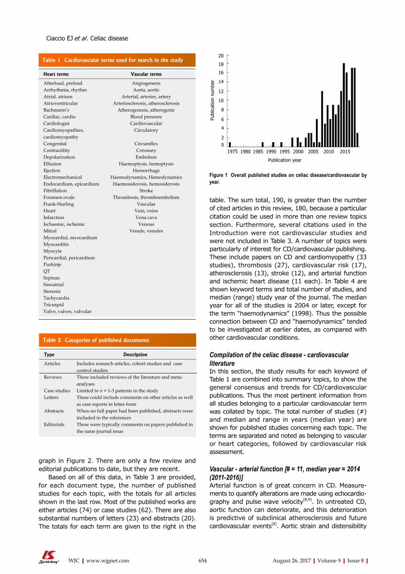
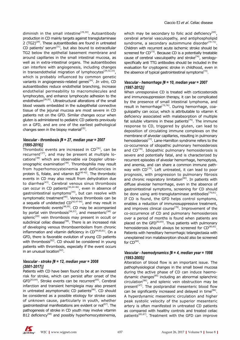
PUBLICATION SUMMARY STATISTICSIn Figure 1 is presented a graph of the number of publications per year in which CD and cardiovascular terms appeared. The earliest studies in which CD was investigated for cardiovascular function were published in 1970. However until the year 1998, only a handful of such studies were published, after which there began a substantial increase in the publishing of CD/cardiovascular studies. Although there were fluctuations in the number of studies in the 2000s and 2010s, the overall trend was a sharp upward swing in published studies. The data for 2017 only includes the first few weeks of the year.
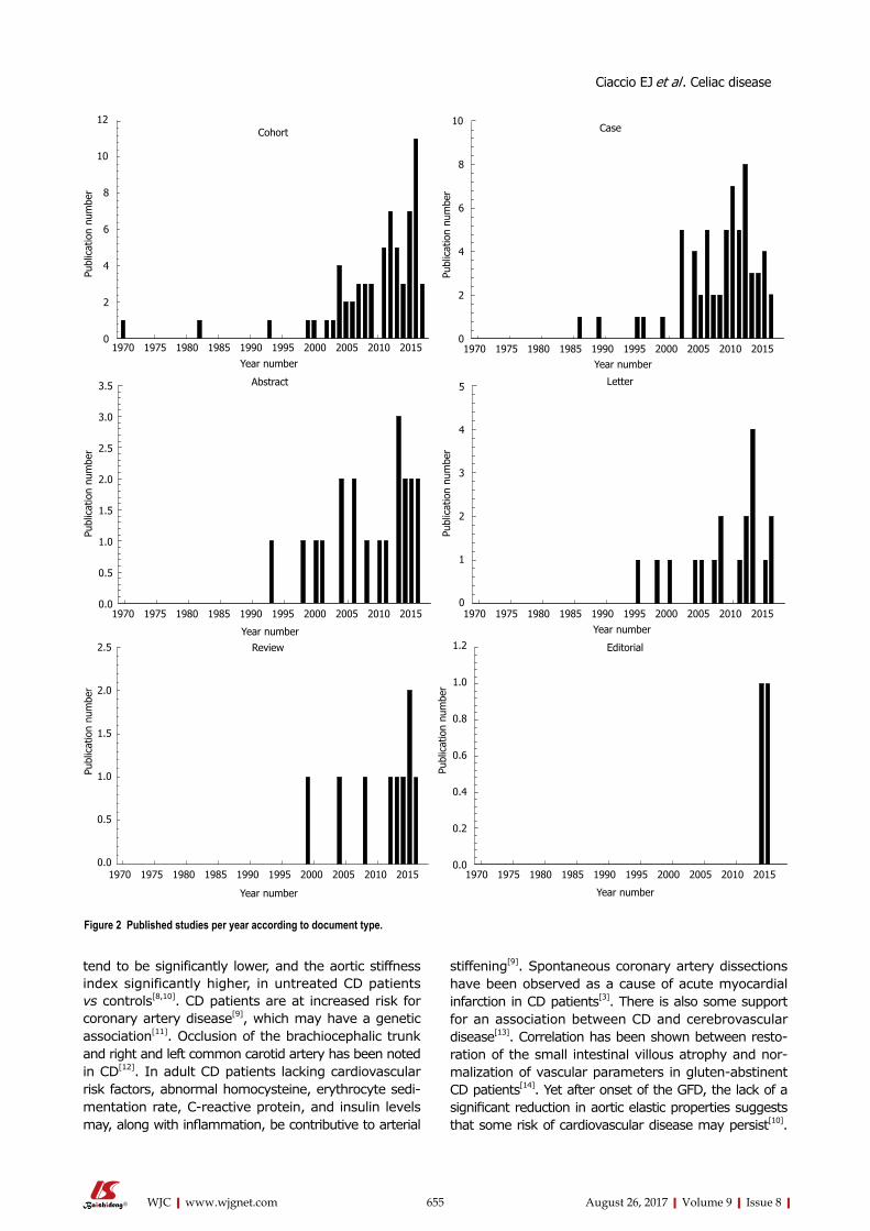
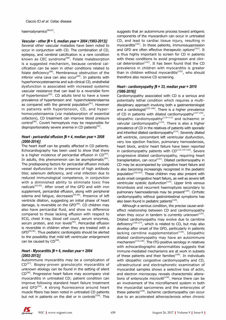
The number of studies for selected types that was published per year is shown in bar graph form in Figure 2. The results are shown for articles (cohort), case studies, abstracts, and letters, and review and editorial publications. Many of the CD/cardiovascular published studies were either articles or case studies. As for graphs of the total studies published that were shown in Figure 1, the graphs of individual published document types in Figure 2 begin to exhibit substantial increases about the year 2000. There were also a number of abstracts and case reports published in the late 1980s and 1990s, as is notable in the case reports
653 August 26, 2017|Volume 9|Issue 8|WJC|www.wjgnet.com
Ciaccio EJ et al . Celiac disease
graph in Figure 2. There are only a few review and editorial publications to date, but they are recent.
Based on all of this data, in Table 3 are provided, for each document type, the number of published studies for each topic, with the totals for all articles shown in the last row. Most of the published works are either articles (74) or case studies (62). There are also substantial numbers of letters (23) and abstracts (20). The totals for each term are given to the right in the
654 August 26, 2017|Volume 9|Issue 8|WJC|www.wjgnet.com
table. The sum total, 190, is greater than the number of cited articles in this review, 180, because a particular citation could be used in more than one review topics section. Furthermore, several citations used in the Introduction were not cardiovascular studies and were not included in Table 3. A number of topics were particularly of interest for CD/cardiovascular publishing. These include papers on CD and cardiomyopathy (33 studies), thrombosis (27), cardiovascular risk (17), atherosclerosis (13), stroke (12), and arterial function and ischemic heart disease (11 each). In Table 4 are shown keyword terms and total number of studies, and median (range) study year of the journal. The median year for all of the studies is 2004 or later, except for the term “haemodynamics” (1998). Thus the possible connection between CD and “haemodynamics” tended to be investigated at earlier dates, as compared with other cardiovascular conditions.
Compilation of the celiac disease - cardiovascular literatureIn this section, the study results for each keyword of Table 1 are combined into summary topics, to show the general consensus and trends for CD/cardiovascular publications. Thus the most pertinent information from all studies belonging to a particular cardiovascular term was collated by topic. The total number of studies (#) and median and range in years (median year) are shown for published studies concerning each topic. The terms are separated and noted as belonging to vascular or heart categories, followed by cardiovascular risk assessment.
Vascular - arterial function [# = 11, median year = 2014 (2011-2016)]Arterial function is of great concern in CD. Measure-ments to quantify alterations are made using echocardio-graphy and pulse wave velocity[8,9]. In untreated CD, aortic function can deteriorate, and this deterioration is predictive of subclinical atherosclerosis and future cardiovascular events[8]. Aortic strain and distensibility
Ciaccio EJ et al . Celiac disease
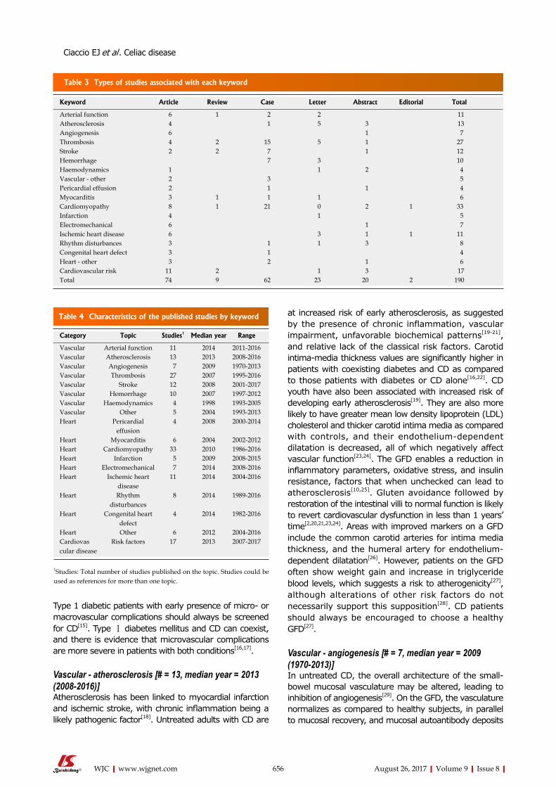
Table 2 Categories of published documents
Type Description
Articles Includes research articles, cohort studies and case control studies
Reviews These included reviews of the literature and meta-analyses
Case studies Limited to n = 1-3 patients in the studyLetters These could include comments on other articles as well
as case reports in letter formAbstracts When no full paper had been published, abstracts were
included in the references Editorials These were typically comments on papers published in
the same journal issue
Figure 1 Overall published studies on celiac disease/cardiovascular by year.
20
14
16
18
4
6
8
10
12
20
Publ
icat
ion
num
ber
1975 1995199019851980 2000 2005 2010 2015
Publication year
Table 1 Cardiovascular terms used for search in the study
655 August 26, 2017|Volume 9|Issue 8|WJC|www.wjgnet.com
stiffening[9]. Spontaneous coronary artery dissections have been observed as a cause of acute myocardial infarction in CD patients[3]. There is also some support for an association between CD and cerebrovascular disease[13]. Correlation has been shown between resto-ration of the small intestinal villous atrophy and nor-malization of vascular parameters in gluten-abstinent CD patients[14]. Yet after onset of the GFD, the lack of a significant reduction in aortic elastic properties suggests that some risk of cardiovascular disease may persist[10].
tend to be significantly lower, and the aortic stiffness index significantly higher, in untreated CD patients vs controls[8,10]. CD patients are at increased risk for coronary artery disease[9], which may have a genetic association[11]. Occlusion of the brachiocephalic trunk and right and left common carotid artery has been noted in CD[12]. In adult CD patients lacking cardiovascular risk factors, abnormal homocysteine, erythrocyte sedi-mentation rate, C-reactive protein, and insulin levels may, along with inflammation, be contributive to arterial
Year number
10
8
6
4
2
0
5
4
3
2
1
0
1.2
1.0
0.8
0.6
0.4
0.2
0.0
Case
Letter
Editorial
Figure 2 Published studies per year according to document type.
656 August 26, 2017|Volume 9|Issue 8|WJC|www.wjgnet.com
at increased risk of early atherosclerosis, as suggested by the presence of chronic inflammation, vascular impairment, unfavorable biochemical patterns[19-21], and relative lack of the classical risk factors. Carotid intima-media thickness values are significantly higher in patients with coexisting diabetes and CD as compared to those patients with diabetes or CD alone[16,22]. CD youth have also been associated with increased risk of developing early atherosclerosis[19]. They are also more likely to have greater mean low density lipoprotein (LDL) cholesterol and thicker carotid intima media as compared with controls, and their endothelium-dependent dilatation is decreased, all of which negatively affect vascular function[23,24]. The GFD enables a reduction in inflammatory parameters, oxidative stress, and insulin resistance, factors that when unchecked can lead to atherosclerosis[10,25]. Gluten avoidance followed by restoration of the intestinal villi to normal function is likely to revert cardiovascular dysfunction in less than 1 years’ time[2,20,21,23,24]. Areas with improved markers on a GFD include the common carotid arteries for intima media thickness, and the humeral artery for endothelium-dependent dilatation[26]. However, patients on the GFD often show weight gain and increase in triglyceride blood levels, which suggests a risk to atherogenicity[27], although alterations of other risk factors do not necessarily support this supposition[28]. CD patients should always be encouraged to choose a healthy GFD[27].
Vascular - angiogenesis [# = 7, median year = 2009 (1970-2013)]In untreated CD, the overall architecture of the small-bowel mucosal vasculature may be altered, leading to inhibition of angiogenesis[29]. On the GFD, the vasculature normalizes as compared to healthy subjects, in parallel to mucosal recovery, and mucosal autoantibody deposits
Type 1 diabetic patients with early presence of micro- or macrovascular complications should always be screened for CD[15]. Type Ⅰ diabetes mellitus and CD can coexist, and there is evidence that microvascular complications are more severe in patients with both conditions[16,17].
Vascular - atherosclerosis [# = 13, median year = 2013 (2008-2016)]Atherosclerosis has been linked to myocardial infarction and ischemic stroke, with chronic inflammation being a likely pathogenic factor[18]. Untreated adults with CD are
Table 3 Types of studies associated with each keyword
Keyword Article Review Case Letter Abstract Editorial Total
Heart Other 6 2012 2004-2016 Cardiovascular disease
Risk factors 17 2013 2007-2017
1Studies: Total number of studies published on the topic. Studies could be used as references for more than one topic.
Ciaccio EJ et al . Celiac disease
657 August 26, 2017|Volume 9|Issue 8|WJC|www.wjgnet.com
which may be secondary to folic acid deficiency[69], cerebral arterial vasculopathy, and antiphospholipid syndrome, a secondary autoimmune disorder[70-72]. Children with recurrent acute ischemic stroke should be screened for CD[73]. Because CD is a potentially treatable cause of cerebral vasculopathy and stroke[74], serology-specifically anti TTG antibodies should be included in the evaluation for cryptogenic stroke in childhood, even in the absence of typical gastrointestinal symptoms[72].
Vascular - hemorrhage [# = 10, median year = 2007 (1997-2012)]When unresponsive CD is treated with corticosteroids and immunosuppression therapy, it can be complicated by the presence of small intestinal lymphoma, and result in hemorrhage[75,76]. During hemorrhage, coa-gulopathy can occur, which is attributable to vitamin K deficiency associated with malabsorption of multiple fat soluble vitamins in these patients[75]. The immune response to CD, triggered by gluten, can lead to deposition of circulating immune complexes on the membrane of alveolar capillaries, resulting in pulmonary hemosiderosis[77]. Lane-Hamilton syndrome refers to the co-occurrence of idiopathic pulmonary hemosiderosis and CD[78]. Idiopathic pulmonary hemosiderosis is severe and potentially fatal, and is characterized by recurrent episodes of alveolar hemorrhage, hemoptysis, and anemia, and can share a common immune path-way with CD[79]. Left untreated, it can lead to poor prognosis, with progression to pulmonary fibrosis and chronic respiratory limitation[80]. In patients with diffuse alveolar hemorrhage, even in the absence of gastrointestinal symptoms, screening for CD should be done using anti-transglutaminase antibodies[79,81]. If CD is found, the GFD helps control symptoms, enables a reduction of immunosuppressive treatment, and improves clinical course[79]. Improvement of the co-occurrence of CD and pulmonary hemosiderosis over a period of months is found when patients are placed on the GFD[82,83]. Thus patients with pulmonary hemosiderosis should always be screened for CD[80,82]. Patients with hereditary hemorrhagic telangiectasia with unexplained iron malabsorption should also be screened for CD[84].
Vascular - haemodynamics [# = 4, median year = 1998 (1993-2005)]Alteration of blood flow is an important issue. The pathophysiological changes in the small bowel mucosa during the active phase of CD can induce haemo-dynamic changes[85] including an abnormal splanchnic circulation[86], and splenic vein obstruction may be present[47]. The postprandial mesenteric blood flow can be significantly increased and delayed in time[86]. A hyperdynamic mesenteric circulation and higher peak systolic velocity of the superior mesenteric artery is often manifested in untreated CD patients as compared with healthy controls and treated celiac patients[85,87]. Treatment with the GFD can improve
diminish in the small intestine[29,30]. Autoantibody production in CD mainly targets against transglutaminase 2 (TG2)[29]. These autoantibodies are found in untreated CD patients’ serum[31], but also bound to extracellular TG2 below the epithelial basement membrane and around capillaries in the small intestinal mucosa, as well as in extra-intestinal organs. The autoantibodies can interfere with angiogenesis, including changes in transendothelial migration of lymphocytes[29,32,33], which is probably influenced by common genetic variants in angiogenesis-related genes[34]. In vitro, CD autoantibodies reduce endothelial branching, increase endothelial permeability to macromolecules and lymphocytes, and enhance lymphocyte adhesion to the endothelium[29,35]. Ultrastructural alterations of the small blood vessels embedded in the subepithelial connective tissue of the jejunal mucosa are most severe in CD patients not on the GFD. Similar changes occur when gluten is administered to pediatric CD patients previously on a GFD, and are one of the earliest pathological changes seen in the biopsy material[35].
Vascular - thrombosis [# = 27, median year = 2007 (1995-2016)]Thrombotic events are increased in CD[36], can be recurrent[37], and may be present at multiple lo-cations[38] which are observable via Doppler ultras-onographic examination[39]. Thrombophilia may result from hyperhomocysteinemia and deficiencies in protein S, folate, and vitamin B2[40-42]. The thrombotic events in CD may also result from dehydration due to diarrhea[43]. Cerebral venous sinus thrombosis can occur in CD patients[40,44-46], even in absence of gastrointestinal symptoms[45], but can resolve with symptomatic treatment[40]. Venous thrombosis can be a sequela of undetected CD[42,47-54], and may result in thromboembolic events[48,55]. CD may be accompanied by portal vein thrombosis[56,57], and mesenteric[58] or splenic[59] vein thrombosis may present in occult or subclinical celiac disease[58]. There is an increased risk of developing venous thromboembolism from chronic inflammation and vitamin deficiency in CD[44,60,61]. On a GFD, there is favorable evolution of young CD patients with thrombosis[62]. CD should be considered in young patients with thrombosis, especially if the event occurs in an unusual location[62].
Vascular - stroke [# = 12, median year = 2008 (2001-2017)]Patients with CD have been found to be at an increased risk for stroke, which can persist after onset of the GFD[63,64]. Stroke events can be recurrent[65]. Cerebral infarction and transient hemiplegia may also present in untreated asymptomatic CD patients[66]. CD should be considered as a possible etiology for stroke cases of unknown cause, particularly in youth, whether gastrointestinal manifestations are evident or not[67]. The pathogenesis of stroke in CD youth may involve vitamin B12 deficiency[68] and possibly hyperhomocysteinemia,
Ciaccio EJ et al . Celiac disease
658 August 26, 2017|Volume 9|Issue 8|WJC|www.wjgnet.com
suggests that an autoimmune process toward antigenic components of the myocardium can occur in untreated CD, and lead to cardiac tissue injury, resulting in myocarditis[101]. In these patients, immunosuppression and GFD are often effective therapeutic options[101]. It is thus highly important to screen for CD in patients with these conditions to avoid progression and clini-cal deterioration[102]. It has been found that the CD prevalence in children with myocarditis is greater than in children without myocarditis[102], who should therefore also receive CD screening.
Heart - cardiomyopathy [# = 33, median year = 2010 (1986-2016)]Cardiomyopathy associated with CD is a serious and potentially lethal condition which requires a multi-disciplinary approach involving both a gastroenterologist and a cardiologist[103-106]. There is a higher prevalence of CD in patients with dilated cardiomyopathy[107-109], idiopathic cardiomyopathy[110-113] and ischaemic or valvular cardiomyopathy[110]. There is also a higher prevalence of CD in the relatives of patients with sporadic and inherited dilated cardiomyopathy[114]. Severely dilated left ventricle, concomitant left ventricular dysfunction, very low ejection fraction, pulmonary hemosiderosis, heart block, and/or heart failure have been reported in cardiomyopathy patients with CD[112,115-120]. Severe progressive dilated cardiomyopathy, requiring heart transplantation, can occur[121]. Dilated cardiomyopathy in CD may be accompanied by congestive heart failure and is also becoming increasingly recognized in the pediatric population[122,123]. These children may also present with acute onset congestive heart failure, as well as severe left ventricular systolic dysfunction[123]. Upper limb venous thrombosis and recurrent haemoptysis secondary to pulmonary haemosiderosis may be present[123]. Cirrhotic cardiomyopathy without gastrointestinal symptoms has also been found in pediatric patients[124].
Although a serious condition, the precise cause-and-effect relationship between CD and cardiomyopathy when they occur in tandem is currently unknown[125]. Dilated cardiomyopathy may evolve due to carnitine deficiency[126,127], which is related to CD, but may also develop after onset of the GFD, particularly in patients lacking carnitine supplementation[126]. Idiopathic dilated cardiomyopathy may have an autoimmune mechanism[110,128]. The tTG-positive serology in relatives with echocardiographic abnormalities suggests that immune-mediated mechanisms are at work in subsets of these patients and their families[106]. In individuals with idiopathic congestive cardiomyopathy and CD, ultrastructural and electrophoretic examination of myocardial samples shows a selective loss of actin, and electron microscopy reveals characteristic altera-tions of enterocyte microvilli[129]. Hence there can be an involvement of the microfilament system in both the myocardial sarcomeres and the enterocytes of these patients[129]. Ischemic cardiomyopathy can occur due to an accelerated atherosclerosis when chronic
haemodynamics[85,87].
Vascular - other [# = 5, median year = 2004 (1993-2013)]Several other vascular maladies have been noted to occur in conjunction with CD. The combination of CD, epilepsy, and cerebral calcification is a rare condition known as CEC syndrome[88]. Folate malabsorption is a suggested mechanism, because cerebral cal-cification can be seen in other conditions related to folate deficiency[88]. Membranous obstruction of the inferior vena cava can also occur[89]. In patients with hyperhomocysteinaemia and sub-clinical CD, endothelial dysfunction is associated with increased systemic vascular resistance that can lead to a reversible form of hypertension[90]. CD adults tend to have a lower prevalence of hypertension and hypercholesterolaemia as compared with the general population[91]. However in patients with hypertension, CD, and hyper-homocysteinaemia (via malabsorption of essential cofactors), CD treatment can improve blood pressure control[90]. Covert hemoptysis may be responsible for disproportionately severe anemia in CD patients[92].
Heart - pericardial effusion [# = 4, median year = 2008 (2000-2014)]The heart itself can be greatly affected in CD patients. Echocardiography has been used to show that there is a higher incidence of pericardial effusion in CD[93]. In adults, this phenomenon can be asymptomatic[94]. The predisposing factors for pericardial effusion include vessel dysfunction in the presence of high antibody titer, selenium deficiency, and viral infection due to reduced immunological competence, in conjunction with a diminished ability to eliminate toxic free radicals[93,95]. After onset of the GFD and with iron supplement, pericardial effusion, along with peripheral edema and fatigue, decreases[94,96]. Presence of left ventricle dilation, suggesting an initial phase of heart damage, is reversible on the GFD[96]. CD children may also have pericardial fluid, and show no difference compared to those lacking effusion with respect to ECG, chest X-ray, blood cell count, serum enzymes, serum protein, and iron levels[95]. Pericardial effusion is reversible in children when they are treated with a GFD[95,96]. Thus pediatric cardiologists should be alerted to the possibility that mild left ventricular enlargement can be caused by CD[96].
Heart - Myocarditis [# = 6, median year = 2004 (2002-2012)]Autoimmune myocarditis may be a complication of CD[97]. Biopsy-proven granulocytic myocarditis of unknown etiology can be found in the setting of silent CD[98]. Progressive heart failure may accompany viral myocarditis in untreated CD; patient condition can improve following standard heart failure treatment and GFD[99]. A strong fluorescence around heart muscle fibers has been noted in untreated CD patients but not in patients on the diet or in controls[100]. This
Ciaccio EJ et al . Celiac disease
659 August 26, 2017|Volume 9|Issue 8|WJC|www.wjgnet.com
prolapse[142] and subclinical myocardial dysfunction of both ventricles[143] can be present in both the pediatric and adult CD population. In children, significantly lower contractility indices and higher left ventricular dimensions are evident as compared with controls[144]. On the GFD, valve regurgitations resolve, and echocardiographic parameters significantly improve[144].
Heart - ischemic heart disease [# = 11, median year = 2014 (2004-2016)]There is an increased risk of ischemic heart disease and higher cardiovascular mortality in CD[145,146] despite the lack of traditional risk factors[26,147] including blood pressure, body mass index, serum cholesterol, lipids, exercise, and smoking[4,148]. First-degree relatives of CD patients are also at an increased risk of ischemic heart disease, but the excess risk is slight[149]. CD and ischemic heart disease may share a common underlying link, rather than a cause-and-effect relationship[18,150]. The underlying association between CD and ischemic heart disease may be chronic inflammation, a major risk factor in the general population; however, potential confounders may also be involved[18,146,148]. After onset of the GFD, persistent villous atrophy detected during follow-up biopsy was not associated with increased risk of ischemic heart disease[151,152].
Rhythm disturbances [# = 8, median year = 2014 (1989-2016)]Compared to controls, untreated CD patients have increased P-wave dispersion and higher interatrial, intra-left atrial, and intra-right atrial conduction delay[153]. Tp-e interval, Tp-e/QT and Tp-e/QTc ratios are also increased in CD[154]. There is a higher prevalence of atrial fibrillation among CD patients[153,155]. Atrial fibrillation, when it occurs, is associated with an in-creased risk of ischaemic stroke and heart failure[156]. The chronic inflammation that can occur in untreated CD is a recognized risk factor for atrial fibrillation[155,156]. CD patients have slower atrial electrical conduction, which may also increase the risk of atrial fibrillation[153]. However, persistent villous atrophy on follow-up biopsy was not associated with any increased risk of atrial fibrillation[151,152]. It has been reported that CD patients with pulmonary hemosiderosis can develop infranodal heart block, necessitating implantation of a pacemaker[77,157]. These patients may lack digestive manifestations but have iron deficiency and vitamin deficiency anemia[77]. Rhythm alterations in CD can thus result from other pathogenic mechanisms including electrolyte disturbances caused by malabsorption, which can normalize on the GFD[77]. Patients with rhythm disturbances and chronic anemia of unclear origin should therefore be tested for CD[77].
Heart - congenital heart defect [# = 4, median year = 2014 (1982-2016)]CD patients are likely to more commonly have atrial septal defect as compared to controls[158]. Screening
inflammation is present in CD[130]. Compliance with a GFD is mandatory if patients are
to avoid progression of their cardiomyopathy[105,131]. The GFD has been shown to have a beneficial effect on cardiac performance in CD patients with dilated cardiomyopathy[132]. After start of the GFD, abnormal left ventricular dimensions, and diminished cardiac function, including decreased ejection fraction, can improve markedly and may even be completely reversible[103,108,118,128,133]. After two years on a GFD, patients presenting with dilated cardiomyopathy associated with CD show progressive increase in mean serum carnitine levels as compared to values observed prior to the diet[127]. Children with CD and dilated cardiomyopathy also have improved cardiac function once adherent to the GFD[109]. In CD patients on the GFD, there is no association with later onset of myocarditis, cardiomyopathy or pericarditis[134].
Screening for CD in patients with dilated cardio-myopathy, pulmonary haemosiderosis, and iron deficiency anemia in the absence of known etiology is advisable regardless of the intestinal symptoms[105,108,116,123,133]. Serologic tests for IgA-EmA and tissue transglutaminase antibodies should be used to screen for CD[106]. Co-morbidities including iron-deficiency anemia in patients with dilated cardiomyopathy should arouse suspicion of CD[117]. All patients diagnosed with cardiomyopathy and CD should be offered an endomyocardial biopsy for better histological and diagnostic definition[128]. It is beneficial to assess CD in children with dilated cardiomyopathy in the absence of known etiologies[104,135].
Heart - infarction [# = 5, median year = 2009 (2008-2015)]Acute myocardial infarction with ST-elevation and spontaneous coronary artery dissection can occur in young patients with CD[3,136]. However, chronic hypocalcemia in untreated CD patients due to poor absorption of minerals can result in electrocardiographic changes that mimic acute myocardial infarction[120]. In CD, there is a higher all-cause mortality one year post-myocardial infarction as compared with the general population[20]. Mesenteric infarction has also occurred as a consequence of CD, and clinicians should be aware of this possibility[137].
Heart - electromechanical functioning [# = 7, median year = 2014 (2008-2016)]Measurement of electromechanical parameters is beneficial to determine the degree of cardiac involvement in CD[138]. Atrial conduction delays are significantly higher in untreated CD as compared with healthy individuals, and may lead to atrial fibrillation[139]. Measurement of atrial electromechanical delay parameters might therefore be useful to predict atrial fibrillation risk[139]. Statistically significant differences in left ventricular function as assessed by echocardiography imaging are found in CD patients vs controls[140]. Patients with CD can have impaired diastolic and systolic function as measured by tissue Doppler echocardiography[141]. Mitral valve
Ciaccio EJ et al . Celiac disease
660 August 26, 2017|Volume 9|Issue 8|WJC|www.wjgnet.com
The lack of a uniform set of risk factors can in-fluence whether CVD risk is affected by a GFD[172]. In actuality, both risk and protective factors for CVD are likely to be present in CD, at baseline and also on a GFD[172]. Modifiable risk factors for CVD can include body mass index and cholesterol, which have been shown to increase on the GFD[168]. In CD individuals with type 1 diabetes on a GFD, improvement in HDL-cholesterol, and a lower resting heart rate, has been demonstrated as compared with those CD patients without diabetes[178]. On a GFD, the lipid profile of CD patients can also improve[17]. At one year on a GFD, waist circumference may increase, but without significant rise in total or LDL cholesterol[173,174]. The GFD should therefore ideally go beyond gluten exclusion and include body weight control and high quality nutrients[172]. A relatively high proportion of CD children on the GFD had one or more CVD risk factors[179]. The most common CVD risk factors are high fasting triglycerides, elevated blood pressure, and high LDL cholesterol concentrations[179]. Insulin resistance is also found, underscoring the need for CVD screening and dietary counseling targeting the pediatric CD population[179]. Screening for CD and monitoring of HDL cholesterol is recommended in youth with type 1 diabetes[177]. CVD risk factors also include metabolic disorders caused by malabsorption in pediatric CD patients[180]. Hence timely correction of water and electrolyte imbalance, and administration of cardiometabolic therapy, is necessary[180].
CONCLUSIONPublished studies pertaining to the connection between cardiovascular conditions and CD began in the late 1960’s, consisting of a few studies each year, and was followed by a substantial increase beginning about the year 2000. Many of the published studies are either articles (including cohort and case control studies) or case studies consisting of one to three patients. Often, as might be expected, a number of case studies appeared in the literature prior to the cohort studies. Based on the evidence presented in these papers, it is apparent that cardiovascular involvement in CD is a real phenomenon and that there are many manifestations, owing to the multifaceted, systemic physiological changes that can occur in CD. A number of the cardiovascular issues that can occur in untreated CD patients, will resolve on a GFD, often in conjunction with healing of the small intestinal villi. Cardiomyopathy is the most frequently documented cardiovascular condition observed in conjunction with CD, and seems to mostly or completely resolve with appropriate treatment, including a GFD. However, if CD is left unrecognized until a late stage, damage done to the heart may not be entirely reversible. Similarly, to the present time there has been substantial docu-mentation of a number of other cardiovascular con-ditions found in conjunction with untreated CD including
for CD in children with congenital heart defect is recommended, since serum TTG IgA levels are significantly higher in patients with congenital heart defect vs control children[159]. Down syndrome patients with congenital heart defect have higher CD prevalence as compared to patients without congenital heart defect, and CD prevalence in Down syndrome patients is higher than in controls[160]. In children with congenital heart disease and CD, growth improves on a GFD[161].
Heart - other [# = 6, median year = 2012 (2004-2016)]Chronic hypocalcemia, which may occur in untreated CD due to malabsorption, has been associated with reversible cardiac dysfunction[120]. Untreated CD children tend to show an imbalance of cardiac sympathetic and parasympathetic activity due to enhanced sympathetic tone[162]. This imbalance is still detected after a six months period of GFD[162]. This suggests the presence of a subclinical autonomic nervous system dysfunction[162]. There is a higher prevalence of CD in patients with Postural Orthostatic Tachycardia Syndrome (POTS)[163]; thus these patients should also be screened for CD[164]. Left ventricular hypertrabeculation/noncompaction may be associated with CD[165]. Subclinical systolic dysfunction of the left ventricle may be present in CD children[166].
Cardiovascular disease risk factors [# = 17, median year = 2013 (2007-2017)]Cardiovascular disease (CVD) as a whole has many etiologies and is the leading cause of death in developed countries[167,168]. There is a modestly increased risk of CVD in CD patients[63]. Both male and female CD patients may have higher estimated risk for CVD as compared to controls[169,170]. However, CVD risk factors conferred by CD have not been well-defined[171]. This has led to conflicting evidence as to whether CD patients actually have increased baseline risk[172]. CD patients are susceptible to increased platelet activation and increased mean platelet volume and RDW values, factors that contribute to increased risk[173,174]. Using the Framingham Heart Study (FRS) 10-year general CVD risk score, lower values were found among CD patients as compared with controls, which may be due to a lesser body mass index and reduced tobacco use among CD patients[171]. Risk factors other than those measured by the FRS may be observed as increased risk of CVD in CD patients[171]. There can also be a positive association between CD and CVD risk due to ascertainment bias[175]. In CD children, risk factors can be frequently observed as compared with healthy subjects[18]. Overall, certain CVD risk factors have been found to be higher in CD youth as compared with the general population, although neither blood pressure nor overweight and obesity rates were increased[176]. Youth with type 1 diabetes and CD had lower high density lipoprotein (HDL) cholesterol, increasing CVD risk, as compared with non CD patients[177].
Ciaccio EJ et al . Celiac disease
661 August 26, 2017|Volume 9|Issue 8|WJC|www.wjgnet.com
16 Pitocco D, Giubilato S, Martini F, Zaccardi F, Pazzano V, Manto A, Cammarota G, Di Stasio E, Pedicino D, Liuzzo G, Crea F, Ghirlanda G. Combined atherogenic effects of celiac disease and type 1 diabetes mellitus. Atherosclerosis 2011; 217: 531-535 [PMID: 21601206 DOI: 10.1016/j.atherosclerosis.2011.04.042]
17 Leeds JS, Hopper AD, Hadjivassiliou M, Tesfaye S, Sanders DS. High prevalence of microvascular complications in adults with type 1 diabetes and newly diagnosed celiac disease. Diabetes Care 2011; 34: 2158-2163 [PMID: 21911773 DOI: 10.2337/dc11-0149]
18 Mormile R. Celiac Disease and Ischemic Heart Disease: What is the Link? J Clin Cell Immunol 2013: 4: 2 [DOI: 10.4172/2155-9899.1000173]
19 Mormile R. Celiac disease and atherosclerosis: An immunologic puzzle to be solved? Immunol Lett 2016; 180: 75-76 [PMID: 27743857 DOI: 10.1016/j.imlet.2016.10.003]
20 Rigoni A, De Marchi S, Prior M, Chiarioni G, Arosio E. 100 young adults with celiac disease may be at increased risk of early atherosclerosis. Nutrition, Metabolism Cardiovascular Dis 2008; 18: S58 [DOI: 10.1016/s0939-4753(08)70101-7]
21 De Marchi S, Chiarioni G, Prior M, Arosio E. Young adults with coeliac disease may be at increased risk of early atherosclerosis. Aliment Pharmacol Ther 2013; 38: 162-169 [PMID: 23730933 DOI: 10.1111/apt.12360]
22 Martini F, Pitocco D, Zaccardi F, Giubilato S, Pallano V, Manto A, Cammarota G, Liuzzo G, Crea F, Ghirlanda G. Early Detection of cardiovascular disease in patients with associated type 1 diabetes mellitus and celiac disease. 1701 N Beauregard St, Alexandria, VA 22311-1717. USA: Amer Diabetes Assoc, 2011
23 Valitutti F, Trovato CM, Barbato M, Cucchiara S. Letter: Atherosclerosis and coeliac disease – Another feature of the changing paradigm? Aliment Pharmacol Therap 2013; 38: 559 [DOI: 10.1111/apt.12399]
25 Aguilar EC, Soares FL, Santos LC, Capettini LS, Teixeira LG, Lemos VS, Alvarez-Leite JI. SUN-LB037: Gluten-free diet reduces cardiovascular risk factors and atherosclerosis development in APOE KO mice. Clin Nutr 2015; 34: S248 [DOI: 10.1016/S0261-5614(15)30758-5]
26 Demir AM, Kuloğlu Z, Yaman A, Fitöz S, Nergizoğlu G, Kansu A. Carotid intima-media thickness and arterial stiffness as early markers of atherosclerosis in pediatric celiac disease. Turk J Pediatr 2016; 58: 172-179 [PMID: 27976558 DOI: 10.24953/turkjped.2016.02.008]
27 Bianchi PI, Biagi F, Borsotti E, Marchese A, Vattiato C, Ferretti F, Balduzzi D, Trotta L, Schiepatti A, Corazza GR. P. 05.13 Peripheral artery disease in coeliac disease. Dig Liver Dis 2013; 45: S123. [DOI: 10.1016/S1590-8658(13)60342-4]
28 Zanini B, Mazzoncini E, Lanzarotto F, Ricci C, Cesana BM, Villanacci V, Lanzini A. Impact of gluten-free diet on cardiovascular risk factors. A retrospective analysis in a large cohort of coeliac patients. Dig Liver Dis 2013; 45: 810-815 [PMID: 23688807 DOI: 10.1016/j.dld.2013.04.001]
29 Myrsky E, Caja S, Simon-Vecsei Z, Korponay-Szabo IR, Nadalutti C, Collighan R, Mongeot A, Griffin M, Mäki M, Kaukinen K, Lindfors K. Celiac disease IgA modulates vascular permeability in vitro through the activity of transglutaminase 2 and RhoA. Cell Mol Life Sci 2009; 66: 3375-3385 [PMID: 19680746 DOI: 10.1007/s00018-009-0116-1]
30 Myrsky E, Syrjänen M, Korponay-Szabo IR, Mäki M, Kaukinen K, Lindfors K. Altered small-bowel mucosal vascular network in untreated coeliac disease. Scand J Gastroenterol 2009; 44: 162-167 [PMID: 18985542 DOI: 10.1080/00365520802400875]
31 Myrsky E, Syrjänen M, Kaukinen K, Korponay-Szabo I, Maki M, Lindfors K. Celiac disease IGA class autoantibodies inhibit the recruitment of vascular smooth muscle cells to capillary walls. J Pediatr Gastroenterol Nutr 2006; 42: E1
32 Myrsky E, Kaukinen K, Syrjänen M, Korponay-Szabó IR, Mäki M, Lindfors K. Coeliac disease-specific autoantibodies targeted against transglutaminase 2 disturb angiogenesis. Clin Exp Immunol 2008; 152:
thrombosis and thromboembolism, ischemic heart disease, stroke, and arrhythmia. There has also been significant investigation of CD and risk of cardiovascular disease. On this topic, a current problem is that there is no uniform set of cardiovascular risk factors used for analysis. Future studies should settle the question of how to best treat these co-occurring conditions, and to determine if other cardiovascular manifestations of CD are common phenomena.
REFERENCES1 Lodha A, Haran M, Hollander G, Frankel R, Shani J. Celiac disease
associated with dilated cardiomyopathy. South Med J 2009; 102: 1052-1054 [PMID: 19738524 DOI: 10.1097/SMJ.0b013e3181b64dd9]
2 Rybak A, Cukrowska B, Socha J, Socha P. Long term follow up of celiac disease-is atherosclerosis a problem? Nutrients 2014; 6: 2718-2729 [PMID: 25050927 DOI: 10.3390/nu6072718]
3 Bayar N, Çağırcı G, Üreyen ÇM, Kuş G, Küçükseymen S, Arslan Ş. The relationship between spontaneous multi-vessel coronary artery dissection and celiac disease. Korean Circ J 2015; 45: 242-244 [DOI: 10.4070/kcj.2015.45.3.242]
4 Wang I, Hopper I. Celiac disease and drug absorption: implications for cardiovascular therapeutics. Cardiovasc Ther 2014; 32: 253-256 [PMID: 25284331 DOI: 10.1111/1755-5922.12094]
5 Ciaccio EJ, Bhagat G, Lewis SK, Green PH. Trends in celiac disease research. Comput Biol Med 2015; 65: 369-378 [PMID: 26095989 DOI: 10.1016/j.compbiomed.2015.05.023]
6 Ciaccio EJ, Bhagat G, Lewis SK, Green PH. Trends in gluten research and its relationship to autoimmune and allergic diseases. Informat Med Unlocked 2016; 3: 7-14 [DOI: 10.1016/j.imu.2016.05.001]
7 Ciaccio EJ, Biviano AB, Iyer V, Garan H. Trends in quantitative methods used for atrial fibrillation and ventricular tachycardia analyses. Informat Med Unlocked 2017; 6: 12-27 [DOI: 10.1016/j.imu.2016.12.003]
8 Sari C, Ertem AG, Sari S, Efe TH, Keles T, Durmaz T, Ertem S, Bozkurt E. Impaired aortic function in patients with coeliac disease. Kardiol Pol 2015; 73: 1108-1113 [PMID: 25987403 DOI: 10.5603/KP.a2015.0097]
9 Korkmaz H, Sozen M, Kebapcilar L. Increased arterial stiffness and its relationship with inflammation, insulin, and insulin resistance in celiac disease. Eur J Gastroenterol Hepatol 2015; 27: 1193-1199 [PMID: 26181110 DOI: 10.1097/MEG.0000000000000437]
10 Bayar N, Çekin AH, Arslan Ş, Çağırcı G, Küçükseymen S, Çay S, Harmandar FA, Yeşil B. Assessment of Aortic Elasticity in Patients with Celiac Disease. Korean Circ J 2016; 46: 239-245 [PMID: 27014355 DOI: 10.4070/kcj.2016.46.2.239]
11 Jansen H, Willenborg C, Schlesinger S, Ferrario PG, König IR, Erdmann J, Samani NJ, Lieb W, Schunkert H. Genetic variants associated with celiac disease and the risk for coronary artery disease. Mol Genet Genomics 2015; 290: 1911-1917 [PMID: 25893417 DOI: 10.1007/s00438-015-1045-3]
12 Altabas K, Crne N, Franjić BD, Pavlović D, Josipović J. [Peritoneal dialysis in a patient with right hemiparesis, lupus nephritis, significant insufficiency of arteries of aortic arch and celiac disease: case report]. Acta Med Croatica 2012; 66 Suppl 2: 72-75 [PMID: 23513421]
13 Heikkilä K, Koskinen OA, Agarwal A, Tikkinen KA, Mäki M, Kaukinen K. Associations of coeliac disease with coronary heart disease and cerebrovascular disease: A systematic review and meta-analysis. Nutr Metab Cardiovasc Dis 2015; 25: 816-831 [PMID: 26111459 DOI: 10.1016/j.numecd.2015.05.004]
14 De Marchi S, Chiarioni G, Prior M, Arosio E. Commentary: coeliac disease and atherosclerosis--hand in hand? Authors’ reply. Aliment Pharmacol Ther 2013; 38: 550-551 [PMID: 23937458 DOI: 10.1111/apt.12405]
15 Pitocco D, Zaccardi F, Martini F, Giubilato S, Liuzzo G, Crea F, Ghirlanda G. The cardiovascular relevance of celiac disease. Diabetes Care 2012; 35: e20 [PMID: 22355023 DOI: 10.2337/dc11-2232]
Ciaccio EJ et al . Celiac disease
662 August 26, 2017|Volume 9|Issue 8|WJC|www.wjgnet.com
tb00764.x]50 Casella G, Baldini V, Perego D, Brambills M, Fraterrigo RT. A rare
association between inflammatory bowel disease (IBD), coeliac disease, membranous glomerulonephritis and leg venous thrombosis associated to heterozygosis for V Leiden factor. Dige Liver Dis 2000; 32: A44 [DOI: 10.1016/S1590-8658(00)80219-4]
51 Casella G, Perego D, Baldini V, Monti C, Crippa S, Buda CA. A rare association between ulcerative colitis (UC), celiac disease (CD), membranous glomerulonephritis, leg venous thrombosis, and heterozygosity for factor V Leiden. J Gastroenterol 2002; 37: 761-762 [PMID: 12375154 DOI: 10.1007/s005350200126]
52 Hida M, Erreimi N, Ettair S, Mouane N, Bouchta F. Associated celiac disease and venous thrombosis. Arch Pediatr 2000; 7: 215-216 [DOI: 10.1016/S0929-693X(00)88097-5]
53 Mezalek ZT, Habiba BA, Hicham H, Houda B, Mouna M, Mohamed A, Mohamed A. C0396 Venous thrombosis revealing celiac disease. Three cases. Throm Res 2012; 130: S154-S155 [DOI: 10.1016/j.thromres.2012.08.140]
54 Kremer Hovinga JA, Baerlocher G, Wuillemin WA, Solenthaler M. [Deep venous thrombosis of the leg in acquired thrombophilia--hyperhomocysteinemia as a sequela of undetected celiac disease]. Ther Umsch 1999; 56: 519-522 [PMID: 10517124 DOI: 10.1024/0040-5930.56.9.519]
55 Ludvigsson JF, Welander A, Lassila R, Ekbom A, Montgomery SM. Risk of thromboembolism in 14,000 individuals with coeliac disease. Br J Haematol 2007; 139: 121-127 [PMID: 17854316 DOI: 10.1111/j.1365-2141.2007.06766.x]
56 Karoui S, Sfar S, Kallel M, Boubaker J, Makni S, Filali A. [Antiphospholipid syndrome revealed by portal vein thrombosis in a patient with celiac disease]. Rev Med Interne 2004; 25: 471-473 [PMID: 15158321 DOI: 10.1016/j.revmed.2004.02.014]
57 Zenjari T, Boruchowicz A, Desreumaux P, Laberenne E, Cortot A, Colombel JF. Association of coeliac disease and portal venous thrombosis. Gastroenterol Clin Biol 1995; 19: 953-954 [PMID: 8746058]
58 Azzam NA, Al Ashgar H, Dababo M, Al Kahtani N, Shahid M. Mesentric vein thrombosis as a presentation of subclinical celiac disease. Ann Saudi Med 2006; 26: 471-473 [PMID: 17146205 DOI: 10.5144/0256-4947.2006.471]
59 Khanna S, Chaudhary D, Kumar P, Mazumdar S. Occult celiac disease presenting as splenic vein thrombosis. Indian J Gastroenterol 2008; 27: 38-39 [PMID: 18541941]
60 Ungprasert P, Wijarnpreecha K, Tanratana P. Risk of venous thromboembolism in patients with celiac disease: A systematic review and meta-analysis. J Gastroenterol Hepatol 2016; 31: 1240-1245 [PMID: 26709842 DOI: 10.1111/jgh.13282]
61 Gabrielli M, Santoliquido A, Santarelli L, Fini L, Andrisani MC, Pola P, Gasbarrini G, Gasbarrini A. 6 P Venous thromboembolism, hyperhomocysteinemia and silent coeliac disease: A case report. Digestive and Liver Disease 2002; 34: A27 [DOI: 10.1016/S1590- 8658(02)90103-9]
62 Zoubeidi H, Ben Salem T, Ben Ghorbel I, Houman MH. Central retinal vein occlusion revealing celiac disease. Eur J Case Reports Int Med 2016; 3 [DOI: 10.12890/2016_000492]
63 Emilsson L, Lebwohl B, Sundström J, Ludvigsson JF. Cardiovascular disease in patients with coeliac disease: A systematic review and meta-analysis. Dig Liver Dis 2015; 47: 847-852 [DOI: 10.1016/j.dld.2015.06.004]
64 Ludvigsson JF, West J, Card T, Appelros P. Risk of stroke in 28,000 patients with celiac disease: A nationwide cohort study in Sweden. J Stroke Cerebrovascular Dis 2012; 21: 860-867 [DOI: 10.1016/j.jstrokecerebrovasdis.2011.05.008]
65 Ozge A, Karakelle A, Kaleağasi H. Celiac disease associated with recurrent stroke: a coincidence or cerebral vasculitis? Eur J Neurol 2001; 8: 373-374 [PMID: 11422441 DOI: 10.1046/j.1468- 1331.2001.00233.x]
Perez MA, Martucciello S, Roivainen A, Pelliniemi LJ, Esposito C, Griffin M, Sblattero D, Mäki M, Kaukinen K, Lindfors K, Caja S. Celiac Disease-Specific TG2-Targeted Autoantibodies Inhibit Angiogenesis Ex Vivo and In Vivo in Mice by Interfering with Endothelial Cell Dynamics. PLoS One 2013; 8: e65887 [PMID: 23824706 DOI: 10.1371/journal.pone.0065887]
34 Castellanos-Rubio A, Caja S, Irastorza I, Fernandez-Jimenez N, Plaza-Izurieta L, Vitoria JC, Maki M, Lindfors K, Bilbao JR. Angiogenesis-related gene expression analysis in celiac disease. Autoimmunity 2012; 45: 264-270 [PMID: 22136669 DOI: 10.3109/08916934.2011.637531]
35 Shiner M. Mucosal vascular changes in coeliac disease and in idiopathic steatorrhoea. Micron 1970; 2: 7-18 [DOI: 10.1016/0047-7206(70)90042-7]
36 Boucelma M, Saadi M, Boukrara H, Bensalah D, Hakem D, Berrah A. [Association of celiac disease and cerebral venous thrombosis: report of two cases]. J Mal Vasc 2013; 38: 47-51 [PMID: 23287617 DOI: 10.1016/j.jmv.2012.11.003]
37 Beyrouti R, Mansour M, Kacem A, Derbali H, Mrissa R. Recurrent cerebral venous thrombosis revealing celiac disease: an exceptional case report. Acta Neurol Belg 2017; 117: 341-343 [PMID: 27680734 DOI: 10.1007/s13760-016-0698-y]
38 Poulin W, Gaertner S, Cordeanu EM, Mirea C, Andrès E, Stephan D. Stroke revealing celiac disease associated with multiple arterial thrombotic locations. Presse Med 2015; 44: 537-538 [PMID: 25887453 DOI: 10.1016/j.lpm.2015.01.007]
39 Çelik G, Çiledağ A, Keskin O, Ensari A, Karabörk A, Kaya A. Sarcoidosis, Celiac disease and deep venous thrombosis: A rare association. Balkan Med J 2011; 28: 450-452 [DOI: 10.5174/tutfd.2010.03637.1]
40 Bahloul M, Chaari A, Khlaf-Bouaziz N, Kallel H, Chaari L, Ben Hamida C, Chelly H, Rekik N, Bouaziz M. [Celiac disease, cerebral venous thrombosis and protein S deficiency, a fortuitous association?]. J Mal Vasc 2005; 30: 228-230 [PMID: 16292200 DOI: 10.1038/ajg.2008.48]
41 Kallel L, Matri S, Karoui S, Fekih M, Boubaker J, Filali A. Deep venous thrombosis related to protein S deficiency revealing celiac disease. Am J Gastroenterol 2009; 104: 256-257 [PMID: 19098891 DOI: 10.1038/ajg.2008.48]
42 Baryshnikov EN, Krums LM, Vorob’eva NN, Parfenov AI. [Lower extremity deep vein thrombosis associated with gluten-sensitivity celiac disease]. Ter Arkh 2010; 82: 52-54 [PMID: 20387677]
43 Lee ES, Pulido JS. Nonischemic central retinal vein occlusion associated with celiac disease. Mayo Clin Proc 2005; 80: 157 [PMID: 15704767 DOI: 10.4065/80.2.157]
44 Johannesdottir SA, Erichsen R, Horváth-Puhó E, Schmidt M, Sørensen HT. Coeliac disease and risk of venous thromboembolism: a nationwide population-based case-control study. Br J Haematol 2012; 157: 499-501 [PMID: 22296563 DOI: 10.1111/j.1365- 2141.2012.09030.x]
45 Doğan M, Peker E, Akbayram S, Bektas MS, Basaranoglu M, Cesur Y, Caksen H. Cerebral venous sinus thrombosis in 2 children with celiac disease. Clin Appl Thromb Hemost 2011; 17: 466-469 [PMID: 20530061 DOI: 10.1177/1076029610369799]
46 Grover PJ, Jayaram R, Madder H. Management of cerebral venous thrombosis in a patient with Lane-Hamilton syndrome and coeliac disease, epilepsy and cerebral calcification syndrome. Br J Neurosurg 2010; 24: 684-685 [PMID: 21070152 DOI: 10.3109/02688697.2010.500412]
47 Ghoshal UC, Saraswat VA, Yachha SK. Association of splenic vein obstruction and coeliac disease in an Indian patient. J Hepatol 1995; 23: 358 [PMID: 8551004 DOI: 10.1016/S0168-8278(95)80019-0]
48 Beyan E, Pamukcuoglu M, Beyan C. Deep vein thrombosis associated with celiac disease. Bratisl Lek Listy 2009; 110: 263-264 [PMID: 19507658]
49 Grigg AP. Deep venous thrombosis as the presenting feature in a patient with coeliac disease and homocysteinaemia. Aust N Z J Med 1999; 29: 566-567 [PMID: 10868541 DOI: 10.1111/j.1445-5994.1999.
68 Rachid B, Zouhayr S, Chtaou N, Messouak O, Belahsen F. [Ischemic stroke revealing celiac disease]. Pan Afr Med J 2010; 5: 2 [PMID: 21120001]
69 Gefel D, Doncheva M, Ben-Valid E, el Wahab-Daraushe A, Lugassy G, Sela BA. Recurrent stroke in a young patient with celiac disease and hyperhomocysteinemia. Isr Med Assoc J 2002; 4: 222-223 [PMID: 11908270]
70 Ajello A, Vitullo G. Stroke and coeliac disease: a new face for an old lady. Italian J Pediatr 2006; 32: 259
71 Audia S, Duchêne C, Samson M, Muller G, Bielefeld P, Ricolfi F, Giroud M, Besancenot JF. [Stroke in young adults with celiac disease]. Rev Med Interne 2008; 29: 228-231 [PMID: 17976872 DOI: 10.1016/j.revmed.2007.08.013]
73 Balci O, Sezer T. The Prevalence of Celiac Disease in Children With Arterial Ischemic Stroke. J Pediatr Hematol Oncol 2017; 39: 46-49 [PMID: 27548338 DOI: 10.1097/MPH.0000000000000661]
74 Slimani N, Hakem D, Mansouri B, Berrah A. Ischemic stroke revealing a celiac disease: A case report and review of the literature. J Neurol Sci 2013; 333: e248 [DOI: 10.1016/j.jns.2013.07.959]
75 Chen CS, Cumbler EU, Triebling AT. Coagulopathy due to celiac disease presenting as intramuscular hemorrhage. J Gen Intern Med 2007; 22: 1608-1612 [PMID: 17768663 DOI: 10.1007/s11606-007-0297-y]
76 Gheorghe L, Popescu I, Gheorghe C, Aposteanu G, Popescu C, Oproiu A. Fatal intestinal hemorrhage complicating ileal lymphoma after cyclosporine for unresponsive celiac disease. Hepato-Gastroenterology 1996; 44: 1342-1345 [PMID: 9356852]
77 Callejas Rubio JL, Ortego N, Díez-Ruiz A, Guilarte J, de la Higuera-Torres J. Celiac disease presenting as chronic anemia associated with heart block. Am J Gastroenterol 1998; 93: 1391-1392 [PMID: 9707085 DOI: 10.1111/j.1572-0241.1998.01391.x]
78 Sethi GR, Singhal KK, Puri AS, Mantan M. Benefit of gluten-free diet in idiopathic pulmonary hemosiderosis in association with celiac disease. Pediatr Pulmonol 2011; 46: 302-305 [PMID: 20967850 DOI: 10.1002/ppul.21357]
79 Testa ME, Maffey A, Colom A, Agüero L, Rogé I, Andrewartha MS, Teper A. [Pulmonary hemorrhage associated with celiac disease]. Arch Argent Pediatr 2012; 110: e72-e76 [PMID: 22859336 DOI: 10.5546/aap.2012.e72]
80 Santos JW, Mello Neto AB, Marchiori RC, Michel GT, Fagundes AL, Tagliari LG, Cancian T. Pulmonary hemosiderosis associated with celiac disease: improvement after a gluten-free diet. J Bras Pneumol 2012; 38: 412-414 [PMID: 22782615 DOI: 10.1590/S1806-37132012000300020]
81 Cottin V, Clérici G, Fabien N, Rousset H, Cordier JF. Celiac disease revealed by diffuse alveolar hemorrhage and heart block. Respirat Med Extra 2006; 2: 89-91 [DOI: 10.1016/j.rmedx.2006.06.001]
82 Abuid M, Kopec SE, Fisher K, Fraire A. Alveolar hemorrhage and celiac disease: A case of Lane-Hamilton Syndrome. Am J Respirat Critical Care Med 2010; 181: A4000 [DOI: 10.1164/ajrccm-conference.2010.181.1_MeetingAbstracts.A4000]
83 Aydogan M, Gundogan A, Ucar E, Tasci C, Gumus S, Polat Z, Deniz O, Tozkoparan E, Ozkan M, Bilgic H. Coexistance of idiopathic pulmonary hemosiderosis and celiac disease: Complete remission with gluten free diet. Eur Respiratory J 2012; 40 (Suppl 56): P3684
84 Lapecorella M, Napolitano M, Mariani G. Hereditary hemorrhagic teleangectasia with celiac disease and atrial fibrillation. Eur J Intern Med 2007; 18: 349 [PMID: 17574120 DOI: 10.1016/j.ejim.2006.10.005]
85 Ertem D, Tüney D, Baloglu H, Pehlivanoglu E. Superior mesenteric artery blood flow in children with celiac disease. J Pediatr Gastroenterol Nutr 1998; 26: 140-145 [PMID: 9481627 DOI: 10.1097/00005176-199802000-00004]
86 Alvarez D, Vazquez H, Bai JC, Mastai R, Flores D, Boerr L. Superior mesenteric artery blood flow in celiac disease. Dig Dis Sci 1993; 38:
1175-1182 [DOI: 10.1007/BF01296064]87 Al-Attar WA, Majeed SE, Shubbar AH. Superior mesenteric artery
blood flow in celiac disease. Estimation with Doppler ultrasound”. IJGE 2005; 5: 38-43
88 Bye AM, Andermann F, Robitaille Y, Oliver M, Bohane T, Andermann E. Cortical vascular abnormalities in the syndrome of celiac disease, epilepsy, bilateral occipital calcifications, and folate deficiency. Ann Neurol 1993; 34: 399-403 [PMID: 8363357 DOI: 10.1002/ana.410340316]
89 Martínez F, Berenguer M, Prieto M, Montes H, Rayón M, Berenguer J. Budd-Chiari syndrome caused by membranous obstruction of the inferior vena cava associated with coeliac disease. Dig Liver Dis 2004; 36: 157-162 [PMID: 15002826 DOI: 10.1016/j.dld.2003.06.006]
90 Lim PO, Tzemos N, Farquharson CA, Anderson JE, Deegan P, MacWalter RS, Struthers AD, MacDonald TM. Reversible hypertension following coeliac disease treatment: the role of moderate hyperhomocysteinaemia and vascular endothelial dysfunction. J Hum Hypertens 2002; 16: 411-415 [PMID: 12037696 DOI: 10.1038/sj.jhh.1001404]
91 West J, Logan RF, Card TR, Smith C, Hubbard R. Risk of vascular disease in adults with diagnosed coeliac disease: a population-based study. Aliment Pharmacol Ther 2004; 20: 73-79 [PMID: 15225173 DOI: 10.1111/j.1365-2036.2004.02008.x]
92 Singhal KK, Janmeja AK, Sodhi R, Punia RS. Hemoptysis in patients of celiac disease with disproportionately severe anemia: tip of the iceberg? Multidiscip Respir Med 2013; 8: 25 [PMID: 23514358 DOI: 10.1186/2049-6958-8-25]
93 Ashrafi F, Darakhshandeh A, Heidarpour M, Tavakoli T, Najafian J. Pericardial effusion in celiac disease. Int J Prev Med 2014; 5: 356-359 [PMID: 24829721]
94 Ashrafi F, Darakhshandeh A, Heidarpour M, Tavakoli T, Najafian J. Pericardial effusion in celiac disease: A case report. Int J Prevent Med 2014; 5: 356-359 [PMID: 10689737]
95 Riccabona M, Rossipal E. [Pericardial effusion in celiac disease--an incidental finding?]. Wien Klin Wochenschr 2000; 112: 27-31 [PMID: 10689737]
96 Nenna R, Ferri M, Thanasi E, Luparia R, Versacci P, Anaclerio S, Bonamico M. P0444 The heart in coeliac disease children and adolescents. An ultrasonographic study. J Pediatr Gastroenterol Nutr 2004; 39: S224-S225 [DOI: 10.1097/00005176-200406001-00568]
97 Namendys Silva SA, Posadas Calleja JG, Domínguez Cherit G. [Myocardial depression in a patient with celiac disease. A clinical case report and literature review]. Rev Gastroenterol Mex 2005; 70: 63-66 [PMID: 16170965]
98 Mazurkiewicz L, Bilinska ZT, Witkowski A, Walczak E, Wagner T, Grzybowski J, Bieganowski A, Michalak E, Wozniewicz B, Ruzyllo W. 351 Unexpected granulocytic myocarditis detected with endomyocardial biopsy manifesting as severe heart failure with conduction disease in a young patient with silent celiac disease. Eur J Heart Failure 2004; 3: 89 [DOI: 10.1016/s1567-4215(04)90261-5]
99 Mazurkiewicz L, Bilińska ZT, Witkowski A, Grzybowski J, Michalak E, Bieganowski A, Walczak E, Wagner T, Woźniewicz B, Ruzyłło W. [Myocardial microabscesses detected by endomyocardial biopsy in a patient with dilated cardiomyopathy and celiac disease: a case report]. Kardiol Pol 2006; 64: 733-736 [PMID: 16886131]
100 Sategna-Guidetti C, Franco E, Martini S, Bobbio M. Binding by serum IgA antibodies from patients with coeliac disease to monkey heart tissue. Scand J Gastroenterol 2004; 39: 540-543 [PMID: 15223677 DOI: 10.1080/00365520410008764]
101 Frustaci A, Cuoco L, Chimenti C, Pieroni M, Fioravanti G, Gentiloni N, Maseri A, Gasbarrini G. Celiac disease associated with autoimmune myocarditis. Circulation 2002; 105: 2611-2618 [PMID: 12045166 DOI: 10.1161/01.CIR.0000017880.86166.87]
102 Menezes TM, Motta ME. Celiac disease prevalence in children and adolescents with myocarditis and dilated cardiomiopathy. J Pediatr (Rio J) 2012; 88: 439-442 [PMID: 23093320 DOI: 10.2223/JPED.2219]
103 Goel NK, McBane RD, Kamath PS. Cardiomyopathy associated with celiac disease. Mayo Clin Proc 2005; 80: 674-676 [PMID: 15887437 DOI: 10.4065/80.5.674]
663 August 26, 2017|Volume 9|Issue 8|WJC|www.wjgnet.com
Ciaccio EJ et al . Celiac disease
104 Boskovic A, Kitic I, Prokic D, Stankovic I. Cardiomyopathy associated with celiac disease in childhood. Case Rep Gastrointest Med 2012; 2012: 170760 [PMID: 23094165 DOI: 10.1155/2012/170760]
105 Mikolašević I, Zaninović-Jurjević T, Zaputović L, Mavrić Ž, Mulc S, Licul V, Štimac D, Tomulić V, Mijandrušić-Sinčić B. Celiac disease (gluten-sensitive enteropathy) and dilated cardiomyopathy. In Kongres Hrvatskoga Kardiolokog Društva 2012; 7 (Suppl 1): 63-64
106 De Bem RS, Da Ro Sa Utiyama SR, Nisihara RM, Fortunato JA, Tondo JA, Carmes ER, Souza RA, Pisani JC, Amarante HM. Celiac disease prevalence in Brazilian dilated cardiomyopathy patients. Dig Dis Sci 2006; 51: 1016-1019 [PMID: 16758314 DOI: 10.1007/s10620-006-9337-4]
107 Not T, Faleschini E, Tommasini A, Repetto A, Pasotti M, Baldas V, Spano A, Sblattero D, Marzari R, Campana C, Gavazzi A, Tavazzi L, Biagi F, Roberto Corazza G, Ventura A, Arbustini E. Celiac disease in patients with sporadic and inherited cardiomyopathies and in their relatives. Eur Heart J 2003; 24: 1455-1461 [PMID: 12909075 DOI: 10.1016/S0195-668X(03)00310-5]
108 Curione M, Barbato M, Cugini P, Amato S, Da Ros S, Di Bona S. Association of cardiomyopathy and celiac disease: an almost diffuse but still less know entity. A review. Arch Med Sci 2008; 4: 103-107
109 Gelfond D, Fasano A. Dilated cardiomyopathy and type I diabetes in a patient with celiac disease. J Pediatric Gastroenterol Nutr 2006; 43: E43 [DOI: 10.1097/00005176-200610000-00121]
110 Vizzardi E, Lanzarotto F, Carabellese N, Mora A, Bertolazzi S, Benini F, Nodari S, Dei Cas L, Lanzini A. Lack of association of coeliac disease with idiopathic and ischaemic dilated cardiomyopathies. Scand J Clin Lab Invest 2008; 68: 692-695 [PMID: 18609114 DOI: 10.1080/00365510802085370]
111 Emilsson L, Andersson B, Elfström P, Green PH, Ludvigsson JF. Risk of idiopathic dilated cardiomyopathy in 29 000 patients with celiac disease. J Am Heart Assoc 2012; 1: e001594 [PMID: 23130142 DOI: 10.1161/JAHA.112.001594]
112 Winter Del R JL, Gabrielli N L, Greig D, Inchauste G, Quezada F, Torres M J, Castro G P. [Dilated cardiomyopathy in celiac disease: report of one case]. Rev Med Chil 2009; 137: 1469-1473 [PMID: 20098806]
114 Di Lenarda A, Chicco D, Moretti M, Andrea T, Valentina B, Tarcisio N, Sinagra G. Prevalence of organ-specific autoantibodies and coeliac disease in patients with sporadic and inherited dilated cardiomyopathy and their relatives. J Am College Cardiol 2007; 49: 60A-60
115 Narula N, Rawal P, Kumar RM, Ram Thapa B. Association of celiac disease with cardiomyopathy and pulmonary hemosiderosis. J Trop Pediatr 2010; 56: 201-203 [PMID: 19897848 DOI: 10.1093/tropej/fmp088]
116 Işikay S, Yilmaz K, Kilinç M. Celiac disease with pulmonary haemosiderosis and cardiomyopathy. BMJ Case Rep 2012; 2012: [PMID: 23169927]
117 Khilnani GC, Jain N, Tiwari P, Hadda V, Singh L. A young man with hemoptysis: Rare association of idiopathic pulmonary hemosiderosis, celiac disease and dilated cardiomyopathy. Lung India 2015; 32: 70-72 [PMID: 25624603 DOI: 10.4103/0970-2113.148457]
118 McGrath S, Thomas A, Gorard DA. Cardiomyopathy responsive to gluten withdrawal in a patient with coeliac disease. BMJ Case Reports 2016; 2016: bcr2015213301 [DOI: 10.1136/bcr-2015-213301]
119 Ouali S, Chabrak S, Larbi N, Kafsi N. [Dilated cardiomyopathy and atrio-ventricular block in coeliac disease. Two case reports]. Arch Mal Coeur Vaiss 2006; 99: 1252-1255 [PMID: 18942529]
120 Mavroudis K, Aloumanis K, Stamatis P, Antonakoudis G, Kifnidis K, Antonakoudis C. Irreversible end-stage heart failure in a young patient due to severe chronic hypocalcemia associated with primary hypoparathyroidism and celiac disease. Clin Cardiol 2010; 33: E72-E75 [PMID: 20043335 DOI: 10.1002/clc.20512]
121 Barrio JP, Cura G, Ramallo G, Diez M, Vigliano CA, Katus HA, Mereles D. Heart transplantation in rapidly progressive end-stage heart failure associated with celiac disease. BMJ Case Rep 2011; 2011: bcr1220103624 [PMID: 22696747 DOI: 10.1136/bcr.12.2010.3624]
122 Sidhu SM, Fishel SC, Dunkerley GR, Lindenberg B. End-stage dilated cardiomyopathy in a 25-year-old presenting with symptoms of gluten enteropathy. Southern Med J 2004; 97: S13
123 Poddar B, Shava U, Srivastava A, Kapoor A. Severe heart failure, dilated cardiomyopathy and pulmonary haemosiderosis in coeliac disease: report of two cases. Paediatr Int Child Health 2014; 34: 142-144 [PMID: 24090525 DOI: 10.1179/2046905513Y.0000000078]
124 Roumeliotis N, Hosking M, Guttman O. Celiac disease and cardiomyopathy in an adolescent with occult cirrhosis. Paediatr Child Health 2012; 17: 437-439 [PMID: 24082804 DOI: 10.1093/pch/17.8.437]
125 Lakhhdar R, Ben Slima H, Drissa M, Drissa H. Familial dilated cardiomyopathy associated with celiac disease. Tunis Med 2012; 90: 181-183 [PMID: 22407635]
126 Uslu N, Demir H, Karagöz T, Saltik-Temizel IN. Dilated cardiomyopathy in celiac disease: role of carnitine deficiency. Acta Gastroenterol Belg 2010; 73: 530-531 [PMID: 21299168]
127 Curione M, Danese C, Viola F, Di Bona S, Anastasia A, Cugini P, Barbato M. Carnitine deficiency in patients with coeliac disease and idiopathic dilated cardiomyopathy. Nutr Metab Cardiovasc Dis 2005; 15: 279-283 [PMID: 16054552 DOI: 10.1016/j.numecd.2005.01.002]
128 Romagnoli E, Boldrini E, Pietrangelo A. Association between celiac disease and idiopathic dilated cardiomyopathy: a case report. Intern Emerg Med 2011; 6: 125-128 [PMID: 20737249 DOI: 10.1007/s11739-010-0442-1]
129 Chuaqui B, Garrido J, Casanegra P. Actin-deficient cardiomyopathy coexisting with celiac disease: a chance association? Pathol Res Pract 1986; 181: 604-614 [PMID: 3786252 DOI: 10.1016/S0344-0338(86)80155-8]
130 Ghorbel IB, Hajji R, Feki NB, Salem TB, Lamloum M, Houman MH. Two exceptional complications revealing celiac disease: ischemic cardiomyopathy and pellagra. Int J Celiac Dis 2015; 3: 31-32 [DOI: 10.12691/ijcd-3-1-5]
131 Lerner A, Matthias T. Celiac disease: Intestinal, heart and skin interconnections. Int J Celiac Dis 2015; 3: 28-30 [DOI: 10.12691/ijcd-3-1-6]
132 Curione M, Barbato M, Viola F, Francia P, De Biase L, Cucchiara S. Idiopathic dilated cardiomyopathy associated with coeliac disease: the effect of a gluten-free diet on cardiac performance. Dig Liver Dis 2002; 34: 866-869 [PMID: 12643296 DOI: 10.1016/S1590-8658(02)80258-4]
133 Glover BM, Treanor NJ, McEneaney DJ. A case of dilated cardiomyopathy associated with coeliac disease. Archives Med Sci 2007; 3: 272
134 Elfström P, Hamsten A, Montgomery SM, Ekbom A, Ludvigsson JF. Cardiomyopathy, pericarditis and myocarditis in a population-based cohort of inpatients with coeliac disease. J Intern Med 2007; 262: 545-554 [PMID: 17949363 DOI: 10.1111/j.1365-2796.2007.01843.x]
135 Zahmatkeshan M, Fallahpoor M, Amoozgar H. Prevalence of celiac disease in children with idiopathic dilated cardiomyopathy. Iran J Pediatr 2014; 24: 587-592 [PMID: 25793066]
136 Picchi A, Pasqualini P, D’Aiello I, Cortese B, Micheli A, Limbruno U. Acute ST-elevation myocardial infarction in a 15-year-old boy with celiac disease and multifactorial thrombotic risk. Thromb Haemost 2008; 99: 1116-1118 [PMID: 18521517 DOI: 10.1160/TH08-01-0021]
137 McNeill A, Duthie F, Galloway DJ. Small bowel infarction in a patient with coeliac disease. J Clin Pathol 2006; 59: 216-218 [PMID: 16443742 DOI: 10.1136/jcp.2005.027698]
138 Saylan B, Cevik A, Kirsaclioglu CT, Ekici F, Tosun O, Ustundag G. Subclinical cardiac dysfunction in children with coeliac disease: is the gluten-free diet effective? ISRN Gastroenterol 2012; 2012: 706937 [PMID: 23209919 DOI: 10.5402/2012/706937]
139 Efe TH, Ertem AG, Coskun Y, Bilgin M, Algul E, Beton O, Asarcikli LD, Erat M, Ayturk M, Yuksel I, Yeter E. Atrial Electromechanical Properties in Coeliac Disease. Heart Lung Circ 2016; 25: 160-165 [PMID: 26412487 DOI: 10.1016/j.hlc.2015.08.003]
140 Cenk S, Aylin DB, Fatma Ebru A, Nihal AB, Sevil ÖS, Serdal B, Emine B, Hüseyin A, Telat K, Tahir D, Osman E, Engin B. Assessment of left ventricular function by strain-strain rate echocardiography in patients with celiac disease. Turk J Med Sci 2014; 44: 173-177 [PMID:
664 August 26, 2017|Volume 9|Issue 8|WJC|www.wjgnet.com
Ciaccio EJ et al . Celiac disease
25536720 DOI: 10.3906/sag-1210-21]141 Akin FE, Sari C, Ozer-Sari S, Demirezer-Bolat A, Durmaz T, Keles T,
Ersoy O, Bozkurt E. The evaluation of left ventricular functions with tissue doppler echocardiography in adults with celiac disease. Saudi J Gastroenterol 2016; 22: 116-121 [PMID: 26997217]
142 Cuoco L, Muriago M, Castaman R, Di Muzio D, Vescovo G, Salvagnini M. PA. 35 High prevalence of asymptomatic mitral valve prolapse in untreated celiac disease patients. Dig Liver Dis 2008; 40: S88 [DOI: 10.1016/S1590-8658(08)60230-3]
143 Fathy A, Abo-Haded HM, Al-Ahmadi N, El-Sonbaty MM. Cardiac functions assessment in children with celiac disease and its correlation with the degree of mucosal injury: Doppler tissue imaging study. Saudi J Gastroenterol 2016; 22: 441-447 [PMID: 27976640 DOI: 10.4103/1319-3767.195550]
144 Lionetti E, Catassi C, Francavilla R, Miraglia Del Giudice M, Sciacca P, Arrigo T, Leonardi S, Salpietro A, Salpietro C, La Rosa M. Subclinic cardiac involvement in paediatric patients with celiac disease: a novel sign for a case finding approach. J Biol Regul Homeost Agents 2012; 26: S63-S68 [PMID: 22691252]
145 Ozturk K, Altun B, Kurt O. Arterial stiffness in patients with celiac disease. Eur J Gastroenterol Hepatol 2015; 27: 1479-1480 [PMID: 26513522 DOI: 10.1097/MEG.0000000000000477]
146 Ludvigsson JF, James S, Askling J, Stenestrand U, Ingelsson E. Nationwide cohort study of risk of ischemic heart disease in patients with celiac disease. Circulation 2011; 123: 483-490 [PMID: 21262996 DOI: 10.1161/CIRCULATIONAHA.110.965624]
147 Emilsson L, Carlsson R, Holmqvist M, James S, Ludvigsson JF. The characterisation and risk factors of ischaemic heart disease in patients with coeliac disease. Aliment Pharmacol Ther 2013; 37: 905-914 [PMID: 23451861 DOI: 10.1111/apt.12271]
148 Lagro J, Duijts L, Jaddoe VW. Letter by Lagro et al regarding article, “Nationwide cohort study of risk of ischemic heart disease in patients with celiac disease”. Circulation 2011; 124: e422; author reply e423 [PMID: 22007107 DOI: 10.1161/CIRCULATIONAHA.111.028027]
149 Emilsson L, James S, Ludvigsson JF. Ischaemic heart disease in first-degree relatives to coeliac patients. Eur J Clin Invest 2014; 44: 359-364 [PMID: 24476531 DOI: 10.1111/eci.12242]
150 Bardella MT, Cantù P, Cesana BM, Piodi LP. Severe heart disease in coeliac sprue and inflammatory Bowel disease: a common pathogenesis? Scand J Gastroenterol 2004; 39: 1304-1305 [PMID: 15743011 DOI: 10.1080/00365520410003533]
151 Lebwohl B, Emilsson L, Fröbert O, Einstein AJ, Green PH, Ludvigsson JF. Mucosal healing and the risk of ischemic heart disease or atrial fibrillation in patients with celiac disease; a population-based study. PLoS One 2015; 10: e0117529 [PMID: 25635403 DOI: 10.1371/journal.pone.0117529]
152 Lebwohl B, Emilsson L, Fröbert O, Einstein AJ, Green PH, Ludvigsson J. Su1437 Mucosal Healing and Risk of ischemic heart disease in celiac disease: A population-based cohort study. Gastroenterology 2014; 146: S-469
153 Bayar N, Çekin AH, Arslan Ş, Çağırcı G, Erkal Z, Çay S, Köklü E, Küçükseymen S. Assessment of Left Atrial Function in Patients with Celiac Disease. Echocardiography 2015; 32: 1802-1808 [PMID: 25923824 DOI: 10.1111/echo.12963]
154 Yayla Ç, Yüksel M, Yayla KG, Açikgöz SK, Akboga MK, Kaplan M, Coskun O, Akpinar MY, Yalinkiliç ZM, Turak O, Kayaçetin E. PP-138 Assessment of Tp-e interval and Tp-e/QT Ratio in Patients with Celiac Disease. Am J Cardiol 2016; 117: S88
155 Gajulapalli RD. Prevalence of atrial fibrillation in patient with celiac disease. Circulation 2014; 130 (Suppl 2): A12318
156 Emilsson L, Smith JG, West J, Melander O, Ludvigsson JF. Increased risk of atrial fibrillation in patients with coeliac disease: a nationwide cohort study. Eur Heart J 2011; 32: 2430-2437 [PMID: 21653560 DOI: 10.1093/eurheartj/ehr167]
157 Mah MW, Priel IE, Humen DP, Brown NE, Sproule BJ. Idiopathic pulmonary hemosiderosis, complete heart block and celiac disease. Can J Cardiol 1989; 5: 191-194 [PMID: 2731063]
158 Kumhar M, Kumar C, Kulkarni D. Celiac disease in association with a congenital heart disease. Int J Celiac Dis 2013; 1: 14-16 [DOI: 10.12691/ijcd-1-1-6]
159 Shahramian I, Dehghani SM, Haghighat M, Noori NM, Teimouri A, Sharafi E, Kalili M, Kalvandi G. Serological Evaluation of Celiac Disease in Children with Congenital Heart Defect; A Case Control Study. Middle East J Dig Dis 2015; 7: 98-103 [PMID: 26106470]
160 Noori NM, Teimouri A, Nakhaey Moghaddam M, Shahraki T. The prevalence of celiac disease in down syndrome children with and without congenital heart defects. Int J Pediatr 2016; 4: 2143-2152 [DOI: 10.22038/ijp.2016.7041]
161 Congdon PJ, Fiddler GI, Littlewood JM, Scott O. Coeliac disease associated with congenital heart disease. Arch Dis Child 1982; 57: 78-79 [PMID: 7065700]
162 Barbato M, Viola F, Curione M, Di Bona S, Ambrosini A, Mancini V, Bascietto C, Cucchiara S. P0428 Cardiac sympathetic and parasympathetic balance before and after gluten free diet in children with celiac disease. J Pediatr Gastroenterol Nutr 2004; 39: S218 [DOI: 10.1097/00005176-200406001-00552]
163 Penny HA, Ferrar M, Atkinson J, Hoggard N, Hadjivassiliou M, West JN, Sanders DS. PTU-184 Is there a relationship between gluten ingestion and postural tachycardia syndrome? Gut 2015; 64 (Suppl 1): A144 [DOI: 10.1136/gutjnl-2015-309861.299]
164 Penny HA, Aziz I, Ferrar M, Atkinson J, Hoggard N, Hadjivassiliou M, West JN, Sanders DS. PWE-055 Coeliac disease is more common in subjects with postural orthostatic tachycardia syndrome compared to healthy controls. Gut 2016; 65 (Suppl 1): A165-A166 [DOI: 10.1136/gutjnl-2016-312388.301]
165 Stöllberger C, Finsterer J. Ischemic stroke in left ventricular noncompaction and celiac disease. Int J Cardiol 2014; 176: 534-536 [PMID: 25086466 DOI: 10.1016/j.ijcard.2014.07.033]
166 Polat TB, Urganci N, Yalcin Y, Zeybek C, Akdeniz C, Erdem A, Imanov E, Celebi A. Cardiac functions in children with coeliac disease during follow-up: insights from tissue Doppler imaging. Dig Liver Dis 2008; 40: 182-187 [PMID: 18165163 DOI: 10.1016/j.dld.2007.11.014]
167 Norsa L, Shamir R, Zevit N. Gluten-free diet in celiac disease: protective or providing additive risk factors for the development of cardiovascular disease? Nutr Ther Metabol 2012; 30: 1-9
168 Vanga RR, Kabbani TA, Mukherjee R, Villafuerte-Galvez JA, Pallav K, Dennis M, Kelly CP, Leffler DA. Sa1258 Risk of cardiovascular disease in individuals with celiac disease. Gastroenterology 2013; 144: S-244 [DOI: 10.1016/S0016-5085(13)60864-1]
169 Wei L, Spiers E, Reynolds N, Walsh S, Fahey T, MacDonald TM. The association between coeliac disease and cardiovascular disease. Aliment Pharmacol Ther 2008; 27: 514-519 [PMID: 18162081 DOI: 10.1111/j.1365-2036.2007.03594.x]
170 Zweig BM, Sheth M, Simpson S, Al-Mallah MH. Association of abdominal aortic calcium with coronary artery calcium and obstructive coronary artery disease: a pilot study. Int J Cardiovasc Imaging 2012; 28: 399-404 [PMID: 21347596 DOI: 10.1007/s10554-011-9818-1]
172 Zanini B, Mazzoncini E, Caselani F, Magni A, Ricci C, Lanzini A. Effect of gluten free diet (GFD) on cardiovascular risk factors: Results in a cohort of 765 celiac disease (CD) patients. Gastroenterology 2011; 140: S-444 [DOI: 10.1016/S0016-5085(11)61822-2]
173 Cipil H, Baydar I, Altinkaynak K, Ademoglu E, Laloglu E, Filiz M, Sanibas AV, Yildiz F, Sevimli H, Arslan M, Arikan S. Mean platelet volume in a patient with celiac disease: The relationship between metabolic syndrome, impaired fasting glucose and cardiovascular risk. Endoc Abstr 2016; 41: EP788 [DOI: 10.1530/endoabs.41.ep788]
174 Lewis NR, Logan RF, Sanders DS, Hubbard RB, Holmes GK, West J. Vascular risk profile in incident coeliac disease and change with gluten-free diet. Gut 2011; 60 (Suppl 1): A87-A88 [DOI: 10.1136/gut.2011.239301.181]
175 Ludvigsson JF, de Faire U, Ekbom A, Montgomery SM. Vascular disease in a population-based cohort of individuals hospitalised with coeliac disease. Heart 2007; 93: 1111-1115 [PMID: 17277354 DOI: 10.1136/hrt.2006.097097]
176 Assa A, Frenkel-Nir Y, Tzur D, Katz LH, Shamir R. Cardiovascular Risk Factors in Adolescents With Celiac Disease: A Cross
665 August 26, 2017|Volume 9|Issue 8|WJC|www.wjgnet.com
Ciaccio EJ et al . Celiac disease
Sectional Population Based Study. J Pediatr Gastroenterol Nutr 2016; Epub ahead of print [PMID: 27906796 DOI: 10.1097/MPG.0000000000001487]
177 Warncke K, Liptay S, Fröhlich-Reiterer E, Scheuing N, Schebek M, Wolf J, Rohrer TR, Meissner T, Holl RW. Vascular risk factors in children, adolescents, and young adults with type 1 diabetes complicated by celiac disease: results from the DPV initiative. Pediatr Diabetes 2016; 17: 191-198 [PMID: 25677756 DOI: 10.1111/pedi.12261]
178 Leeds JS, Hopper AD, Hadjivasslliou M, Tesfaye S, Sanders DS. What are the implications of newly-identified coeliac disease
in patients with type 1 diabetes mellitus? Effect on glycaemic control, quality of life, cardiac risk factors and peripheral nerve function. Proceedings Nutr Society 2009; 68: E56 [DOI: 10.1017/S0029665109001979]
179 Norsa L, Shamir R, Zevit N, Verduci E, Hartman C, Ghisleni D, Riva E, Giovannini M. Cardiovascular disease risk factor profiles in children with celiac disease on gluten-free diets. World J Gastroenterol 2013; 19: 5658-5664 [PMID: 24039358 DOI: 10.3748/wjg.v19.i34.5658]
180 Omelchenko OV. Features of functional state of cardiovascular system in children with celiac disease. Childs Health 2013; 45: 66-69
P- Reviewer: Balaban YH S- Editor: Qi Y L- Editor: A E- Editor: Lu YJ
666 August 26, 2017|Volume 9|Issue 8|WJC|www.wjgnet.com