FINE STRUCTURE OF THE SURFACE OF THE CEREBRAL CORTEX OF HUMAN BRAIN HELEN J. RAMSEY From the Department of Neuropathology, Barrow Neurological Institute of St. Joseph's Hospital, Phoenix, Arizona ABSTRACT Evidence is presented for the existence of arborizing cytoplasmic processes extending from the surface of the cerebral cortex of human brain into the surrounding fluid medium. These originate from subpial fibrous astrocytes and contain the usual cytoplasmic organelles of those cells. They are bordered by basement membrane. Their occurrence is localized and variable over the cortical surface. They are more prevalent in pathological human material than in "normal" human brain and somewhat more prevalent in the latter than in normal rat cortex. Some additional information is presented regarding the relationship of lepto- meninges to the cortical surface. The pia mater does not invariably adhere inseparably to the subjacent layer of fibrous astrocytes as generally assumed at present, nor does it always form a continuous layer over the surface of the brain in the material under study. Both collagen and cytoplasmic extensions of astrocytes intervene between these layers. These findings imply that glial elements of the cortex have direct access to the cerebrospinal fluid. The subpial surface of cerebral cortex consists of the somata of fibrous astrocytes together with their intermeshed processes. Most of the literature on the fine structure of this area has concerned cortex of rodents (1-6). An interface has been de- scribed and illustrated between the smooth outer surface of the layer of fibrous astrocytes and the smooth inner surface of the pia mater, separated only by shared basement membrane (2). In a series of rat brains examined in this laboratory this was often seen to be the case, but more frequently the pial and astrocytic layers were separated by considerable space, with collagen appearing be- tween the two layers. Furthermore, the astrocytic surface was sometimes characterized by marked irregularity of outline, followed accurately by basement membrane but not by pia. With some frequency, processes from fibrous astrocytes ex- tended freely into the space. It is well known that this area is difficult to preserve intact, and in the early stages of the investigation these findings were dismissed as artifact. In electron microscope preparations of human cortex obtained at surgery, cytoplasmic extensions of this sort became much more conspicuous, in- creasing the likelihood of their reality. The interval between pia and brain was often large, and both collagen and cytoplasmic processes intervened be- tween the two. These two features are the subject of the present investigation. METHOD The human cortical material studied was obtained at surgery. It included the following: "Normal" human cortex obtained at a distance from electrically abnormal areas in temporal and frontal lobes (3 pa- tients); electrically abnormal areas in epilepsy; con- tused brain (near a hematoma); cortex close to the site of a malignant glial tumor; cortex showing postencephalitic atrophy; and atrophied brain (un- diagnosed). Since the samples for electron microscopy were 323 on April 4, 2019 jcb.rupress.org Downloaded from http://doi.org/10.1083/jcb.26.2.323 Published Online: 1 August, 1965 | Supp Info:
Transcript
F I N E S T R U C T U R E OF T H E S U R F A C E OF
T H E C E R E B R A L C O R T E X OF H U M A N B R A I N
H E L E N J. R A M S E Y
From the Department of Neuropathology, Barrow Neurological Institute of St. Joseph's Hospital,
Phoenix, Arizona
A B S T R A C T
Evidence is presented for the existence of arbor iz ing cytoplasmic processes extending from the surface of the cerebral cortex of h u m a n bra in into the sur rounding fluid medium. These originate f rom subpial fibrous astrocytes and contain the usual cytoplasmic organelles of those cells. They are bordered by basement membrane . The i r occurrence is localized and var iable over the cortical surface. They are more prevalent in pathological h u m a n mater ia l than in " n o r m a l " h u m a n bra in and somewhat more prevalent in the lat ter than in normal rat cortex. Some addi t ional informat ion is presented regarding the relat ionship of lepto- meninges to the cortical surface. The pia ma te r does not invar iab ly adhere inseparably to the subjacent layer of fibrous astrocytes as generally assumed at present, nor does it always form a cont inuous layer over the surface of the b ra in in the mater ia l unde r study. Both collagen and cytoplasmic extensions of astrocytes in tervene between these layers. These findings imply tha t glial elements of the cortex have direct access to the cerebrospinal fluid.
The subpial surface of cerebral cortex consists of the somata of fibrous astrocytes together wi th their in termeshed processes. Most of the l i terature on the fine structure of this area has concerned cortex of rodents (1-6). An interface has been de- scribed and illustrated between the smooth outer surface of the layer of fibrous astrocytes and the smooth inner surface of the pia mater , separated only by shared basement m e m b r a n e (2). In a series of ra t brains examined in this laboratory this
was often seen to be the case, bu t more frequently
the pial and astrocytic layers were separated by
considerable space, with collagen appear ing be-
tween the two layers. Fur thermore , the astrocytic
surface was sometimes character ized by marked
irregulari ty of outline, followed accurately by
basement m e m b r a n e but not by pia. Wi th some
frequency, processes from fibrous astrocytes ex-
tended freely into the space. I t is well known tha t
this area is difficult to preserve intact, and in the
early stages of the investigation these findings were dismissed as artifact.
In electron microscope preparat ions of h u m a n cortex obta ined at surgery, cytoplasmic extensions of this sort became m u c h more conspicuous, in- creasing the likelihood of their reality. The interval between pia and bra in was often large, and bo th collagen and cytoplasmic processes in tervened be- tween the two. These two features are the subject of the present investigation.
M E T H O D
The human cortical material studied was obtained at surgery. It included the following: "Normal" human cortex obtained at a distance from electrically abnormal areas in temporal and frontal lobes (3 pa- tients); electrically abnormal areas in epilepsy; con- tused brain (near a hematoma); cortex close to the site of a malignant glial tumor; cortex showing postencephalitic atrophy; and atrophied brain (un- diagnosed).
Since the samples for electron microscopy were
323
on April 4, 2019jcb.rupress.org Downloaded from http://doi.org/10.1083/jcb.26.2.323Published Online: 1 August, 1965 | Supp Info:
FIGURE 1 A ~-/z section of normal human cortex with leptomeninges, stained with paraphenylene- diamine. The rectangle encloses an area similar to that shown in the succeeding electron mierographs, including a portion of the molecular layer of the brain (M) and the lower part of the meninges. The pia mater (P) is made up of the innermost layer of cells and is overlaid by meningeal blood vessels (V). From the outer layer of cells, the arachnoid mater (AM), a network of fine trabeeulae (T) extends through the subarachnoid space (S). X 145.
removed before surgical excision of cortex, they were relatively undisturbed. All samples were taken from the mid-convexity of cortical convolutions. The fixa- tives used were ice-cold 4 per cent formaldehyde buffered with Millonig's phosphate buffer (7, 8), and 6 per cent glutaraldehyde similarly buffered. The samples were recut and selected while in the fixative, remained there for 10 minutes, and were then trans- ferred to cold 1 per cent OsO4 buffered with Millonig's buffer (8). Normal rat cortex was fixed by perfusion with the formaldehyde fixative followed by postfixation by immersion in osmium tetroxide. After an hour in OsO4, all samples were dehydrated through a graded series of alcohols and stained with uranyl acetate. They were treated with propylene oxide and embedded in Vestopal. The plane of sec- tion was generally oriented at right angles to the cortical surface. Thin sections were stained with lead tartrate stain according to the method of Millonig (9) and examined in a Philips EM200 microscope.
O B S E R V A T I O N S
Cytoplasmic Extensions
Cytoplasmic extensions from subpial fibrous astrocytes ranged in form from moderate irregular-
ities of surface contour (Figs. 2 and 3) to elaborate arborizing structures 10/z in extent (Figs. 4 to 6). They varied in size from roughly that of a mito- chondrion to that of a red blood cell, and were, therefore, theoretically within the range of the light microscope. Nevertheless, their fragile nature and their situation among similar structures made them virtually unrecognizable at the light micro- scope level. Fig. 1 serves to point out an area sim- ilar to the areas shown in the succeeding electron micrographs. As seen with the electron micro- scope, the extensions were variable in configura- tion but often consisted of an irregularly shaped base or narrow stem bearing secondary and some- times tertiary branches. The terminals were usu- ally straight and blunt, about 2 # long and 0.1 to 0.5 # broad, having no consistent orientation. The great irregularity in form sometimes resulted in the inclusion, in some sections, of closed circular or irregular spaces lined with basement mem- brane.
Such eruptions by no means showed uniform distribution over a given brain specimen. They occurred as outgrowths either extending freely out
324 THE JOURNAL OF CELL BIOLOGY • VOLUME ~6, 1965
beyond the level of the general b ra in surface or occupying depressions in it so tha t their outer limits were even with the over-all surface level. They sometimes bent to extend parallel to the surface for a distance of several microns. They carr ied basement m e m b r a n e on all free bound- aries. The i r cytoplasmic content was essentially identical with tha t of the feltwork of astrocytic processes from which they arose; it included fila- ments, occasional glycogen particles, and a few mitochondria . In many instances, the cytoplasm appeared denser than tha t of the parent cell. This is probably a characterist ic of surface layers, whether extended or not. Cytoplasmic extensions were composed either of a single b ranched or un-
b ranched cell process or of m a n y processes to- gether. In the lat ter case, the gap between proc- esses was closed by a t ight junc t ion at its te rmina- tion. T igh t junct ions were also seen between processes along the level surface of the glial felt- work from which cytoplasmic extensions arose.
The distr ibut ion and complexity of these ex- tensions were var iable and bore no relat ionship to the state of preservation nor to spatial relations with the leptomeninges. They were only part ly related to the cytology of under lying cortex. Oc- casionally, a large ascending b r a n c h of a fibrous astrocyte approaching the surface from below terminated in such structures. I t was sometimes clear tha t an area of surface irregulari ty repre-
FmunE ~ Cortical surface in an adult rat, composed of the cytoplasm of a single subpial fibrous astrocyte (A) whose external surface is bordered by basement membrane (BM). A large penetrating vein (v) appears at the left with a perivascular cell (PV). Pial cells (P) form a discontinuous covering over the surface. Two protrusions (arrows) arise from the astrocytic cytoplasm and indent the pial layer. Collagen (C) appears between pial and astrocytic layers. Further cytoplasmic extensions (E) occur along the blood vessel. X 19,500.
HELEN J. RAMSEY Fine Structure of Human Cerebral Cortex 325
FIGURE 3 Cortical surface of h u m a n brain showing mild irregularity of contour. Pia mater is not con- t inuous nor adherent to cortex. From a case of postencephalitic atrophy. P, pial cell; BM, basement membrane ; Ar, meningeal artery; C, collagen. X 13,500.
326 THE JOURNAL OF CELL BIOLOGY • VOLUME 26, 1965
sented a single cell, while neighboring cells were essentially unmodified in this respect.
Much of the human material studied was path- ological. While cytoplasmic extensions could be found in all cases examined, they existed in most exaggerated form in cases of serious disorder, with the most conspicuous examples being in cortex near the site of a malignant glial tumor. Contused brain showed only the moderate amount charac- teristic of normal brain. While most cells display- ing surface irregularity were cytologically normal in appearance, astrocytes which were clearly reac- tive also formed surface extensions.
No correlation with age was observed, nor any with the presence of disease-altered meninges. No difference was seen whether the pia was stripped off, dislocated, or undisturbed in the course of preparation.
Cytoplasmic processes from glial cells also oc- curred for a short distance along blood vessels entering and leaving the cortex from the meninges (Fig. 7). These were small and simple in form. Since they extended into a relatively narrow and stable space otherwise filled with extracellular material, some further support is lent to the as- sumption that they are non-artifactual in origin.
Pia-Glial Relations
Not unexpectedly, the leptomeningcs of human brain were found to be much thicker, denser, and more complex than those observed in the rat brain. The amount of collagen seen in the arachnoid layer of human brain was often as great as that of the rat dura, and fibrocytes appeared occasionally among the collagen fibers.
Two characteristic features became apparent. First, all specimens of human brain studied con- tained areas in which pial cells formed a continu- ous thin covering, and also areas in which the cells were separated and scattered, often at a consider- able distance from the glial surface. In the latter case, the surrounding spaces were filled with col- lagen and other structures, indicating a minimum of dislocation in preparation, as in Fig. 3. While considerable caution is indicated in accepting this disjunction of cells as the condition existing in life, the observation was nevertheless made consistently in all material.
Secondly, the pia mater in human brain did not always cling to the subjacent glial surface in the manner considered usual in the rat brain. This was
shown not only by the frequent wide separation of the two (which is open to question on the grounds of artifact), but also by the non-corre- spondence of the topography of the two and especially by the intervention of collagen in con- siderable amounts and of cell processes between the two layers. The separation of pia and glia became unquestionable at the site of the astrocytic cyto- plasmic extensions described above.
Incidental to these observations is the realization that the basement membrane between the two layers is proper to the layer of astrocytes, not to the pia, since it obviously surrounds every process and depression of the former and is never seen along the pia except when the two layers are con- tiguous.
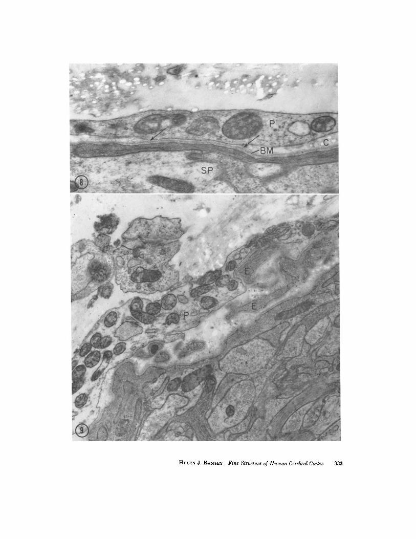
Fig. 8 shows a section of pia-glial border in which the two layers are closely adherent, sepa- rated only by basement membrane. At both ends of this bond the two layers become separated and a few fibers of collagen can be seen between them. Few such areas were seen in the present material. In Fig. 9 another pia-glial approximation is s~.own; in this situation the pia is stretched out as a smooth sheet over several cytoplasmic processes, whose connection with the astrocytic layer falls out of the section. Both of these areas are con- sidered undisturbed.
D I S C U S S I O N
While it is inevitable that samples of brain ob- tained at surgery are more liable to distortion than experimental brain material, several con- sistent observations emerge suggesting that the usual interpretation of the relation of meninges to cortical surface may be subject to modification.
The term "pia mater" was formerly taken to include both the innermost layer of meningeal cells and the network of blood vessels lying over it. ~hc term is usually now applied in a more restrictive sense to the thin layer of cells investing the brain, the " in t ima pia" of Key and Retzius (10), and excludes the overlying vasculature and its adnexa referred to more correctly by Millen and Woollam (l l) as "epi-pial tissue." Electron microscope findings are in agreement with this view. Many investigators consider that no cyto- logical distinction can be made between pial and arachnoidal cells other than that of form and loca- tion. Pease and Schultz (2) subscribed to this view. They also demonstrated that arachnoid trabeculae in the rat meninges consist simply of assemblies of
HELEN J. ~,AMSEY Fine Structure of Human Cerebral Cortex 327
arachnoidal ceils traversing the subarachnoid space. The subarachnoid space has always been considered to be limited externally by the arach- noid mater and internally by the pia mater. In the present study, the apparent partial discontinu- ity of this internal covering, its frequent physical separation from underlying brain, and the pres- ence of cytoplasmic extensions from the astrocytic layer all suggest that the internal limit of the sub- arachnoid space is, in fact, the subpial fibrous astrocytes of the brain itself, and that the cerebro- spinal fluid bathes the basement membrane- in- vested surface of these cells directly.
Cytoplasmic processes of various types are common throughout biological material seen at the ultrastructural level (t2, 13). While many are permanent formed elements, some are considered to be temporary or variable. This is, of course, the case with ameboid cells, including phagocytes. Among cells having a permanent location, but bearing variable extensions is the endothelial cell with its fingers of cytoplasm reaching into the vessel lumen (14). These are thought to function by enclosing small amounts of fluid to be incor- porated subsequently into the cell (15). This ap- pearance is not duplicated in the case of astrocytic extensions whose arborizations include organelles and end bluntly rather than in a curving tongue. No vacuolization or pinocytosis was seen. No other
instance comes to mind of extensions of this com- plexity arborizing freely into a fluid medium.
What cellular function may be subserved by the cytoplasmic extensions can at present only be sur- mised, but it seems reasonable to assume that some exchange occurs here and is facilitated by the in- crease of surface produced.
It is suggested that the brain surface may be fluctuating in the details of its topography. Oc- casionally, a large glial process within the sub- stance of the cortex could be seen extending up toward the surface and there spreading out to occupy a surprisingly broad area and extruding cytoplasmic excrescences into the surrounding fluid medium. This leads to the further speculation that such processes may be extended according to the requirements of the cell, and that the source- composition of the uppermost components of the superficial glial meshwork may be fluctuating as well, with first one and then another process usurping much of the superficial area.
In this connection it is interesting to recall that living glial cells in tissue culture (16) possess un- dulating membranes which are transient and mobile in nature.
This research was supported by United States Public Health Service Grant B-3678.
Received for publication, October 26, 1964.
R E F E R E N C E S
1. FARQUHAR, M. G., and HARTMANN, J. F., Neu- roglial structure and relationships as revealed by electron microscopy, J. Neuropath. and Exp. Neurol., 1957, 11, 18.
2. PEASE, D. C., and SCHULTZ, R. g., Electron microscopy of rat cranial meninges, Am. J. Anat., 1958, 102, 301.
3. giN, H.-S., DUNCAN, D., and ALEXANDER, R. S., A note on the subpial cytoplasm of the central nervous system: an electron microscope study, Texas Rep. Biol. and Med., 1960, 18, 620.
4. DUNCAN, D., and ALEXANDER, R., An electron microscope study of the supraoptic nucleus of the rat, Anat. Rec., 1961, 139, 222.
5. NELSON, E., BLINZlNGER, K., and HAGER, H., Electron microscopic observations on sub- arachnoid and pcrivascular spaces of the Syrian hamster brain, Neurol., 1961, 11, 285.
6. NELSON, E., BLINZINGER, K., and HAGER, H., An electron-microscopic study of bacterial meningitis, AMA Arch. Neurol., 1962, 6, 390.
7. PEASE, D. C., Buffered formaldehyde as a killing
FmVRE 4 An arborizing cytoplasmic extension from the superficial astrocytic meshwork in human cortex. Cytoplasm in the expansion resembles that of the cell from which it originates; it contains astrocytic fibrils and mitochondria. All surfaces are bordered by basement membrane (BM). Unstained collagen (C) is scattered through the surrounding space. From parietal cortex in the vicinity of a malignant glial tumor. )< 40,500.
FIGURE 5 A more complex configuration of cytoplasmic extensions from the cortical surface in human brain. X £8,000.
328 TIIE JOURNAL OF CELL BIOLOGY • VOLUME ~6, 1965
HELEN J. RA~sEY Fine Structure of Human Cerebral Cortex 329
FIaURE 6 A group of processes from subpial fibrous astrocytes in human brain. The cytoplasm contains an assortment of fibrils (F), glycogen particles (g) and mitochondria (m), and is similar to that of the astrocytic layer at the left of the figure. Basement membrane (BM) outlines the processes. X ~4,000.
FIGURE 7 A small vein close to the cortical surface in human brain. The lumen curves out of the section in the center of the figure. Many small processes (arrows) can be seen extending from glial cytoplasm (G) into the perivascular area which is otherwise filled with extracellular material. X 14,000.
agent and primary fixative for electron mi- croscopy, Anat. Rec., 1962, 146, 342.
8. MILLONm, G., Advantages of a phosphate buffer for OsO4 solutions in fixation, J. Appl. Phys., 1961, 32, 1637.
9. MmLONm, G., A modified procedure for lead staining of thin sections, J. Biophysic. and Biochem. Cytol., 1961, 11, 736.
I0. KEY, A., and RETZmS, G., Studien in der Anatomie des Nervensystems und des Binde- gewebes, Stockholm, P. A. Norstedt and S6ner, 1875.
11. MILLEN, J. W., and WOOLLAM, D. H. M., Ob- servations on the nature of pia mater, Brain, 1961, 84, 514.
12. POLmARD, A., and BAun, C. A., La surface de la cellule, in Les Structures Inframicrosco-
piques Normales et Pathologiques des Cellules et des Tissus, Paris, Masson et Cie, 1958, chap. 4.
13. POLmARD, A., and BESSrS, M., Sur un mode d'incorporation des macromolecules par la ceIlule, visible au microscope electronique: la rhopheocytose, Compt. rend. Acad. sc., 1958, 246, 3194.
14. FAWCET% D. W., and WITTENBURO, J., Struc- tural specializations of endothelial cell june- tions, Anat. Rec., 1962, 142, 231.
15. FAWCETT, D. W., in Physiology and Pathology of Peripheral Blood Vessels, Baltimore, Williams & Wilkins, 1963, chap. 2.
16. PO~ma~AT, C. M., Dynamic Neurogliology, Texas Rep. Biol. Med., 1951, 10, 885.
HELEN J. RAMSEY Fine Structure of Human Cerebral Cortex 331
Fmun~ 8 Conjunction between pial (P) and subpial (SP) layers. At the center of the figure (between arrows) these layers are separated only by shared basement membrane. At left and right the layers separate, with the basement membrane (BM) adhering to the astrocytic layer, and a few cross-sections of collagen (C) appearing in the space so formed. )< 41,5o{).
FIGURI~ 9 An area in which a smooth sheet of pial cytoplasm (P) overlies but does not follow the irregularities of astrocytic extensions (E). The stem by which the latter are joined to their cell of origin lies out of the section. X ~4,000.
332 THE JOURNAL OF CELL BIOLOGY - VOLUME ~6, 1965
HELEN J. RAMSEY Fine Structure of Human Cerebral Cortex 333